
Endostatin and Cancer Therapy: A Novel Potential Alternative to Anti-VEGF Monoclonal Antibodies

 ,
,  , ,
, ,  and
and
Abstract
:1. Introduction
2. Angiogenesis
3. Anti VEGF Monoclonal Antibodies
Adverse Events
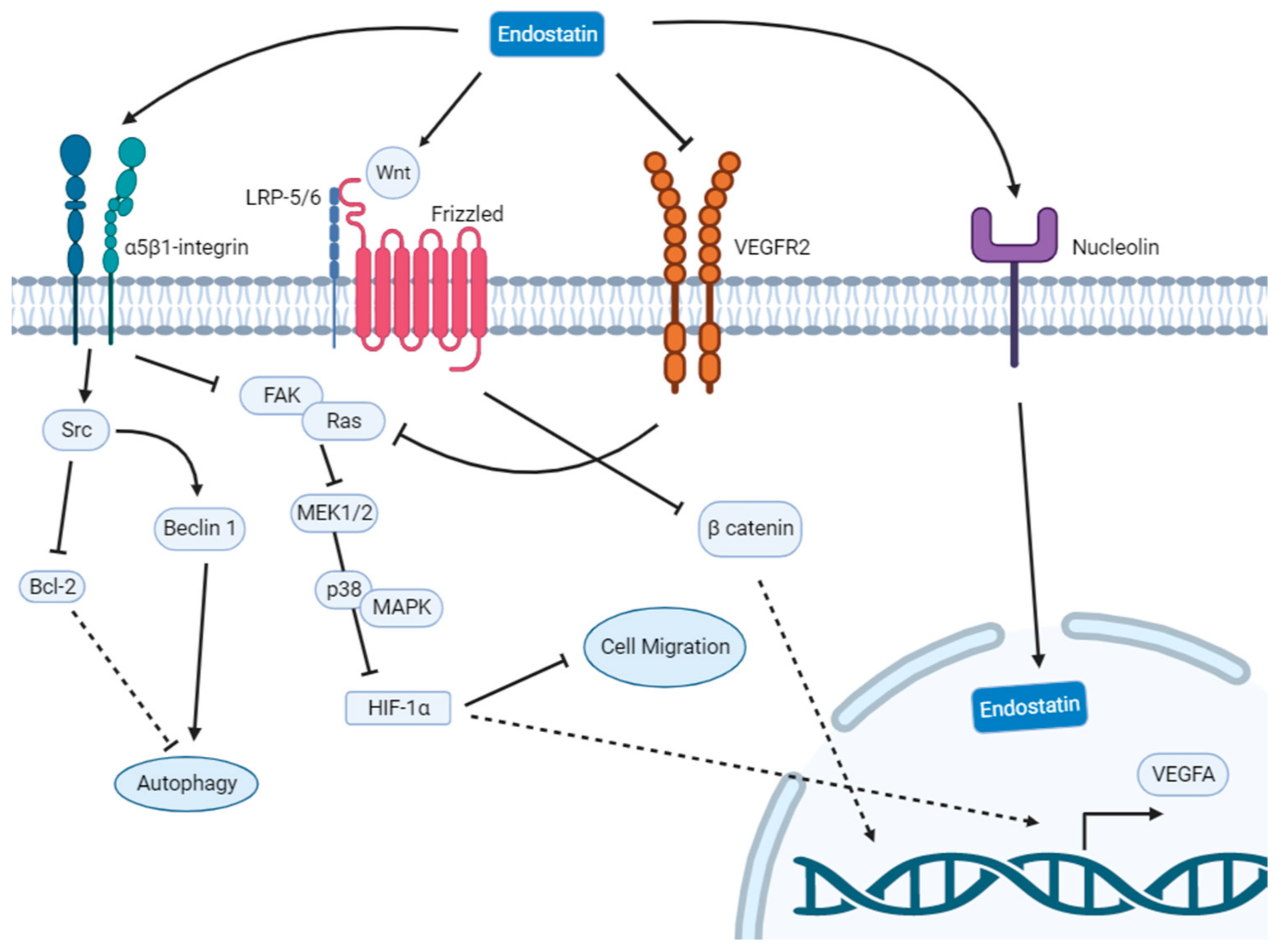
4. Endostatin
4.1. Pulmonary Cancer
4.2. Gastric Cancer
4.3. Esophageal Cancer
4.4. Colorectal Cancer
4.5. Nasopharyngeal Cancer
4.6. Breast Cancer
5. Discussion
Author Contributions
Funding
Institutional Review Board Statement
Informed Consent Statement
Data Availability Statement
Conflicts of Interest
References
- Mattiuzzi, C.; Lippi, G. Current Cancer Epidemiology. J. Epidemiol. Glob. Health 2019, 9, 217–222. [Google Scholar] [CrossRef] [PubMed] [Green Version]
- Yahya, E.B.; Alqadhi, A.M. Recent Trends in Cancer Therapy: A Review on the Current State of Gene Delivery. Life Sci. 2021, 269, 119087. [Google Scholar] [CrossRef]
- Li, T.; Kang, G.; Wang, T.; Huang, H. Tumor Angiogenesis and Anti-Angiogenic Gene Therapy for Cancer (Review). Oncol. Lett. 2018, 16, 687–702. [Google Scholar] [CrossRef] [PubMed] [Green Version]
- Adair, T.H.; Montani, J.-P. Angiogenesis; Morgan & Claypool Life Sciences: San Rafael, CA, USA, 2010. [Google Scholar]
- Ramjiawan, R.R.; Griffioen, A.W.; Duda, D.G.; Steele, E.L. Anti-Angiogenesis for Cancer Revisited: Is There a Role for Combinations with Immunotherapy? HHS Public Access. Angiogenesis 2017, 20, 185–204. [Google Scholar] [CrossRef] [PubMed]
- Du, R.; Lu, K.V.; Petritsch, C.; Liu, P.; Ganss, R.; Passegué, E.; Song, H.; Vandenberg, S.; Johnson, R.S.; Werb, Z.; et al. HIF1α Induces the Recruitment of Bone Marrow-Derived Vascular Modulatory Cells to Regulate Tumor Angiogenesis and Invasion. Cancer Cell 2008, 13, 206–220. [Google Scholar] [CrossRef] [PubMed] [Green Version]
- Viallard, C.; Larrivée, B. Tumor Angiogenesis and Vascular Normalization: Alternative Therapeutic Targets. Angiogenesis 2017, 20, 409–426. [Google Scholar] [CrossRef]
- Carmeliet, P.; Jain, R.K. Principles and Mechanisms of Vessel Normalization for Cancer and Other Angiogenic Diseases. Nat. Rev. Drug Discov. 2011, 10, 417–427. [Google Scholar] [CrossRef]
- Carmeliet, P. VEGF as a Key Mediator of Angiogenesis in Cancer. Oncology 2005, 69, 4–10. [Google Scholar] [CrossRef]
- Kerbel, R.S. Tumor Angiogenesis. N. Engl. J. Med. 2008, 358, 2039–2049. [Google Scholar] [CrossRef] [Green Version]
- Stanca Melincovici, C.; Boşca, A.B.; Şuşman, S.; Mărginean, M.; Mihu, C.; Istrate, M.; Moldovan, I.-M.; Roman, A.L.; Mihu, C.M. Vascular Endothelial Growth Factor (VEGF)-Key Factor in Normal and Pathological Angiogenesis. Rom. J. Morphol. Embryol. 2018, 59, 455–467. [Google Scholar]
- Apte, R.S.; Chen, D.S.; Ferrara, N. VEGF in Signaling and Disease: Beyond Discovery and Development. Cell 2019, 176, 1248–1264. [Google Scholar] [CrossRef] [Green Version]
- Oguntade, A.S.; Al-Amodi, F.; Alrumayh, A.; Alobaida, M.; Bwalya, M. Anti-Angiogenesis in Cancer Therapeutics: The Magic Bullet. J. Egypt. Natl. Cancer Inst. 2021, 33, 15. [Google Scholar] [CrossRef]
- Garcia, J.; Hurwitz, H.I.; Sandler, A.B.; Miles, D.; Coleman, R.L.; Deurloo, R.; Chinot, O.L. Bevacizumab (Avastin®) in Cancer Treatment: A Review of 15 Years of Clinical Experience and Future Outlook. Cancer Treat. Rev. 2020, 86, 102017. [Google Scholar] [CrossRef]
- Riccardi, C.; Napolitano, E.; Platella, C.; Musumeci, D.; Melone, M.A.B.; Montesarchio, D. Anti-VEGF DNA-Based Aptamers in Cancer Therapeutics and Diagnostics. Med. Res. Rev. 2021, 41, 464–506. [Google Scholar] [CrossRef]
- Goldberg, R.M.; Sargent, D.J.; Morton, R.F.; Fuchs, C.S.; Ramanathan, R.K.; Williamson, S.K.; Findlay, B.P.; Pitot, H.C.; Alberts, S.R. A Randomized Controlled Trial of Fluorouracil plus Leucovorin, Irinotecan, and Oxaliplatin Combinations in Patients with Previously Untreated Metastatic Colorectal Cancer. J. Clin. Oncol. 2004, 22, 23–30. [Google Scholar] [CrossRef]
- Ferrara, N.; Hillan, K.J.; Novotny, W. Bevacizumab (Avastin), a Humanized Anti-VEGF Monoclonal Antibody for Cancer Therapy. Biochem. Biophys. Res. Commun. 2005, 333, 328–335. [Google Scholar] [CrossRef]
- Papachristos, A.; Kemos, P.; Katsila, T.; Panoilia, E.; Patrinos, G.P.; Kalofonos, H.; Sivolapenko, G.B. VEGF-A and ICAM-1 Gene Polymorphisms as Predictors of Clinical Outcome to First-Line Bevacizumab-Based Treatment in Metastatic Colorectal Cancer. Int. J. Mol. Sci. 2019, 20, 5791. [Google Scholar] [CrossRef] [Green Version]
- Gerger, A.; El-Khoueiry, A.; Zhang, W.; Yang, D.; Singh, H.; Bohanes, P.; Ning, Y.; Winder, T.; Labonte, M.J.; Wilson, P.M.; et al. Pharmacogenetic Angiogenesis Profiling for First-Line Bevacizumab plus Oxaliplatin-Based Chemotherapy in Patients with Metastatic Colorectal Cancer. Clin. Cancer Res. 2011, 17, 5783–5792. [Google Scholar] [CrossRef] [Green Version]
- Schneider, B.P.; Wang, M.; Radovich, M.; Sledge, G.W.; Badve, S.; Thor, A.; Flockhart, D.A.; Hancock, B.; Davidson, N.; Gralow, J.; et al. Association of Vascular Endothelial Growth Factor and Vascular Endothelial Growth Factor Receptor-2 Genetic Polymorphisms With Outcome in a Trial of Paclitaxel Compared With Paclitaxel Plus Bevacizumab in Advanced Breast Cancer: ECOG 2100. J. Clin. Oncol. 2008, 26, 4672–4678. [Google Scholar] [CrossRef]
- Etienne-Grimaldi, M.-C.; Formento, P.; Degeorges, A.; Pierga, J.-Y.; Delva, R.; Pivot, X.; Dalenc, F.; Espié, M.; Veyret, C.; Formento, J.-L.; et al. Prospective Analysis of the Impact of VEGF-A Gene Polymorphisms on the Pharmacodynamics of Bevacizumab-Based Therapy in Metastatic Breast Cancer Patients. Br. J. Clin. Pharmacol. 2011, 71, 921–928. [Google Scholar] [CrossRef] [Green Version]
- Papachristos, A.; Kemos, P.; Kalofonos, H.; Sivolapenko, G. Correlation Between Bevacizumab Exposure and Survival in Patients with Metastatic Colorectal Cancer. Oncologist 2020, 25, 853–858. [Google Scholar] [CrossRef] [PubMed] [Green Version]
- Porta, M.; Striglia, E. Intravitreal Anti-VEGF Agents and Cardiovascular Risk. Intern. Emerg. Med. 2019, 15, 199–210. [Google Scholar] [CrossRef] [PubMed]
- Kamba, T.; McDonald, D.M. Mechanisms of Adverse Effects of Anti-VEGF Therapy for Cancer. Br. J. Cancer 2007, 96, 1788–1795. [Google Scholar] [CrossRef] [PubMed]
- Ngo Ntjam, N.; Thulliez, M.; Paintaud, G.; Salvo, F.; Angoulvant, D.; Pisella, P.J.; Bejan-Angoulvant, T. Cardiovascular Adverse Events with Intravitreal Anti-Vascular Endothelial Growth Factor Drugs: A Systematic Review and Meta-Analysis of Randomized Clinical Trials. JAMA Ophthalmol. 2021, 139, 610–619. [Google Scholar] [CrossRef]
- Falavarjani, K.G.; Nguyen, Q.D. Adverse Events and Complications Associated with Intravitreal Injection of Anti-VEGF Agents: A Review of Literature. Eye 2013, 27, 787–794. [Google Scholar] [CrossRef] [Green Version]
- Seebacher, N.A.; Stacy, A.E.; Porter, G.M.; Merlot, A.M. Clinical Development of Targeted and Immune Based Anti-Cancer Therapies. J. Exp. Clin. Cancer Res. 2019, 38, 156. [Google Scholar] [CrossRef] [Green Version]
- Lacal, P.M.; Graziani, G. Therapeutic Implication of Vascular Endothelial Growth Factor Receptor-1 (VEGFR-1) Targeting in Cancer Cells and Tumor Microenvironment by Competitive and Non-Competitive Inhibitors. Pharmacol. Res. 2018, 136, 97–107. [Google Scholar] [CrossRef]
- Escudier, B.; Eisen, T.; Stadler, W.M.; Szczylik, C.; Oudard, S.; Siebels, M.; Negrier, S.; Chevreau, C.; Solska, E.; Desai, A.A.; et al. Sorafenib in Advanced Clear-Cell Renal-Cell Carcinoma. N. Engl. J. Med. 2007, 356, 125–134. [Google Scholar] [CrossRef]
- Wakelee, H.A.; Dahlberg, S.E.; Keller, S.M.; Tester, W.J.; Gandara, D.R.; Graziano, S.L.; Adjei, A.A.; Leighl, N.B.; Aisner, S.C.; Rothman, J.M.; et al. Adjuvant Chemotherapy with or without Bevacizumab in Patients with Resected Non-Small-Cell Lung Cancer (E1505): An Open-Label, Multicentre, Randomised, Phase 3 Trial. Lancet Oncol. 2017, 18, 1610–1623. [Google Scholar] [CrossRef]
- Yao, J.; Fan, L.; Peng, C.; Huang, A.; Liu, T.; Lin, Z.; Yang, Q.; Zhang, T.; Ma, H. Clinical Efficacy of Endostar Combined with Chemotherapy in the Treatment of Peritoneal Carcinomatosis in Gastric Cancer: Results from a Retrospective Study. Oncotarget 2017, 8, 70788–70797. [Google Scholar] [CrossRef] [Green Version]
- Faye, C.; Moreau, C.; Chautard, E.; Jetne, R.; Fukai, N.; Ruggiero, F.; Humphries, M.J.; Olsen, B.R.; Ricard-Blum, S. Molecular Interplay between Endostatin, Integrins, and Heparan Sulfate. J. Biol. Chem. 2009, 284, 22029–22040. [Google Scholar] [CrossRef] [Green Version]
- Wickström, S.A.; Alitalo, K.; Keski-Oja, J. Endostatin Associates with Integrin 5 1 and Caveolin-1, and Activates Src via a Tyrosyl Phosphatase-Dependent Pathway in Human Endothelial Cells. Cancer Res. 2002, 62, 5580–5589. [Google Scholar]
- Poluzzi, C.; Iozzo, R.V.; Schaefer, L. Endostatin and Endorepellin: A Common Route of Action for Similar Angiostatic Cancer Avengers. Adv. Drug Deliv. Rev. 2016, 97, 156–173. [Google Scholar] [CrossRef] [Green Version]
- Matsumoto, G.; Hirohata, R.; Hayashi, K.; Sugimoto, Y.; Kotani, E.; Shimabukuro, J.; Hirano, T.; Nakajima, Y.; Kawamata, S.; Mori, H. Control of Angiogenesis by VEGF and Endostatin-Encapsulated Protein Microcrystals and Inhibition of Tumor Angiogenesis. Biomaterials 2014, 35, 1326–1333. [Google Scholar] [CrossRef] [Green Version]
- Ning, T.; Jiang, M.; Peng, Q.; Yan, X.; Lu, Z.J.; Peng, Y.L.; Wang, H.L.; Lei, N.; Zhang, H.; Lin, H.; et al. Low-Dose Endostatin Normalizes the Structure and Function of Tumor Vasculature and Improves the Delivery and Anti-Tumor Efficacy of Cytotoxic Drugs in a Lung Cancer Xenograft Murine Model. Thorac. Cancer 2012, 3, 229–238. [Google Scholar] [CrossRef]
- Wang, H.-L.; Ning, T.; Li, M.; Lu, Z.-J.; Yan, X.; Peng, Q.; Lei, N.; Zhang, H.; Luo, F. Effect of Endostatin on Preventing Postoperative Progression of Distant Metastasis in a Murine Lung Cancer Model. Tumori J. 2011, 97, 787–793. [Google Scholar] [CrossRef]
- Ding, Y.; Wang, Y.; Cui, J.; Si, T. Endostar Blocks the Metastasis, Invasion and Angiogenesis of Ovarian Cancer Cells. Neoplasma 2020, 67, 595–603. [Google Scholar] [CrossRef]
- Mohajeri, A.; Sanaei, S.; Kiafar, F.; Fattahi, A.; Khalili, M.; Zarghami, N. The Challenges of Recombinant Endostatin in Clinical Application: Focus on the Different Expression Systems and Molecular Bioengineering. Adv. Pharm. Bull. 2017, 7, 21–34. [Google Scholar] [CrossRef]
- Jiang, W.; Sun, W.; Li, W.; Gao, J.; Wang, H.; Zhou, W.; Liang, J.; Aa, L.; Wang, L. Real-World Treatment Pattern and Comprehensive Comparative Effectiveness of Endostar plus Different Chemotherapy in Advanced Patients with Non-Small Cell Lung Cancer. Sci. Rep. 2022, 12, 10841. [Google Scholar] [CrossRef]
- Han, B.; Xiu, Q.; Wang, H.; Shen, J.; Gu, A.; Luo, Y.; Bai, C.; Guo, S.; Liu, W.; Zhuang, Z.; et al. A Multicenter, Randomized, Double-Blind, Placebo-Controlled Study to Evaluate the Efficacy of Paclitaxel-Carboplatin Alone or with Endostar for Advanced Non-Small Cell Lung Cancer. J. Thorac. Oncol. 2011, 6, 1104–1109. [Google Scholar] [CrossRef] [Green Version]
- Jiang, X.-D.; Dai, P.; Wu, J.; Song, D.-A.; Yu, J.-M. Clinical Investigation: Thoracic Cancer Effect of Recombinant Human Endostatin on Radiosensitivity in Patients With NoneSmall-Cell Lung Cancer Radiation Oncology. Int. J. Radiat. Oncol. Biol. Phys. 2012, 83, 1272–1277. [Google Scholar] [CrossRef] [PubMed]
- Hu, W.; Fang, J.; Nie, J.; Dai, L.; Zhang, J.; Chen, X.; Ma, X.; Tian, G.; Wu, D.; Han, S.; et al. Efficacy and Safety of Extended Use of Platinum-Based Doublet Chemotherapy plus Endostatin in Patients with Advanced Nonsmall Cell Lung Cancer. Medicine 2016, 95, e4183. [Google Scholar] [CrossRef] [PubMed]
- Zhu, Q.; Zang, Q.; Jiang, Z.M.; Wang, W.; Cao, M.; Su, G.Z.; Zhen, T.C.; Zhang, X.T.; Sun, N.B.; Zhao, C. Clinical Application of Recombinant Human Endostatin in Postoperative Early Complementary Therapy on Patients with Non-Small Cell Lung Cancer in Chinese Mainland. Asian Pac. J. Cancer Prev. 2015, 16, 4013–4018. [Google Scholar] [CrossRef] [PubMed] [Green Version]
- Bao, Y.; Peng, F.; Zhou, Q.-C.; Yu, Z.-H.; Li, J.-C.; Cheng, Z.-B.; Chen, L.; Hu, X.; Chen, Y.-Y.; Wang, J.; et al. Phase II Trial of Recombinant Human Endostatin in Combination with Concurrent Chemoradiotherapy in Patients with Stage III Non-Small-Cell Lung Cancer. Radiother. Oncol. 2015, 114, 161–166. [Google Scholar] [CrossRef]
- Zhao, X.; Su, Y.; You, J.; Gong, L.; Zhang, Z.; Wang, M.; Zhao, Z.; Zhang, Z.; Li, X.; Wang, C. Combining Antiangiogenic Therapy with Neoadjuvant Chemotherapy Increases Treatment Efficacy in Stage IIIA (N2) Non-Small Cell Lung Cancer without Increasing Adverse Effects. Oncotarget 2016, 7, 62619–62626. [Google Scholar] [CrossRef] [Green Version]
- Jiang, X.; Guan, W.; Li, M.; Liang, W.; Qing, Y.; Dai, N.; Zhang, S.; Deng, Y.; Meng, H.; Yang, Y.; et al. Endostatin Combined with Platinum-Based Chemo-Radiotherapy for Advanced Non-Small Cell Lung Cancer. Cell Biochem. Biophys. 2015, 71, 571–577. [Google Scholar] [CrossRef]
- Ma, H.; Peng, F.; Xu, Y.; Bao, Y.; Hu, X.; Wang, J.; Fang, M.; Kong, Y.; Dong, B.; Chen, M. Five-Year Survival Rate Analysis: The Combination of Fortnightly-Administration of Endostar and Concurrent Chemoradiotherapy versus Concurrent Chemoradiotherapy in the Treatment of Inoperable Locally Advanced Non-Small Cell Lung Cancer. Ann. Palliat. Med. 2021, 10, 7560–7570. [Google Scholar] [CrossRef]
- Zhao, X.; Mei, K.; Cai, X.; Chen, J.; Yu, J.; Zhou, C.; Li, Q. A Randomized Phase II Study of Recombinant Human Endostatin plus Gemcitabine/Cisplatin Compared with Gemcitabine/Cisplatin Alone as First-Line Therapy in Advanced Non-Small-Cell Lung Cancer. Investig. New Drugs 2012, 30, 1144–1149. [Google Scholar] [CrossRef]
- Yang, X.; Wang, X.; Wang, N.; Jiang, W.; Li, Y.; Yang, X.; Yin, B. A Study on the Efficacy of Recombinant Human Endostatin Combined with Chemotherapy in Treating Advanced Non-Small-Cell Lung Cancer. JBUON 2019, 24, 2263–2269. [Google Scholar]
- Zhao, J.; Cheng, Y.; He, L.; Wang, J.; Xi, Q.; Gong, C. Extended Use of Rh-Endostatin Improves Prognosis in Patients with Advanced Non-Small Cell Lung Cancer: An Analysis of Retrospective Study. J. Thorac. Dis. 2022, 14, 4416–4426. [Google Scholar] [CrossRef]
- Wang, X.J.; Miao, K.; Luo, Y.; Li, R.; Shou, T.; Wang, P.; Li, X. Randomized Controlled Trial of Endostar Combined with Cis-Platin/Pemetrexed Chemotherapy for Elderly Patients with Advanced Malignant Pleural Effusion of Lung Adenocarcinoma. JBUON 2018, 23, 92–97. [Google Scholar]
- Jiang, X.; Ding, M.; Qiao, Y.; Liu, Y.; Liu, L. Recombinant Human Endostatin Combined with Radiotherapy in the Treatment of Brain Metastases of Non-Small Cell Lung Cancer. Clin. Transl. Oncol. 2014, 16, 630–636. [Google Scholar] [CrossRef]
- Chen, L.; Tong, F.; Peng, L.; Huang, Y.; Yin, P.; Feng, Y.; Cheng, S.; Wang, J.; Dong, X. Efficacy and Safety of Recombinant Human Endostatin Combined with Whole-Brain Radiation Therapy in Patients with Brain Metastases from Non-Small Cell Lung Cancer. Radiother. Oncol. 2022, 174, 44–51. [Google Scholar] [CrossRef]
- Zhou, Z.T.; Zhou, F.X.; Wei, Q.; Zou, L.Y.; Qin, B.F.; Peng, X.S. Phase II Study of Cisplatin/Etoposide and Endostar for Extensive-Stage Small-Cell Lung Cancer. Cancer Chemother. Pharmacol. 2011, 68, 1027–1032. [Google Scholar] [CrossRef]
- Zhao, Y.; Zhang, X.; Jin, C.; Yu, X.; Zhang, M.; Cao, Y.; Li, Y.; Wang, A.; Shan, X.; Zhang, J.; et al. Efficacy and Safety of Endostatin in Combination with Chemotherapy in Small Cell Lung Cancer: A Phase 2 Single-Arm Multicenter Open-Label Trial. Ann. Palliat. Med. 2021, 10, 3277–3285. [Google Scholar] [CrossRef]
- Lu, S.; Li, L.; Luo, Y.; Zhang, L.; Wu, G.; Chen, Z.; Huang, C.; Guo, S.; Zhang, Y.; Song, X.; et al. A Multicenter, Open-Label, Randomized Phase II Controlled Study of Rh-Endostatin (Endostar) in Combination with Chemotherapy in Previously Untreated Extensive-Stage Small-Cell Lung Cancer. J. Thorac. Oncol. 2015, 10, 206–211. [Google Scholar] [CrossRef] [Green Version]
- Sexton, R.E.; Al Hallak, M.N.; Diab, M.; Azmi, A.S. Gastric Cancer: A Comprehensive Review of Current and Future Treatment Strategies. Cancer Metastasis Rev. 2020, 39, 1179–1203. [Google Scholar] [CrossRef]
- Van Cutsem, E.; Muro, K.; Cunningham, D.; Bodoky, G.; Sobrero, A.; Cascinu, S.; Ajani, J.; Oh, S.C.; Al-Batran, S.E.; Wainberg, Z.A.; et al. Biomarker Analyses of Second-Line Ramucirumab in Patients with Advanced Gastric Cancer from RAINBOW, a Global, Randomized, Double-Blind, Phase 3 Study. Eur. J. Cancer 2020, 127, 150–157. [Google Scholar] [CrossRef] [Green Version]
- Yang, H.; Sui, Y.; Guo, X.; Tan, X.; Li, Y.; Wang, M. Endostar Continuous Intravenous Infusion Combined with S-1 and Oxaliplatin Chemotherapy Could Be Effective in Treating Liver Metastasis from Gastric Cancer. J. Cancer Res. Ther. 2018, 14, S1148–S1151. [Google Scholar] [CrossRef]
- Wang, L.; Li, W.; Liu, Y.; Zhang, C.; Gao, W.; Gao, L. Clinical Study on the Safety, Efficacy, and Prognosis of Molecular Targeted Drug Therapy for Advanced Gastric Cancer. Am. J. Transl. Res. 2021, 13, 4704. [Google Scholar]
- Uhlenhopp, D.J.; Then, E.O.; Sunkara, T.; Gaduputi, V. Epidemiology of Esophageal Cancer: Update in Global Trends, Etiology and Risk Factors. Clin. J. Gastroenterol. 2020, 13, 1010–1021. [Google Scholar] [CrossRef] [PubMed]
- Huang, F.L.; Yu, S.J. Esophageal Cancer: Risk Factors, Genetic Association, and Treatment. Asian J. Surg. 2018, 41, 210–215. [Google Scholar] [CrossRef] [PubMed]
- Deng, W.Y.; Song, T.; Li, N.; Luo, S.X.; Li, X. Clinical Observation and Therapeutic Evaluation of Rh-Endostatin Combined with DP Regimen in Treating Patients with Advanced Esophageal Cancer. Asian Pac. J. Cancer Prev. 2014, 15, 6565–6570. [Google Scholar] [CrossRef] [PubMed] [Green Version]
- Hu, Z.; Sun, S.; Zhao, X.; Yu, H.; Wu, X.; Wang, J.; Chang, J.; Wang, H. Rh-Endostatin Plus Irinotecan/Cisplatin as Second-Line Therapy for Advanced Esophageal Squamous Cell Carcinoma: An Open-Label, Phase II Study. Oncologist 2022, 27, 253–e312. [Google Scholar] [CrossRef]
- Wang, Z.-Q.; Wang, D.-S.; Wang, F.-H.; Ren, C.; Tan, Q.; Li, Y.-H. PHASE II STUDIES Recombinant Human Endostatin plus Paclitaxel/Nedaplatin for Recurrent or Metastatic Advanced Esophageal Squamous Cell Carcinoma: A Prospective, Single-Arm, Open-Label, Phase II Study. Investig. New Drugs 2021, 39, 516–523. [Google Scholar] [CrossRef]
- Zhong, Z.; Zhang, Z.; Wang, D.; Qing, Y.; Dai, N. Recombinant Human Endostatin Combined with Definitive Chemoradiotherapy as Primary Treatment for Patients with Unresectable but without Systemic Metastatic Squamous Cell Carcinoma of the Oesophagus. Br. J. Radiol. 2012, 85, 1104–1109. [Google Scholar] [CrossRef] [Green Version]
- WCRF. Colorectal Cancer Statistics|WCRF International. Available online: https://www.wcrf.org/cancer-trends/colorectal-cancer-statistics/ (accessed on 20 January 2023).
- Ridouane, Y.; Lopes, G.; Ku, G.; Masud, H.; Haaland, B. Targeted First-Line Therapies for Advanced Colorectal Cancer: A Bayesian Meta-Analysis. Oncotarget 2017, 8, 66458–66466. [Google Scholar] [CrossRef] [Green Version]
- Macedo, L.T.; da Costa Lima, A.B.; Sasse, A.D. Addition of Bevacizumab to First-Line Chemotherapy in Advanced Colorectal Cancer: A Systematic Review and Meta-Analysis, with Emphasis on Chemotherapy Subgroups. BMC Cancer 2012, 12, 89. [Google Scholar] [CrossRef] [Green Version]
- Xu, H.-X.; Huang, X.-E.; Qian, Z.-Y.; Li, X.X.Y.; Li, C.-G. Clinical Observation of Endostar® Combined with Chemotherapy in Advanced Colorectal Cancer Patients. Asian Pac. J Cancer Prev. 2011, 12, 3087–3090. [Google Scholar]
- Chen, Z.; Guo, W.; Cao, J.; Lv, F.; Zhang, W.; Qiu, L.; Li, W.; Ji, D.; Zhang, S.; Xia, Z.; et al. Endostar in Combination with Modified FOLFOX6 as an Initial Therapy in Advanced Colorectal Cancer Patients: A Phase i Clinical Trial. Cancer Chemother. Pharmacol. 2015, 75, 547–557. [Google Scholar] [CrossRef]
- Li, B.L.; Hu, X.L.; Zhao, X.H.; Sun, H.G.; Zhou, C.Y.; Zhang, Y. Endostar Combined with Irinotecan/Calcium Folinate/5-Fluorouracil (FOLFIRI) for Treating Advanced Colorectal Cancer: A Clinical Study. J. Chemother. 2015, 27, 301–306. [Google Scholar] [CrossRef]
- Zhou, J.F.; Bai, C.M.; Wang, Y.Z.; Li, X.Y.; Cheng, Y.J.; Chen, S.C. Endostar Combined with Chemotherapy for Treatment of Metastatic Colorectal and Gastric Cancer: A Pilot Study. Chin. Med. J. 2011, 124, 4299–4302. [Google Scholar] [CrossRef]
- Chen, J.; Qi, J.; Yu, B.-L.; Peng, X.-H.; Wang, F.; Tan, J.-J.; Chen, Q.-Q.; Peng, X.-Y.; Zeng, F.-F.; Liu, X. A Retrospective Study to Compare Five Induction Chemotherapy Regimens Prior to Radiotherapy in the Reduction of Regional Lymph Node Size in Patients with Nasopharyngeal Carcinoma. Med. Sci. Monit. 2018, 24, 2562–2568. [Google Scholar] [CrossRef] [Green Version]
- Guan, Y.; Li, A.; Xiao, W.; Liu, S.; Chen, B.; Lu, T.; Zhao, C.; Han, F. The Efficacy and Safety of Endostar Combined with Chemora diotherapy for Patients with Advanced, Locally Recurrent Nasopharyngeal Carcinoma. Oncotarget 2015, 6, 33926–33934. [Google Scholar] [CrossRef] [Green Version]
- Li, Y.; Tian, Y.; Jin, F.; Wu, W.; Long, J.; Ouyang, J.; Zhou, Y. A Phase II Multicenter Randomized Controlled Trial to Compare Standard Chemoradiation with or without Recombinant Human Endostatin Injection (Endostar) Therapy for the Treatment of Locally Advanced Nasopharyngeal Carcinoma: Long-Term Outcomes Update. Curr. Probl. Cancer 2020, 44, 100492. [Google Scholar] [CrossRef]
- Yin, Y.; Zhou, Z.; Li, Z.; Shen, M.; Qin, Y.; Yang, C.; Wang, R.; Kang, M. Efficacy of Concurrent Chemoradiotherapy plus Endostar Compared with Concurrent Chemoradiotherapy in the Treatment of Locally Advanced Nasopharyngeal Carcinoma: A Retrospective Study. Radiat. Oncol. 2022, 17, 135. [Google Scholar] [CrossRef]
- Chen, W.; Wang, F.; Yang, Z.; Zhang, T.; Shen, M.; Wang, R.; Kang, M. Long-Term Efficacy and Adverse Reactions of IMRT Combined with Endostar versus IMRT Combined with Chemotherapy for Locally Advanced Nasopharyngeal Carcinoma: A Retrospective Study. Ann. Palliat. Med. 2021, 10, 11891–11900. [Google Scholar] [CrossRef]
- Siegel, R.L.; Miller, K.D.; Wagle, N.S.; Ahmedin, J.; Siegel, R.L. Cancer Statistics, 2023. Cancer J. Clin. 2023, 73, 17–48. [Google Scholar] [CrossRef]
- Criscitiello, C.; Azim, H.A.; Schouten, P.C.; Linn, S.C.; Sotiriou, C. Understanding the Biology of Triple-Negative Breast Cancer. Ann. Oncol. 2012, 23 (Suppl. 6), vi13–vi18. [Google Scholar] [CrossRef]
- Zhao, S.; Zuo, W.-J.; Shao, Z.-M.; Jiang, Y.-Z. Molecular Subtypes and Precision Treatment of Triple-Negative Breast Cancer. Ann. Transl. Med. 2020, 8, 499. [Google Scholar] [CrossRef]
- Wang, X.; Shao, X.; Huang, J.; Lei, L.; Huang, Y.; Zheng, Y.; Cao, W.; Chen, Z. Exploring the Concepts and Practices of Advanced Breast Cancer Treatment: A Narrative Review. Ann. Transl. Med. 2021, 9, 721. [Google Scholar] [CrossRef] [PubMed]
- Zhang, X.; Zhang, Z.; Cao, M.; Liu, B.; Mori, M.; Luoh, S.W.; Bergan, R.; Liu, Y.; Liu, Y. A Randomized Parallel Controlled Phase II Trial of Recombinant Human Endostatin Added to Neoadjuvant Chemotherapy for Stage III Breast Cancer. Clin. Breast Cancer 2020, 20, 291–299. [Google Scholar] [CrossRef] [PubMed]
- Huang, Y.; Zheng, Y.; Cao, W.-M.; Chen, Z.; Fu, J.; Li, G.; Jia, W.X. A Phase II Study of Rh-Endostatin in Combination with Chemotherapy in Human Epidermal Growth Factor Receptor 2 (HER-2) Negative Advanced Breast Cancer (ABC). J. Clin. Oncol. 2020, 38 (Suppl. 15), 1071. [Google Scholar] [CrossRef]
- Huang, W.; Liu, J.; Wu, F.; Chen, K.; Li, N.; Hong, Y.; Huang, C.; Zhen, H.; Lin, L. The Efficacy and Safety of Endostar Combined with Taxane-Based Regimens for HER-2-Negative Metastatic Breast Cancer Patients. Oncotarget 2016, 7, 31501. [Google Scholar] [CrossRef] [PubMed] [Green Version]
- Tan, A.; Wang, H.; Nong, L.; Jia, Y.; Liu, Y.; Zhong, W.; Qin, F.; Wang, H.; Tang, J.; Zhou, W.; et al. Efficacy and Safety of Continuous Infusion of Rh-Endostatin Combined with Platinum-Based Chemotherapy for Advanced Triple-Negative Breast Cancer. Ann. Palliat. Med. 2021, 10, 12101–12112. [Google Scholar] [CrossRef]
- Chen, J.; Yao, Q.; Huang, M.; Wang, B.; Zhang, J.; Wang, T.; Ming, Y.; Zhou, X.; Jia, Q.; Huan, Y.; et al. A Randomized Phase III Trial of Neoadjuvant Recombinant Human Endostatin, Docetaxel and Epirubicin as First-Line Therapy for Patients with Breast Cancer (CBCRT01). Int. J. Cancer 2018, 142, 2130–2138. [Google Scholar] [CrossRef] [Green Version]
- Chen, J.; Yao, Q.; Li, D.; Zhang, J.; Wang, T.; Yu, M.; Zhou, X.; Huan, Y.; Wang, J.; Wang, L. Neoadjuvant Rh-Endostatin, Docetaxel and Epirubicin for Breast Cancer: Efficacy and Safety in a Prospective, Randomized, Phase II Study. BMC Cancer 2013, 13, 248. [Google Scholar] [CrossRef] [Green Version]
- Jia, Q.; Xu, J.; Jiang, W.; Zheng, M.; Wei, M.; Chen, J.; Wang, L.; Huan, Y. Dynamic Contrast-Enhanced MR Imaging in a Phase Ⅱ Study on Neoadjuvant Chemotherapy Combining Rh-Endostatin with Docetaxel and Epirubicin for Locally Advanced Breast Cancer. Int. J. Med. Sci. 2013, 10, 110–118. [Google Scholar] [CrossRef] [Green Version]
- Shi, H.; Chen, L.; Che, Y.; Sun, W.; Niu, X.; Lu, W. Efficacy of Endostatin Combined with Continuous Transcatheter Arterial Infusion and Chemoembolization on Gastric Cancer with Liver Metastasis and Analysis of Prognosis. JBUON 2020, 25, 1469–1475. [Google Scholar]
- Zhai, Y.; Ma, H.; Hui, Z.; Zhao, L.; Li, D.; Liang, J.; Wang, X.; Xu, L.; Chen, B.; Tang, Y.; et al. HELPER Study: A Phase II Trial of Continuous Infusion of Endostar Combined with Concurrent Etoposide plus Cisplatin and Radiotherapy for Treatment of Unresectable Stage III Non-Small-Cell Lung Cancer. Radiother. Oncol. 2019, 131, 27–34. [Google Scholar] [CrossRef]
- Guo, F.; Chen, C.; Liang, Y.; Ma, S.; Zou, W. Efficacy of the Combination of Endostar with Chemotherapy on Stage IVb and Recurrent Metastatic Cervical Cancer. J. Cent. South Univ. 2020, 45, 1412–1418. [Google Scholar] [CrossRef]
- Chen, X.; Nie, J.; Dai, L.; Hu, W.; Zhang, J.; Han, J.; Ma, X.; Tian, G.; Han, S.; Wu, D.; et al. Comparison of Endostatin Combined with Pt-Dc versus Bevacizumab Combined with Pt-Dc in the First-Line Treatment of Advanced Lung Adenocarcinoma: A Retrospective Propensity Score-Matched Cohort Study. Ann. Palliat. Med. 2021, 10, 7847–7856. [Google Scholar] [CrossRef]
- Shi, X.; Dong, X.; Young, S.; Chen, A.; Liu, X.; Zheng, Z.; Huang, K.; Lu, D.; Feng, S.; Morahan, G.; et al. The Impact of Angiogenesis Inhibitors on Survival of Patients with Small Cell Lung Cancer. Cancer Med. 2019, 8, 5930–5938. [Google Scholar] [CrossRef]
- Guan, X.; Li, W.; Wang, Y.; Zhao, Q.; Yu, X.; Jiang, J.; Bian, W.; Xu, C.; Sun, Y.; Zhang, C. The Mechanism of Rh-Endostatin-Induced Cardiotoxicity and Its Protection by Dihydromyricetin [in Vivo/in Vitro, C57BL/6 Mice, AC16 and HiPSC-CMs]. Toxicol. Lett. 2023, 377, 29–37. [Google Scholar] [CrossRef]
- Guo, L.; Xu, B.; Zhou, D.; Chang, G.; Fu, Y.; Liu, L.; Luo, Y. Biophysical and Biological Characterization of PEGylated Recombinant Human Endostatin. Clin. Exp. Pharmacol. Physiol. 2019, 46, 920–927. [Google Scholar] [CrossRef]
- Geng, X.; Guo, L.; Liu, L.; Wang, C.; Peng, Q.; Qi, W.; Sun, L.; Liu, X.; Miao, Y.; Lin, Z.; et al. A Pre-Clinical Safety Study of PEGylated Recombinant Human Endostatin (M 2 ES) in Sprague Dawley Rats. Regul. Toxicol. Pharmacol. 2018, 95, 190–197. [Google Scholar] [CrossRef]
- Rezaei, N.; Mehrnejad, F.; Vaezi, Z.; Sedghi, M.; Mohsen Asghari, S.; Naderi-Manesh, H. Encapsulation of an Endostatin Peptide in Liposomes: Stability, Release, and Cytotoxicity Study. Colloids Surf. B Biointerfaces 2019, 185, 110552. [Google Scholar] [CrossRef]


| Study Design | Radio/Chemotherapy Used | Endostatin Scheme | Main Outcomes | Adverse Events | Ref. |
|---|---|---|---|---|---|
| Breast Cancer | |||||
| Phase 2 RCT, n = 67, stage IIA to IIIC, first-line treatment | DTX EPR | 7.5 mg/m2 for 14 days every 3 weeks | ORRs 90.9% vs. 67.7% in control group pCR was identified in 5 (15.2%) vs. 2 (6.5%) | No significant difference was found between groups. | [89] |
| Phase 2 RCT, n = 87, stage III, | DTX EPR CTX | 15 mg/day i.v. for 14 days every 3 weeks | ORRs 81.82% vs. 58.14% in control group Median RFS 67.3 m vs. 55.0 m in control group Median OS 74.2 m vs. 59.1 m in control group 3- and 5-year OS 88.5% and 82.8% vs. 76.7% and 54.4% in control group | No significant difference was found between groups. | [84] |
| Phase 3 RCT, n = 803, stage IIA to IIIC BC, first-line treatment | DTX EPR | 7.5 mg/m2 for 14 days every 3 weeks | ORRs 91.0% vs. 77.9% in control group pCR was identified in 43 (10.7%) patients vs. 31(7.7%) in the control group, but no significant difference was found. | No significant difference was found between groups. | [88] |
| RCT, n = 64, stage IIA to IIIC | DTX EPR | 7.5 mg/m2 i.v. for 14 days every 3 weeks | ORRs 90.9% vs. 67.7% in control group Mean tumor size change was 21.18 cm3 ± 7.32 vs. 15.95 cm3 ± 4.32 in control group | Not reported | [90] |
| Prospective study, n = 57, HER-2 negative metastatic BC | TAX ABX TAX + GEM DTX + CPC | 7.5 mg/m2 i.v. for 14 days and was continued until progressive disease | ORRs was 68.4% for the whole population and 79.3%, 54.5%, and 16.7% for first-line, second-line, and third-line or beyond treatment group, respectively. Median PFS was 10.8 m | Grade 3–4 adverse events: neutropenia (80.7%), leukopenia (77.2%), liver dysfunction (10.5%), and peripheral neurotoxicity (8.8%). | [86] |
| Prospective study, n = 21, advanced TNBC | CB/CP + GEM DTX PEM | 30 mg/d i.v. for 7 days in addition to the chemotherapy cycle | ORRs was 47.6% for the whole population, while it was 50% and 44.4% for first-line and second-line or beyond treatment group, respectively. OS 13.3 months PFS 8.8 months | Grade 3–4 adverse events: neutropenia (14.3%), anemia (14.3%), leukopenia (9.5%), thrombocytopenia (9.5%), febrile neutropenia (4.8%), and hypertension (4.8%). | [87] |
| Colorectal cancer | |||||
| Pilot study, n = 24, metastatic CCR and GC | CAPIRI GP XELOX DCF FOLFIRI FOLFOX4 | 15 mg daily i.v. for 14 days every 3 weeks or 7 days every 2 weeks | ORR 19% and 57.1% in first-line treatment group Disease control rate 47.6% | Grade 3–4 adverse events: Leukopenia (30.4%), Neutropenia (34.8%), Thrombocytopenia (17.4%), Anemia (13.0%) Cardiac adverse events: Three patients presented transient sinus bradycardia with spontaneous remission. | [74] |
| Retrospective controlled study, n = 36, no metastatic CCR | FOLFOX-4 | 15 mg daily i.v. for 14 days | ORR 38.9% vs. 22.2% in control group OS 12.1 m vs. 11.4 m in control group PFS 6.4 m vs. 3.8 m in control group | Grade 3–4 adverse events: Leukopenia (16.7% vs. 11%) Thrombocytopenia (5.6% vs. 0%) Nausea/vomiting (5.6% vs. 5.6%) Cardiac adverse events Hypertension 17.7% vs. 5.6% in the control group Cardiac ischemia 11.1% vs. 0.0% in the control group. | [71] |
| RCT, n = 38, advanced CRC | IR + 5FU + CF | 15 mg i.v. daily | OR 42.9% vs. 29.4% in control group median TTP 14.5 months vs. 11 in control group | Cardiac adverse events: Three patients presented grade 1 electrocardiogram abnormalities and two presented grade 1–2 hypertension. No significant difference in the other adverse events between groups. | [73] |
| Phase 1 trial, n = 21, advanced CRC | OX + FU + FA (FOLFOX) | ascending doses from 7.5 to 75 mg/m2/day | Endostar was generally safe and well tolerated | Grade 3–4 adverse events: Neutropenia (23.8%), leucopenia (9.5%), thrombocytopenia (4.8%). Cardiac adverse events: Two patients presented ventricular arrhythmia. | [72] |
| Upper gastrointestinal tract cancer | |||||
| RCT, n = 96, gastric cancer with liver metastases | FU + OX + CF | 15 mg i.v. in addition to chemotherapy cycles | ORR 70.8% vs. 47.9% in control group OS and PFS significantly higher in E supplemented group | No significant difference in adverse events between groups. | [91] |
| RCT, n = 38, metastatic squamous cell carcinoma of the esophagus | 5FU + CP + RT | 15 mg i.v. daily | 1-year OS 72% vs. 50% in control group Median survival 18.2 m vs. 11.6 m in control group | No significant difference in adverse events between groups. | [67] |
| Lung cancer | |||||
| Phase 2 trial, n= 126, treatment-naive NSCLC | TAX + CPT | 7.5 mg/m2/d i.v. | ORR 39.3% vs. 23% in control group DCR: 90.2% vs. 67.2% in control group | Slight decrease in overall incidence rate of adverse events in E treatment group. Not statistically significant. | [41] |
| Prospective study, n = 50, hypoxia positive stage I-III NSCLC | RT | 15 mg i.v. | Total effective rate: 80% vs. 44% in control group | No significant difference in adverse events between groups. | [42] |
| RCT, n = 75, IB-IIIA NSCLC | CP/DXT/CPT/GEM/PEM/NVB following NCCN guidelines, following surgery | 7.5 mg/m2 i.v. | Average PFS increased by 9.8 m 5-year OS 59.3 vs. 42.1 in control group | No significant difference in adverse events between groups. | [44] |
| Phase 2 trial, n = 50, stage III NSCLC | DXT + CP, followed by RT | 7.5 mg/m2/d i.v. | PFS 9.9 m 3-year control rate 51% Median OS 24 months | All toxicities were tolerable with proper treatment. | [45] |
| RCT, n = 30, stage IIIA NSCLC | CP + NVB | 7.5 mg/m2 i.v. | Tumor regression rate increased approximately in 12% vs. control group OS 19 m vs. 16 m in control group | No significant difference in adverse events between groups. No serious adverse events or death were reported. | [46] |
| Retrospective study, n = 71, stage III/IV NSCLC | PEM + GEM + TAX + INN, followed by 2 cycles of RT | 15 mg i.v. in addition to chemotherapy cycles | PFS 12 m vs. 7 m in control group Non-specified higher OS vs. control group | CT + E group’s higher OS equaled higher rate of anemia, thrombocytopenia, nausea/vomiting, diarrhea, and fatigue. | [47] |
| Phase 2 trial, n = 73, unresectable NSCLC | EP + CP + RT | 7.5 mg/m2/24 h 120 h, 14 days/cycle | PFS 13.3 m OS 34.7 m 51 patients achieved objective response | The most common adverse event was leukopenia. 33 patients had grade 3 or more hematologic events. | [92] |
| Phase 2 trial, n = 193, treatment-naive locally advanced NSCLC | EP + CP, DXT + CP or CPT TAX + CP or CPT NVB + CP PEM + CP GEM + CP; RT | 7.5 mg/m2 | mean OS 29.7 m vs. 21.3 m in control group Hazard Ratio between E and control group: 0.697 | The incidence of grade 1 and 2 injury was 33.7% and 14.4%; 9.1% and 3.8% vs. 14.4%; in the E supplemented vs. control group, respectively. | [48] |
| Phase 2 trial, n = 69, stage IIIB/IV NSCLC | GEM + CP | 7.5 mg/m2 i.v. | PFS 6.8 m vs. 4.3 m in control group Survival rate at 12 m 51.6% vs. 38.7% in control group OS 12.4 months vs. 9.8 in control group | The addition of E to GEM/CP did not increase hematological toxicities. | [49] |
| Retrospective study, n = 136, stage IIIB-IV NSCLC | CP + TAX or PEM or GEM or DXT | 15 mg/day | ORR 48.5% vs. 29.5% in control group DCR 91.2% vs. 75% in control group | No significant difference in adverse events between groups. | [50] |
| Retrospective study, n = 115, stage IIIB-IV NSCLC | PEM + CP or INN GEM + CP or INN IR + CP | 7.5 mg/m2 | PFS 8.9 m in extended E group vs. 2 m in non-extended group of patients with squamous cell carcinoma OS 27.2 m in extended E group vs. 10.8 m in non-extended group with squamous cell carcinoma | No difference was found between both groups in the incidence of adverse effects. 12.5% and 12.2% had grade 3 or 4 adverse events. | [51] |
| RCT, n = 200, stage IIIB-IV NSCLC | PEM + CP GEM + CP IR + CP PEM + INN INN + GEM | 7.5 mg/m2 daily | PFS 8 m in extended E group vs. 5.8 m in non-extended group OS 23 m in extended E group vs. 14 m in non-extended group | There were no statistically significant differences in grade 3 to 4 toxicities overall between the 2 treatment groups. | [43] |
| RCT, n = 128, lung adenocarcinoma with malignant pleural effusion | CP + PEM | 45 mg intracavitary | Higher and stronger effect on control of the malignant pleural effusion and disease control rate vs. control group | There was no increase of adverse reactions relative to the control group. | [52] |
| RCT, n = 80, NSCLC with brain metastases | RT | 7.5 mg/m2/day during radiotherapy | Decrease in brain edema on E treated group No significant differences on OS | No significant difference in adverse events between groups. The most common reaction was granulopenia. | [53] |
| RCT, n = 43, NSCLC with brain metastases | RT | 30 mg/day | median PFS 8.1 m vs. 4.9 m in control group OS 14.2 m vs. 6.4 m in control group | No significant difference in adverse events between groups. | [54] |
| Phase 2 trial, n = 33, chemotherapy-naive extended SCLC | CP + EP | 15 mg i.v. | Median PFS 5 m Median OS 11.5 m | 57.6% developed neutropenia. | [55] |
| Phase 2 trial, n = 22, SCLC | EP + CP or CPT | 30 mg/day 3 days prior CT and 4 days post CT | PFS 8 m OS 13.6 m ORR 61.9% DCR 95.2% | The main adverse reactions were myelosuppression, albuminuria, nausea, and vomiting. All patients tolerated the treatment. | [56] |
| Phase 2 trial, n = 140, advanced treatment-naive SCLC | EP + CPT | 7.5 mg/m2 i.v. | PFS 7.3 m vs. 3.9 m in control group ORR 21% higher vs. control group OS similar to control group QOL higher vs. control group | No differences in toxicity vs. control group. | [57] |
| Head and neck cancer | |||||
| Clinical report, n = 22, recurrent grade III-IVB NPC | DXT + CP or DXT + CP + 5FU or GEM + CP; + IMRT | 105 mg/m2 | CR was achieved in 20 patients 1-year OS 93.3% 1-year PFS 92.3% 1-year Distant metastasis free-survival 90% | There were no reports of grade 5 toxicities. Incidence of radiation injury was substantially lower than previous studies. | [76] |
| Phase 2 trial, n = 114, locally advanced NPC | DXT + CP followed by CP + IMRT | 7.5 mg/m2 | CR was achieved in 91.1% of patients E trial group improved the complete remission rate of cervical lymph node metastasis | No significant difference in adverse events between groups. The most frequently observed acute toxicities were neutropenia, vomiting, and mucositis | [77] |
| Retrospective study, n = 23, locally advanced NPC | CP + IMRT | 15 mg/day | No significant differences in OS, PFS nor ORR | Incidence of xerostomia, difficulty in mouth opening and subcutaneous soft tissue fibrosis was lower on E group. | [79] |
| RCT, n = 44, recurrent metastatic cervical cancer | GEM + CP or DTX+ CP | 7.5 mg/m2 | median PFS 7.2 m vs. 5.1 m in control group | No significant difference in adverse events between groups. | [93] |
Disclaimer/Publisher’s Note: The statements, opinions and data contained in all publications are solely those of the individual author(s) and contributor(s) and not of MDPI and/or the editor(s). MDPI and/or the editor(s) disclaim responsibility for any injury to people or property resulting from any ideas, methods, instructions or products referred to in the content. |
© 2023 by the authors. Licensee MDPI, Basel, Switzerland. This article is an open access article distributed under the terms and conditions of the Creative Commons Attribution (CC BY) license (https://creativecommons.org/licenses/by/4.0/).
Share and Cite
Méndez-Valdés, G.; Gómez-Hevia, F.; Lillo-Moya, J.; González-Fernández, T.; Abelli, J.; Cereceda-Cornejo, A.; Bragato, M.C.; Saso, L.; Rodrigo, R. Endostatin and Cancer Therapy: A Novel Potential Alternative to Anti-VEGF Monoclonal Antibodies. Biomedicines 2023, 11, 718. https://doi.org/10.3390/biomedicines11030718
Méndez-Valdés G, Gómez-Hevia F, Lillo-Moya J, González-Fernández T, Abelli J, Cereceda-Cornejo A, Bragato MC, Saso L, Rodrigo R. Endostatin and Cancer Therapy: A Novel Potential Alternative to Anti-VEGF Monoclonal Antibodies. Biomedicines. 2023; 11(3):718. https://doi.org/10.3390/biomedicines11030718
Chicago/Turabian StyleMéndez-Valdés, Gabriel, Francisca Gómez-Hevia, José Lillo-Moya, Tommy González-Fernández, Joaquin Abelli, Antonia Cereceda-Cornejo, Maria Chiara Bragato, Luciano Saso, and Ramón Rodrigo. 2023. "Endostatin and Cancer Therapy: A Novel Potential Alternative to Anti-VEGF Monoclonal Antibodies" Biomedicines 11, no. 3: 718. https://doi.org/10.3390/biomedicines11030718