Article

Molecular Measurable Residual Disease Assessment before Hematopoietic Stem Cell Transplantation in Pediatric Acute Myeloid Leukemia Patients: A Retrospective Study by the I-BFM Study Group

1
Department of Women’s and Children’s Health, Haematology-Oncology Clinic and Lab, University of Padova, 35128 Padova, Italy
2
Department of Pediatric Hematology and Oncology and Cell and Gene Therapy, IRCCS Ospedale Pediatrico Bambino Gesù, Sapienza University of Rome, 00165 Rome, Italy
3
Department of Pediatric Hematology and Oncology, University Hospital Essen, 45147 Essen, Germany
4
Department of Pediatrics, Aarhus University Hospital, 8000 Aarhus, Denmark
5
Department of Pediatrics, Institute of Clinical Sciences, “Sahlgrenska Academy” University of Gothenburg, 40010 Gothenburg, Sweden
6
Princess Máxima Center for Pediatric Oncology, 1013 GM Utrecht, The Netherlands
7
Department of Clinical Chemistry, Sahlgrenska University Hospital, 40010 Gothenburg, Sweden
8
Department of Laboratory Medicine, Institute of Biomedicine, University of Gothenburg, 40010 Gothenburg, Sweden
*
Authors to whom correspondence should be addressed.
These authors contributed equally to this work.
Biomedicines 2022, 10(7), 1530; https://doi.org/10.3390/biomedicines10071530
Submission received: 20 May 2022 / Revised: 14 June 2022 / Accepted: 24 June 2022 / Published: 28 June 2022
(This article belongs to the Special Issue Molecular Research on Acute Myeloid Leukemia (AML))

Abstract

:
Hematopoietic stem cell transplantation (HSCT) is a curative post-remission treatment in patients with acute myeloid leukemia (AML), but relapse after transplant is still a challenging event. In recent year, several studies have investigated the molecular minimal residual disease (qPCR-MRD) as a predictor of relapse, but the lack of standardized protocols, cut-offs, and timepoints, especially in the pediatric setting, has prevented its use in several settings, including before HSCT. Here, we propose the first collaborative retrospective I-BFM-AML study assessing qPCR-MRD values in pretransplant bone marrow samples of 112 patients with a diagnosis of AML harboring t(8;21)(q22; q22)RUNX1::RUNX1T1, or inv(16)(p13q22)CBFB::MYH11, or t(9;11)(p21;q23)KMT2A::MLLT3, or FLT3-ITD genetic markers. We calculated an ROC cut-off of 2.1 × 10⁻⁴ that revealed significantly increased OS (83.7% versus 57.1%) and EFS (80.2% versus 52.9%) for those patients with lower qPCR-MRD values. Then, we partitioned patients into three qPCR-MRD groups by combining two different thresholds, 2.1 × 10⁻⁴ and one lower cut-off of 1 × 10⁻², and stratified patients into low-, intermediate-, and high-risk groups. We found that the 5-year OS (83.7%, 68.6%, and 39.2%, respectively) and relapse-free survival (89.2%, 73.9%, and 67.9%, respectively) were significantly different independent of the genetic lesion, conditioning regimen, donor, and stem cell source. These data support the PCR-based approach playing a clinical relevance in AML transplant management.

Graphical Abstract

1. Introduction

Hematopoietic stem cell transplantation (HSCT) is the most effective option for preventing relapse in patients with acute myeloid leukemia (AML) achieving complete remission [1]. Prognosis of pediatric AML patients has improved over the last decade, with current long-term survival rates reaching 70% [2,3] and HSCT has contributed substantially to this improvement [2,4,5]. Nevertheless, relapse after HSCT remains the main cause of treatment failure [3,6]. Transplantation rates in ongoing trials with refined indications to HSCT vary from 8 to 29% and, notably, recent results have supported the use of HSCT in AML patients in first complete remission (CR) if they show features predicting high risk of recurrence [7]. Achievement of morphological CR, defined as the presence of normal hematopoiesis and ≤5% blasts in the bone marrow, is important to optimize the outcome of patients given HSCT. However, to date, no studies in pediatric AML have analyzed the impact of molecular minimal residual disease (MRD) measurement before HSCT on transplant outcome. Moreover, given the availability of different methods to evaluate MRD (with different sensitivity), distinguishing diverse subgroups with different risks of post-transplant relapse could help implement novel intervention strategies aimed at reducing treatment failure. Measurable residual disease determined by quantitative polymerase chain reaction (qPCR-MRD) has increased the ability to monitor therapy response in AML [8,9,10]; in clinical trials conducted in adults, there is robust evidence that measurable residual disease determined by quantitative polymerase chain reaction (qPCR-MRD) increased the ability to monitor therapy response and improved prognostic precision [8,9,10] with respect to other techniques, including flow cytometry, leading to a refined risk stratification [11,12]. Indeed, recently, it has been demonstrated in CBF-rearranged and NPM1-mutated patients that qPCR-MRD played an independent prognostic role when assessed at different timepoints, including before transplant [13]. However, despite initial promising results for certain genetic lesions [14], the role of qPCR-MRD monitoring in childhood AML to define a deeper remission status is still controversial, and currently defined by flow cytometry [15,16,17]. By the way, there is a proportion of patients with MRD negativity experiencing relapse which can be due to (i) the acquisition of new disease markers/immunophenotype shift or (ii) low sensitivity and specificity of the method used (e.g., limit of manual gating strategies used to analyze multidimensional data) [15]. Over the last two decades, great advances in deciphering the genetic landscape of pediatric AML have been achieved [18,19], and therefofe, a suitable qPCR-MRD biomarker related to recurrent chromosomal rearrangements and gene mutations is now available in up to 75% of patients [19,20]. The role of MRD measurement before HSCT has been established for several hematological malignancies and in adult AML [21,22,23,24], but, so far, no data have been reported in pediatric AML. Furthermore, the concept of “molecular remission”, introduced for the first time in 2003 by the International Working Group guidelines to refine treatment response adults in AML [20], has never reached a consensus within large cooperative pediatric groups.
Here, we describe the results of an international retrospective BFM collaborative retrospective study in a large cohort of AML pediatric patients on qPCR-MRD monitoring and its prognostic role before HSCT. This analysis was possible due to harmonization of methods, and reports qPCR-MRD analyses by different MRD thresholds, finding that disease levels are associated with different probabilities of overall survival, relapse and transplant-related mortality, to be further considered in multidisciplinary discussion before transplant.

2. Materials and Methods

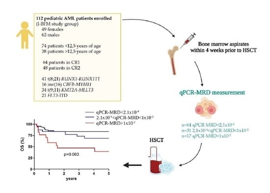
2.1. Patients and Samples

Included in the study are patients (i) with de novo AML (n = 112) other than acute promyelocytic leukemia; (ii) aged 0–18 years; (iii) diagnosed between 2002 and 2016 in one of the three consortia participating in this collaborative study; (iv) harboring isolated molecular lesion among t(8;21)RUNX1::RUNX1T1, inv(16)CBFB::MYH11, t(9;11)KMT2A::MLLT3, or FLT3-ITD. The AIEOP (Associazione Italiana Ematologia Oncologia Pediatrica) included n = 63 patients, Berlin–Frankfurt–Münster (BFM) study group included n = 11 patients, and the NOPHO (Nordic Society of Paediatric Haematology and Oncology) included n = 38 patients. The approval for this study was obtained from the institutional review board of each participating center. All patients provided written informed consent within each trial in accordance with the Declaration of Helsinki, obtained from patients’ parents or legal guardians. French-American-British (FAB) morphological classification, immune-phenotype analysis, and molecular characterization were performed at the laboratory of Pediatric Hematology of the University Hospital in Padova for the AIEOP cases and patients were enrolled in the AIEOP-AML2002/01 trial. For the BFM-SG group, samples were provided by the Clinic for Pediatrics III of the University Hospital of Essen (and patients were treated according to AML-BFM 2012 trial). As regards NOPHO patients, samples were collected at the Department of Clinical Chemistry at the Sahlgrenska University Hospital in Gothenburg or at the Department of Hematology Hemodiagnostic Laboratory of the Aarhus University Hospital (and patients were included in NOPHO AML-2004 or NOPHO-DBH AML-2012 trials). Post-remission treatment was determined according to genetic and cytogenetic criteria, reserving HSCT in first or second CR (CR1, or after a morphological relapse when in second remission phase, CR2). For this study, MRD results were included if the sample was taken within 4 weeks before HSCT and patients did not receive any further therapy between sampling and the start of conditioning.

2.2. RNA Extraction and Retro-Transcription

Mononuclear cells from bone marrow (BM) samples were isolated by density gradient using Lymphoprep™ (Stemcell Technologies, Vancouver, BC, Canada). RNA extraction and reverse transcription were performed by groups, as previously reported [25].

2.3. Molecular Marker Identification by Polymerase Chain Reaction

All patients were screened at diagnosis for t(8;21)RUNX1::RUNX1T1, inv(16)CBFB::MYH11, t(9;11)KMT2A::MLLT3, and FLT3-ITD by reverse transcription polymerase chain reaction (RT-PCR) with specific primers previously validated to detect the lesions [26,27,28,29]. Shifted primers were used to confirm the positivity of the rearrangement/mutation and to define the breakpoint for each case. All AML cases underwent karyotyping or FISH at diagnosis to confirm the lesion and characterize blasts’ karyotypes.

2.4. qPCR-MRD Detection

For each rearrangement included in the study (namely t(8;21) RUNX1::RUNX1T1, inv(16)CBFB::MYH11, t(9;11)KMT2A::MLLT3, and FLT3-ITD), we set up a real-time quantitative reverse transcription polymerase chain reaction (qPCR) to quantify AML-specific transcripts and monitor qPCR-MRD, as previously described [25,26,27,28,29]. The local laboratories tested the performance of all assays by evaluating each new batche of primer/probe sets by standard curve dilutions. Efficiency was in a dynamic range of at least 4 logs, with 6 logs as the maximum sensitivity reached.

2.5. Quality Control of Standard Operating Procedures (SOPs)

Patients’ samples were exchanged among laboratories to compare SOP performances. In particular, samples of one patient at diagnosis and follow-up for each genetic lesion from each reference laboratory were sent to the other laboratories. Samples were used to perform cDNA preparation locally following each lab’s SOPs, as well as qPCR. Quantification cycle (Cq) values together with calculated ∆Cq and qPCR-MRD values were disclosed and compared among laboratories to validate the previously discussed and harmonized methods.

2.6. Statistical Methods

Significance among groups was analyzed using the χ2 test and Fischer’s exact test. Overall survival (OS) was calculated from the date of the HSCT to the time of death due to any cause or time of last contact. Event-free survival (EFS) was calculated from the date of the HSCT to the last follow-up or first event (failure to engraft, relapse, death in remission, secondary malignancies, whichever occurs first). Relapse-free survival (RFS) was calculated from the date of the HSCT to the last follow-up or relapse event. OS, EFS, and RFS were estimated using the Kaplan–Meier method and groups were compared by using the log-rank test. Treatment-related mortality (TRM) and cumulative incidence of relapse (CIR) were calculated as cumulative incidence curves to adjust the estimates for the appropriate competing risks. Gray’s test was used to assess, in univariate analyses, differences between cumulative incidences using the “cmprsk” package for R software. A multivariable analysis was performed with Cox regression analysis considering the ages of patients at HSCT, type of donor, CR status, and qPCR-MRD reduction. Graphs and associated statistical analyses were generated using GraphPad Prism 8. A p-value <0.05 was considered to be statistically significant.

3. Results

3.1. Inter-Laboratory Quality Control (QC)

This study was retrospective in nature, and all study groups had developed their methodology independently. Therefore, we formed a network of reference laboratories within iBFM, EuMolNet, to share methods and expertise. To verify uniformity among the laboratories, we performed four different QC rounds that consisted of exchanging RNA among labs and performing the qPCR-MRD measurements of different molecular aberrations. The QC results showed that inter-laboratory testing of collected patients’ samples were not statistically different and qPCR-MRD values remained in the same log reduction in each lab that performed the analysis (Figure S1). These findings guaranteed that the results obtained were robust and suitable for the collaborative qPCR-MRD study presented here.

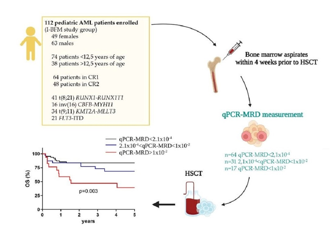
3.2. Survival According to the Type of Remission

Given the retrospective nature of this study, we enrolled patients for whom genetic screening was available at diagnosis and material biobanked before transplant. Therefore, AML harboring t(8;21), inv(16), t(9;11), and FLT3-ITD lesions were available among all institutions. Clinical and molecular details of the patients enrolled in the studies are shown in Table 1 (Figure S2).
Sixty-four out of 112 patients were transplanted in CR1 and 48 in CR2. With a median follow-up after transplant of 3.1 years (range 0.1–16.3 years); 29 patients (26%) died, 15 cases because of transplant-related causes and the remaining 14 cases because of leukemia recurrence. The five-year OS of the entire cohort was 71.4% (95% CI 61.1–79.4), while the 5-year EFS was 67.6% (95% CI 57.1–76) (Figure 1A). The 64 patients transplanted in CR1 showed a 5-year OS of 84% (95% CI 68.9–92.2), while the 48 children who receive HSCT in CR2 had an OS of 53.9% (95% CI 38.3–67.2, p < 0.0001). Similarly, the 5-year EFS was 78.6% (95% CI 63.4–88) for patients given HSCT in CR1 and 52.3% (95% CI 36.9–65.6) for CR2 patients (p = 0.0005, Figure 1B). TRM was 13.5% and CIR was 17.1% for the whole cohort (Figure 1C); TRM significantly increased to 24.4% (95% CI 12.9–37.9) for patients transplanted in CR2 with respect to a value of 8.8% observed in patients given HSCT in CR1 (95% CI 2.4–20.4, p = 0.0012, Figure 1D). CIR was 12.7% (95% CI 5.5–23.3) and 23.3% (95% CI 12.3–36.2) for CR1 and CR2 patients, respectively (p = 0.079, Figure 1E). The source of stem cells did not have impact on outcome, whereas the type of donor did; indeed, patients transplanted from a matched-related donor had a better OS as compared with those transplanted from a matched unrelated donor, as observed in those few ones who received an autologous transplant (n = 8) (p = 0.034, Figure S3A,B). Moreover, there was no influence of the type of conditioning regimen used (all enrolled patients received a myeloablative preparation) (Figure S3C). When considering age, we did not find significant impact on outcomes (Figure S4).

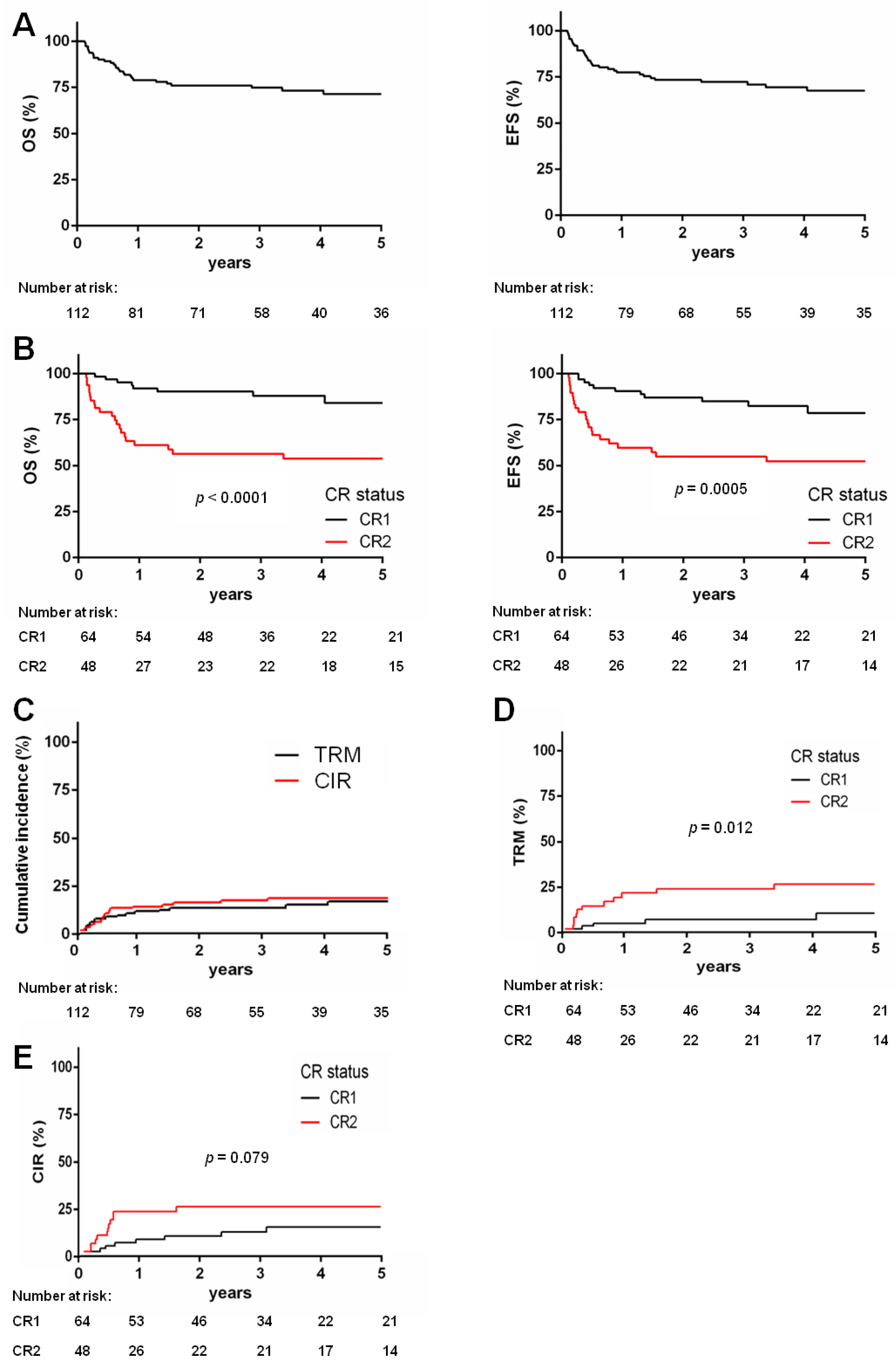
3.3. qPCR-MRD Threshold 2.1 × 10⁻⁴ Is Associated with HSCT Outcome

Evaluable pre-HSCT BM samples collected within 4 weeks before HSCT were measured for residual disease by qPCR. Given the lack of reported qPCR-MRD threshold associated with HSCT outcomes, we performed an ROC analysis, finding that qPCR-MRD values below 2.1 × 10⁴ (or undetectable associated to values below the limit of detection) for 64 patients (57%) was associated with an improved outcome. Indeed, those 64 patients with qPCR-MRD values below 2.1 × 10⁴ had a 5-year OS of 83.7% (95% CI 71.7–90.9), whereas those with qPCR-MRD values above the cut-off had an OS of 57.1% (95% CI 40.6–70.6, p = 0.008, Figure 2A) Similar significant results were obtained for EFS in these two groups (p = 0.007, Figure 2A). Eighteen (16%) patients relapsed at a median time of 5.2 months after transplantation (75% within the first year). Among them, children who had qPCR-MRD values above 2.1 × 10⁴ had a higher risk to relapse (26%) as compared with those with lower qPCR-MRD values (qPCR-MRD < 2.1 × 10⁴, 10%, p = 0.031, Figure 2B). Of note, the type of genetic lesion, namely CBFr, KMT2Ar, and FLT3-ITD, did not correlate with outcome (p = ns, Figure 2C and Figure S5A). Analyzing the influence of qPCR-MRD values on outcome for each single rearrangement, we found that qPCR-MRD values below 2.1 × 10⁴ significantly influenced OS and EFS in FLT3-ITD patients (OS 100% versus 62.5%, p = 0.019, Figure 2D, EFS in Figure S5B), and in t(8;21)RUNX1::RUNX1T1-rearranged patients (OS 83.8% versus 49.7%, p = 0.045, Figure 2E, EFS in Figure S5C). On the contrary, for the remaining genetic lesions, the qPCR-MRD value did not play a significant role (Figure S5D–F), however, the power of these analyses is limited by a reduced sample size.

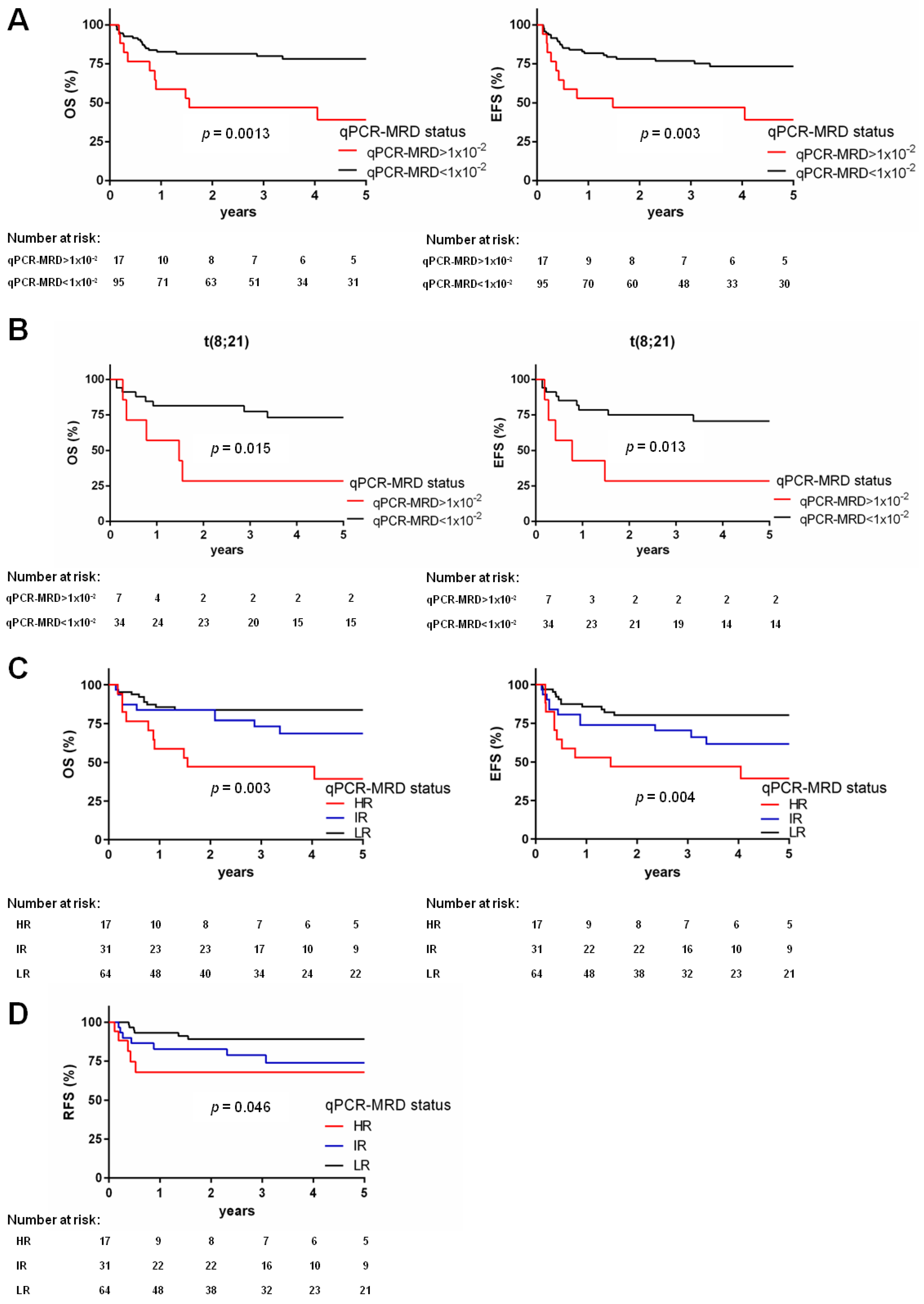
3.4. qPCR-MRD Threshold 1 × 10⁻² Refines HSCT Outcome Prediction

Among the patients who underwent HSCT with high detectable qPCR-MRD values (n = 48), we observed very heterogeneous qPCR-MRD values. Thus, we stratified patients at a higher qPCR-MRD threshold, namely qPCR-MRD values above 1 × 10². This value identified a cohort of 17 very high-risk patients (HR) with worse OS and EFS (OS 39.2%, versus 78.2% for the other 95 patients, p = 0.0013, and EFS 39.2% versus 73.4%, p = 0.003, Figure 3A). This cut-off clearly defined that these patients (15% of the total cohort) underwent HSCT with a low qPCR-MRD reduction, resulting in a dismal outcome. These patients were equally distributed in CR1 (n = 8) or CR2 (n = 9); they were heterogeneous for genetic markers (t(8;21) n = 7, inv(16) n = 2, t(9;11) n = 5, and FLT3-ITD n = 3), and for the type of HSCT received (MUD n = 8, matched family donor n = 9) (Table S1). Analyzing outcomes according to the 1 × 10² qPCR-MRD reduction threshold in each genetic class, we found that among the HR patients for qPCR-MRD before HSCT, those patients harboring t(8;21)RUNX1::RUNX1T1 had the worst prognosis (OS 28.6% for qPCR-MRD values above 1 × 10² versus 73.2% for qPCR-MRD values below 1 × 10², p = 0.015; and EFS 28.6% versus 70.7%, respectively, p = 0.013, Figure 3B); notably, most of these cases received HSCT while in CR2 (six out of seven cases). Regarding FLT3-ITD, t(9;11)KMT2A::MLLT3, and inv(16)CBFB::MYH11 only three, five, and two cases, respectively, were included in the HR class (Table S1) because of a very good blast clearance and transplantation in CR1 for most of these patients (Figures S2 and S6).

3.5. Model of Pre-HSCT Risk Stratification by qPCR-MRD

With the purpose to better predict outcome after HSCT, we analyzed the influence of these two different qPCR-MRD thresholds on outcome, proposed a model where the two significant cut-offs generated three risk groups, namely low (LR), intermediate (IR), or high (HR). This stratification defined patients with qPCR-MRD negative or <2.1 × 10⁴ values as belonging to the LR group (n = 64, 57% of the whole cohort); in this group, 6 out of 64 (9%) patients experienced relapse and ten patients died. Seventeen patients with qPCR-MRD values above 1 × 10² (15%) were allocated to the HR group, 5/17 patients relapsed (29%) experiencing a poor outcome (EFS 39.2%) (Table S2). Finally, in the IR group (n = 31, 28%), the qPCR-MRD values were in between the two cut-offs; 7 out of these 31 patients (22%) had a relapse. In detail, the estimated 5-year OS of the three risk groups was 83.7% for LR, 68.6% for IR, and 39.2% for HR, while the 5-year EFS was 80.2%, 61.6%, and 39.2% (p = 0.003 and p = 0.004, respectively, Figure 3C). Notably, this model confirmed a significantly reduced relapse-free survival for those patients with higher qPCR-MRD values (HR, RFS = 67.9%; IR, RFS = 73.9%; LR, RFS = 89.2%, p = 0.046, Figure 3D and Figure S7). Finally, by multivariate Cox regression analysis, our proposed qPCR-MRD stratification model was found to be an independent factor (hazard ratio 0.47, p = 0.0011) together with disease status at time of allograft (hazard ratio 4.5 for CR2, p = 0.0006) influencing OS and EFS (Table S3).

4. Discussion

Advances in genetic profiling have, in part, clarified the complex dynamics of AML. Moreover, a large number of molecular markers that not only influence risk stratification at diagnosis, but also may help in monitoring response to therapy are now available [18]. In this regard, protocols used by different cooperative groups differ in genetic marker selection [30,31], risk stratification, MRD monitoring, and indications to HSCT [24,32]. In this study, we managed some issues related to qPCR-MRD, trying to assess whether it could improve risk stratification to ultimately personalize post-transplant strategies. We assessed and established optimal qPCR assays validated independently by five reference laboratories being included in the BFM-pediatric AML study group (Italy, Germany, Sweden, the Netherlands, and Denmark), following the adult AML landmark experience in this methodology implementation, based on the Europe Against Cancer (EAC) program and the European Leukemia Network (ELN) [31,33,34,35]. We defined a common interpretation of qPCR-MRD, resulting into high sensitivity and high specificity to detect blasts and determining MRD values, bypassing the more heterogeneous results obtained by multiparameter flow cytometry analysis (MFC) due to site-specific differences in MFC methodology and operators, antigens, and fluorochromes used, methods for cell lysis, number of events collected, and AML immunophenotype shift [36]. This collaborative international study retrospectically collected qPCR-MRD values of 112 AML cases harboring a recurrent molecular marker, being monitored before transplant received in CR1 for high-risk features or in CR2 after a disease recurrence, accordingly to each different national trial stratification. The associations between qPCR-MRD and post-HSCT relapse and outcome were robust, and seen within all cases, regardless of the genetic risk and trial the patients were enrolled in. However, given the nature of this study of qPCR-MRD validation, we were unable to account for inter-study differences in the selection of patients for HSCT, different post-remission treatments, heterogeneity in transplant conditioning regimens, graft characteristics, and immunosuppression.
We found that patients with undetectable qPCR-MRD had a superior long-term survival, independent from the state of CR1 or CR2 remission at HSCT, the type of conditioning regimen, donor employed, and genetic rearrangement and that the use of HSCT in CR2 was independently associated with a worse outcome in this subgroup of AML patients [37,38,39,40,41,42,43]. We documented that detectable higher qPCR-MRD values had an overall adverse effect on transplant outcome. In particular, we determined that the most informative cut-off (2.1 × 10⁻⁴) was able to discriminate patients with a low risk to relapse (10.4%) and an extremely good OS (82.8%). We also explored a higher cut-off equivalent to 1 × 10⁻² of qPCR-MRD that allowed the identification of a group of 17 patients with high qPCR-MRD values (>1 × 10⁻²) and very poor OS (i.e., below 40%). Thanks to this novel partitioning of AML cases according to qPCR-MRD values, we proposed a pre-HSCT allocation to low-, intermediate-, and high-risk groups. This stratification, supported by relapse free survival, could help personalizing transplant management in order to improve clinical outcome. Of interest, the 17 patients defined as HR cases could be recognized exclusively by the qPCR-MRD values. Notably, analyzing subgroup of patients according to their genetic lesion, we showed that qPCR-MRD reduction in t(8;21)-rearranged (n = 41) and FLT3-ITD (n = 21) patients significantly predicted patients outcome, as previously found after induction course [14,29,44]. For the KMT2A-rearranged AML cases (n = 34), we did not find a significant correlation with qPCR-MRD values; however, most of these patients had low qPCR-MRD values at the time of HSCT. This observation of a good qPCR-MRD reduction by both the FLT3-ITD and t(9;11)KMT2A::MLLT3 subgroups, together with the fact that, in this setting, patients were mainly transplanted in CR1, fosters the relevance of CR1 and qPCR-MRD in planning an HSCT with high rate of success in both subgroups [7]. Management of patients with low qPCR-MRD reduction remains controversial, and a key question is whether the type of conditioning can affect residual disease, and thereby improve outcome. Studies mostly in adult AML patients have provided conflicting results due to specific transplant conditioning [45,46]. A recent report by Hourigan and colleagues on adult AML patients in CR randomized to receive either myeloablative or reduced-intensity conditioning (RIC) documented that patients with measurable MRD had an increased risk of relapse if treated with RIC [47]. Here, all pediatric patients received a myeloablative treatment, and we observed no effect on survival according to different types of conditioning.
Our study establishes the importance of pre-HSCT qPCR-MRD, attempting a qPCR-MRD based risk-stratification to identify the majority of patients experiencing adverse events after HSCT, and supports that they might be discussed for alternative approaches either before (such as the use of refined conditioning regimens, immunotherapy, or targeted therapy to reduce the qPCR-MRD value) [48] or after HSCT (such as the rapid tapering of immunosuppression, the use of donor-lymphocyte infusions, or specific agents such as tyrosine-kinase inhibitors).
Finally, this study supports that MRD detection has strong clinical relevance during the whole AML course and that technological advances in the molecular field allowed to increase sensitivity and specificity, making it an essential tool with the ability to complement morphology and immunophenotypic analysis, particularly for compelling clinical decisions. However, since previous studies have shown a high concordance between qPCR and MFC residual disease values, we support that the prognostic role of MRD is not influenced by the detection method [15,29], thus, we consider the MRD risk model described here to be a further tool to be validated in larger cohorts.

5. Conclusions

This international retrospective I-BFM-AML group study, regardless of its limitations, strengthens the need for collaborative networks and the comparison with other techniques supports the role of pediatric residual disease monitoring before HSCT to refine therapeutic decisions. Furthermore, new challenges in molecular approaches to improve qPCR are being faced by using next-generation sequencing [49,50], where multiplex quantification of mutations [51] could improve the ability to track molecular AML residual disease, as well as cover all patients lacking, for now, a qPCR marker.

Supplementary Materials

The following supporting information can be downloaded at: https://www.mdpi.com/article/10.3390/biomedicines10071530/s1. Figure S1: Inter-laboratory quality control (QC); Figure S2: Schematic diagram showing the number of patients in each part of the study; Figure S3: Effect of transplant-related variables on survival; Figure S4: Impact of patients’ age on clinical outcome; Figure S5: Genetics and qPCR-MRD status role in HSCT; Figure S6: Bar representation of the percentage of patients belonging to LR, IR and HR groups according to their genetic lesions; Figure S7: Relapse-free survival from HSCT according to qPCR-MRD values; Table S1: Clinical and biological characteristics of high risk patients for qPCR-MRD reduction before HSCT; Table S2: Clinical, molecular, and transplant variables in each qPCR-MRD-defined group; Table S3: Multivariate analyses.

Author Contributions

Conceptualization, M.P., F.L., H.H., D.R., N.V.N., L.F. and J.A.; methodology, M.B., C.W., M.H., E.S., C.T., A.D.R. and K.P.; formal analysis, P.M., L.S. and M.B.; investigation, M.B. and M.P.; resources, F.L. and M.P.; data curation, P.M.; writing—original draft preparation, M.B. and P.M.; writing—review and editing, M.P. and F.L.; funding acquisition, F.L. and M.P. All authors have read and agreed to the published version of the manuscript.

Funding

This work was supported by grants from Associazione Italiana Contro Leucemia Linfomi e Mieloma (AIL-Treviso section) to M.P., from the AIRC Fundation (Associazione Italiana Ricerca sul Cancro, Special Grant “5xmille”−9962 to F.L., AND AIRC-IG20562 to M.P.). M.B. is supported by an AIRC fellowship for Italy. C.T. is supported by Fondazione Umberto Veronesi.

Institutional Review Board Statement

Children with AML were enrolled by three different consortia, Associazione Italiana Ematologia Oncologia Pediatrica (AIEOP AML2002/01 trial, EudraCT:2014-000976-25), Berlin-Frankfurt-Münster (AML-BFM 2012 trial, EudraCT:2013-000018-39), and Nordic Society of Paediatric Haematology and Oncology (NOPHO AML-2004 or NOPHO-DBH AML-2012 trials), and treated according to national clinical trials. The approval for this study was obtained from the institutional review board of each participating center.

Informed Consent Statement

In accordance with the Declaration of Helsinki, informed consent to provide samples for biological studies was obtained from patients’ parents or legal guardians.

Data Availability Statement

Data sharing is not applicable to this article as no datasets were generated or analyzed during the current study.

Acknowledgments

We thank the AIEOP reference laboratory of Padova for morphology, cytogenetic, and immunophenotype analyses, and the BioBanking (BBOP) of children’s samples enrolled in the AML 2002/01 protocol at the Hematology-Oncology Clinic, University-Hospital of Padova, the staff at the unit for gene analyses at the Department of Clinical Chemistry, Sahlgrenska University Hospital. Our thanks also to all clinicians working at the participating AIEOP, BMF, and NOPHO centers. This manuscript is in loving memory of Professor Giuseppe Basso.

Conflicts of Interest

The authors declare no conflict of interest.

References

  1. Döhner, H.; Weisdorf, D.J.; Bloomfield, C.D. Acute Myeloid Leukemia. N. Engl. J. Med. 2015, 373, 1136–1152. [Google Scholar] [CrossRef] [Green Version]
  2. Rasche, M.; Zimmermann, M.; Borschel, L.; Bourquin, J.-P.; Dworzak, M.; Klingebiel, T.; Lehrnbecher, T.; Creutzig, U.; Klusmann, J.-H.; Reinhardt, D. Successes and Challenges in the Treatment of Pediatric Acute Myeloid Leukemia: A Retrospective Analysis of the AML-BFM Trials from 1987 to 2012. Leukemia 2018, 32, 2167–2177. [Google Scholar] [CrossRef] [PubMed] [Green Version]
  3. Pession, A.; Masetti, R.; Rizzari, C.; Putti, M.C.; Casale, F.; Fagioli, F.; Luciani, M.; Lo Nigro, L.; Menna, G.; Micalizzi, C.; et al. Results of the AIEOP AML 2002/01 Multicenter Prospective Trial for the Treatment of Children with Acute Myeloid Leukemia. Blood 2013, 122, 170–178. [Google Scholar] [CrossRef] [PubMed] [Green Version]
  4. Kaspers, G.J.L.; Zimmermann, M.; Reinhardt, D.; Gibson, B.E.S.; Tamminga, R.Y.J.; Aleinikova, O.; Armendariz, H.; Dworzak, M.; Ha, S.Y.; Hasle, H.; et al. Improved Outcome in Pediatric Relapsed Acute Myeloid Leukemia: Results of a Randomized Trial on Liposomal Daunorubicin by the International BFM Study Group. J. Clin. Oncol. 2013, 31, 599–607. [Google Scholar] [CrossRef] [PubMed]
  5. O’Hare, P.; Lucchini, G.; Cummins, M.; Veys, P.; Potter, M.; Lawson, S.; Vora, A.; Wynn, R.; Peniket, A.; Kirkland, K.; et al. Allogeneic Stem Cell Transplantation for Refractory Acute Myeloid Leukemia in Pediatric Patients: The UK Experience. Bone Marrow Transpl. 2017, 52, 825–831. [Google Scholar] [CrossRef] [Green Version]
  6. Lucchini, G.; Labopin, M.; Beohou, E.; Dalissier, A.; Dalle, J.H.; Cornish, J.; Zecca, M.; Samarasinghe, S.; Gibson, B.; Locatelli, F.; et al. Impact of Conditioning Regimen on Outcomes for Children with Acute Myeloid Leukemia Undergoing Transplantation in First Complete Remission. An Analysis on Behalf of the Pediatric Disease Working Party of the European Group for Blood and Marrow Transplanta. Biol. Blood Marrow Transpl. 2017, 23, 467–474. [Google Scholar] [CrossRef] [Green Version]
  7. Locatelli, F.; Masetti, R.; Rondelli, R.; Zecca, M.; Fagioli, F.; Rovelli, A.; Messina, C.; Lanino, E.; Bertaina, A.; Favre, C.; et al. Outcome of Children with High-Risk Acute Myeloid Leukemia given Autologous or Allogeneic Hematopoietic Cell Transplantation in the Aieop AML-2002/01 Study. Bone Marrow Transpl. 2015, 50, 181–188. [Google Scholar] [CrossRef] [Green Version]
  8. Ley, T.J.; Miller, C.; Ding, L.; Raphael, B.J.; Mungall, A.J.; Robertson, G.; Hoadley, K.; Triche, T.J.; Laird, P.W.; Baty, J.D.; et al. Genomic and Epigenomic Landscapes of Adult de Novo Acute Myeloid Leukemia. N. Engl. J. Med. 2013, 368, 2059–2074. [Google Scholar] [CrossRef] [Green Version]
  9. Papaemmanuil, E.; Gerstung, M.; Bullinger, L.; Gaidzik, V.I.; Paschka, P.; Roberts, N.D.; Potter, N.E.; Heuser, M.; Thol, F.; Bolli, N.; et al. Genomic Classification and Prognosis in Acute Myeloid Leukemia. N. Engl. J. Med. 2016, 374, 2209–2221. [Google Scholar] [CrossRef]
  10. Tyner, J.W.; Tognon, C.E.; Bottomly, D.; Wilmot, B.; Kurtz, S.E.; Savage, S.L.; Long, N.; Schultz, A.R.; Traer, E.; Abel, M.; et al. Functional Genomic Landscape of Acute Myeloid Leukaemia. Nature 2018, 562, 526–531. [Google Scholar] [CrossRef]
  11. Leung, W.; Pui, C.H.; Coustan-Smith, E.; Yang, J.; Pei, D.; Gan, K.; Srinivasan, A.; Hartford, C.; Triplett, B.M.; Dallas, M.; et al. Detectable Minimal Residual Disease before Hematopoietic Cell Transplantation Is Prognostic but Does Not Preclude Cure for Children with Very-High-Risk Leukemia. Blood 2012, 120, 468–472. [Google Scholar] [CrossRef] [PubMed] [Green Version]
  12. Grimwade, D.; Freeman, S.D. Defining Minimal Residual Disease in Acute Myeloid Leukemia: Which Platforms Are Ready for “Prime Time”? Blood 2014, 124, 3345–3355. [Google Scholar] [CrossRef] [PubMed] [Green Version]
  13. Dillon, R.; Hills, R.; Freeman, S.; Potter, N.; Jovanovic, J.; Ivey, A.; Kanda, A.S.; Runglall, M.; Foot, N.; Valganon, M.; et al. Molecular MRD Status and Outcome after Transplantation in NPM1-Mutated AML. Blood 2020, 135, 680–688. [Google Scholar] [CrossRef] [PubMed] [Green Version]
  14. Pigazzi, M.; Manara, E.; Buldini, B.; Beqiri, V.; Bisio, V.; Tregnago, C.; Rondelli, R.; Masetti, R.; Caterina Putti, M.; Fagioli, F.; et al. Minimal Residual Disease Monitored after Induction Therapy by Rq-Pcr Can Contribute to Tailor Treatment of Patients with t(8;21) Runx1-Runx1t1 Rearrangement. Haematologica 2015, 100, e99–e101. [Google Scholar] [CrossRef]
  15. Buldini, B.; Rizzati, F.; Masetti, R.; Fagioli, F.; Menna, G.; Micalizzi, C.; Putti, M.C.; Rizzari, C.; Santoro, N.; Zecca, M.; et al. Prognostic Significance of Flow-Cytometry Evaluation of Minimal Residual Disease in Children with Acute Myeloid Leukaemia Treated according to the AIEOP-AML 2002/01 Study Protocol. Br. J. Haematol. 2017, 177, 116–126. [Google Scholar] [CrossRef]
  16. Rubnitz, J.E.; Inaba, H.; Dahl, G.; Ribeiro, R.C.; Bowman, W.P.; Taub, J.; Pounds, S.; Razzouk, B.I.; Lacayo, N.J.; Cao, X.; et al. Minimal Residual Disease-Directed Therapy for Childhood Acute Myeloid Leukaemia: Results of the AML02 Multicentre Trial. Lancet Oncol. 2010, 11, 543–552. [Google Scholar] [CrossRef] [Green Version]
  17. Van Der Velden, V.H.J.; Van Der Sluijs-Geling, A.; Gibson, B.E.S.; Te Marvelde, J.G.; Hoogeveen, P.G.; Hop, W.C.J.; Wheatley, K.; Bierings, M.B.; Schuurhuis, G.J.; De Graaf, S.S.N.; et al. Clinical Significance of Flowcytometric Minimal Residual Disease Detection in Pediatric Acute Myeloid Leukemia Patients Treated according to the DCOG ANLL97/MRC AML12 Protocol. Leukemia 2010, 24, 1599–1606. [Google Scholar] [CrossRef] [Green Version]
  18. Bolouri, H.; Farrar, J.E.; Triche, T.; Ries, R.E.; Lim, E.L.; Alonzo, T.A.; Ma, Y.; Moore, R.; Mungall, A.J.; Marra, M.A.; et al. The Molecular Landscape of Pediatric Acute Myeloid Leukemia Reveals Recurrent Structural Alterations and Age-Specific Mutational Interactions. Nat. Med. 2018, 24, 103–112. [Google Scholar] [CrossRef] [Green Version]
  19. Grimwade, D. The Changing Paradigm of Prognostic Factors in Acute Myeloid Leukaemia. Best Pract. Res. Clin. Haematol. 2012, 25, 419–425. [Google Scholar] [CrossRef]
  20. Cheson, B.D.; Bennett, J.M.; Kopecky, K.J.; Büchner, T.; Willman, C.L.; Estey, E.H.; Schiffer, C.A.; Doehner, H.; Tallman, M.S.; Lister, T.A.; et al. Revised Recommendations of the International Working Group for Diagnosis, Standardization of Response Criteria, Treatment Outcomes, and Reporting Standards for Therapeutic Trials in Acute Myeloid Leukemia. J. Clin. Oncol. 2003, 21, 4642–4649. [Google Scholar] [CrossRef]
  21. Czyz, A.; Nagler, A. The Role of Measurable Residual Disease (MRD) in Hematopoietic Stem Cell Transplantation for Hematological Malignancies Focusing on Acute Leukemia. Int. J. Mol. Sci. 2019, 20, 5362. [Google Scholar] [CrossRef] [PubMed] [Green Version]
  22. Buckley, S.A.; Wood, B.L.; Othus, M.; Hourigan, C.S.; Ustun, C.; Linden, M.A.; Defor, T.E.; Malagola, M.; Anthias, C.; Valkova, V.; et al. Minimal Residual Disease Prior to Allogeneic Hematopoietic Cell Transplantation in Acute Myeloid Leukemia: A Meta-Analysis. Haematologica 2017, 102, 865–873. [Google Scholar] [CrossRef] [PubMed] [Green Version]
  23. Walter, R.B.; Gooley, T.A.; Wood, B.L.; Milano, F.; Fang, M.; Sorror, M.L.; Estey, E.H.; Salter, A.I.; Lansverk, E.; Chien, J.W.; et al. Impact of Pretransplantation Minimal Residual Disease, as Detected by Multiparametric Flow Cytometry, on Outcome of Myeloablative Hematopoietic Cell Transplantation for Acute Myeloid Leukemia. J. Clin. Oncol. 2011, 29, 1190–1197. [Google Scholar] [CrossRef] [Green Version]
  24. Kayser, S.; Benner, A.; Thiede, C.; Martens, U.; Huber, J.; Stadtherr, P.; Janssen, J.W.G.; Röllig, C.; Uppenkamp, M.J.; Bochtler, T.; et al. Pretransplant NPM1 MRD Levels Predict Outcome after Allogeneic Hematopoietic Stem Cell Transplantation in Patients with Acute Myeloid Leukemia. Blood Cancer J. 2016, 6, e449. [Google Scholar] [CrossRef] [PubMed]
  25. Juul-Dam, K.L.; Ommen, H.B.; Nyvold, C.G.; Walter, C.; Vålerhaugen, H.; Kairisto, V.; Abrahamsson, J.; Alm, S.J.; Jahnukainen, K.; Lausen, B.; et al. Measurable Residual Disease Assessment by QPCR in Peripheral Blood Is an Informative Tool for Disease Surveillance in Childhood Acute Myeloid Leukaemia. Br. J. Haematol. 2020, 190, 198–208. [Google Scholar] [CrossRef] [PubMed]
  26. Van Dongen, J.J.M.; Macintyre, E.A.; Gabert, J.A.; Delabesse, E.; Rossi, V.; Saglio, G.; Gottardi, E.; Rambaldi, A.; Dotti, G.; Griesinger, F.; et al. Standardized RT-PCR Analysis of Fusion Gene Transcripts from Chromosome Aberrations in Acute Leukemia for Detection of Minimal Residual Disease. Report of the BIOMED-1 Concerted Action: Investigation of Minimal Residual Disease in Acute Leukemia. Leukemia 1999, 13, 1901–1928. [Google Scholar] [CrossRef]
  27. Pigazzi, M.; Manara, E.; Bisio, V.; Aveic, S.; Masetti, R.; Menna, G.; Zecca, M.; Pession, A.; Locatelli, F.; Basso, G. Screening of Novel Genetic Aberrations in Pediatric Acute Myeloid Leukemia: A Report from the AIEOP AML-2002 Study Group. Blood 2012, 120, 3860–3862. [Google Scholar] [CrossRef] [Green Version]
  28. Pigazzi, M.; Masetti, R.; Bresolin, S.; Beghin, A.; Di Meglio, A.; Gelain, S.; Trentin, L.; Baron, E.; Giordan, M.; Zangrando, A.; et al. MLL Partner Genes Drive Distinct Gene Expression Profiles and Genomic Alterations in Pediatric Acute Myeloid Leukemia: An AIEOP Study. Leukemia 2011, 25, 560–563. [Google Scholar] [CrossRef] [Green Version]
  29. Manara, E.; Basso, G.; Zampini, M.; Buldini, B.; Tregnago, C.; Rondelli, R.; Masetti, R.; Bisio, V.; Frison, M.; Polato, K.; et al. Characterization of Children with FLT3-ITD Acute Myeloid Leukemia: A Report from the AIEOP AML-2002 Study Group. Leukemia 2017, 31, 18–25. [Google Scholar] [CrossRef] [Green Version]
  30. Testi, A.M.; Pession, A.; Diverio, D.; Grimwade, D.; Gibson, B.; Cardoso de Azevedo, A.; Moran, L.; Leverger, G.; Elitzur, S.; Hasle, H.; et al. Risk-Adapted Treatment of Acute Promyelocytic Leukemia: Results from the International Consortium for Childhood APL. Blood 2018, 132, 405–412. [Google Scholar] [CrossRef]
  31. Grimwade, D.; Vyas, P.; Freeman, S. Assessment of Minimal Residual Disease in Acute Myeloid Leukemia. Curr. Opin. Oncol. 2010, 22, 656–663. [Google Scholar] [CrossRef] [PubMed]
  32. Kayser, S.; Schlenk, R.F.; Grimwade, D.; Yosuico, V.E.D.; Walter, R.B. Evidence-Based Focused Review Minimal Residual Disease—Directed Therapy in Acute Myeloid Leukemia. Blood 2015, 125, 2331–2336. [Google Scholar] [CrossRef] [PubMed] [Green Version]
  33. Gabert, J.; Beillard, E.; van der Velden, V.H.J.; Bi, W.; Grimwade, D.; Pallisgaard, N.; Barbany, G.; Cazzaniga, G.; Cayuela, J.M.; Cavé, H.; et al. Standardization and Quality Control Studies of “real Time” Quantitative Reverse Transcriptase Polymerase Chain Reaction of Fusion Gene Transcripts for Residual Disease Detection in Leukemia—A Europe Against Cancer Program. Leukemia 2003, 17, 2318–2357. [Google Scholar] [CrossRef] [PubMed]
  34. Döhner, H.; Estey, E.; Grimwade, D.; Amadori, S.; Appelbaum, F.R.; Ebert, B.L.; Fenaux, P.; Larson, R.A.; Levine, R.L.; Lo-coco, F.; et al. Diagnosis and Management of AML in Adults: 2017 ELN Recommendations from an International Expert Panel. Blood 2017, 129, 424–448. [Google Scholar] [CrossRef] [Green Version]
  35. Heuser, M.; Freeman, S.D.; Ossenkoppele, G.J.; Buccisano, F.; Hourigan, C.S.; Ngai, L.L.; Tettero, J.M.; Bachas, C.; Baer, C.; Béné, M.C.; et al. 2021 Update on MRD in Acute Myeloid Leukemia: A Consensus Document from the European LeukemiaNet MRD Working Party. Blood 2021, 138, 2753–2767. [Google Scholar] [CrossRef]
  36. Buccisano, F.; Maurillo, L.; Del Principe, M.I.; Del Poeta, G.; Sconocchia, G.; Lo-Coco, F.; Arcese, W.; Amadori, S.; Venditti, A. Prognostic and Therapeutic Implications of Minimal Residual Disease Detection in Acute Myeloid Leukemia. Blood 2012, 119, 332–341. [Google Scholar] [CrossRef] [Green Version]
  37. Sison, E.A.R.; Brown, P. Does Hematopoietic Stem Cell Transplantation Benefit Infants with Acute Leukemia? Hematol. Am. Soc. Hematol. Educ. Program 2013, 2013, 601–604. [Google Scholar] [CrossRef] [Green Version]
  38. Fagioli, F.; Zecca, M.; Locatelli, F.; Lanino, E.; Uderzo, C.; Di Bartolomeo, P.; Berger, J.M.; Favre, C.; Rondelli, R.; Pession, A.; et al. Allogeneic Stem Cell Transplantation for Children with Acute Myeloid Leukemia in Second Complete Remission. J. Pediatr. Hematol. Oncol. 2008, 30, 575–583. [Google Scholar] [CrossRef] [Green Version]
  39. Sander, A.; Zimmermann, M.; Dworzak, M.; Fleischhack, G.; Von Neuhoff, C.; Reinhardt, D.; Kaspers, G.J.L.; Creutzig, U. Consequent and Intensified Relapse Therapy Improved Survival in Pediatric AML: Results of Relapse Treatment in 379 Patients of Three Consecutive AML-BFM Trials. Leukemia 2010, 24, 1422–1428. [Google Scholar] [CrossRef] [Green Version]
  40. Karlsson, L.; Forestier, E.; Hasle, H.; Jahnukainen, K.; Jónsson, Ó.G.; Lausen, B.; Norén Nyström, U.; Palle, J.; Tierens, A.; Zeller, B.; et al. Outcome after Intensive Reinduction Therapy and Allogeneic Stem Cell Transplant in Paediatric Relapsed Acute Myeloid Leukaemia. Br. J. Haematol. 2017, 178, 592–602. [Google Scholar] [CrossRef] [Green Version]
  41. Creutzig, U.; Zimmermann, M.; Ritter, J.; Reinhardt, D.; Hermann, J.; Henze, G.; Jürgens, H.; Kabisch, H.; Reiter, A.; Riehm, H.; et al. Treatment Strategies and Long-Term Results in Paediatric Patients Treated in Four Consecutive AML-BFM Trials. Leukemia 2005, 19, 2030–2042. [Google Scholar] [CrossRef] [PubMed] [Green Version]
  42. Gibson, B.E.S.; Webb, D.K.H.; Howman, A.J.; de Graaf, S.S.N.; Harrison, C.J.; Wheatley, K. Results of a Randomized Trial in Children with Acute Myeloid Leukaemia: Medical Research Council AML12 Trial. Br. J. Haematol. 2011, 155, 366–376. [Google Scholar] [CrossRef] [PubMed]
  43. Nakayama, H.; Tabuchi, K.; Tawa, A.; Tsukimoto, I.; Tsuchida, M.; Morimoto, A.; Yabe, H.; Horibe, K.; Hanada, R.; Imaizumi, M.; et al. Outcome of Children with Relapsed Acute Myeloid Leukemia Following Initial Therapy under the AML99 Protocol. Int. J. Hematol. 2014, 100, 171–179. [Google Scholar] [CrossRef]
  44. Viehmann, S.; Teigler-Schlegel, A.; Bruch, J.; Langebrake, C.; Reinhardt, D.; Harbott, J. Monitoring of Minimal Residual Disease (MRD) by Real-Time Quantitative Reverse Transcription PCR (RQ-RT-PCR) in Childhood Acute Myeloid Leukemia with AML1/ETO Rearrangement. Leukemia 2003, 17, 1130–1136. [Google Scholar] [CrossRef] [Green Version]
  45. Walter, R.B.; Gyurkocza, B.; Storer, B.E.; Godwin, C.D.; Pagel, J.M.; Buckley, S.A.; Sorror, M.L.; Wood, B.L.; Storb, R.; Appelbaum, F.R.; et al. Comparison of Minimal Residual Disease as Outcome Predictor for AML Patients in First Complete Remission Undergoing Myeloablative or Nonmyeloablative Allogeneic Hematopoietic Cell Transplantation. Leukemia 2015, 29, 137–144. [Google Scholar] [CrossRef] [PubMed] [Green Version]
  46. Ustun, C.; Courville, E.L.; DeFor, T.; Dolan, M.; Randall, N.; Yohe, S.; Bejanyan, N.; Warlick, E.; Brunstein, C.; Weisdorf, D.J.; et al. Myeloablative, but Not Reduced-Intensity, Conditioning Overcomes the Negative Effect of Flow-Cytometric Evidence of Leukemia in Acute Myeloid Leukemia. Biol. Blood Marrow Transpl. 2016, 22, 669–675. [Google Scholar] [CrossRef] [Green Version]
  47. Hourigan, C.S.; Dillon, L.W.; Gui, G.; Logan, B.R.; Fei, M.; Ghannam, J.; Li, Y.; Licon, A.; Alyea, E.P.; Bashey, A.; et al. Impact of Conditioning Intensity of Allogeneic Transplantation for Acute Myeloid Leukemia with Genomic Evidence of Residual Disease. J. Clin. Oncol. 2020, 38, 1273–1283. [Google Scholar] [CrossRef]
  48. Pastore, F.; Dufour, A.; Benthaus, T.; Metzeler, K.H.; Maharry, K.S.; Schneider, S.; Ksienzyk, B.; Mellert, G.; Zellmeier, E.; Kakadia, P.M.; et al. Combined Molecular and Clinical Prognostic Index for Relapse and Survival in Cytogenetically Normal Acute Myeloid Leukemia. J. Clin. Oncol. 2014, 32, 1586–1594. [Google Scholar] [CrossRef] [Green Version]
  49. Hayes, D.N.; Kim, W.Y.; Hayes, D.N.; Kim, W.Y. The next Steps in Next-Gen Sequencing of Cancer Genomes Find the Latest Version: The next Steps in next-Gen Sequencing of Cancer Genomes. J. Clin. Invest. 2015, 125, 462–468. [Google Scholar] [CrossRef] [Green Version]
  50. Delsing Malmberg, E.; Rehammar, A.; Pereira, M.B.; Abrahamsson, J.; Samuelsson, T.; Ståhlman, S.; Asp, J.; Tierens, A.; Palmqvist, L.; Kristiansson, E.; et al. Accurate and Sensitive Analysis of Minimal Residual Disease in Acute Myeloid Leukemia Using Deep Sequencing of Single Nucleotide Variations. J. Mol. Diagnostics 2019, 21, 149–162. [Google Scholar] [CrossRef] [Green Version]
  51. Jongen-Lavrencic, M.; Grob, T.; Hanekamp, D.; Kavelaars, F.G.; Al Hinai, A.; Zeilemaker, A.; Erpelinck-Verschueren, C.A.J.; Gradowska, P.L.; Meijer, R.; Cloos, J.; et al. Molecular Minimal Residual Disease in Acute Myeloid Leukemia. N. Engl. J. Med. 2018, 378, 1189–1199. [Google Scholar] [CrossRef] [PubMed]
Figure 1. Survival of the whole patients’ cohort according to the remission status before HSCT: (A) Kaplan–Meier estimates showing 5-year probability of overall survival (OS, left, survival 71.4%) and event-free survival (EFS, right, survival 67.6%) for the whole cohort of patients enrolled (n = 112); (B) five-year probability of OS (left) for children receiving HSCT in CR1 (n = 64) or CR2 (n = 48) (84% vs. 53.9%, p < 0.0001) and EFS for patients transplanted in CR1 or CR2 (78.6% vs. 52.3%, p = 0.0005); (C) cumulative incidence of treatment-related mortality (TRM) in the whole population of children given HSCT (black line, n = 112, 13.5%), and cumulative incidence of relapse (CIR) in the whole population of AML patients given transplantation (red line, n = 112, 17.1%); (D) cumulative incidence of transplant-related mortality in patients who underwent transplantation in CR1 (n = 64) or CR2 (n = 48) (8.8% vs. 24.4%, p = 0.012); (E) cumulative incidence of relapse in patients who underwent transplantation in CR1 (n = 64) or CR2 (n = 48) (12.7% vs. 23.3%, p = 0.079).
Biomedicines 10 01530 g001
Figure 2. qPCR-MRD values above 2.1 × 10⁻⁴ impact on transplantation outcome: (A) Kaplan–Meier curves showing that patients who underwent HSCT with qPCR-MRD values above 2.1 × 10⁻⁴ (n = 48) have a worse OS with respect to patients receiving transplant with qPCR-MRD values below 2.1 × 10⁻⁴ (n = 64) (57.1% vs. 83.7%, p = 0.008), EFS for the same groups of patients (52.9% vs. 80.2%, p = 0.007); (B) cumulative incidence of relapse for cases with qPCR-MRD values above 2.1 × 10⁻⁴ (n = 48) and patients with qPCR-MRD values below 2.1 × 10⁻⁴ (n = 64) (26% vs. 10%, p = 0.031); (C) five-year OS for patients according to their genetics: CBFr (n = 57), KMT2Ar (n = 34), and FLT3-ITD (n = 21) (survival 69.6% vs. 62.7% vs. 85.5%, respectively, p = 0.39); (D) Kaplan_Meier survival curves of OS for children with FLT3-ITD mutation given transplantation with qPCR-MRD values above 2.1 × 10⁻⁴ (n = 8) or with qPCR-MRD values below 2.1 × 10⁻⁴ (n = 13) (62.5% vs. 100%, p = 0.019); (E) five-year probability of OS for children with t(8;21)RUNX1::RUNX1T1 rearrangement given HSCT with qPCR-MRD values above 2.1 × 10⁻⁴ (n = 21) or with qPCR-MRD values below 2.1 × 10⁻⁴ (n = 20) (49.7% vs. 83.8%, p = 0.045).
Biomedicines 10 01530 g002
Figure 3. Two qPCR-MRD thresholds generate a risk stratification model predicting the survival of patients: (A) Survival estimates for patients receiving HSCT with qPCR-MRD values above 1 × 10⁻² (n = 17) and qPCR-MRD values below 1 × 10⁻² (n = 95). Curves show OS (left, 39.2% vs. 78.2%, p = 0.0013) and EFS (right, 39.2% vs. 73.4%, p = 0.003); (B) five-year survival curves for patients harboring t(8;21)RUNX1::RUNX1T1 rearrangement given HSCT with qPCR-MRD values above 1 × 10⁻² (n = 7) and qPCR-MRD values below 1 × 10⁻² (n = 34): OS (28.6% vs. 73.2%, p = 0.015) and EFS (28.6% vs. 70.7%, p = 0.013); (C) Kaplan–Meier curves of OS or patients who underwent HSCT with qPCR-MRD values above 1 × 10⁻² (HR, n = 17), 2.1 × 10⁻⁴ < qPCR-MRD values < 1 × 10⁻² (IR, n = 31), and qPCR-MRD values below 2.1 × 10⁻⁴ (LR, n = 64) (survival 39.2% vs. 68.6% vs. 83.7%, respectively, p = 0.003) and EFS (survival 39.2% vs. 61.6% vs. 80.2%, p = 0.004); (D) relapse-free survival of patients according to three risk stratification model: qPCR-MRD values above 1 × 10⁻² (HR, n = 17), 2.1 × 10⁻⁴ < qPCR-MRD values < 1 × 10⁻² (IR, n = 31), and qPCR-MRD values below 2.1 × 10⁻⁴ (LR, n = 64) (67.9% vs. 73.9% vs. 89.2%, respectively, p = 0.046).
Biomedicines 10 01530 g003
Table 1. Patients’ characteristics.
AIEOPNOPHOBFMTotal
n° pts633811112
Age (average)7.99.19.88.9
Gender
male3719763
female2619449
WBC (average) (n = 107)50,38259,24552,63553,347
Genetics
Standard risk
t(8;21)RUNX1::RUNX1T12017441
inv(16)CBFB::MYH1186216
High risk
t(9;11)KMT2A::MLLT31910534
FLT3-ITD165021
Karyotype (n = 82)
complex68115
Type of remission
CR1537464
CR21031748
pts status
relapse post HSCT (n°)711018
death (n°)818329
Median Follow-up (years)3.32.31.43.1
range 0.3–140.1–160.1–40.1–16
Type of HSCT (n = 108)
MUD-ALLO2128453
SIBLING-RELATED3410347
AUTOLOGOUS8008
Source (n = 94)
BM4512461
PB1514130
CB3003
Preparative regimen before HSCT
BUS-based5211265
TBI-based6006
other515525
Publisher’s Note: MDPI stays neutral with regard to jurisdictional claims in published maps and institutional affiliations.

