Article

Prevalence and Antimicrobial Susceptibility of Bovine Mycoplasma Species in Egypt

by
Ahmed M. Ammar
1,†,
Marwa I. Abd El-Hamid
1,†,
Yousreya H. Mohamed
2,
Heba M. Mohamed
1,
Dalal H. M. Al-khalifah
3,
Wael N. Hozzein
4,
Samy Selim
5,
Wafaa M. El-Neshwy
6 and
Rania M. S. El-Malt
7,*
1
Department of Microbiology, Faculty of Veterinary Medicine, Zagazig University, Zagazig 44519, Egypt
2
Department of Mycoplasma Research, Animal Health Research Institute, Agriculture Research Center, Giza 12622, Egypt
3
Department of Biology, College of Science, Princess Nourah Bint Abdulrahman University, P.O. Box 84428, Riyadh 11671, Saudi Arabia
4
Botany and Microbiology Department, Faculty of Science, Beni-Suef University, Beni-Suef 62511, Egypt
5
Department of Clinical Laboratory Sciences, College of Applied Medical Sciences, Jouf University, Sakaka 72388, Saudi Arabia
6
Department of Animal Medicine, Infectious Diseases, Faculty of Veterinary Medicine, Zagazig University, Zagazig 44519, Egypt
7
Department of Bacteriology, Zagazig Branch, Animal Health Research Institute, Agriculture Research Center, Zagazig 44516, Egypt
*
Author to whom correspondence should be addressed.
These authors contributed equally to this work.
Biology 2022, 11(7), 1083; https://doi.org/10.3390/biology11071083
Submission received: 26 May 2022 / Revised: 13 July 2022 / Accepted: 15 July 2022 / Published: 20 July 2022
(This article belongs to the Special Issue Microbial Diversity and Microbial Resistance)

Abstract

:

Simple Summary

Bovine Mycoplasma species, particularly antimicrobial resistant Mycoplasma bovis are important causes of bovine respiratory disease (BRD) in cattle, which causes major economic losses worldwide. Thus, the current study aimed to determine the prevalence and antimicrobial resistance profiles of bovine Mycoplasma spp. isolated from cattle’s respiratory tracts, in addition to evaluating the fluoroquinolone resistance in the recovered isolates using broth microdilution and conventional PCR techniques in Egypt. Our result showed that M. bovis was the most common spp. (61%), followed by M. bovirhinis (15%). In total, mycoplasma isolates were more prevalent among all examined lung tissues (38%), followed by nasal swabs (35%), tracheal tissues (28%), and tracheal swabs (27%). All the examined mycoplasma isolates (n = 76) were 100% susceptible to spectinomycin, tulathromycin, spiramycin, and tylosin, but high doxycycline and enrofloxacin minimum inhibitory concentrations (MICs) values were observed among 43.4% and 60.5% of the tested isolates, respectively. Three and two mycoplasma isolates with high enrofloxacin MICs were confirmed to be M. bovis and M. bovirhinis, respectively, by PCR assays. All molecularly confirmed mycoplasma isolates (n = 5) were positive for the gyrA gene (100%), meanwhile, three isolates (60%) were positive for the parC gene. In conclusion, understanding antimicrobial resistance mechanisms is a significant tool for the future development of genetic-based diagnostic techniques for the rapid detection of resistant mycoplasma strains.

Abstract

Among many bovine Mycoplasma species (spp.), Mycoplasma bovis is recognized as a significant causative agent of respiratory diseases in cattle. In recent years, resistant M. bovis isolates, especially to fluoroquinolones, have been reported globally as a result of the extensive usage of antimicrobials in the treatment of bovine pneumonia. Therefore, the aim of this study is to investigate the prevalence and antimicrobial susceptibility patterns of bovine Mycoplasma spp. isolated from the respiratory tracts of cattle in Egypt and to assess the fluoroquinolones resistance in the recovered mycoplasma isolates via broth microdilution and conventional PCR techniques. Conventional phenotypic methods identified 128 mycoplasma isolates (32%) from 400 different samples, with M. bovis being the predominant spp. (61%), followed by M. bovirhinis (15%). Of note, mycoplasma isolates were rarely isolated from total healthy lung tissues (7/55, 12.7%), but they were frequently isolated from pneumonic lungs (31/45, 68.9%). All the examined mycoplasma isolates (n = 76) were sensitive to tilmicosin, tylosin, tulathromycin, spiramycin, and spectinomycin (100% each), while 60.5% and 43.4% of the examined isolates had high minimum inhibitory concentration (MIC) values to enrofloxacin and doxycycline, respectively. Three and two mycoplasma isolates with high enrofloxacin MICs were confirmed to be M. bovis and M. bovirhinis, respectively, by PCR assays. All molecularly confirmed mycoplasma isolates (n = 5) were positive for the gyrA gene (100%); meanwhile, three isolates (60%) were positive for the parC gene. In conclusion, our findings revealed alarming resistance to enrofloxacin and doxycycline antibiotics; thus, antimicrobial usage must be restricted and molecular techniques can help in the rapid detection of the resistant strains.

1. Introduction

Mycoplasma bovis (M. bovis) is a cell wall-less bacterium, which belongs to the class Mollicutes [1] and it is characterized by its small genome size and low G + C content (23–40%) [2]. It is considered to be a major cause of respiratory illnesses in young calves, and it is also considered to be one of the major causative agents of bovine respiratory disease (BRD), which is characterized by pneumonia in the form of low-grade fever, loss of appetite, mild depression, runny eyes, nasal discharge, dyspnea, hyperpnoea, and mild to continuous cough [3]. Additionally, M. bovis is considered the second most pathogenic bovine mycoplasma after M. mycoides subsp. mycoides, the causative agent of contagious bovine pleuropneumonia [4]. Mycoplasma bovis was first recognized from a case in the USA in 1961 [5], and since then, it has been correlated with a variety of clinical infections such as BRD, genital disorders, otitis media, mastitis, and arthritis [6]. Mycoplasma bovis is the most common species (spp.) isolated from cattle pneumonic lungs with case fatality rates as high as 45%. It can also persist in a herd for very long periods (a few weeks to several months) and it can be disseminated by the infected animals [7,8,9]. Healthy cattle exposed to M. bovis via the respiratory system may become chronic carriers but seldom develop diseases in the absence of stressors or other co-infecting microorganisms [10]. Of note, bovine pneumonic pasteurellosis, and bovine enzootic bronchopneumonia are caused by several organisms such as Pasteurella spp., Haemophilus somnus, Actinomyces pyogenes, and M. bovis may exacerbate the disease condition [10,11,12]. Moreover, mastitis due to M. bovis is a significant problem for milk production and animal welfare in large dairy herds around the world. It causes severe damage to the udder of cattle and it is largely untreatable by chemotherapy. The clinical signs include swelling and induration of the udder following a marked decrease in milk yield in dairy cows, calf mortality, and weight loss in surviving calves [6]. Furthermore, arthritis caused M. bovis mostly occurs in pre-weaned calves; however, it can occur in cattle at any age, and it is usually correlated with respiratory infection and it usually affects the big rotator joints (stifle, hock, shoulder, elbow, carpal, and hip). Additionally, the acute phase of the disease is characterized by pain, joint swelling, fever, and lameness with poor response to antimicrobial agents [10,12]. Otitis media interna in calves is believed to be caused by M. bovis either alone or in correlation with other microorganisms and it is characterized by poor appetite, conjunctival discharge, and pain with different degrees from pyrexia to neurologic manifestations [6,12].
Since no efficient vaccines are available, sanitary control measures and antimicrobial treatment remain the primary options for controlling M. bovis infections either as a preventive measure or in the early stages of the disease. Extended-spectrum fluoroquinolones such as marbofloxacin, enrofloxacin, and danofloxacin, long-lasting macrolides (tildipirosin, tulathromycin, and gamithromycin), florfenicol, and broad-spectrum cephalosporins (ceftiofur and cefquinome) are commonly used to treat or prevent BRD [13]. Additionally, M. bovis has recently become resistant to several antimicrobial classes such as fluoroquinolones, macrolides, and tetracyclines as a result of the uncontrolled usage of antimicrobial agents in the animal industry, which leads to economic losses due to treatment limitations by these antimicrobials [1].
Fluoroquinolones are the therapy of choice for a wide range of clinical and veterinary infections including M. bovis infection in cattle. Fluoroquinolones act via inhibiting the DNA gyrase and topoisomerases IV, which is fundamental for the replication of DNA [14]. Resistance to fluoroquinolone in many M. bovis strains is due to the chromosomal mutation in the quinolone resistance-determining regions (QRDRs) of the parC gene encoding topoisomerase IV and the gyrA gene encoding the DNA-gyrase, which is responsible for the high minimum inhibitory concentrations (MICs) of these antibiotics [15,16]. No alterations correlated to fluoroquinolone resistance have been reported so far in parE or gyrB genes in any clinical mycoplasma isolates. Meanwhile, two hot spots for mutations in gyrA and parC genes were reported for M. bovis isolates, which were responsible for the high minimum inhibitory concentrations (MICs) of these antibiotics [13].
Molecular techniques such as PCR are useful, rapid, and widely used for the detection of fluoroquinolone-resistant M. bovis strains using specific primers targeting DNA gyrase (gyrA gene) and/or topoisomerase IV (parC gene) [17,18,19]. Additionally, DNA sequencing is a direct and accurate method used for detecting the mutations in specific resistance genes such as gyrA and parC, which is important in understanding the antibiotic-resistant mechanisms that help in controlling the infection caused by fluoroquinolones resistant M. bovis strains [20]. Few reports have investigated the acquisition and the mechanisms of fluoroquinolone resistance in M. bovis in cattle [21]. Moreover, there is a lack of information about the antimicrobial resistance, especially to fluoroquinolones in bovine mycoplasma isolates in Egypt. Thus, the present work aimed to investigate the prevalence and the antimicrobial resistance patterns of bovine Mycoplasma spp. in the respiratory tracts of cattle, in addition to assessing the fluoroquinolone resistance in mycoplasma isolates via broth microdilution and conventional PCR techniques in Egypt.

2. Materials and Methods

2.1. Sample Collection and Study Area

In the current work, 100 animals of various sex, breeds, and ages (6–12 months old calves and 2–3 years old adult cattle) from three different abattoirs in Sharkia Governorate, Egypt, were sampled over a two-year period (August 2018–December 2020). A total of 400 samples (four different samples from each animal) were collected from apparently healthy (n = 55) and clinically diseased (n = 45) cattle, which all had respiratory symptoms including coughing, nasal discharges, abnormal lung sounds, with or without fever (Table 1). Before slaughtering, a clinical examination was performed to document age, sex, body condition scores, respiratory rate, and any obvious signs of the disease. After slaughtering, the animals were subjected to a post-mortem (PM) examination in accordance with the Egyptian guidelines for the inspection of cattle with special attention given to lung and tracheal tissues. Ethical approval was not required for the current work because all samples were obtained during routine diagnostic examinations and necropsies.
Among apparently healthy cattle (n = 55), 30 calves and 25 adult cattle were sampled. The collected samples (n = 220) comprised nasal and tracheal swabs and tracheal and lung tissues (n = 55 each). Moreover, among clinically diseased cattle (n = 45), 25 calves and 20 adult cattle were sampled and those animals showed respiratory symptoms (cough, abnormal lung sound, increased respiratory rate with or without fever), which are regarded as significant signs for mycoplasma infection. The collected samples (n = 180) included nasal and tracheal swabs and tracheal and lung tissues (multiple necrotic foci with caseous material and extensive fibrosis) (n = 45 each). Specimens were obtained under aseptic precautions utilizing a sterile scalpel to avoid surface contamination. Finally, the collected samples were transported to the laboratory within 2 h of sampling in special ice-filled containers.

2.2. Isolation and Identification of Mycoplasma Species

Mycoplasma isolation and identification were attempted from live and PM samples, which included materials from lung and trachea areas at the interface between healthy tissues and lesions [22]. Half a gram of each tissue sample was cut into small pieces and ground with sterile sand to be inoculated in pleuropneumonia-like organism (PPLO) broth (Oxoid, Basingstoke, UK) with mycoplasma-selective supplement G (Oxoid, Basingstoke, UK) as previously pronounced [23]. The PPLO agar (Oxoid, Basingstoke, UK) plates were inoculated with a loopful of the inoculated broth showing mycoplasma growth and the plates were incubated at 37 °C in microaerophilic humid conditions in an incubator with 10% CO2 and 95% N2 for 14 days, and the inoculated Petri dishes were examined every second day by stereomicroscope for determining the characteristic mycoplasma colonies (fried-egg appearance). Samples with no visible growth within the first week of inoculation were incubated until 21 days before being considered as mycoplasma negative. After that, the digitonin test was applied on the fried-egg-shaped colonies to differentiate among genus Acholeplasma and Mycoplasma utilizing dried filter paper discs impregnated with 0.2 mL of 1.5% ethanolic solution of digitonin. Acholeplasma spp. were digitonin resistant, while Mycoplasma spp. were sensitive [24]. Moreover, identification of Mycoplasma spp. was made by biochemical tests including arginine deamination, glucose fermentation, and film and spot formation [25]. Serological confirmation of Mycoplasma spp. was performed according to the procedures described previously [26]; meanwhile, species-specific identification was conducted using anti-M. bovis, anti-M. bovirhinis, anti-M. bovigenitalium, and anti-M. arginini hyperimmune sera via the growth inhibition methods using dried antisera impregnated paper discs [27].

2.3. Antimicrobial Susceptibility Testing via Broth Microdilution Method

Antimicrobial susceptibility procedure was conducted via broth microdilution technique [28] using customized 96-well microtiter plates (Trek Diagnostics, Independence, OH, USA) and eight antimicrobials representing five different antimicrobial classes approved for therapeutic applications in the veterinary field in Egypt. The used antimicrobials were fluoroquinolones (enrofloxacin (INVESA, Spain)), macrolides (tulathromycin (Zoeits, Canada), tilmicosin (ELANCO, Geneva, Switzerland), tylosin and spiramycin (ELANCO, Greenfield, IN, USA)), tetracyclines (doxycycline (Oxoid, Basingstoke, UK)), aminoglycosides (spectinomycin (Estonia, Poland)) and phenicols (florfenicol (Oxoid, Basingstoke, UK)).
The MIC values of the antimicrobial agents against each isolate were determined by the broth microdilution method using PPLO broth medium (pH 7.8) (Difco™, BD Diagnostic Systems, Sparks, MD, USA) supplemented with 0.5% sodium pyruvate and 0.004% phenol red according to the method recommended by Hannan [28]. Briefly, each isolate was cultivated into 4 mL of the prepared broth and incubated aerobically for 48–72 h at 37 °C. A double-fold serial dilution was performed for each isolate utilizing the prepared broth with 200 μL total volume in each of the 12 wells. Ten microliters of the first six dilutions were cultivated onto the surface of PPLO agar plates (Difco™, Detroit, MI, USA) and the serial dilutions and the inoculated agar plates were incubated at 37 °C for 48–72 h with 80–100% relative humidity and 5–7% CO2 in the CO2 incubator. After incubation, the lowest dilution had a blue-red color change, and the colonies counted under stereomicroscope were used for determining the colony forming unit (CFU) and color-changing unit (CCU) counts of the isolates accordingly. Aliquots of mycoplasma isolates were prepared in PPLO broth to achieve 103–105 CCU/mL dilution. Double-fold serial dilutions of the tested antimicrobials were prepared and one-tenth milliliter of each antimicrobial dilution was added to 9 consecutive wells in the microtiter plates. MIC testing was performed by cultivating 200 μL of 103–105 CCU/mL of the mycoplasma isolate into every well of the microtiter plates. The microtiter plates were incubated for 24–72 h at 37 °C with 80–100% relative humidity and 5–7% CO2 until the positive control wells gave a blue-red color change. After that, the microtiter plates were read and any well that gave a blue-red color change was determined. The MIC was determined as the lowest concentration of the antimicrobial agent that completely inhibited the growth in the broth after one week (no color change). The CLSI has no approved MIC breakpoint values for the mycoplasmas of livestock [29]; thus, the results were interpreted according to previous publications [28,30,31,32] (Table 2). Additionally, MIC50 and MIC90 were determined as the lowest concentrations of the antimicrobial agents that inhibited 50% and 90% of the tested isolates, respectively [33].

2.4. Molecular Identification of Enrofloxacin-Resistant Mycoplasma Species by Conventional PCR and DNA Sequencing Techniques

2.4.1. DNA Extraction

Total DNA extraction was performed as previously described [34]. Briefly, the bacterial lysates, which were utilized as templates for PCR assays were prepared via suspending a loopful of bacteria from a fresh overnight culture on a tryptic soy agar plate (Difco™, Detroit, MI, USA) in 200 μL of sterile water. After that, the mixture was boiled at 100 °C for five minutes to release the DNA, and then it was centrifuged. The supernatant was utilized as a template for all PCR assays.

2.4.2. Conventional PCR Assays and Cycling Parameters

Three pairs of oligonucleotide primers were used in conventional PCR assays for amplification of 16S rRNA genes of genus Mycoplasma, M. bovis, and M. bovirhinis. Additionally, another two primer pairs were used for the detection of the most important quinolone resistance-determining regions (QRDRs) genes (gyrA and parC). All PCR reactions were performed, in triplicate, using HotStarTaq Master Mix (Fermentanas, Thermoscientific, Waltham, MA, USA) following the manufacturer’s guidelines. The sequences of oligonucleotide primers utilized in all PCR assays are presented in Table 3. Agarose gel electrophoresis and ethidium bromide staining (Sigma, Burlington, MA, USA) for visualization of PCR products were performed as previously pronounced [35]. PCR-grade water (no template DNA) was utilized as a negative control, and reference strains of M. bovis (ATCC 25523) and M. bovirhinis (ATCC 27748) were utilized as positive controls in PCR assays.

2.5. Statistical Analysis

Our findings were analyzed utilizing SPSS Inc. version 26 (IBM Corp., Armonk, NY, USA). The Chi-square test was utilized to analyze the differences in the occurrence of Mycoplasma spp. from various origins and to assess the variations in the antimicrobial resistance rates of the obtained isolates from different sources and species. If p values were less than 0.05, they were recognized as statistically significant. All figures were performed via GraphPad Prism software version 8 (San Diego, CA, USA).

3. Results

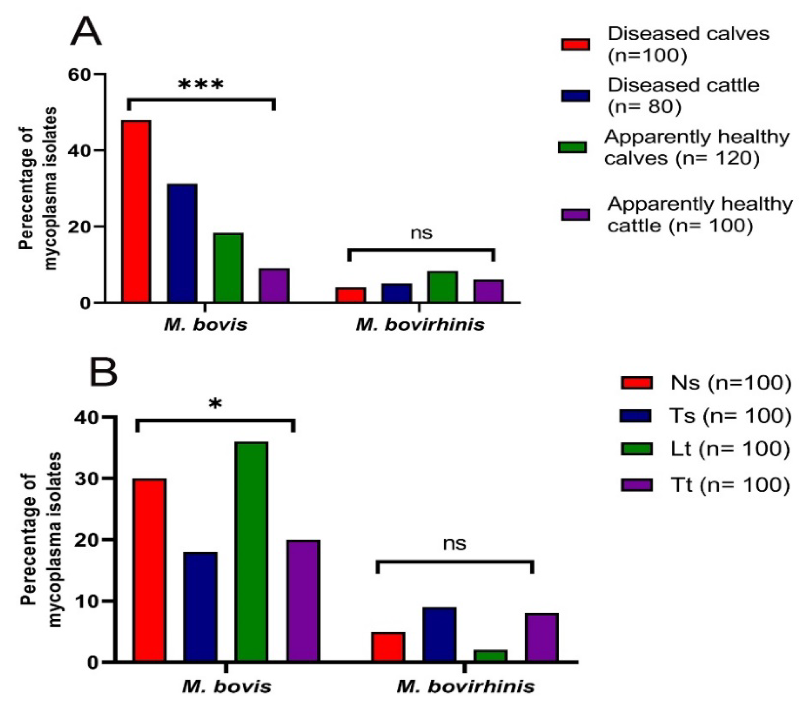
3.1. Prevalence of Mycoplasma Species in Cattle

Regarding the phenotypic identification of Mycoplasma species, a total of 140 isolates (35%) from various 400 samples showed fried egg and/or centerless granular colonies on PPLO agar plates. Additionally, a total of 128 mycoplasma isolates (32%) representing multiple isolates from the same animal were digitonin positive, and they were identified phenotypically as the genus Mycoplasma (Table 4). Meanwhile, 12 isolates (3%) were digitonin negative and they were identified as the genus Acholeplasma; five were obtained from the nasal swabs of apparently healthy adult cattle and seven were obtained from the lung tissues of apparently healthy calves. Of note, a total of 76 mycoplasma isolates were obtained from 100 investigated animals (76%); each isolate represented a single animal.
Considering the identification of multiple isolates from the same animal, Mycoplasma spp. were more prevalent among the collected samples from diseased calves (52%), followed by those from diseased cattle (36.3%) and apparently healthy calves and cattle (26.7 and 15%, respectively) (Figure 1A). Among the collected samples from apparently healthy calves and cattle, mycoplasma isolates were more prevalent among nasal swabs (30 and 24%, respectively), followed by tracheal tissues (26.7 and 20%, respectively) and tracheal swabs (26.7 and 16%, respectively), while lung tissues collected from apparently healthy cattle were mycoplasma negative. Among diseased calves and cattle, mycoplasma isolates were more prevalent among lung tissues (76 and 60%, respectively), followed by nasal swabs (48 and 40%, respectively), tracheal tissues (44 and 20%, respectively), and tracheal swabs (40 and 25%, respectively). In total, mycoplasma isolates were more prevalent among all examined lung tissues (38%), followed by nasal swabs (35%), tracheal tissues (28%), and tracheal swabs (27%) (Table 4 and Figure 1B). It was noted that mycoplasma isolates were rarely isolated from total healthy lung tissues (7/55, 12.7%), but they were frequently isolated from pneumonic lungs (31/45, 68.9%) (Table 4).
Regarding the identification of all mycoplasma isolates at the spp. level, M. bovis was the predominant spp. (61%), followed by M. bovirhinis (15%). Mycoplasma bovis was the predominant spp. among the collected samples from diseased calves (48%), followed by those from diseased cattle (31.3%) and apparently healthy calves and cattle (18.3 and 9%, respectively). Meanwhile, the highest isolation rates of M. bovirhinis were observed among the collected samples from apparently healthy calves (8.3%), followed by apparently healthy cattle (6%) and diseased cattle and calves (5 and 4%, respectively) (Figure 1A). The highest isolation rates of M. bovis isolates were observed among lung tissues (36%), followed by nasal swabs (30%), tracheal tissues (20%), and tracheal swabs (18%). On the other hand, the highest isolation rates of M. bovirhinis isolates were noticed among tracheal swabs (9%), followed by tracheal tissues (8%), nasal swabs (5%), and lung tissues (2%). Of note, highly statistical significance variations in the occurrence of total mycoplasma isolates and M. bovis were recognized among various samples’ sources (p ˂ 0.001); meanwhile, no statistically significant variations were recognized in the occurrence of M. bovirhinis among different samples’ sources (p = 0.59) (Figure 1A). Additionally, there were no statistically significant variations in the occurrence of total mycoplasma isolates and M. bovirhinis among various samples’ types (p = 0.28 and 0.16, respectively); however, there were statistically significant differences in the prevalence of M. bovis among different samples’ types (p = 0.01) (Figure 1B).

3.2. Antimicrobial Susceptibility Testing of Mycoplasma Isolates

Of the 128 recovered mycoplasma isolates, 76 were tested against 8 antibiotics by broth microdilution method for determination of their MICs. Those 76 isolates were randomly picked to represent all sample types and each isolate represented a single animal, and those isolates were recovered from apparently healthy calves (n = 20), apparently healthy cattle (n = 11), diseased calves (n = 25), and diseased cattle (n = 20) and each isolate represented a single animal. Interestingly, analysis of the antimicrobial susceptibility of the 76 examined isolates showed that all the tested isolates had low MICs values (sensitive) to tulathromycin, tilmicosin, tylosin, spiramycin, and spectinomycin (100% each), while 46 mycoplasma isolates (60.5%) had high MICs values (resistant) to enrofloxacin, and 33 mycoplasma isolates (43.4%) had high MICs values (resistant) to doxycycline (Table 5 and Table 6).
Regarding the species level of the recovered mycoplasma isolates, M. bovis isolates had high MICs values (resistant) to enrofloxacin (67.2%) and doxycycline (54.1%) antibiotics, while M. bovirhinis isolates had high MICs values (resistant) only to the enrofloxacin (33.3%) antibiotic (Table 6). There were statistically significant variations in the resistance rates of M. bovis and M. bovirhinis isolates against enrofloxacin (p = 0.01).
Regarding the sources of the samples, high prevalence rates of enrofloxacin and doxycycline resistance were detected among mycoplasma isolates recovered from diseased calves (100 and 60%) and cattle (100 and 90%), respectively (Figure 2). There were highly statistically significant (p < 0.001) variations in the resistance rates of mycoplasma isolates from different samples’ sources against enrofloxacin and doxycycline (Figure 2).

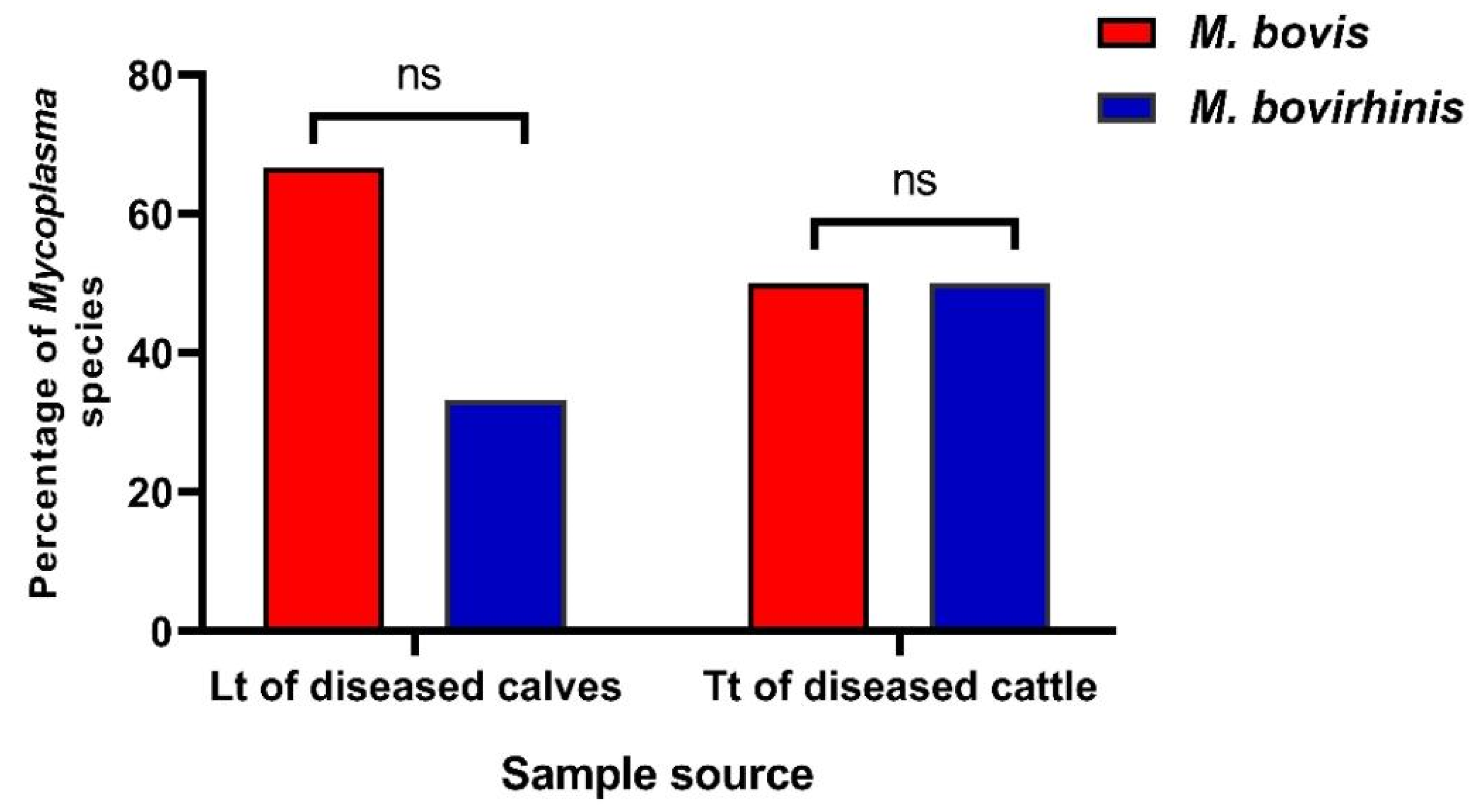
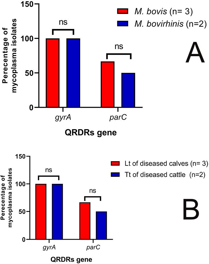
3.3. Molecular Identification of Enrofloxacin-Resistant Mycoplasma Isolates from Different Sources

Conventional PCR assays were carried out on five mycoplasma isolates with high enrofloxacin MIC values (≥4 µg/mL); three isolates were obtained from lung tissues of diseased calves and two isolates were obtained from tracheal tissues of diseased cattle. All the tested mycoplasma isolates (100%) were recognized as the genus Mycoplasma as they all harbored genes specific 16s rRNA gene. Moreover, three isolates (60%) were confirmed to be M. bovis, while the remaining two isolates (40%) were confirmed to be M. bovirhinis as they were all positive for the presence of 16s rRNA genes specific for each species (Figure 3). These findings were correlated with those of the phenotypic identification techniques. Of the three M. bovis isolates, two isolates were recovered from lung tissues of diseased calves, while the remained one isolate was obtained from the tracheal tissue of diseased cattle (Figure 3). Moreover, two M. bovirhinis isolates were obtained from lung tissue of a diseased calf and tracheal tissue of a diseased cattle (Figure 3). Moreover, no statistically significant variations were recognized in the occurrence of M. bovis and M. bovirhinis among different investigated samples’ types (p = 0.5 and 0.833, respectively) (Figure 3).
All molecularly confirmed mycoplasma isolates (n = 5) were examined for the presence of gyrA and parC genes of the quinolone resistance-determining regions. All the tested isolates were positive for the gyrA gene (100%). Meanwhile, three isolates (60%) were positive for the parC gene; two M. bovis isolates were recovered from lung tissues of diseased calves, and the one remaining M. bovirhinis isolate was recovered from tracheal tissue of diseased cattle (Figure 4). Of note, the M. bovis isolate with the highest enrofloxacin MIC value (8 ug/mL) was positive for both the gyrA and parC genes. Moreover, there were no statistically significant variations in the occurrence of the parC gene among mycoplasma isolates from different samples’ sources (p = 1) (Figure 4).

4. Discussion

It was pronounced that multiple global crises were emerging because of the extensive spreading of resistant bacteria such as Mycoplasma spp., Klebsilla spp., vancomycin-resistant Staphylococcus Aureus, and methicillin-resistant Staphylococcus Aureus, in addition to zoonotic foodborne bacteria such as Salmonella Typhimurium, Salmonella Enteritdis, and Campylobacter spp. [41,42,43,44,45,46,47,48,49,50,51,52,53]. The increase in the antimicrobial resistance rate of bovine Mycoplasma spp. to fluoroquinolone and tetracyclines is of great significance to the cattle industry globally. Herein, we determined a high prevalence rate of bovine Mycoplasma spp. (32%) among different samples’ types obtained from the respiratory tracts of cattle in Sharkia Governorate, Egypt. This result is partially similar to other studies conducted in Europe (34.3%) [54] and Turkey (35.4%) [55], and it was higher than the results of a previous study conducted in Nigeria (7.7%) [56] but lower than those results observed in other studies conducted in the USA (92%) [57] and Egypt (67.5%) [58]. Our findings demonstrated a high incidence of bovine Mycoplasma spp. recovered from lung tissues (38%). This result is partially similar to those of other studies conducted in Egypt, (40.9%) [59] and (36.8%) [60] and the USA (41.1%) [61], and it was higher than those recognized in previous studies conducted in Egypt, (31.66%) [62] and (29.6%) [63] but lower than those observed in other studies conducted in Canada (98%) [64], Denmark (86.8%) [65], the United Kingdom (86.4%) [66], and Argentina (70%) [67]. In the present study, bovine Mycoplasma spp. were more prevalent among lung tissues (38%) than nasal swabs (35%). This result is in complete agreement with those reported in previous studies conducted in Europe (60 and 24.4%, respectively) [54] and Canada (36 and 8.8%, respectively) [68], but it was in contrast with previous studies conducted in Europe, where Mycoplasma spp. were more prevalent among nasal swabs than lung tissues [69]. Herein, Mycoplasma spp. were more distributed among the samples collected from diseased calves (52%), followed by those obtained from diseased cattle (36.3%). These results were in complete agreement with those observed in recent studies conducted in Egypt (36 and 8.9%, respectively) [70] and Europe (17.1 and 11.4%, respectively) [54]. In the current study, M. bovis was the most predominant recovered spp. (61%) among the collected samples as documented previously in Iran (100%) [71], Britain (86.4%) [66], and Mexico (55%) [71,72]. Generally, the wide differences in the occurrence of bovine Mycoplasma spp. among different studies may be attributed to the environmental conditions, isolation and identification techniques, hygienic measures, types of the examined samples, animal breeds, time of sampling, management factors, stresses, and the geographical locations [70].
Recently, increased attention has been given to the significant role of M. bovis in BRD cases, which have become resistant to traditional antimicrobial therapy [29]. In the present study, 46 mycoplasma isolates (60.5%) were resistant to enrofloxacin with MICs values ranging from 2 to 8 μg/mL. These results were higher than those observed in other studies conducted in Europe (8.6%); (MICs ≥ 10 μg/mL) [73] and Spain (1.4%); (MICs ≤ 64 μg/mL) [74], but they were lower than those observed in a recent study conducted in Canada (100%); (MICs ≤ 128 μg/mL) [29]. Herein, all the tested mycoplasma isolates had low MICs values (sensitive) to macrolides (tilmicosin, tulathromycin, spiramycin, and tylosin); (100% each). This is in contrast with a recent study carried out in Switzerland, where high resistance rates (100%) were recorded against the macrolide group of antibiotics [75]. Moreover, all our examined mycoplasma isolates had low MICs values sensitive to spectinomycin. This is in contrast with a previous study conducted in Europe, which declared that 54.3% of the tested isolates had high MICs values (resistant) to spectinomycin [73]. Herein, mycoplasma isolates recovered from diseased cattle were more resistant to enrofloxacin (100%) than the apparently healthy ones. This result is in agreement with the findings of a previous study conducted in Canada, which reported that mycoplasma isolates from diseased cattle were more resistant to enrofloxacin (41.2%) than the healthy ones (23.3%) [29]. Additionally, doxycycline resistance was detected among 90% of our examined mycoplasma isolates that were recovered from diseased cattle; this result is in contrast with the findings of a previous study conducted in Israel, which reported that all the tested mycoplasma isolates were sensitive to doxycycline [32]. The high resistance rates of M. bovis isolates in developing countries may have resulted from the uncontrolled usage of antimicrobial agents in veterinary medicine as growth promoters, in cattle treatment in the case of respiratory diseases without any prescription, and as a control measure in farms, because there are no commercial vaccines available for bovine Mycoplasma spp. Therefore, antimicrobial usage must be controlled in the cattle industry [12]. Moreover, it is fundamental to utilize alternative drugs from medicinal plants to control resistant pathogens [76,77,78,79,80,81].
The PCR assays allow specific and quick detection of several bacterial pathogens associated with cattle pneumonia compared with the traditional isolation procedures [82]. In the present work, 60% of the tested isolates were identified as M. bovis and 40% were identified as M. bovirhinis. Interestingly, there was a 100% correlation between molecular and conventional identification results of the Mycoplasma species. These results are in agreement with a previous study conducted in Italy, where 70.3% of the tested isolates were identified as M. bovis, while 17.18% of the examined isolates were identified as M. bovirhinis [83]. Routine methods of using molecular techniques such as PCR and DNA sequencing can help in the rapid detection of the resistant mycoplasma strains. Thus, we recommend further large-scale studies to understand the different resistance mechanisms of fluoroquinolones and tetracyclines in mycoplasma strains obtained from different samples and animals of various health conditions.

5. Conclusions

Our findings revealed a high prevalence of bovine Mycoplasma spp. in the respiratory tract of cattle in Egypt. Moreover, our results showed alarming resistance to enrofloxacin and doxycycline in the examined mycoplasma isolates. Therefore, current antimicrobial usage rules must be followed to avoid an increase in antibiotic resistance, and fluoroquinolones and tetracyclines should be reserved for the treatment of serious diseases that have failed to respond to other antimicrobial classes. A major limitation in our approach is the need for DNA sequencing of gyrA and parC genes on large numbers of investigated isolates, in addition to fluoroquinolones sensitive and resistant strains, to understand the mechanism of fluoroquinolone resistance in bovine mycoplasma strains, which can help in the rapid detection of resistant mycoplasma strains and the management of mycoplasma infection.

6. Supplementary Aspect of Research

Sequencing of the PCR amplified products of gyrA and parC genes were conducted using an ABI 3730xl DNA sequencer (Thermo Fisher, Waltham, MA, USA). The amplified fragments were purified using the Gene Jet PCR purification kit (Cat. no. K0701, Thermoscientific, Waltham, MA, USA), and the sequencing of the purified products was performed with Big Dye Terminator V3.1 cycle sequencing kit (Applied Biosystems, PerkinElmer, Foster City, CA, USA) in an ABI 3130 automated DNA sequencer (Applied Biosystems, Carlsbad, CA, USA) according to the manufacturer’s guidelines. Sequence editing, consensus, and alignment construction were conducted utilizing BioEdit software package version 7.0.4.1. The phylogenetic trees were conducted utilizing MegAlign module for tree reconstruction of sequences by the Neighbor-joining technique based on ClustalW. Of note, the amino acid numbering was referred to Escherichia coli and it is based on the E. coli K-12 sequences for the parC (OM326889) and gyrA (OM326888) genes. The comparisons of obtained nucleotide sequences and multiple alignments were conducted utilizing the BioEdit sequence alignment editor (7.0.4.1) (Ibis bioscience, Inc., Carlsbad, CA, USA). Sequences of gyrA and parC genes were then submitted to NCBI GenBank utilizing BankIt (http://www.ncbi.nlm.nih.gov/WebSub/?tool=genbank) (accessed on 16 January 2022). under the accession numbers of OM326888 and OM326889, respectively.
One M. bovis isolate with high enrofloxacin MICs values (resistant) (MIC = 8 µg/mL), which was obtained from the lung tissue of a diseased calf, was subjected to DNA sequence analysis of gyrA and parC genes. Sequences of gyrA and parC genes of M. bovis isolate were submitted to the GeneBank database under the accession nos. of OM326888 and OM326889, respectively. The gyrA amino acid sequence of M. bovis isolate revealed 100% identity with those of other M. bovis isolates, M. bovis PG45 (reference strain; enrofloxacin sensitive), and E. coli K12 (Supplementary Figures S1–S3). Additionally, amino acids sequencing of the parC gene of M. bovis isolate revealed 100% identity when compared to other M. bovis isolates, and all these isolates showed 98% amino acid identity with E. coli K12 and 99% amino acid identity with M. bovis PG45 (control negative; enrofloxacin sensitive) (Supplementary Figures S4–S6). Additionally, the prediction protein of parC showed novel amino acid substitution at positions 2 (Gln→Arg); (CAG→CGT) in comparison with M. bovis PG45 (reference strain; enrofloxacin sensitive), as a first report in Egypt; thus, it might be considered the main cause of fluoroquinolone resistance in the examined isolate. This novel mutation is of interest and requires further scientific investigation on more isolates across the spectrum of fluoroquinolone susceptibility, and then a statistical inference is needed to show if the isolates that are resistant to fluoroquinolones are more statistically likely to have a mutation at position 2 than isolates that are susceptible.
Although fluoroquinolones are recognized to be effective antimicrobial agents in the treatment of bovine mycoplasmosis, mycoplasma isolates with high MICs (≤8 µg/mL) have been described in several countries [13]. Fluoroquinolone resistance usually develops as a result of target mutations in quinolone resistance-determining regions of the genes encoding gyrase and topoisomerases enzymes (gyrA and parC, respectively) [84]. In the current study, no amino acid substitutions were found within the quinolone resistance-determining regions of the gyrA gene, which is similar to a previous study carried out in France [13], but it was in contrast with a previous study conducted in Russia, where there was amino acid substitution in gyrA at positions 81 and 83 [85]. Additionally, a comparison of the parC quinolone resistance-determining regions in our tested isolates with the M. bovis PG45 strain (negative control; enrofloxacin sensitive) revealed the presence of amino acid substitutions at positions 2 (Gln→Arg), which, to the best of our knowledge, were described for the first time in Egypt; thus, it might be considered the main cause of fluoroquinolone resistance in the examined isolate. This result was consistent with previous studies conducted in several countries, which reported that fluoroquinolone resistance was due to mutations in the parC gene at positions 78, 80, 81, and 84 [85] and at position 84 [1,18]. Routine methods of using molecular techniques such as DNA sequencing can help in detecting mutations’ sites, which is important in understanding the resistance mechanism and can be helpful in the rapid detection of the resistant mycoplasma strains. Thus, we recommend further large-scale studies as an immediate future aspect of research to understand the different resistance mechanisms of fluoroquinolones and tetracyclines in M. bovis strains that have been obtained from different samples and animals of various health conditions.

Supplementary Materials

The following supporting information can be downloaded at: https://www.mdpi.com/article/10.3390/biology11071083/s1, Supplementary Figure S1. Phylogenetic tree of the examined M. bovis isolate based on gyrA sequence.; Supplementary Figure S2. Amino acid sequence alignments of gyrA gene of the examined M. bovis isolate.; Supplementary Figure S3. Amino acid identity percentages between gyrA genes of E. coli K12, M. bovis gyrA (OM326888), M. bovis PG45 (control negative; enrofloxacin sensitive), and other M. bovis isolates on GenBank.; Supplementary Figure S4. Phylogenetic tree of the examined M. bovis isolate based on parC sequence.; Supplementary Figure S5. Amino acid sequence alignments of parC gene of the examined M. bovis isolate.; Supplementary Figure S6. Amino acid identity percentages between ParC genes of E. coli K12, M. bovis ParC (OM326889), M. bovis PG45 (control negative; enrofloxacin sensitive), and other M. bovis isolates on GeneBank.

Author Contributions

Conceptualization, A.M.A.; R.M.S.E.-M. and M.I.A.E.-H.; methodology, H.M.M., M.I.A.E.-H., R.M.S.E.-M. and Y.H.M.; software, R.M.S.E.-M.; validation, H.M.M. and Y.H.M.; formal analysis, H.M.M., M.I.A.E.-H.; R.M.S.E.-M. and Y.H.M.; investigation, A.M.A., R.M.S.E.-M. and M.I.A.E.-H.; resources, H.M.M. and Y.H.M.; data curation, A.M.A., H.M.M., M.I.A.E.-H. and R.M.S.E.-M.; writing—original draft preparation, H.M.M.; writing—review and editing; A.M.A.; R.M.S.E.-M. and M.I.A.E.-H.; visualization, D.H.M.A.-k.; S.S.; W.M.E.-N. and W.N.H.; supervision, A.M.A., M.I.A.E.-H., R.M.S.E.-M. and Y.H.M.; project administration, D.H.M.A.-k.; S.S.; W.M.E.-N. and W.N.H.; funding acquisition, D.H.M.A.-k.; S.S.; W.M.E.-N. and W.N.H. All authors have read and agreed to the published version of the manuscript.

Funding

This research was funded by Faculty of Veterinary Medicine, Zagazig University, Egypt and Animal Health Research Institute, Egypt. Also it was funded by Princess Nourah bint Abdulrahman University Researchers Supporting Project number PNURSP2022R15, Princess Nourah bint Abdulrahman University, Riyadh, Saudi Arabia.

Institutional Review Board Statement

Ethical review and approval were waived for this study because all samples were obtained during routine diagnostic examinations and necropsies.

Informed Consent Statement

Not applicable.

Data Availability Statement

Data sharing is not applicable to this article.

Acknowledgments

We want to thank Faculty of Veterinary Medicine, Zagazig University, Egypt and Animal Health Research Institute, Egypt for their support of our research. Also, we want to extend our thanks to Princess Nourah bint Abdulrahman University Researchers Supporting Project number PNURSP2022R15, Princess Nourah bint Abdulrahman University, Riyadh, Saudi Arabia.

Conflicts of Interest

The authors declare no conflict of interest.

References

  1. Lysnyansky, I.; Ayling, R.D. Mycoplasma bovis: Mechanisms of resistance and trends in antimicrobial susceptibility. Front. Microbiol. 2016, 7, 595. [Google Scholar] [CrossRef] [PubMed]
  2. Parker, A.M.; Sheehy, P.A.; Hazelton, M.S.; Bosward, K.L.; House, J.K. A review of mycoplasma diagnostics in Cattle. J. Vet. Intern. Med. 2018, 32, 1241–1252. [Google Scholar] [CrossRef]
  3. Calcutt, M.J.; Lysnyansky, I.; Sachse, K.; Fox, L.K.; Nicholas, R.A.J.; Ayling, R.D. Gap analysis of Mycoplasma bovis disease, diagnosis and control: An aid to identify future development requirements. Transbound. Emerg. Dis. 2018, 65, 91–109. [Google Scholar] [CrossRef] [PubMed] [Green Version]
  4. Isa, F.M. Mycoplasma bovis: A Neglected Pathogen in Nigeria-A Mini Review. Dairy Vet. Sci. J. 2017, 4, 555633. [Google Scholar]
  5. Hale, H.H.; Helmboldt, C.F.; Plastridge, W.N.; Stula, E.F. Bovine mastitis caused by a Mycoplasma spp. Cornell Vet. 1962, 52, 582–591. [Google Scholar] [PubMed]
  6. Tardy, F.; Aspan, A.; Autio, T.; Ridley, A.; Tricot, A.; Colin, A.; Strube, M.L. Mycoplasma bovis in nordic european countries: Emergence and dominance of a new clone. J. Pathog. 2020, 9, 875. [Google Scholar] [CrossRef]
  7. Punyapornwithaya, V.; Fox, L.K.; Hancock, D.D.; Gay, J.M.; Alldredge, J.R. Association between an outbreak strain causing Mycoplasma bovis mastitis and its asymptomatic carriage in the herd: A case study from Idaho, USA. Prev. Vet. Med. 2010, 93, 66–70. [Google Scholar] [CrossRef]
  8. Hazelton, M.S.; Morton, J.M.; Parker, A.M.; Sheehy, P.A.; Bosward, K.L.; Malmo, J.; House, J.K. Whole dairy herd sampling to detect subclinical intramammary Mycoplasma bovis infection after clinical mastitis outbreaks. Vet. Microbiol. 2020, 244, 108662. [Google Scholar] [CrossRef]
  9. Oliveira, T.E.S.; Pelaquim, I.F.; Flores, E.F.; Massi, R.P.; Valdiviezo, M.J.J.; Pretto-Giordano, L.G.; Headley, S.A. Mycoplasma bovis and viral agents associated with the development of bovine respiratory disease in adult dairy cows. Trans. Emerg. Dis. 2020, 67, 82–93. [Google Scholar] [CrossRef]
  10. Bras, A.L.; Barkema, H.W.; Woodbury, M.R.; Ribble, C.S.; Perez-Casal, J. Windeyer, M. Clinical presentation, prevalence and risk factors associated with Mycoplasma bovis-associated disease in farmed bison (Bison bison) herds in western Canada. J. Am. Vet. Med. Assoc. 2017, 250, 1167–1175. [Google Scholar] [CrossRef]
  11. Ozdemir, U.; Turkyilmaz, M.; Nicholas, A. Detection and antibiotic susceptibility of Mycoplasma bovis and other respiratory disease pathogens from pneumonic lung samples in a calf rearing unit. Madr. J. Vet. Med. Res. 2019, 1, 8–12. [Google Scholar]
  12. Ammar, A.M.; Abd El-Hamid, M.I.; Hashem, Y.M.; El-Malt, R.M.S.; Mohamed, H.M. Mycoplasma bovis: Taxonomy, Characteristics, Pathogenesis and Antimicrobial Resistance. Zagazig Vet. J. 2021, 49, 444–461. [Google Scholar] [CrossRef]
  13. Khalil, D.; Becker, C.A.; Tardy, F. Alterations in the quinolone resistance-determining regions and fluoroquinolones resistance in clinical isolates and laboratory-derived mutants of Mycoplasma bovis not all genotypes may be equal. Appl. Environ. Microb. 2016, 82, 1060–1068. [Google Scholar] [CrossRef] [PubMed] [Green Version]
  14. Ammar, A.M.; Abd El-Hamid, M.I.; El-Malt, R.M.S.; Azab, D.S.; Albogami, S.; Bendary, M.M. Molecular detection of fluoroquinolone resistance among multidrug-, extensively drug-, and pan-drug-resistant Campylobacter species in Egypt. J. Antibiot. 2021, 10, 1342. [Google Scholar] [CrossRef]
  15. Sato, T.; Okubo, T.; Usui, M.; Higuchi, H.; Tamura, Y. Amino acid substitutions in gyrA and parC are associated with fluoroquinolones resistance in Mycoplasma bovis isolates from Japanese dairy calves. J. Vet. Med. Sci. 2013, 75, 1063–1065. [Google Scholar] [CrossRef] [Green Version]
  16. Redgrave, L.S.; Sutton, S.B.; Webber, M.A.; Piddock, L.J. Fluoroquinolones resistance: Mechanisms, impact on bacteria, and role in evolutionary success. Trend. Microbiol. 2014, 22, 438–445. [Google Scholar] [CrossRef]
  17. Megid, R.; Nicholas, R.A.J.; Miles, R.J. Biochemical characterization of Mycoplasma bovirhinis, Mycoplasma dispar and recent bovine isolates of Mycoplasma canis. Vet. Res. Commun. 2001, 25, 1–12. [Google Scholar] [CrossRef]
  18. Lysnyansky, I.; Mikula, I.; Gerchman, I.; Levisohn, S. Rapid detection of a point mutation in the parC gene associated with decreased susceptibility to fluoroquinolones in Mycoplasma bovis. Antimicrob. Agents Chemother. 2009, 53, 4911–4914. [Google Scholar] [CrossRef] [Green Version]
  19. Kairova, M.; Rakhimzhanova, D. Molecular genetic techniques and oligonucleotides for mycoplasma identification—A review. Acta Vet. Brno 2021, 89, 317–332. [Google Scholar] [CrossRef]
  20. Heaton, V.J.; Ambler, J.E.; Fisher, L.M. Potent antipneumococcal activity of gemifloxacin is associated with dual targeting of gyrase and topoisomerase IV, an in vivo target preference for gyrase and enhanced stabilization of cleavable complexes in vitro. Antimicrob. Agents Chemother. 2000, 44, 3112e7. [Google Scholar] [CrossRef] [Green Version]
  21. Sulyok, K.M.; Kreizinger, Z.; Wehmann, E.; Lysnyansky, I.; Bányai, K.; Marton, S.; Jerzsele, Á.; Rónai, Z.; Turcsányi, I.; Makrai, L.; et al. Mutations associated with decreased susceptibility to seven antimicrobial families in field and laboratory-derived Mycoplasma bovis strains. Antimicrob. Agents Chemother. 2017, 61, e01983-16. [Google Scholar] [CrossRef] [PubMed] [Green Version]
  22. Nicholas, R.; Baker, S. Recovery of mycoplasmas from animals. Mycoplasma protocols. Methods Mol. Biol. 1998, 104, 37–43. [Google Scholar] [PubMed]
  23. Shahzad, W.; Munir, R.; Rana, M.Y.; Ahmad, R.; Khan, M.S.; Akbar, G.; Ijaz, M.; Mehmood, F. Prevalence, molecular diagnosis and treatment of Mycoplasma conjunctivae isolated from infectious keratoconjunctivitis affected Lohi sheep maintained at Livestock Experiment Station, Bahadurnagar, Okara, Pakistan. Trop. Anim Health Prod. 2013, 45, 737–742. [Google Scholar] [CrossRef]
  24. Freundt, E.A.; Andrews, B.E.; Erno, H.; Kunze, M.; Black, F.T. The sensitivity of Mycoplasmatales to sodium polyanethol sulfonate and digitonin. Zentralbl. Bakteriol. Parasitenkd. Infektioskr. Hyg. Abt. Orig. 1973, 225, 104–112. [Google Scholar]
  25. Poveda, J.B.; Nicholas, R. Serological identification of mycoplasmas by growth and metabolism inhibition tests. Mycoplasma Protoc. Methods Mol. Biol. 1998, 104, 105–111. [Google Scholar]
  26. Clyde, W.A. Mycoplasma species identification based upon growth inhibition by specific antisera. J. Immunol. 1964, 92, 958–965. [Google Scholar] [PubMed]
  27. Lauerman, L.H. Mycoplasmas of the bovine respiratory tract. In Mycoplasmosis in Animals: Laboratory Diagnosis; Whitford, H.W., Rosenbusch, R.F., Lauerman, L.H., Eds.; Iowa State University Press: Ames, IA, USA, 1994; pp. 50–56. [Google Scholar]
  28. Hannan, P.C. Guidelines and recommendations for antimicrobial minimum inhibitory concentration (MIC) testing against veterinary Mycoplasma species. Vet. Res. 2000, 31, 373–395. [Google Scholar] [CrossRef] [Green Version]
  29. Jelinski, M.; Kinnear, A.; Gesy, K.; Andrés-Lasheras, S.; Zaheer, R.; Weese, S.; McAllister, T.A. Antimicrobial sensitivity testing of Mycoplasma bovis isolates derived from Western Canadian feedlot cattle. Microorganisms 2020, 8, 124. [Google Scholar] [CrossRef] [Green Version]
  30. NCCLS (National Committee for Clinical Laboratory Standards). Performance Standards for Antimicrobial Disk and Dilution Susceptibility Tests for Bacteria Isolated from Animals; Approved Standard—Second Edition; NCCLS Document M31-A2; National Committee for Clinical Laboratory Standards: Wayne, PA, USA, 2002; Volume 22, p. 57. [Google Scholar]
  31. Godinho, K.S. Susceptibility testing of tulathromycin: Interpretative breakpoints and susceptibility of field isolates. Vet. Microbiol. 2008, 129, 426–432. [Google Scholar] [CrossRef] [Green Version]
  32. Gerchman, I.; Levisohn, S.; Mikula, I. In vitro antimicrobial susceptibility of Mycoplasma bovis isolated in Israel from local and imported cattle. Vet. Microbiol. 2009, 137, 268–275. [Google Scholar] [CrossRef]
  33. Catania, S.; Bottinelli, M.; Fincato, A.; Gastaldelli, M.; Barberio, A.; Gobbo, F.; Vicenzoni, G. Evaluation of Minimum Inhibitory Concentrations for 154 Mycoplasma synoviae isolates from Italy collected during 2012–2017. PLoS ONE 2019, 14, e0224903. [Google Scholar] [CrossRef] [PubMed] [Green Version]
  34. González, Y.R.; Ros Bascuñana, C.; Bölske, G.; Mattsson, J.G.; Fernandez Molina, C.; Johansson, K.E. In vitro amplification of the 16S rRNA genes from Mycoplasma bovis and Mycoplasma agalactiae by PCR. Vet. Microbiol. 1995, 47, 183–190. [Google Scholar] [CrossRef]
  35. Sambrook, J.; Fritsch, E.F.; Maniatis, T. Molecular Cloning: A Laboratory Manual; Cold Spring Harbor Laboratory: Cold Spring Harbor, NY, USA, 1989; Volume 1. [Google Scholar]
  36. Alberto, A.; Addis, M.F.; Chessa, B.; Cubaddu, T.; Profiti, M.; Rosati, S.; Ruiu, A.; Pittau, M. Molecular and antigenic characterization of a Mycoplasma bovis strain causing an outbreak of infectious keratoconjunctivitis. J. Vet. Diagn. Investig. 2006, 18, 41–51. [Google Scholar] [CrossRef] [PubMed] [Green Version]
  37. Salama, S.E.M.; Kholy, M.M.; Talkhan, O.F.A.; Mosallam, S.A.; Atwa, E.I. Bacterial agent of respiratory manifestation in cattle and associated biochemical alterations in Menoufiea Governorate. Nat. Sci. 2009, 7, 26-30.–30. [Google Scholar]
  38. Kobayashi, H.; Hirose, K.; Worarach, A.; Paugtes, P.; Ito, N.; Morozumi, T.; Yamamoto, K. In vitro amplification of the 16S rRNA genes from Mycoplasma bovirhinis, Mycoplasma alkalescens and Mycoplasma bovigenitalium by PCR. J. Vet. Med. Sci. 1998, 60, 1299–1303. [Google Scholar] [CrossRef] [Green Version]
  39. Al-Farha, A.A.B.; Hemmatzadeh, F.; Khazandi, M.; Hoare, A.; Petrovski, K. Evaluation of effects of Mycoplasma mastitis on milk composition in dairy cattle from South Australia. BMC Vet. Res. 2017, 13, 351. [Google Scholar] [CrossRef] [Green Version]
  40. Hata, E.; Harada, T.; Itoh, M. Relationship between antimicrobial susceptibility and multilocus sequence type of Mycoplasma bovis isolates and development of a method for rapid detection of point mutations involved in decreased susceptibility to macrolides, lincosamides, tetracyclines, and spectinomycin. Appl. Environ. Microbiol. 2019, 85, e00575-19. [Google Scholar]
  41. Abd El-Hamid, M.I.; Bendary, M.M. Comparative phenotypic and genotypic discrimination of methicillin resistant and susceptible Staphylococcus aureus in Egypt. Cell. Mol. Biol. 2015, 61, 101–112. [Google Scholar]
  42. Ammar, A.M.; Abd El-Hamid, M.I.; Eid, S.E.A.; El Oksh, A.S. Insights into antimicrobial resistance and virulence genes of emergent multidrug resistant avian pathogenic Escherichia coli in Egypt: How closely related are they? Revue Méd. Vét. 2015, 166, 304–314. [Google Scholar]
  43. Ammar, A.M.; Attia, A.M.; Abd El-Aziz, N.K.; Abd El Hamid, M.I.; El-Demerdash, A.S. Class 1 integron and associated gene cassettes mediating multiple-drug resistance in some foodborne pathogens. Int. Food Res. J. 2016, 23, 332–339. [Google Scholar]
  44. Ammar, A.M.; Attia, A.M.; Marwa, I.; Abd El-Hamid, M.I.; El-Shorbagy, I.M.; Shaimaa, A.; Abd El-Kader, S.A. Genetic basis of resistance waves among methicillin resistant Staphylococcus aureus isolates recovered from milk and meat products in Egypt. Cell. Mol. Biol. 2016, 62, 7–15. [Google Scholar] [PubMed]
  45. Bendary, M.M.; Solyman, S.M.; Azab, M.M.; Mahmoud, N.F.; Hanora, A.M. Genetic diversity of multidrug resistant Staphylococcus aureus isolated from clinical and non clinical samples in Egypt. Cell. Mol. Biol. 2016, 62, 55–61. [Google Scholar] [PubMed]
  46. Abd El-Aziz, N.K.; Abd El-Hamid, M.I.; Bendary, M.M.; El-Azazy, A.A.; Ammar, A.M. Existence of vancomycin resistance among methicillin resistant S. aureus recovered from animal and human sources in Egypt. Slov. Vet. Res. 2018, 55, 221–230. [Google Scholar]
  47. Abd El-Hamid, M.I.; Awad, N.F.S.; Abo-Shama, U.H.; Yousreya, M.H.; Abdel-Rahman, M.A. In vitro evaluation of various antimicrobials against field avian Mycoplasma gallisepticum and Mycoplasma synoviae isolates in Egypt. Poult. Sci. J. 2019, 98, 6281–6288. [Google Scholar] [CrossRef]
  48. Abd El-Hamid, M.I.; Bendary, M.M.; Merwad, A.M.A.; Elsohaby, I.; Ghaith, D.M.; Alshareef, W.A. What is behind phylogenetic analysis of hospital-, community and livestock-associated methicillin-resistant Staphylococcus aureus? Transbound. Emerg. Dis. 2019, 66, 1506–1517. [Google Scholar] [PubMed]
  49. Awad, N.F.S.; Abd El-Hamid, M.I.; Hashem, Y.M.; Erfan, A.M.; Abdelrahman, B.A.; Mahmoud, H.I. Impact of single and mixed infections with Escherichia coli and Mycoplasma gallisepticum on Newcastle disease virus vaccine performance in broiler chickens: An in vivo perspective. J. Appl. Microbiol. 2019, 127, 396–405. [Google Scholar] [CrossRef]
  50. Ammar, A.M.; El-Naenaeey, E.S.Y.; El-Malt, R.M.S.; El-Gedawy, A.A.; Khalifa, E.; Elnahriry, S.S.; Abd El-Hamid, M.I. Prevalence, antimicrobial susceptibility, virulence and genotyping of Campylobacter jejuni with a special reference to the anti-virulence potential of Eugenol and beta-resorcylic acid on some multi-drug resistant isolates in Egypt. J. Anim. Sci. 2020, 11, 3. [Google Scholar] [CrossRef] [PubMed]
  51. Ammar, A.M.; El-Naenaeey, E.S.Y.; El-Hamid, M.I.A.; El-Gedawy, A.A.; El-Malt, R.M.S. Campylobacter as a Major Foodborne Pathogen: A Review of Its Characteristics, Pathogenesis, Antimicrobial Resistance and Control. J. Microbiol. Biotechnol. Food Sci. 2021, 10, 609–619. [Google Scholar] [CrossRef]
  52. Bendary, M.M.; Ibrahim, D.; Mosbah, R.A.; Mosallam, F.; Hegazy, W.A.H.; Awad, N.F.S.; Alshareef, W.A.; Alomar, S.Y.; Zaitone, S.A.; Abd El-Hamid, M.I. Thymol Nanoemulsion: A new therapeutic option for extensively drug resistant foodborne pathogens. J. Antibiot. 2021, 10, 25. [Google Scholar] [CrossRef]
  53. Ibrahim, D.; Abdelfattah-Hassan, A.; Badawi, M.; Ismail, T.A.; Bendary, M.M.; Abdelaziz, A.M.; Mosbah, R.A.; Mohamed, D.; Arisha, A.H.; El-Hamid, M.I.A. Thymol nanoemulsion promoted broiler chicken’s growth, gastrointestinal barrier and bacterial community and conferred protection against Salmonella Typhimurium. Sci. Rep. 2021, 11, 7742. [Google Scholar] [CrossRef]
  54. Gabinaitiene, A.; Siugzdaite, J.; Zilinskas, H.; Siugzda, R.; Petkevicius, S. Mycoplasma bovis and bacterial pathogens in the bovine respiratory tract. J. Vet. Med. 2011, 56, 28–34. [Google Scholar] [CrossRef] [Green Version]
  55. Müştak, H.K. Diagnosis of Mycoplasma bovis infection in cattle by ELISA and PCR. Kafkas Üniversitesi Vet. Fakültesi Derg. 2014, 20, 249–252. [Google Scholar] [CrossRef] [Green Version]
  56. Francis, M.I.; Ankeli, P.I.; Kwanashie, C.N.; Adamu, J.; Allam, L.; Raji, M.A.; Scacchia, M. Detection of Mycoplasma bovis from cattle presented for slaughter in Adamawa and Taraba States, Northeastern Nigeria. Vet. Sci. 2020, 6, 109–115. [Google Scholar] [CrossRef]
  57. Boonyayatra, S.; Fox, L.K.; Gay, J.M.; Sawant, A.; Besser, T.E. Discrimination between Mycoplasma and Acholeplasma species of bovine origin using digitonin disc diffusion assay, nisin disc diffusion assay, and conventional polymerase chain reaction. J. Vet. Diagn. Investig. 2012, 24, 7–13. [Google Scholar] [CrossRef] [Green Version]
  58. Abd El-Tawab, A.; Awad, A.; Elhofy, F.; Hassan, N.I.; Ramdan, M. Identification and genetic characterization of Mycoplasma spp. affecting respiratory system in Egyptian cattle. Benha Vet. Med. J. 2021, 40, 21–26. [Google Scholar] [CrossRef]
  59. García-Galán, A.; De la Fe, C.; Gomis, J.; Bataller, E.; Sánchez, A.; Quereda, J.J.; Gómez-Martín, A. The addition of Lactobacillus spp. negatively affects Mycoplasma bovis viability in bovine cervical mucus. BMC Vet. Res. 2020, 16, 251. [Google Scholar] [CrossRef]
  60. El-Shafey, D.Y. Advanced Studies on Mycoplasma bovis in Cattle. Ph.D. Thesis, Cairo University, Giza, Egypt, 2005. [Google Scholar]
  61. Soehnlen, M.K.; Kariyawasam, S.; Lumadue, J.A.; Pierre, T.A.; Wolfgang, D.R.; Jayarao, B.M. Molecular epidemiological analysis of Mycoplasma bovis isolates from the Pennsylvania Animal Diagnostic Laboratory showing genetic diversity. J. Dairy Sci. 2011, 94, 1893–1899. [Google Scholar] [CrossRef] [Green Version]
  62. Fawkia, I.A.E.R. In vitro antibiotic sensitivity of different strains of Mycoplasma and Acholeplasma isolated from feeder calves. Proceeding Third Sci. Congr. JESCD 1995, 1, 173–177. [Google Scholar]
  63. El-Shabiny, L.M.; Abou El-Makarem, M.M.; Nada, H.S. Mycoplasma isolated from cattle lungs and their Pathogenicity study. Egypt. J. Agric. Res. 1999, 77, 421–431. [Google Scholar]
  64. Gagea, M.I.; Bateman, K.G.; Shanahan, R.A.; van Dreumel, T.; McEwen, B.J.; Carman, S.; Archambault, M.; Caswell, J.L. Natural occurring Mycoplasma bovis associated pneumonia and polyarthritis in feedlot beef calves. J. Vet. Diagn. Investig. 2006, 18, 29–40. [Google Scholar] [CrossRef] [Green Version]
  65. Kusiluka, L.J.; Kokotovic, B.; Ojeniyi, B.; Friis, N.F.; Ahrens, P. Genetic variations among Mycoplasma bovis strains isolated from Danish cattle. FEMS Microbiol. Lett. 2000, 192, 113–118. [Google Scholar] [CrossRef] [PubMed]
  66. Dudek, K.; Nicholas, R.A.; Szacawa, E.; Bednarek, D. Mycoplasma bovis infections—Occurrence, diagnosis and control. J. Pathog. 2020, 9, 640. [Google Scholar] [CrossRef] [PubMed]
  67. Margineda, C.A.; Zielinski, G.O.; Jurado, S.B.; Ferrella, A.; Mozgovoj, M.; Alcaraz, A.C.; Lopez, A. Mycoplasma bovis pneumonia in feedlot cattle and dairy calves in Argentina. Brazil. J. Vet. Pathol. 2017, 10, 79–86. [Google Scholar] [CrossRef]
  68. Li, C.; Zaheer, R.; Kinnear, A.; Jelinski, M.; McAllister, T.A. Comparative Microbiomes of the respiratory tract and joints of feedlot cattle mortalities. Microorganisms 2022, 10, 134. [Google Scholar] [CrossRef] [PubMed]
  69. Rifatbegovic, M.; Assunção, P.; Poveda, J.B.; Pasic, S. Isolation of Mycoplasma bovis from the respiratory tract of cattle in Bosnia and Herzegovina. Vet. Rec. 2007, 160, 484. [Google Scholar] [CrossRef]
  70. Hashem, Y.M.; Mousa, W.S.; Abdeen, E.E.; Abdelkhalek, H.M.; Nooruzzaman, M.; El-Askary, A.; Wareth, G. Prevalence and molecular characterization of Mycoplasma species, Pasteurella multocida, and Staphylococcus aureus isolated from calves with respiratory manifestations. J. Anim. 2022, 12, 312. [Google Scholar] [CrossRef]
  71. Maunsell, F.P.; Donovan, G.A. Mycoplasma bovis Infections in Young Calves. Food Anim. Pract. 2009, 25, 139–177. [Google Scholar] [CrossRef]
  72. Nicholas, R.A.J.; Fox, L.K.; Lysnyansky, I. Mycoplasma mastitis in cattle: To cull or not to cull. Vet. J. 2016, 216, 142–147. [Google Scholar] [CrossRef]
  73. Sulyok, K.M.; Kreizinger, Z.; Fekete, L.; Hrivnák, V.; Magyar, T.; Jánosi, S.; Gyuranecz, M. Antibiotic susceptibility profiles of Mycoplasma bovis strains isolated from cattle in Hungary, Central Europe. BMC Vet. Res. 2014, 10, 256. [Google Scholar] [CrossRef] [Green Version]
  74. Klein, U.; de Jong, A.; Moyaert, H.; El Garch, F.; Leon, R.; Richard-Mazet, A.; Ayling, R.D. Antimicrobial susceptibility monitoring of Mycoplasma hyopneumoniae and Mycoplasma bovis isolated in Europe. Vet. Microbiol. 2017, 204, 188–193. [Google Scholar] [CrossRef]
  75. Khalil, D.; Becker, C.A.; Tardy, F. Monitoring the decrease in susceptibility to ribosomal RNAs targeting antimicrobials and its molecular basis in clinical Mycoplasma bovis isolates over time. Microb. Drug Resist. 2017, 23, 799–811. [Google Scholar] [CrossRef] [PubMed]
  76. Abd El-Hamid, M.I.; El-Sayed, M.E.; Ali Aisha, R.; Abdallah, H.M.; Marwa, I.A.; El-mowalid, G.A. Marjoram extract down-regulates the expression of Pasteurella multocida adhesion, colonization and toxin genes: A potential mechanism for its antimicrobial activity. Comp. Immunol. Microbiol. Infect. Dis. 2019, 62, 101–108. [Google Scholar] [CrossRef] [PubMed]
  77. Elmowalid, G.A.; Marwa, I.; Abd El-Wahab, A.M.; Atta, M.; Abd El-Naser, G.; Attia, A.M. Garlic and ginger extracts modulated broiler chicks innate immune responses and enhanced multidrug resistant Escherichia coli O78 clearance. Comp. Immunol. Microbiol. Infec Dis. 2019, 66, 101334. [Google Scholar] [CrossRef] [PubMed]
  78. Abd El-Hamid, M.I.; El-Naenaeey, E.Y.; Kandeel, T.M.; Hegazy, W.A.H.; Mosbah, R.A.; Nassar, M.S.; Bakhrebah, M.A.; Abdulaal, W.H.; Alhakamy, N.A.; Bendary, M.M. Promising Antibiofilm Agents: Recent Breakthrough against Biofilm Producing Methicillin-Resistant Staphylococcus aureus. Antibiotics 2020, 9, 667. [Google Scholar] [CrossRef] [PubMed]
  79. Aljazzar, A.; Abd El-Hamid, M.I.; El-Malt, R.M.S.; El-Gharreb, W.R.; Abdel-Raheem, S.M.; Ibrahim, A.M.; Abdelaziz, A.M.; Ibrahim, D. Prevalence and Antimicrobial Susceptibility of Campylobacter Species with Particular Focus on the Growth Promoting, Immunostimulant and Anti-Campylobacter jejuni Activities of Eugenol and Trans-Cinnamaldehyde Mixture in Broiler Chickens. Animals 2022, 12, 905. [Google Scholar] [CrossRef]
  80. Mosallam, F.M.; Helmy, E.A.; Bendary, M.M.; El-Batal, I.A. Potency of a novel synthesized Ag- eugenol nanoemulsion for treating some bacterial and fungal pathogens. J. Mater. Res. 2021, 36, 1524–1537. [Google Scholar] [CrossRef]
  81. Ghaly, M.; Shaheen, A.; Bouhy, A.; Bendary, M. Alternative therapy to manage otitis media caused by multidrug-resistant fungi. Arch. Microbiol. 2020, 1, 10. [Google Scholar] [CrossRef]
  82. Bell, C.J.; Blackburn, P.; Elliott, M.; Patterson, T.I.; Ellison, S.; Lahuerta-Marin, A.; Ball, H.J. Investigation of polymerase chain reaction assays to improve detection of bacterial involvement in bovine respiratory disease. J. Vet. Diagn. Investig. 2014, 26, 631–634. [Google Scholar] [CrossRef] [Green Version]
  83. Ayling, R.D.; Bashiruddin, S.E.; Nicholas, R.A. Mycoplasma species and related organisms isolated from ruminants in Britain between 1990 and 2000. Vet. Rec. 2004, 155, 413–416. [Google Scholar] [CrossRef]
  84. Piddock, L.J. Mechanisms of fluoroquinolone resistance: An update 1994–1998. Drugs 1999, 58, 11–18. [Google Scholar] [CrossRef]
  85. Chernova, O.A.; Medvedeva, E.S.; Mouzykantov, A.A.; Baranova, N.B.; Chernov, V.M. Mycoplasmas and their antibiotic resistance: The problems and prospects in controlling infections. Acta Nat. 2016, 8, 24–34. [Google Scholar] [CrossRef] [Green Version]
Figure 1. Prevalence of M. bovis and M. bovirhinis isolates in various samples’ sources (A) and types (B). Ns: nasal swabs, Ts: tracheal swabs, Lt: lung tissues; Tt: tracheal tissues, * p < 0.05, *** p < 0.001, ns: non-significant.
Biology 11 01083 g001
Figure 2. Prevalence rates of enrofloxacin and doxycycline among mycoplasma isolates from different samples’ sources. Ns: nasal swabs, Ts: tracheal swabs, Lt: lung tissue, Tt: tracheal tissue, *** p < 0.001.
Biology 11 01083 g002
Figure 3. Prevalence of molecularly identified Mycoplasma species in the collected samples from different sources. The prevalence rate for each species was calculated regarding the total number of the examined samples from different sources. ns: non-significant, Lt: lung tissue, Tt: tracheal tissue.
Biology 11 01083 g003
Figure 4. Prevalence of gyrA and parC genes in two bovine Mycoplasma species (A) and mycoplasma isolates from different samples’ sources (B). Lt: lung tissue, Tt: tracheal tissue, QRDRs: quinolone resistance-determining regions, ns: non-significant.
Biology 11 01083 g004
Table 1. Distribution of different samples collected from apparently healthy and diseased cattle in Sharkia Governorate, Egypt.
Sample Type (Symbol)Animal Health ConditionTotal
Apparently Healthy (n = 55)Diseased (n = 45)
Calve
(n = 30)
Adult Cattle
(n = 25)
Calve
(n = 25)
Adult Cattle
(n = 20)
Nasal swab (Ns)30252520100
Tracheal swab (Ts)30252520100
Lung tissue (Lt)30252520100
Tracheal tissue (Tt)30252520100
Total12010010080400
Table 2. Interpretative minimum inhibitory concentrations’ breakpoints of different antimicrobial agents against Mycoplasma species.
Antimicrobial ClassAntimicrobial Agent (Symbol)MIC Breakpoint (µg/mL)Reference
SensitiveIntermediate SensitiveResistant
FluroquinolonesEnrofloxacin (ENR)≤0.250.5−1≥2[32]
MacrolidesTulathromycin (TUL)≤1632≥64[31]
Tilmicosin (TIL)≤816≥32[32]
Tylosin (TYL)≤8-≥16[28]
Spiramycin (SP)≤2−8->8
TetracyclinesDoxycycline (DO)≤12−4≥8
AminoglycosidesSpectinomycin (SPT)≤3264≥128[30]
PhenicolsFlorfenicol (FF)≤24≥8
Table 3. Sequence of oligonucleotide primers and amplified PCR products for five target genes of Mycoplasma species.
Specificity
(Target Gene)
Primer Sequence (5′-3′)PCR Amplified Product (bp)Reference
Genus Mycoplasma
(16S rRNA)
F: AGACTCCTACGGGAGGCAGCA
R: ACTAGCGAT TCCGACTTCATG
1000[36]
M. bovis
(16S rRNA)
F: CCTTTTAGATTGGGATAGCGGATG
R: CCGTCAAGGTAGCATCATTTCCTAT
360[34,37]
M. bovirhinis
(16S rRNA)
F: GCTGATAGAGAGGTCTATCG
R: ATTACTCGGGCAGTCTCC
316[38,39]
QRDRs
(gyrA)
F: GACGAATCATCTAGCGAG
R: GCCTTCTAGCATCAAAGTAGC
531[18,40]
QRDRs
(parC)
F: GAGCAACAGTTAAACGATTTG
R: GGCATAACAACTGGCTCTT
488[18,40]
QRDRs: Quinolone resistance determining region, bp: base pair.
Table 4. Prevalence of bovine Mycoplasma species in various samples at Sharkia Governorate, Egypt.
Sample Type
(Symbol, No.)
** Total No. of Mycoplasma Isolates from Various Samples of Investigated Animals (%)Total *
Diseased Calves
(n = 100)
Diseased Cattle
(n = 80)
Apparently Healthy Calves
(n = 120)
Apparently Healthy Cattle
(n = 100)
Nasal swab (Ns, 100)12 (48)8 (40)9 (30)6 (24)35 (35)
Tracheal swab (Ts, 100)10 (40)5 (25)8 (26.7)4 (16)27 (27)
Lung tissue (Lt, 100)19 (76)12 (60)7 (23.3)0 (0)38 (38)
Tracheal tissue (Tt, 100)11 (44)4 (20)8 (26.7)5 (20)28 (28)
* Total (400)52 (52)29 (36.3)32 (26.7)15 (15)128 (32)
* The isolation rates were calculated concerning the total number of the examined samples from each source and each animal. ** The isolation rates were calculated concerning the number of the samples from each animal.
Table 5. Minimal inhibitory concentrations of the tested antimicrobials against mycoplasma isolates.
Antimicrobial ClassAntimicrobial Agent (Symbol)No. of Mycoplasma Isolates Showing MIC Values of the Tested Antimicrobials (μg/mL) *MIC50MIC90
0.00780.01560.03120.06250.1250.250.51248
FluoroquinolonesEnrofloxacin (ENR)-----51015414122
MacrolidesTulathromycin (TUL)-7645141624---0.51
Tilmicosin (TIL)1329277-------0.01560.0312
Tylosin (TYL)----17182216 3-0.51
Spiramycin (SP)--37336------0.06250.125
TetracyclinesDoxycycline (DO)-------53353348
AminoglycosidesSpectinomycin (SPT)-66---10122814-24
PhenicolsFlorfenicol (FF)------6261727-24
* The dark vertical lines denote the resistance breakpoint, MIC: minimum inhibitory concentration, MIC50 = (n × 0.5), MIC90 = (n × 0.9).
Table 6. Antibiotic susceptibility patterns of total mycoplasma, Mycoplasma bovis, and Mycoplasma bovirhinis isolates.
No. of Mycoplasma Species and Total Mycoplasma Isolates (%) Showing Susceptibility Patterns Against the Examined Antibiotic
Antimicrobial ClassAntimicrobial Agent (Symbol)M. bovis
(n = 61)
M. bovirhinis
(n = 15)
Total Mycoplasma Isolates
(n = 76)
RISRISRIS
FluoroquinolonesEnrofloxacin (ENR)41 (67.2)20
(32.8)
-5
(33.3)
10
(66.7)
-46
(60.5)
30
(39.5)
-
MacrolidesTulathromycin (TUL)--61 (100)--15
(100)
--76
(100)
Tilmicosin (TIL)--61 (100)--15
(100)
--76
(100)
Tylosin (TYL)--61
(100)
--15
(100)
--76
(100)
Spiramycin (SP)--61
(100)
--15
(100)
--76
(100)
TetracyclinesDoxycycline (DO)33 (54.1)28
(45.9)
--10
(66.7)
5
(33.3)
33
(43.4)
38
(50)
5
(6.6)
AminoglycosidesSpectinomycin (SPT)--61
(100)
--15
(100)
--76
(100)
PhenicolsFlorfenicol (FF)-21
(34.4)
40
(65.6)
-10
(66.7)
5
(33.3)
-31
(40.8)
45
(59.2)
R: resistant, I: intermediate sensitive, S: sensitive.
Publisher’s Note: MDPI stays neutral with regard to jurisdictional claims in published maps and institutional affiliations.

