Article

Hepatoprotective Effects of Garlic Extract against Carbon Tetrachloride (CCl4)-Induced Liver Injury via Modulation of Antioxidant, Anti-Inflammatory Activities and Hepatocyte Architecture

1
Department of Medical Laboratories, College of Applied Medical Sciences, Qassim University, Buraydah 52571, Saudi Arabia
2
Department of Basic Health Sciences, College of Applied Medical Sciences, Qassim University, Buraydah 52571, Saudi Arabia
*
Author to whom correspondence should be addressed.
Appl. Sci. 2020, 10(18), 6200; https://doi.org/10.3390/app10186200
Submission received: 26 July 2020 / Revised: 1 September 2020 / Accepted: 2 September 2020 / Published: 7 September 2020
(This article belongs to the Section Applied Biosciences and Bioengineering)

Abstract

:
The current study aims to explore the hepatoprotective mechanisms of garlic extract through in vivo and in vitro assays. The in vitro investigation of antioxidant and anti-inflammatory potential showed maximum 67.5% of free radical scavenging and 71.36% albumin denaturation inhibition by 600 μg/mL garlic extract. To explore the hepatoprotective activity by in vivo experiments, the animals were orally intoxicated with 150 μL of CCl4 (1:1 v/v in olive oil) and treated with garlic extract (75 mg/kg b.w.) 3 times/week, for eight successive weeks. The administration of garlic extract significantly ameliorated CCl4 induced increment in amounts of serum Alanine aminotransferase (ALT), Alkaline phosphatase (ALP) and Aspartate transaminaseas (106.7, 116.3, 136.4 U/L) as compared to disease control which showed increased level (140.5, 156.2, 187.6 U/L). Besides, significant reduction of Superoxide dismutase (SOD), Glutathione peroxidases (GPx), and Glutathione (GSH) (29.3, 48.4, and 25.9 U/mg protein) was noticed in CCl4 induced animals, respectively. Likewise, garlic extract treatment facilitated a significant increment in all tested antioxidant enzymes levels (41.6, 63.3, and 32.5 U/mg protein), respectively. Additionally, Tumor necrosis factor⍺ (TNF-⍺), C-reactive protein (CRP), Interleukin-1β (IL-1β), Interleukin 6 (IL-6) and ICAM-1 (Intercellular Adhesion Molecule 1) level (63.79, 580.2, 18.3, 63.74 and 148.4 pg/mL) were increased significantly in CCl4-induced group, while garlic extract treatment decreased these pro inflammatory marker levels (40.24, 460.4, 15.4, 45.14, and 125.3 pg/mL). The animals exposed to CCl4 showed various types of alterations like lymphocytes infiltration, edema and congestion, while the animals treated with garlic extract plus CCl4 showed amelioration of the hepatocytes architectures. Thus, our finding advocates that the consumption of garlic can be a potential therapeutic remedy in the inhibition of liver ailments.

1. Introduction

Since ancient times, natural products including plants, microorganisms, animals and marine organisms have been used by human beings in traditional medicines to treat and alleviate different diseases. With the passage of time, synthetic medicines became more popular, irrespective of having various side effects. Several commonly used medicines like ibuprofen, warfarin, diclofenac, etc., have been found to be associated with some severe complications. There are numerous medicinal plants which possess a broad range of medicinal applications and are nowadays being more commonly used because of their very minimal side effects, lesser toxicity and most effective dynamic healing effects as compared to synthetic counterparts [1]. Therefore, the search for more effective agents with less side effects among alternative medicines and natural products is a new field of interest [2]. Garlic (Allium sativum) is a perennial herb that belongs to the amaryllidaceae plant family. It is one of the most multipurpose medicinal plants used as a traditional herbal medicine to prevent and treat a broad range of diseases, including cardiovascular diseases, atherosclerosis, hyperlipidaemia, thrombosis, hypertension and diabetes [3].
The liver, with its multiple functions, is one of the most important organs. It plays an active role in metabolism as it secretes bile that breaks down fats in the small intestine during digestion, stores and releases glucose and synthesizes different types of proteins. In addition, the liver converts harmful ammonia into urea, processes haemoglobin, clears bilirubin, fights infections and detoxifies medicines and other toxic chemicals. Liver diseases are one of the chief reasons of morbidity and mortality worldwide. Several factors, directly or indirectly, cause liver diseases like virus, exposure to drugs or chemicals, obesity or diabetes. Besides this, different autoimmune disorders and untreated liver diseases can lead to malignancy and liver cancer and eventually death [4]. The liver is the centre for metabolism of all xenobiotic substances, including drugs and chemicals. During the different detoxification activities on most of the drugs, the liver experiences various types of diseases. There are at least six mechanisms involved in hepatocyte injury [5].
Carbon tetrachloride (CCl4) is one of the best known hepatic toxins that is used in animal research work to investigate the hepatoprotective effect of various medicinal plants and natural products. Moreover, it has been reported that severe liver necrosis, as well as steatosis can be induced by a direct single exposure to CCl4 because it is a powerful hepatotoxic and xenobiotic agent [6,7]. Depending on the dose and duration, various types of liver pathogenesis are induced upon exposure to CCl4. This toxin causes the damage of cells either by covalent binding of its reactive intermediates to cell components or by triggering peroxidation of membrane lipids through interaction with free radical intermediates. This leads to damage of lipids, predominantly unsaturated phospholipids that result in damage to intracellular membranes [8]. Besides, reductive dehalogenation of CCl4 by p450 enzymes leads to the formation of trichloromethyl radicals which can react with the sulfhydryl groups of glutathione and protein thiols.
Besides this, the components of the antioxidant defence system of the liver like superoxide dismutase (SOD), catalase (CAT), glutathione peroxidase (GPx), glutathione reductase (GR) etc. are reported to be altered due to intoxication with CCl4 [9]. The resulting overproduction of reactive oxygen species (ROS) including superoxide anion, hydroxyl radical, hydrogen peroxide, and other radicals due to CCl4 results in oxidative stress [10]. Oxidative stress is a commonly used term that refers to a state when the cellular redox balance is altered [11] and it plays an important role in various types of diseases [12].
The lipid peroxidation products exhibit chemotactic activities and activate pro-inflammatory cytokines. In addition, these products also stimulate hepatic collagen-producing stellate cells and induce mixed lesions called as steatohepatitis which has some specific distinct features including degeneration and hepatocyte necrosis, inflammatory infiltrate and fibrosis [13,14]. It has been noticed that the level of pro-inflammatory markers also becomes altered in liver damage and other liver disorders. Nuclear factor-κB (NF-κB) is a family of dimeric transcription factors that promotes the expression of inflammation associated genes in response to stimulation of inflammation [15]. In this context, many medicinal plants or active compound of herbs have been reported to neutralise the hepatotoxic activity of CCl4 due to their ability to inhibit the formation of free radicals, and their hepatoprotective activity has been suggested to be linked with their antioxidant potential.
Additionally, antioxidants have been detailed to protect cell organelles, organs, and tissues against oxidative stress and to prevent the pathogenesis of oxidative damage. Garlic contains various valuable chemical compounds and its health benefits have been primarily attributed to its organo-sulfur components such as alliin, γ-glutamylcysteine, and their derivatives [16]. Garlic and its active compounds have often been described to have key role in disease management because of its anti-inflammatory, antioxidant, anti-cancer and other various health-promoting activities. Garlic oil has been shown to have hepatoprotective ability against CCl4-induced hepatotoxicity [17].
Therefore, the current study was designed to explore the in vitro and in vivo antioxidant, anti-inflammatory and hepatoprotective activities of garlic methanolic extract in rat models against CCl4- induced hepatotoxicity. The in vitro studies involved the use of bovine serum album in anti-inflammatory experiments as it is reported that Bovine serum albumin (BSA) can be a good model for anti-inflammatory studies in the search of potential anti-inflammatory drugs. In the search of anti-inflammatory nature of garlic, inhibition of protein denaturation and antiproteinase action, were conducted as it has been reported that the antioxidant capacity is responsible for health beneficial effects of any natural product. Therefore, the 2,2-diphenyl-1-picryl-hydrazyl-hydrate (DPPH) assay and H2O2 reducing tests were performed to evaluate its antioxidant potential. For further confirmation of hepatoprotective role of garlic, liver function test, anti-oxidant enzymes, and anti-inflammatory assay in animal model. Further the hepatocyte architecture was evaluated through the histopathological staining. The novelty of our findings lies in the comparative analysis of in vitro and in vivo studies which supports the hepatoprotective potential of garlic.

2. Materials and Methods

2.1. Chemicals

CCl4, Folin-Ciocalteau reagent, 2,2-diphenyl-1-picrylhydrazyl (DPPH), ferric chloride, potassium ferricyanide, gallic acid, trichloroacetic acid, ascorbic acid, and chloroform were purchased from Sigma–Aldrich (St. Louis, MO, USA). Diagnostic kits for serum alanine aminotransferase (ALT), aspartate aminotransferase (AST), alkaline phosphatase (ALP) were purchased from Randox Laboratories Ltd. (London, UK). Other chemicals, including hydrogen peroxide, ethanol, acetone, thiobarbituric acid, bovine serum albumin, ferric sulfate, ferric chloride, sodium acetate, and butanol were purchased from Merck (Darmstadt, Germany). Antioxidant enzyme kits including GPx, SOD and GSH were bought from Abcam (Cambridge, UK). Inflammatory markers like IL-6, CRP, TNF-⍺ and ICAM1 were also purchased from Abcam, as was antibody Vascular Endothelial Growth Factor (VEGF). All other reagents and chemicals used in the current work were analytical grade and obtained from their authorised distributors in Saudi Arabia.

2.2. Preparation of Crude Methanolic Garlic Extract and Reference Drug Preparation

Pure dry powder of garlic raw bulblet was purchased from the local market of Buraydah, Qassim, KSA. The methanolic extract of garlic was prepared in accordance with the Eidi et al. method [18]. The garlic bulbs were chopped into very small pieces and allowed to dry to minimize the moisture. The methanolic garlic extract was made through addition of 1:3 ratios of garlic and methanol, and subjected to extraction for 48 h according to a published method with minor modifications [8]. At the end of extraction, it was passed through Whatman filter paper No.1 (Whatman Ltd., Maidstone, UK). This methanolic filtrate was concentrated under reduced pressure on rotary evaporator at 40 °C and then stored at 4 °C for further use. The filtrate was reconstituted in desired amount of 1% DMSO according to our need in sterile vials to obtain methanol extract of known concentration. The percentage of yield extracted was calculated as % yield:
%   yield = [ Weight   of   sample   extract / Initial   weight   of   sample ] × 100
Ibuprofen and diclofenac sodium were used as reference drugs and were crushed into a fine powder. About 200 milligrams of these drug powders had been weighed out utilizing a digital analytical balance and we mixed these powder into 20.0 mL of double distilled water, respectively. The solutions had been mixed well utilizing a vortex. A serial dilution of ibuprofen and diclofenac sodium was carried by using phosphate buffer, pH 6.4 for heat-induced denaturation inhibition, while, for anti-proteinase action evaluation, diclofenac sodium was serially diluted in Tris HCl buffer, pH 7.4.

2.3. In Vitro Study

2.3.1. Confirmatory Tests for Flavonoids (Alkaline Reagent Test) and Phenolics (FeCl3 Test)

The presence or absence of different flavonoid compounds in methanolic extract of garlic (1 mg/mL) were checked and confirmed simply by the use of few drops of NaOH [12]. Appearance of intense yellow colour by NaOH which becomes colourless after the addition of few drops of dilute HCl is a confirmatory test of the presence of different flavonoids in garlic extract. The formation of red, blue, green or purple coloration by addition of few drops of 1% FeCl3 in garlic extract dissolved in methanol is an indication of the presence of other phenolic compounds.

2.3.2. Determination of Total Phenolic Content by Folin-Ciocalteu Reagent

Total phenolic content was investigated by Folin-Ciocalteu reagent method with some minor modifications [12]. To 0.5 mL of crude methanolic extract (1 mg/mL) in a test tube, 2.5 mL of 10% Folin-Ciocalteu reagent was added. Finally, 2 mL of 7.5% sodium carbonate was included in it and mixed well. The test tubes were incubated in a dark at room temperature for 30 min and the absorbance was measured at spectrophotometrically a constant wavelength 760 nm by using spectrophotometer (1240 UV mini spectrophotometer, Shimadzu, Kyota, Japan). A standard calibration plot was prepared with different concentrations of gallic acid (50–250 μg/mL). The concentration of total phenolic content in the methanolic extract was calculated using this standard. Total phenolic content was expressed as mg Gallic Acid Equivalents (GAE). All tests were performed in triplicates and the results were expressed as mg Gallic acid equivalents per gm sample extract.
Following equation was used to calculate total phenolic content:
Total   Phenolic   Content = X × Vol / Wt
where X = Concentration of gallic acid (mg/mL), Vol = Volume of garlic extract in mL and Wt = Weight of garlic extract in g.

2.3.3. Total Flavonoid Content

Total flavonoid content of garlic extract had been determined by employing the aluminium chloride colorimetric test [12,19]. In different test tubes, 500 μL of garlic extract with a concentration of 50 µg/mL or quercetin solution of varying concentration from 20 to 250 µg/mL were mixed and 500 μL of 2% AlCl3 in ethanol solution was included in each tube. The solutions were mixed well and then incubated at room temperature for sixty minutes. After completion of incubation, a yellow color appeared, indicative of the presence of flavonoids. The absorbance was recorded at 420 nm against a blank. Using a quercetin calibration curve, the total flavonoid content was calculated and expressed as mg of quercetin equivalent per g of crude extract (mg QUE/g) by the following equation:
Total   flavonoid   content = X × Vol / Wt
where X = Concentration of quercetin (mg/mL), Vol = Volume of garlic extract in mL and Wt = Weight of pure garlic extract in g

2.3.4. Estimation of DPPH Scavenging Ability

A 1,1-diphenyl-2-picrylhydrazyl (DPPH) assay with slight modifications was employed to investigate the antioxidant ability of the garlic extract [12]. Briefly, different concentrations (50–600 μg/mL) of garlic extract were produced through serial dilution of a known concentration (10 mg/mL) stock solution in methanol. DPPH (0.3 mM) was prepared in absolute methanol and 1 mL was mixed with 2500 μL various dilutions of garlic extract. The reaction mixture was well mixed and the solutions were stored in the absence of sunlight for 30 min at room temperature. The absorbance of each solution had been recorded at 517 nm against a blank which contained only methanol. The percentage of DPPH scavenging ability of garlic extract was determined in accordance with the following equation:
Percent   DPPH   scavenging   ability = [ ( Xc Xs ) / Xc ] × 100
where Xc = Absorbance of control and Xs = Absorbance in presence of extract.

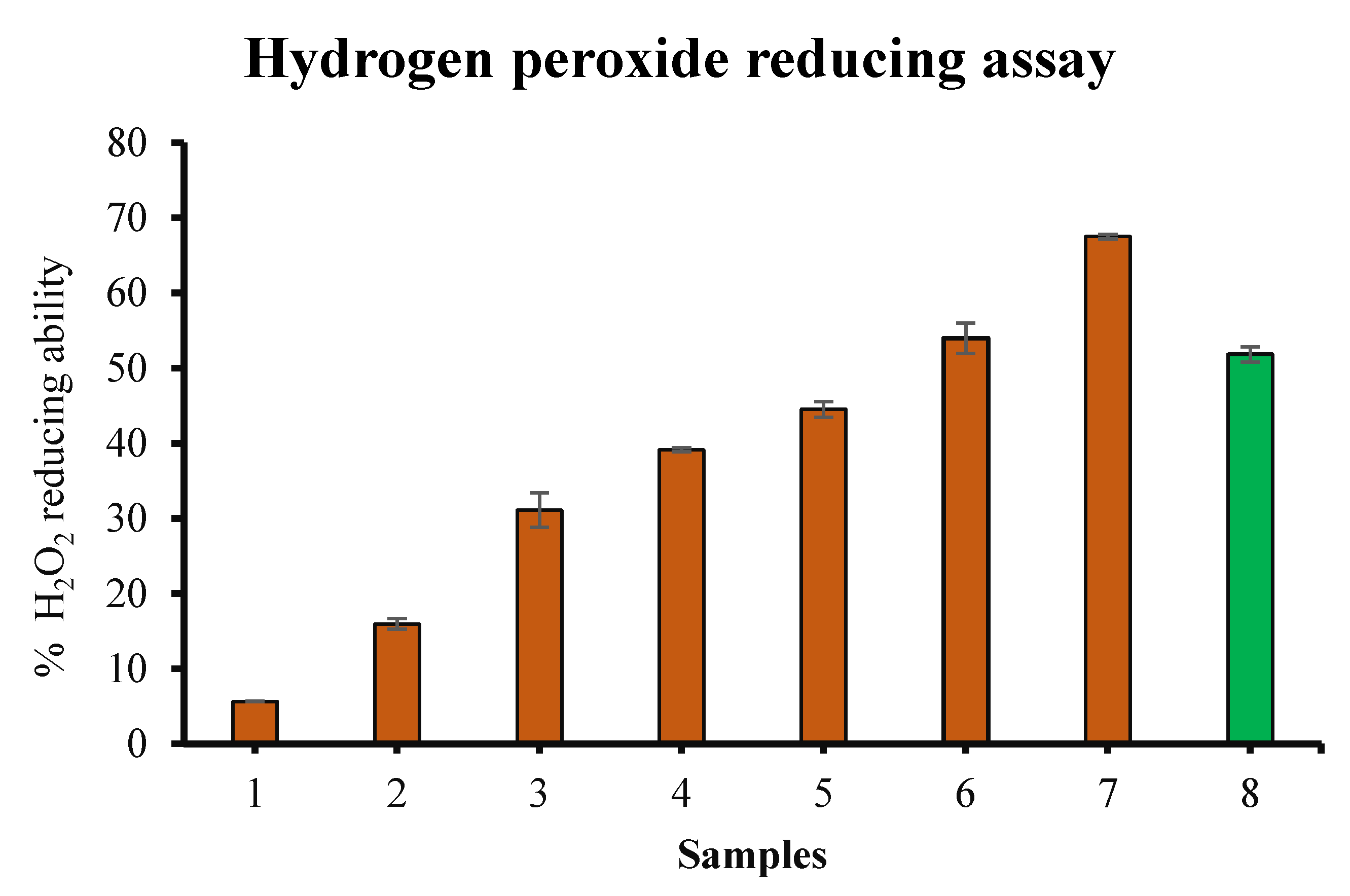
2.3.5. Hydrogen Peroxide Reducing Ability

To validate the antioxidant activity, the hydrogen peroxide reducing ability of garlic extract was evaluated utilizing a slightly changed method of Ruch et al. [20]. For this purpose, 1 × phosphate buffer saline (pH 7.4) had been utilized to produce 40 mM hydrogen peroxide. One mL of this hydrogen peroxide solution was added to different dilutions of garlic extract (50–600 µg/mL) kept in the dark. The tubes were kept for 10 min and the absorbance of dilutions was calculated employing a UV visible spectrophotometer at 230 nm against a blank. The blank had phosphate buffer without hydrogen peroxide. The results were compared with the percentage hydrogen peroxide reducing ability of ascorbic acid as reference (100 µg/mL). Experiments were done in triplicate. The percentage hydrogen peroxide reducing ability was evaluated in accordance with the following equation:
Percentage   hydrogen   peroxide   reducing   ability = [ ( Xc Xs ) / Xc ] × 100
where Xc = Absorbance of control and Xs = Absorbance in the presence of garlic extract.

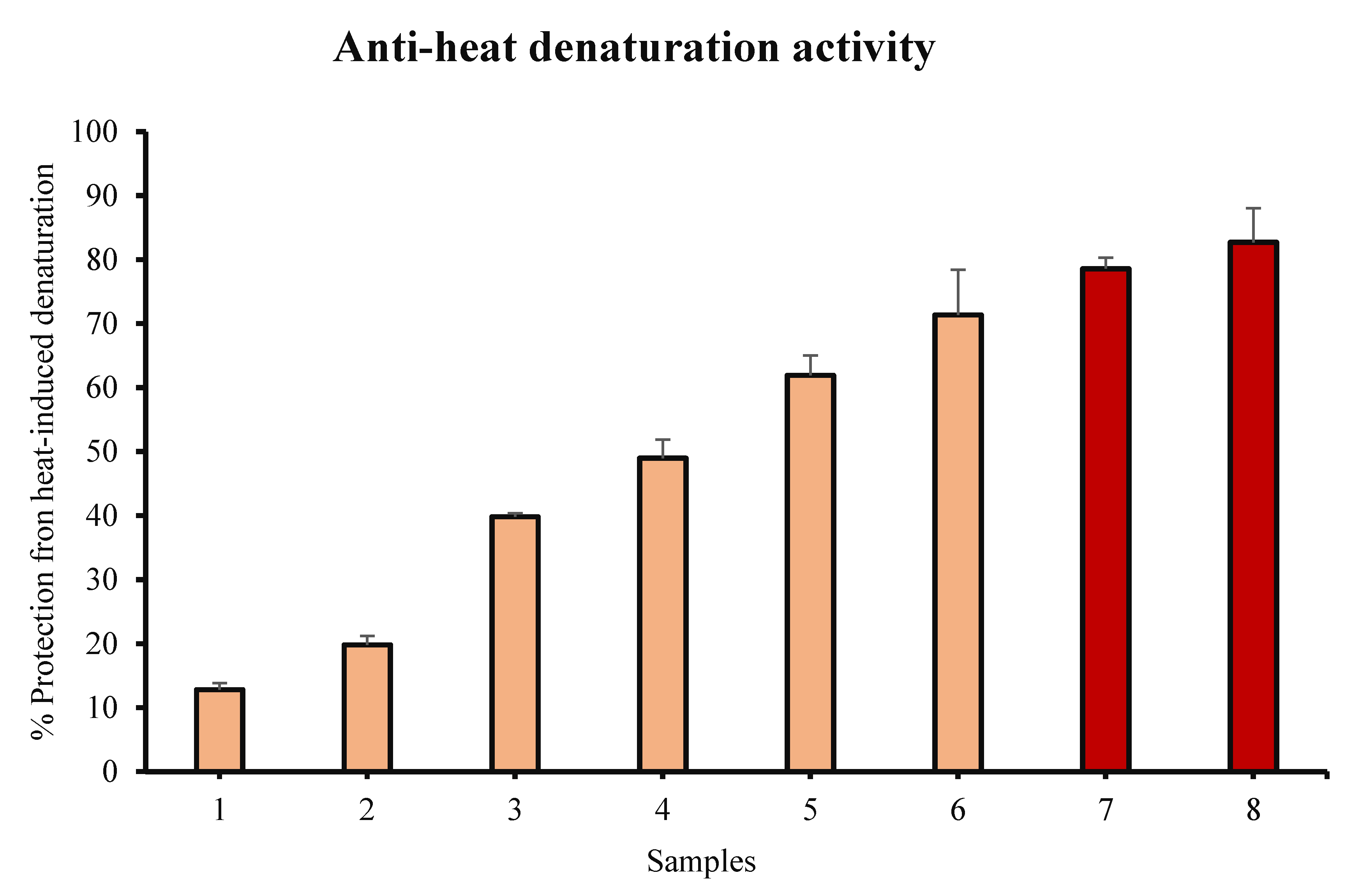
2.3.6. Inhibition of Bovine Serum Albumin Denaturation

The in vitro anti-inflammatory action of garlic extract had been evaluated by albumin denaturation inhibition using a slightly modified method of Sakat et al. [21]. One % aqueous bovine serum albumin (500 μL) in different test tubes was mixed with 100 μL of various dilutions (100–600 µg/mL) of garlic extract, and 100 and 200 µg/mL of ibuprofen. The test tubes were then stored at room temperature for 20 min. After completion of incubation, the test tubes were heated (51 °C) for 30 min. The heating was followed by cooling down of samples and the turbidity of each sample was evaluated at 660 nm by employing a spectrophotometer. Experiments were done in triplicate. The % inhibition of bovine serum albumin denaturation had been evaluated utilizing the following formula:
%   Inhibition = [ ( Xc Xs ) / Xc ] × 100
where Xc = Absorbance of control and Xs = Absorbance in presence of extract/ibuprofen.

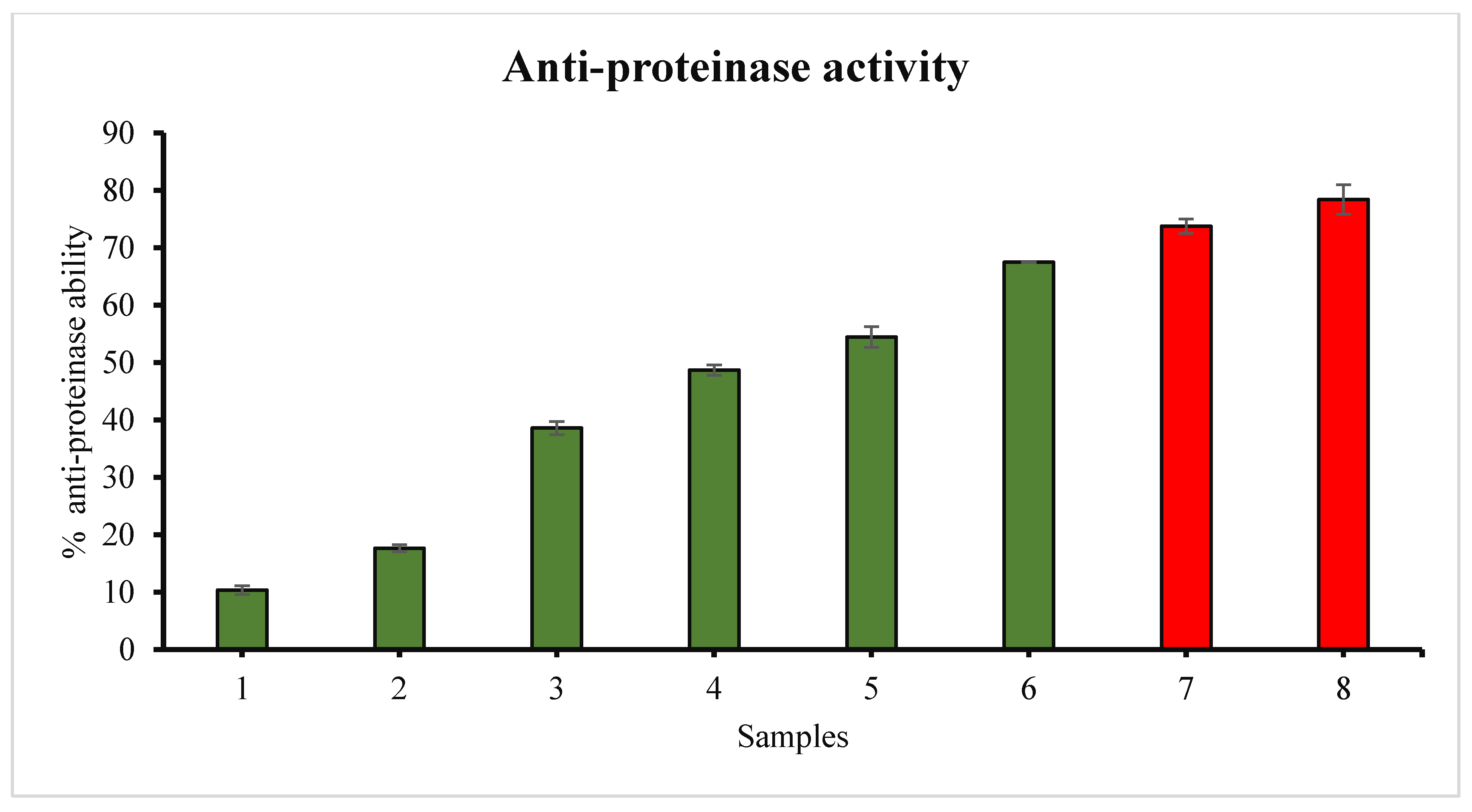
2.3.7. Anti-Proteinase Action

For further confirmation of the anti-inflammatory potential of garlic extract, the anti-proteinase action of garlic was explored using a modification of the method of Sakat et al. [21]. For this, 1 mL of 0.06 mg trypsin in 20 mM Tris HCl buffer, pH 7.4 and 1 mL of various dilutions (100–600 μg/mL) of garlic extract or diclofenac sodium (100 and 200 μg/mL) in a 2 mL reaction mixture had been added in different tubes. The tubes were first stored at room temperature for 5 min, after which 1 mL of 0.8% (w/v) casein was added to this reaction mixture. The mixtures were further incubated for 20 min. Later, 2 mL of 70% perchloric acid were added to stop the reaction. Centrifugation was carried out for five minutes at 2500 rpm. The absorbance of the supernatant was recorded on a UV-visible spectrophotometer at 210 nm against buffer as blank. Experiments were done in triplicate. The % anti-proteinase action was calculated by following formula:
Percentage   anti - proteinase   action = [ ( Xc Xs ) / Xc ] × 100
where Xc = Absorbance of control and Xs = Absorbance in the presence of extract/diclofenac sodium.

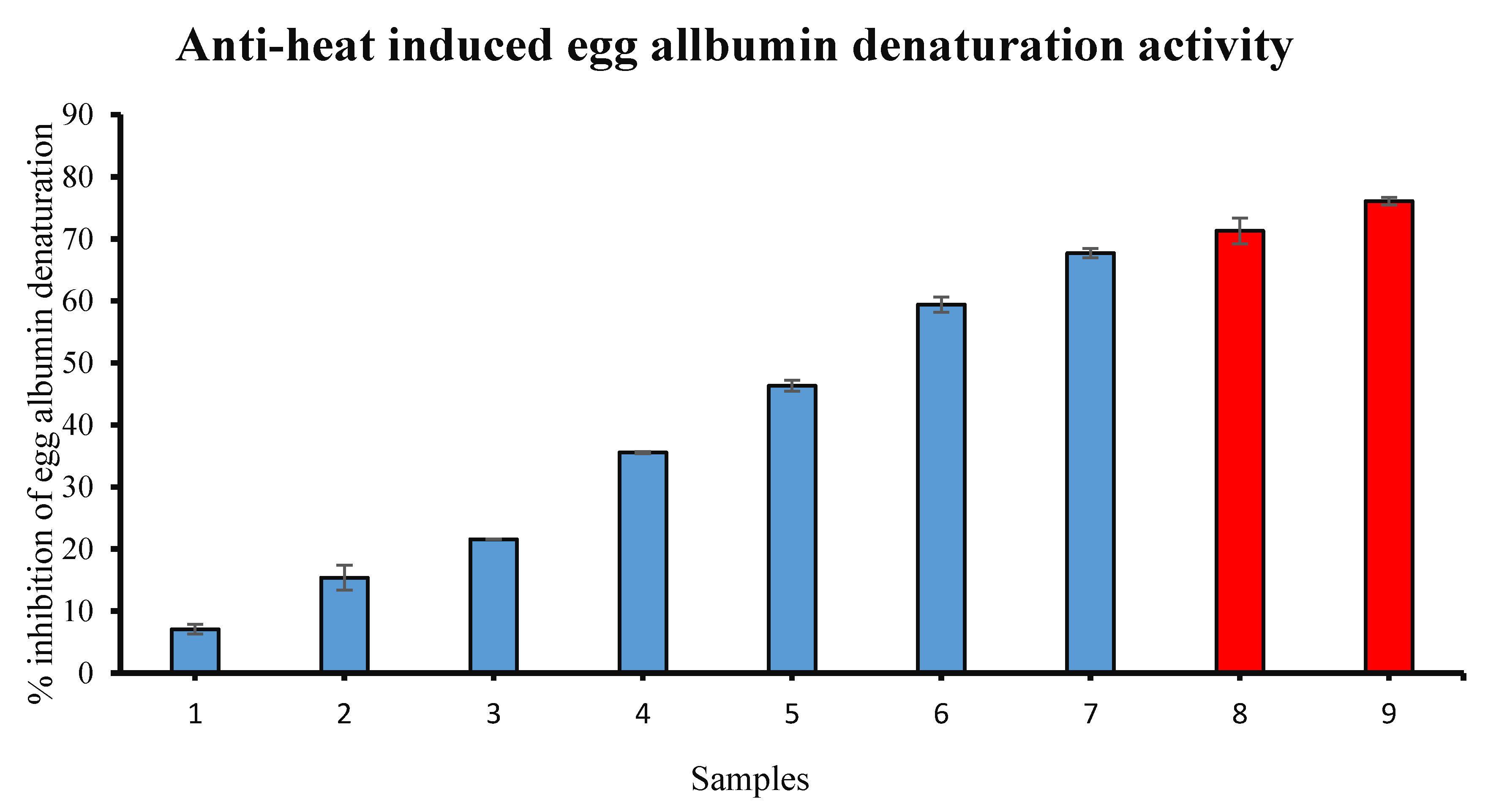
2.3.8. Inhibition of Egg Albumin Denaturation

Reaction mixtures containing 200 μL of fresh hen egg albumin, 2800 μL of phosphate buffer (pH 6.4), and two mL of different dilutions (50–600 µg/mL) of garlic extract and standard drug (diclofenac sodium) were placed in separate test tubes, respectively [22]. The mixtures were incubated for 15 min at 37 ± 2 °C. Later, mixtures were subjected to heat (70 °C) for five minutes, cooled down and the samples’ absorbance was assessed at 660 nm using a spectrophotometer. Buffer was used as blank. The % egg albumin denaturation inhibition had been measured using the following equation:
Percentage   Inhibition = [ ( Xc Xs ) / Xc ] × 100
where Xc = Absorbance of control and Xs = Absorbance in presence of extract/diclofenac sodium.

2.4. In Vivo Studies

2.4.1. Animals

Adult healthy male albino white rats (32 in total) with body weights of 175–200 g were obtained from King Saud University, Saudi Arabia and were arbitrarily partitioned into four groups of eight rodents in each group (n = 8), one week after adjustment to the research facility environment. The animals were placed in apolycarbonate cages with a room temperature (20–25 °C) and a 12 h light-twelve-hour dark cycle. The animals had free access to standard rat chow and fresh water. This research had been endorsed by the Ethics Committee of College of Applied Medical Sciences, Qassim University (cams1-2018-1-14-s-3360).

2.4.2. Induction of Liver Damage

To trigger the hepatic injury, CCl4 was diluted in olive oil in a proportion of 1:1 (v/v) [23] and 150 µL of the mixture was administrated orally thrice per week for total eight weeks.

2.4.3. Animals Grouping

As discussed above, the animals were randomly categorized into four groups to achieve the objectives of the study. Group 1 (normal control) animals were given normal saline as vehicle and this group. Group 2 was the CCl4 group and this group received 1:1 (v/v) CCl4 in olive oil orally thrice per week. Group 3 (co-treatment group) received CCl4 and garlic extract given at a dosage of 75 mg/kg b.w and 1:1 (v/v) CCl4 in olive oil. Group IV animals received garlic extract at a dose of 75 mg/kg b.w orally three times a week.

2.4.4. Blood Sampling

Twenty four h after the last dosing of CCl4, all animals were subjected to anesthesia by chloroform inhalation and killed by cervical decapitation. Blood samples were obtained and centrifuged at 3000 rpm for 10 min at 4 °C to isolate the serum. The collected serum was kept at −20 °C for further analysis.

2.4.5. Measurement of Serum Aminotransferase Activities, Lipid Profile and Total Protein

The activities of liver function enzymes such as alanine aminotransferase (ALT), aspartate aminotransferase (AST) and alkaline phosphatase (ALP) in animal serum were determined using an automatic biochemical analyser (Hospitex Diagnostic, Eos- Bravo, Florence, Italy) as directed by the manufacturer and the results were interpreted correspondingly. Commercial kits (Abcam) were used to determine the concentrations of cholesterol, triglyceride, total protein and albumin.

2.4.6. Measurement of Antioxidant Enzymes

The livers from each group of animals were quickly excised and homogenized using chilled phosphate buffer saline (pH 7.4). The supernatants were collected after centrifugation of homogenates at 4500× g for 20 min at 4 °C and stored at −20 °C for future analysis of antioxidant enzymes. The activities of hepatic antioxidant enzymes like glutathione peroxidase (GPx), superoxide dismutase (SOD) and glutathione level (GSH) and Total Antioxidant Capacity (T-AOC) were investigated using diagnostic kits (Abcam) according to the manufacturer’s instructions.

2.4.7. Assay of C-Reactive Protein (CRP), TNF-Alpha, IL-6 and ICAM1

An enzyme-linked immunosorbent assay (ELISA) of C-reactive protein, TNF-α, IL-6, ICAM1 Kit (Abcam) in blood serum was used to measure the concentration of all markers.

2.4.8. Haematoxylin and Eosin (H&E) Staining for Histological Examinations

Liver tissues were extracted for histological analyses and processed according to a standardized method with minor modifications [24]. Further, they were washed with cold saline, dried using filter paper, weighed and then treated with 10% phosphate-buffered formalin. Tissues were dehydrated in different grades of ethanol, cleaned in xylene and tissue was processed using an automated tissue processor (Leica, Nussloch, Germany). The tissues were embedded in paraffin wax via an embedding station (Leica) and 5 μm thick sections were prepared and mounted on polylysine-coated glass slides. The sections were stained using haematoxylin and eosin staining and tissues architecture was evaluated under a light microscope (Olympus, Tokyo, Japan). Images were captured at ×100 magnification and the results were interpreted accordingly.

2.4.9. Masson Trichrome Staining

Masson trichrome staining was done as prescribed by the kit’s manufacturer. Briefly, deparaffinising of the sections was performed and preheated Bouin’s fluid was added for 60 min, followed by a 10 min cooling period. The slides were washed in tap water till the section became fully clear. The entire slide was stained with working Wiegert’s Iron Haematoxylin for 5 min. Biebrich Scarlet/Acid Fuchsin Solution was then applied to the slides for 15 min. Differentiation in phosphomolybdic/phosphotungstic acid solution was made for 10–15 min or until collagen did not become red. Without rinsing, aniline blue solution was applied to the slides for 5–10 min. Acetic acid solution (1%) was applied to the slides for 3–5 min. Dehydration was performed very quickly in two changes of 95% ethyl alcohol, followed by two changes of absolute alcohol and the slides were cleared via xylene and mounting was performed.

2.4.10. Expressional Evaluation of VEGF Protein

VEGF protein immunoreactivity was examined in all experimental animals and the expression pattern was evaluated using previously described methods with slight modifications [25,26]. In brief, xylene was used to deparaffinise the samples and graded alcohol was used for rehydration and phosphate buffer saline was used three times for washing of sections. Unmasking of antigen sites was done by utilizing 50 mM citrate buffer (pH 6.0) via microwave method then tissues were kept in 3% hydrogen peroxide for 20 min for blocking of endogenous peroxidase. Later, sections were kept in blocking agent for 60 min at room temperature to block the undesirable sites in humidified atmosphere. Then, VEGF antibodies had been implemented on the tissue overnight at 4 °C. Later, incubation was carried out with secondary biotinylated antibody for two hours. The sections were washed in phosphate buffer saline thrice and were incubated with streptavidin peroxidase for 1 h. At that point, diaminobenzidine (DAB) chromogen was used and sections were counterstained with haematoxylin and the expression pattern was analysed under a light microscope and images was captured to analyse the results.
The immunostained sections were examined using an Olympus (Tokyo, Japan) microscope and results interpreted accordingly. Evaluations of VEGF protein expression was made based on cytoplasmic staining. Quantification of positively cytoplasmic stained hepatocytes was evaluated by a pathologist blinded to the different animal groups using a light microscope and measured IHC positivity. A total of 500 cells was counted manually at high power (×400) after identifying at low power (×100) in five selected fields. The positively stained cells were expressed as a percentage of the total cells counted in each case and results are expressed as mean plus or minus standard deviation.

2.4.11. Statistical Analysis

All numerical data was interpreted in terms of mean ± SD expressed and all comparisons were made by one-way analysis of variance (ANOVA) test. Statistical analysis was done by utilizing SPSS software. p < 0.05 was considered to be statistically significant.

3. Results

3.1. In Vitro Studies

3.1.1. Preliminary Study

The presence of flavonoids and phenolic compounds was confirmed by the respective assays as described above. The appearance, smell, and the percentage yield of garlic methanol extract were found to be brownish, pungent, and 6.32%, respectively. Further, total phenolic compounds in the garlic extract was determined to be 18.45 ± 0.08 mg gallic acid equivalent/g dry weight of extract that is nearly same as detailed by previous researchers [27]. It is indeed important to keep in mind that phenolic contents show the means of protection against infections or environmental stress, and therefore, are not related with growth activity and plant tissue development [12]. Moreover, any presence of sulfur compounds probably decreases the total phenolic and flavonoid content.
The chemical composition of garlic (Allium sativum) extract has been investigated previously. Phenolic acids, including vanillic acid, caffeic acid, p-coumaric acid and ferulic acid, anthocyanins like cyanidin-3-(6′-malonyl)-glucoside) and organosulfur compounds such as L-alliin and methiin were shown to be the major components of garlic extract [28]. HPLC GC/MS analysis identified luteolin-7-glucoside, luteolin, apifenin-7-glucoside, hyperoside, rutin, caffeic acid, chlorogenic acid and rosmarinic acid in as the major phenolic compounds methanolic garlic extract [29]. However, another study revealed that pyrogallol (139.35 to 418.80 μg/g), rutin (51.35 to 90.45 μg/g) protocatechuic acid (31.75 to 76.25 μg/g), β-resorcyclic acid (313.5 to 452.µg/mL), gallic acid (67.40 to 196.80 μg/g) and quercetin (25.70 to 56.65 μg/g) were the principal phenolic constituents in garlic extract [30].
To calculate the total flavonoid content of the extract, the colorimetric AlCl3 test employing quercetin as reference was used. The total flavonoid content was found to be 13.58 ± 0.01 mg quercetin equivalents (QE)/g dry weight of the extract.

3.1.2. α,α-Diphenyl-β-Picrylhydrazyl (DPPH) Free Radical Scavenging Method

The potential of giving electron, and reducing DPPH free radical is exploited in this assay to evaluate the antioxidant activity of garlic extract. It was seen that the addition of garlic extract to DPPH solution reduced the DPPH free radical to the corresponding hydrazine. DPPH reduction was investigated by recording the drop in absorbance at 517 nm. Our results suggest that garlic has an excellent DPPH free radical scavenging activity and this ability at 600 μg/mL was shown to be the most notable. Our data suggest that garlic extract might be a rich source of anti-oxidant compounds and its DPPH scavenging activity increases dose-dependently. The DPPH radical scavenging test results are shown in Figure 1.

3.1.3. Hydrogen Peroxide (H2O2) Radical Reducing Ability

Because of its antioxidant nature, garlic extract is capable of reducing reactive oxygen species like H2O2. We show the % hydrogen peroxide reducing ability of garlic extract in Figure 2. In the current study, we have investigated the percentage hydrogen peroxide reducing ability of different concentrations of garlic extract as well as ascorbic acid. Our data shows that hydrogen peroxide reducing ability of garlic extract increases in a concentration-dependent way. Garlic shows the highest reducing ability at a dosage of 600 µg/mL. Ascorbic acid exhibited significant hydrogen peroxide reducing ability at a dosage of 100 µg/mL. This ability of garlic extract confirms the presence of significant amount of polyphenolic substances in it.

3.1.4. Inhibition of Bovine Serum Albumin Denaturation

During denaturation, some external factors disrupt the native structures of proteins. The denaturation of bovine serum albumin (BSA) is very important because it induces the expression of antigens linked with type III hypersensitivity reaction. Ultimately, it results in the initiation of an inflammatory response. Hence, the anti-inflammatory ability of garlic extract was explored by assessing its protein denaturation protection potential. The data of our experiments indicate that garlic extract has an excellent ability to protect towards denaturation of bovine serum albumin induced by heat, and the percent protection increases with the increase in the amount of garlic extract. The most considerable anti-inflammatory activity was shown by the garlic extract at 600 µg/mL. The presence of antioxidant compounds can be suggested to be responsible for the protective ability of garlic extract against heat-induced BSA denaturation. Ibuprofen is a very common anti-inflammatory medicine that exhibited the highest percent protection at 200 µg/mL (Figure 3).

3.1.5. Anti-Proteinase Ability

There are certain enzymes called proteinases which are known to be involved in arthritic reactions. Proteinases cause tissue damage so the anti-proteinase ability of a natural product can be considered to be associated with inflammatory disorders.
Our results suggest garlic has a noteworthy anti-proteinase ability and this ability had been found to increase with the amount administered. The highest anti-proteinase ability of garlic was observed at 600 µg/mL. The reference drug diclofenac sodium exhibited maximum inhibition at 200 µg/mL (Figure 4).

3.1.6. Inhibition of Egg Albumin Denaturation

The anti-inflammatory effect of garlic was appraised against egg albumin denaturation (Figure 5). Plant extract displayed egg albumin denaturation inhibition in an amount-dependent way (50–600 μg/mL). The findings suggest that garlic is very remarkably efficient towards denaturation of egg albumin induced by heat. The extract at 100–600 µg/mL demonstrated anti-inflammatory activity because it was able to prevent the denaturation of egg albumin induced by heat which might be attributed to the presence of some anti-oxidant polyphenolic substances. The reference medicine diclofenac displayed the highest inhibition at 200 µg/mL.

3.2. In Vivo Studies

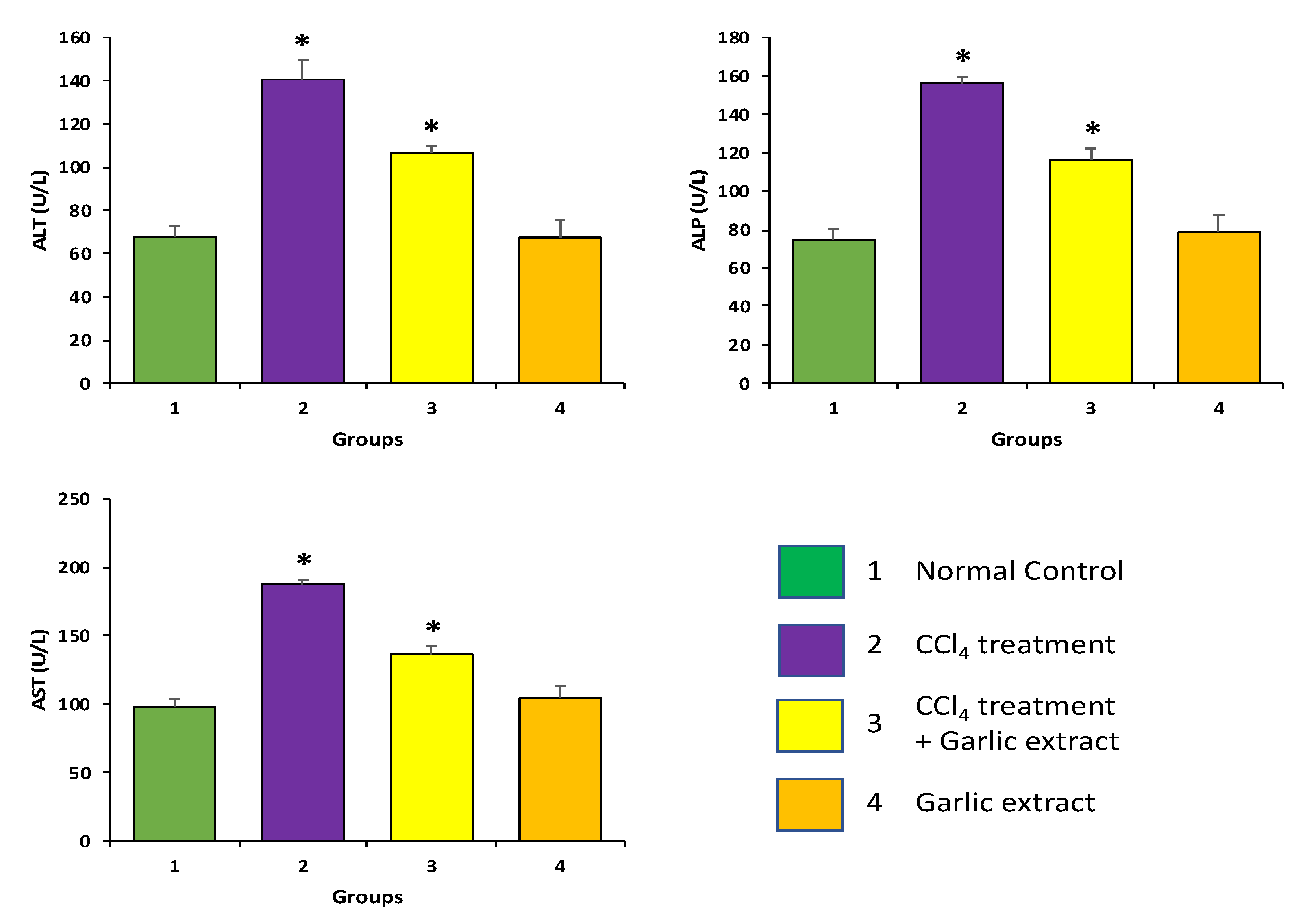
3.2.1. The Effect of Garlic Extract on Liver Function Enzymes in Animals Exposed to CCl4

The effects of garlic extract were evaluated in the experimental group for biochemical parameters. In the current study, it was noticed that CCl4 induction showed significantly increased levels of liver function enzymes, including ALP, ALT, and AST, in contrast to control animals (p < 0.05). Furthermore, our findings also demonstrated that treatment of CCl4-induced animals with garlic extract led to a considerable decrease in the levels of these enzymes and near to normal levels were achieved, which establishes the hepatoprotective potential of garlic extract (p < 0.05) (Figure 6).

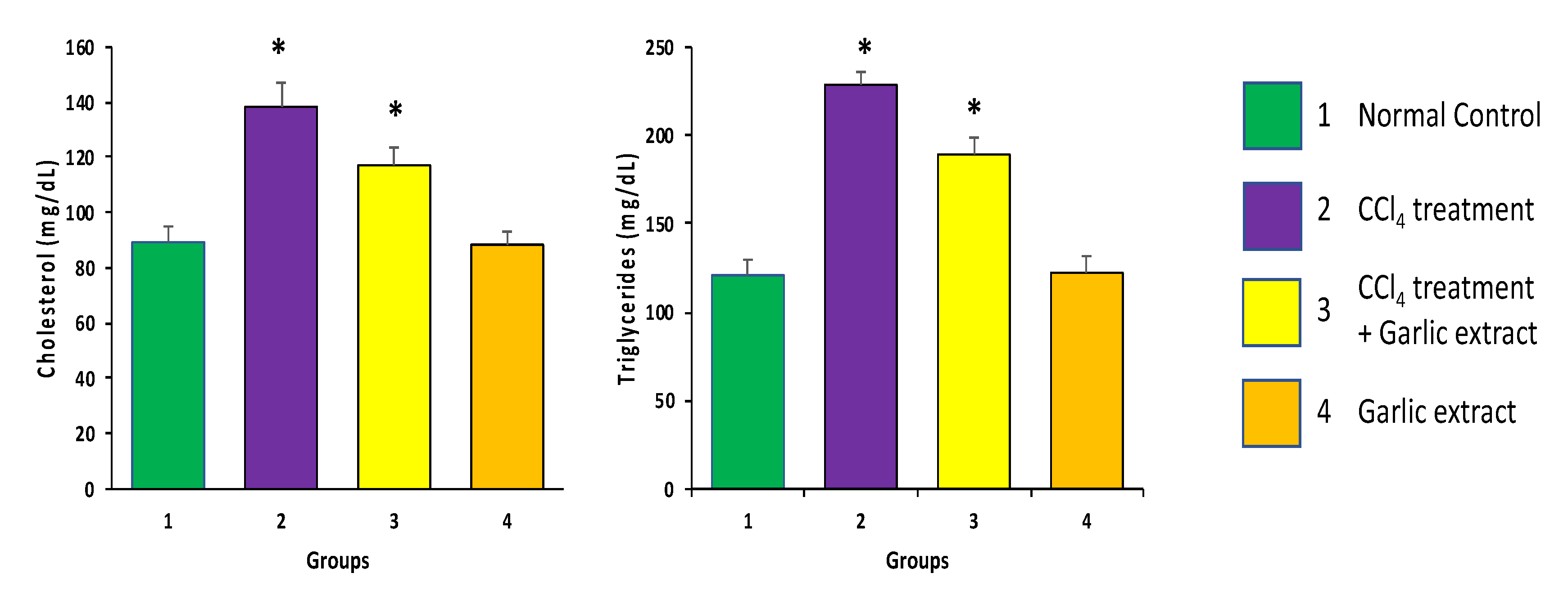
3.2.2. Effect of Garlic Extract on Lipid Profile in Animals Exposed to CCl4

CCl4 induced animals showed a substantial decrease in serum lipids e.g., cholesterol and triglycerides in contrast to control animals. Giving garlic extract at a dosage of 75 mg/kg with CCl4 maintained the serum lipid and level had been close to normal range in contrast to animals having CCl4 (Figure 7) (p < 0.05).

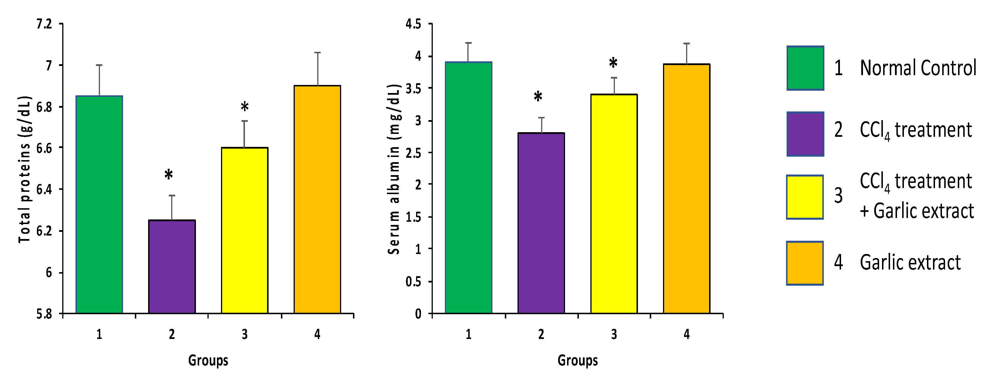
3.2.3. Effect of Garlic Extract on the Total Protein and Albumin in Animals Exposed to CCl4

CCl4-induced group animals exhibited a considerable decrease in albumin and overall protein content as compared to the control animals. Co-administrating garlic extract and CCl4 together maintains the increase in overall protein content, and albumin relative to the group having CCl4 only (Figure 8) (p < 0.05).

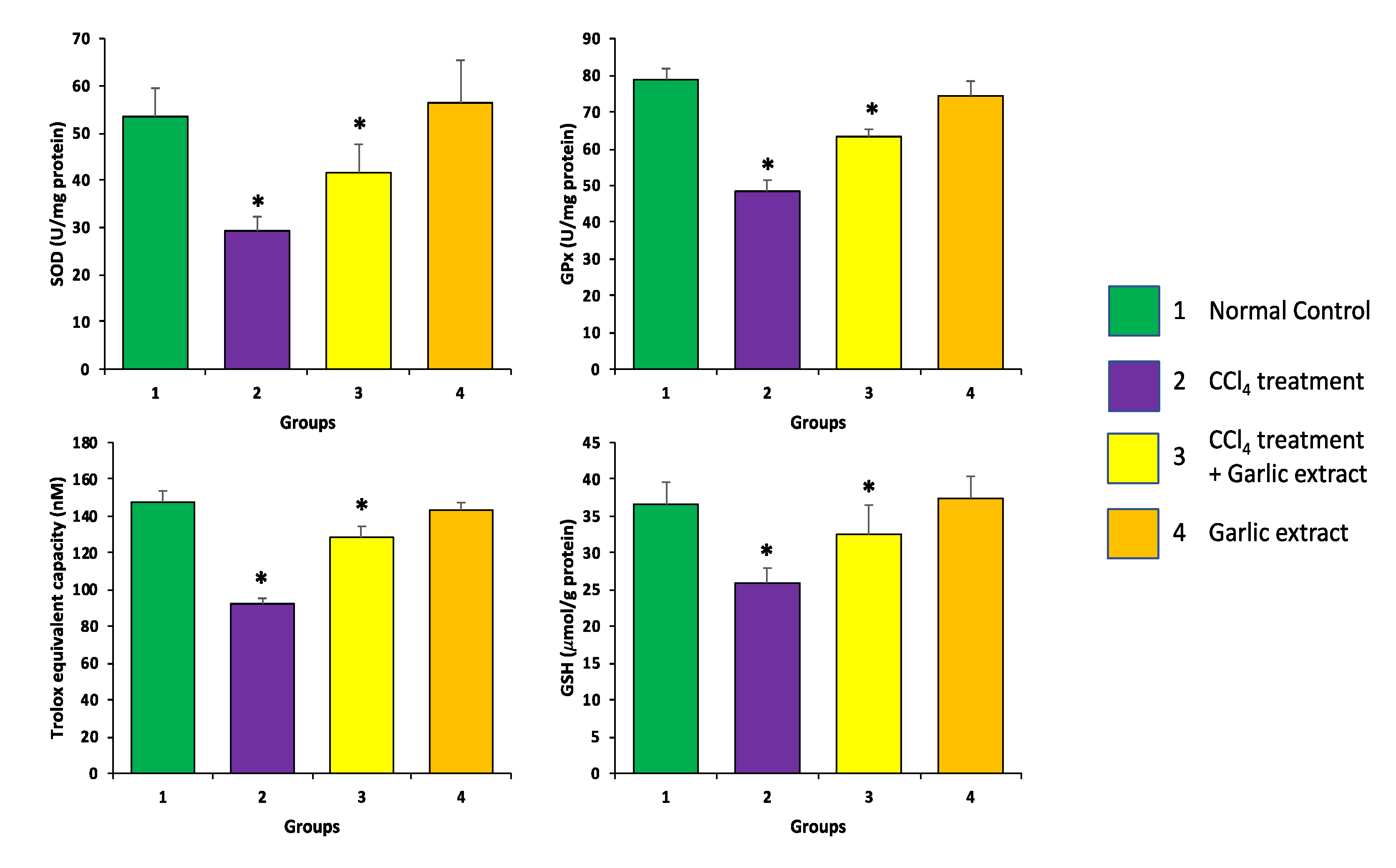
3.2.4. Effect of Garlic Extract on Antioxidant Enzymes Levels in Animals Exposed to CCl4

A considerable decrease in levels of antioxidant enzymes including superoxide dismutase (SOD), glutathione (GSH), glutathione peroxidase (GPx), and T-AOC was observed in CCl4-induced animals when compared with the control (Figure 9). Moreover, garlic extract treatment facilitated a significant increase in the all tested antioxidant enzymes level when compared with the CCl4-induced group. Besides, in animals treated with garlic extract only, it was observed that the antioxidant enzymes level was almost in the control group range. However, it was noticed that the difference in antioxidant enzymes levels in CCl4 induced animals group and control group was statically significant (p < 0.05) which establishes the hepatoprotective potential of garlic extract through modulation of the antioxidant enzyme activity.

3.2.5. Effect of Garlic Extract on Inflammatory Markers in Animals Exposed to CCl4

It was observed that levels of C-reactive protein, TNF-alpha, IL-6, ICAM1 and IL-1β were considerably increased in CCl4-induced animals in contrast to control animals (Figure 10). The administration of garlic extract at a dosage of 75 mg/kg body weight decreased the TNF-α level significantly relative to the CCl4-treated animals (p < 0.05).
The amounts of IL-6, IL-1β and CRP was enhanced in animals having CCl4 relative to the control group (p < 0.05) (Figure 10). In animals administered both garlic extract and CCl4, the amounts of IL-1β, IL-6, and CRP was decreased noticeably relative to the CCl4 group (p < 0.05) (Figure 10). Moreover, ICAM1 was also measured in the treated and untreated groups of animals and it was observed that the levels of ICAM1 were high in animals administrated with CCl4 relative to control animals. In the animal group having both garlic extract and CCl4, the level of ICAM1 was decreased considerably in contrast to animals having CCl4 (p < 0.05).
The findings showed that C-reactive protein, TNF-alpha, IL-6, ICAM1 and IL-1β level in the CCl4 -induced animals were increased considerably in contrast to the control group (Figure 10). The supply of garlic extract at a dosage of 75 mg/kg has shown to decrease TNF-α level significantly relative to CCl4 treated animals (p < 0.05).

3.2.6. Effect of Garlic Extract on Hepatocytes Architecture

Liver sections of the control animals exhibits typical cellular architectures with intact cytoplasm, sinusoidal spaces, prominent nucleus, and central vein. Liver sections of animals exposed to CCl4 shows various types of alterations such as infiltration of lymphocytes, edema and congestion. The animal group treated with garlic extract plus CCl4 showed amelioration of the hepatocyte architecture truncated by CCl4 induction (Figure 11). The animals in the garlic extract-treated group revealed significantly low alteration as compared to the extensive liver damage found in the CCl4 group. Garlic administration showed prevention of the swelling, lymphocytes infiltration, necrosis, congestion and haemorrhages caused by CCl4.

3.2.7. Masson Trichrome Staining

Masson’s trichrome staining had been carried out on all experimental group, and the findings revealed a significant increase in collagen deposition in group treated with CCl4 relative to control animals. Following treatment with garlic extract, collagen deposition was reduced (Figure 12).

3.2.8. Effect of Garlic Extract on Expression Pattern of VEGF Protein

A total of five fields from each section were selected, and 100 cells from each area were counted and the mean percentage positivity was calculated. Expression of VEGF was considered positive if more than 5% of cells showed positivity and less than 5% positivity was taken as the negative case [26]. The percentage of IHC staining intensity of cytoplasmic VEGF was considered mild when positive staining is < 25%, moderate when positive staining is 25–50% and strong when positive staining is in > 50%. The VEGF protein staining among the treatment group was either negative, weakly positive or strong positive. The cytoplasmic expression of VEGF protein was seen in 50% (4/8) cases of CCl4 treated group (Figure 13), whereas the control group did not show any expression of VEGF protein. Moreover, the expression of VEGF protein was also noted 25% (2/8) of the CCl4 and garlic extract treated group, but the intensity of positivity was weak. Besides, VEGF protein did not show any expression in the group treated with garlic extract only (Figure 13).

4. Discussion

Since ancient times, medicinal plants have been used worldwide as traditional medicines in the management of various diseases. There are many studies exhibiting that some promising phytochemicals can be developed as an alternative for synthetic medicines for health problems [31]. Among these, many plants have been reported to have considerable hepatoprotective activity. Still the search for new innovative drugs for the management of liver diseases is needed. Garlic is cultivated worldwide and is possibly effective for the management of various illnesses. Our research investigated its antioxidant, anti-inflammatory, as well the hepatoprotective effect against CCl4-induced liver injury to verify its health promoting abilities.
The liver is the second largest and one of the most important organs in our body which plays a significant role in regulating different biological processes. The liver has diverse functions such as metabolism, synthesis of many important biomolecules, detoxification, secretion and storage, etc. Further, the maintenance, performance, and regulation of body homeostasis is helped by the liver. Almost all the biochemical pathways including growth, fighting against disease, nutrient supply, energy provision and reproduction, metabolism of carbohydrate, protein and fat, detoxification, secretion of bile and storage of vitamins are carried out by liver, which also helps in the detoxification and removal of foreign substances. Hence, the liver is the prime target for attack by foreign substances which leads to liver disorders and damage.
These days, liver diseases are a focus of more research because they cause notable morbidity and mortality worldwide. Some specific reasons of liver diseases include exposure to chemicals, excess consumption of alcohol, infections and autoimmune disorders [32]. It is well-known that chemicals like carbon tetrachloride (CCl4) can lead to excessive formation of ROS which results in oxidative damage and lipid peroxidation. These are key factors for liver damage induced by chemicals. CCl4 is a very common hepatic toxin in the animal that is used to study the hepatoprotective effect of various medicinal plants as well as natural compounds, as a single exposure to CCl4 may lead to severe liver necrosis, as well as steatosis [6,7].
Elevated or abnormal levels of liver function enzymes are considered to be significant markers of liver damage. In the current study, administration of CCl4 was found to increase the levels of ALP, ALT and AST significantly, as compared to control group. However, our results also indicated that treatment of CCl4-induced animals with garlic extract caused a significant decrease of ALT, AST and ALP enzyme levels. Therefore, our results indicate that garlic extract has a significant hepatoprotective potential. The findings of current study were in accordance with a previous study which has also confirmed that levels of AST and ALT became increased considerably after CCl4 treatment, and this was suppressed by aged black garlic treatment [33]. Further, the administration of a single garlic clove resulted in a significant decrease of serum ALP, AST and ALT [34].
Oxidative stress is generated due to the imbalance in the production of cellular oxidants and their removal due to a poor antioxidant defense system. Oxidative stress and oxidative damages have been reported to be linked with hepatic injury. Consequently, antioxidant supplementation has become a notable therapeutic strategy for decreasing the risk of liver disease caused due to superfluous free radicals [35,36]. Various types of antioxidants have the ability to reduce or neutralize free radicals produced by toxic materials. Moreover, oxidizing GSH to glutathione disulfide causes reduction of hydrogen peroxide as well as hydroperoxide. In the current study, our data indicated that antioxidant enzymes levels were significantly reduced in animals treated with CCl4 when compared with the control group. Moreover, garlic extract treatment facilitated a significant increase in all tested antioxidant enzymes levels when compared with CCl4-induced groups. This supports the notion that garlic extract possesses hepatoprotective potential which may be due to its potential to increase the activity of antioxidant enzymes. Further, our study supports a previous study which suggested that CCl4 treatment led to a significant reduction of GSH levels when compared to the normal group and GSH levels were increased significantly by treatment with extract [37]. Similarly, it has been documented that hepatic SOD, CAT and GPx are reduced by cyclophosamide. However, the treatment with black garlic extract reverses the reduction of hepatic SOD, CAT and GPx which occurred due to treatment with cyclophosamide [38]. Further, our study is in accordance with another previous study which has described that lipid peroxidation becomes significantly reduced because of the administration of garlic and silymarin. On the other hand, endogenous antioxidants like SOD, CAT, GSH have been reported to be considerably increased due to administration of garlic and silymarin [39]. SOD is an important line of defense against oxidative stress [40].
Inflammation is considered to be one of the root causes of liver tissue damage and hence, it contributes significantly to non-alcoholic fatty liver diseases, severe fibrinogenesis, and hepatocellular carcinoma. Inflammation has been reported to be activated by CCl4-induced liver toxicity which leads to the release of pro-inflammatory cytokine including IL-lβ and TNF-α [34]. IL-1β and TNF-α are the main cytokines in inflammatory process and hepatic injury is linked with increased levels of IL-1β and TNF-α [41]. CRP (C-reactive protein) is an important substance which is released by the liver and it plays an important role in the inflammatory process. CRP has been known to be involved in opsonization as well as activation of the complement system in response to IL-6 secretion [24]. In addition, CRP is considered to be a sensitive marker of inflammation and it has been reported to be increased intensely in other conditions including trauma, burns, myocardial infarction and cancer [42].
It has been documented that increased level of various pro-inflammatory cytokines is associated with the liver damage. Hence, increased level of pro-inflammatory markers might be involved in initiation of the pathogenesis of various liver diseases. In the current study, it was observed that CRP, TNF-α, IL-6, ICAM1 levels in animal treated with CCl4, were increased significantly as compared to control group. The treatment with garlic extract at a dose of 75 mg/kg b.w, was shown to significantly decrease the inflammatory marker level. A pioneering study based on garlic extract reported that levels of TNF-α and IL-1β in the animals treated with CCl4 group were significantly increased. However, our data indicates that treatment with garlic extract resulted in the reduction of TNF-α and IL-1β [37].
Further, the infiltration of lymphocytes, inflammation, edema, haemorrhages and necrosis in the CCl4-treated group have been found to be linked with hepatic cell injury. These changes were significantly attenuated by treatment with garlic extract. This study was similar to previous finding related to histopathological examination of the liver sections of CCl4/DEN administrated animals with hepatocyte ballooning, multiple centrilobular necrosis, and infiltration of inflammatory cells into the portal tract as well as sinusoid [43,44,45]. Another study also detailed that extract of black garlic could decrease CCl4 administration-induced hepatotoxicity and hepatitis. Further, CCl4 induction leads to hepatic inflammation with infiltration of mononuclear cell surrounding hepatic veins accompanied by slight damage of neighboring tissues. Inflammation reactions and typical structure of hepatocytes and portal space have been reported to become significantly improved by treatment of single clove of garlic [46]. Angiogenesis signalling is involved in the wound mending response in hepatic fibrosis, and not only to Extracellular matrix (ECM) deposition, but also to portal hypertension [47]. VEGF may induce growth of new blood vessels as a response to hepatic injury, which is essential for HF [48]. In the current study, our data shows that animals treated with CCl4 shows high VEGF protein cytoplasmic expression. Whereas, VEGF expression was low in the group administrated with CCl4 plus garlic extract. Moreover, garlic and its active compound have established their potential of cancer management via altering different cell signalling molecules, including VEGF [49].
Our data from in vivo experiments have indicated that garlic extract possess anti-inflammatory potential. This is in accordance with current in vitro results which confirm the anti-inflammatory property of garlic extract by inhibiting heat-induced egg albumin and bovine serum albumin denaturation, as well as by its ability against proteinase. Denaturation of proteins is an important motivating factor for inflammation and proteinase are enzymes that are reported to be associated with tissue damage. Our data shows that garlic extract has considerable potential to inhibit heat induced denaturation and it displays a significant anti-proteinase activity. Thus, the in vitro anti-inflammatory results support our in vivo anti-inflammatory results. It has been shown that garlic extract has excellent anti-inflammatory activity that is basically equivalent to the reference anti-inflammatory treatment, i.e., ibuprofen and diclofenac sodium. Further, in vitro data indicates that garlic has strong antioxidant potential and in vivo data shows that administration of garlic extract improves the antioxidant status of animals treated with CCl4. Hence, the strong antioxidant nature of garlic extract may be considered as an important mechanism for its health beneficial activities. In 2017, Naji and colleagues reported that garlic is a good source of natural antioxidants and it has protective effect against chemicals such as CCl4 induced hepatotoxicity in rabbits [34]. Thus, our study confirms the findings of Naji et al., and proposes that administration of garlic extract can have some significant role in prevention of CCl4 induced liver damages.

5. Conclusions

Garlic is commonly used as a food additive and traditional medicine since ancient times. Our findings indicate that garlic extract possesses substantial antioxidant activity that can be responsible for prevention of reactive oxygen species-mediated disorders including inflammation and liver damage. In addition, our investigation confirms the hepatoprotective nature of garlic extract against CCl4-induced liver damage as noted by a noteworthy decrease in liver enzymes, reduced oxidative damage and suppressed inflammatory responses. In addition, the in vitro heat-induced denaturation inhibition activities and anti-proteinase potential further evidence its hepatoprotective role. Moreover, this data reports that the administration of garlic extract improves the hepatocytes architecture through maintenance of histopathological changes and reduction of collagen fiber which confirms its health promoting effects. The outcomes of the current research support the idea that garlic extract has excellent antioxidant, and anti-inflammatory effects and have the ability to manage the liver pathogenesis. Thus, our finding advocates that the consumption of garlic can be a potential therapeutic remedy in the inhibition of liver ailments. Further, it is recommended to isolate and characterize the chief constituents present in the garlic that contributes to its hepatoprotective potentials.

Author Contributions

Conceptualization, S.A.A., and A.H.R.; methodology, A.H.R., A.A.K., S.A.; validation, M.A.A., F.A.; formal analysis, A.A. investigation, S.A.A., A.A., A.A.K. and A.H.R.; resources, S.A.A., S.A., and M.A.A.); data curation, F.A. and A.A. writing—original draft preparation, A.H.R., and S.A.; writing—review and editing, A.A., M.A.A., F.A., A.A.K.; supervision, M.A.A. and A.A.; funding acquisition, S.A., M.A.A., A.A. All authors have read and agreed to the published version of the manuscript.

Funding

This research received no external funding.

Acknowledgments

Researchers would like to thank the Deanship of Scientific Research, Qassim University for funding publication of this project.

Conflicts of Interest

The authors declare no conflict of interest.

References

  1. Nisar, B.; Sultan, A.; Rubab, S.L. Comparison of Medicinally Important Natural Products versus Synthetic Drugs-A Short Commentary. Nat. Prod. Chem. Res. 2018, 6, 1–2. [Google Scholar] [CrossRef]
  2. Khan, M.A.; Anwar, S.; Aljarbou, A.N.; Al-Orainy, M.; Aldebasi, Y.H.; Islam, S.; Younus, H. Protective effect of thymoquinone on glucose or methylglyoxal-induced glycation of superoxide dismutase. Int. J. Biol. Macromol. 2014, 65, 16–20. [Google Scholar] [CrossRef] [PubMed]
  3. Anwar, S.; Younus, H. Inhibitory effect of alliin from Allium sativum on the glycation of superoxidedismutase. Int. J. Biol. Macromol. 2017, 103, 182–193. [Google Scholar] [CrossRef] [PubMed]
  4. Sivakrishnan, S. Liver diseases—An overview. World J. Pharm. Pharm. Sci. 2019, 8, 1385–1395. [Google Scholar]
  5. Lee, W.M. Drug-Induced Hepatotoxicity. N. Engl. J. Med. 2003, 349, 474–485. [Google Scholar] [CrossRef]
  6. Brautbar, N.; Williams, J. Industrial solvents and liver toxicity: Risk assessment, risk factors and mechanisms. Int. J. Hyg. Environ. Health 2002, 205, 479–491. [Google Scholar] [CrossRef]
  7. Manibusan, M.K.; Odin, M.; Eastmond, D.A. Postulated Carbon Tetrachloride Mode of Action: A Review. J. Environ. Sci. Health Part C 2007, 25, 185–209. [Google Scholar] [CrossRef]
  8. Cheeseman, K.H.; Albano, E.F.; Tomasi, A.; Slater, T.F. Biochemical studies on the metabolic activation of halogenated alkanes. Environ. Health Perspect. 1985, 64, 85–101. [Google Scholar] [CrossRef]
  9. Srivastava, A.; Shivanandappa, T. Hepatoprotective effect of the root extract of Decalepis hamiltonii against carbon tetrachloride-induced oxidative stress in rats. Food Chem. 2010, 118, 411–417. [Google Scholar] [CrossRef]
  10. Ritter, C.; Reinke, A.; Andrades, M.; Martins, M.R.; Rocha, J.; Menna-Barreto, S.; Quevedo, J.; Moreira, J.C.F.; Dal-Pizzol, F. Protective effect of N-acetylcysteine and deferoxamine on carbon tetrachloride-induced acute hepatic failure in rats. Crit. Care Med. 2004, 32, 2079–2083. [Google Scholar] [CrossRef]
  11. Mccord, J.M. The evolution of free radicals and oxidative stress. Am. J. Med. 2000, 108, 652–659. [Google Scholar] [CrossRef]
  12. Anwar, S.; Almatroudi, A.; Allemailem, K.S.; Joseph, R.J.; Khan, A.A.; Rahmani, A.H. Protective Effects of Ginger Extract against Glycation and Oxidative Stress-Induced Health Complications: An In Vitro Study. Processes 2020, 8, 468. [Google Scholar] [CrossRef]
  13. Castellano, G. Esteatohepatitis no alcohólica. Gastroenterol. Hepatol. 1999, 22, 13–19. [Google Scholar]
  14. Angulo, P. Medical progress: Nonalcoholic fatty liver disease. N. Engl. J. Med. 2002, 346, 1221–1231. [Google Scholar] [CrossRef] [Green Version]
  15. Luedde, T.; Schwabe, R.F. NF-κB in the liver-linking injury, fibrosis and hepatocellular carcinoma. Nat. Rev. Gastroenterol. Hepatol. 2011, 8, 108–118. [Google Scholar] [CrossRef] [Green Version]
  16. Raghu, R.; Lu, K.-H.; Sheen, L.-Y. Recent Research Progress on Garlic (dà suàn) as a Potential Anticarcinogenic Agent Against Major Digestive Cancers. J. Tradit. Complement. Med. 2012, 2, 192–201. [Google Scholar] [CrossRef] [Green Version]
  17. Abdel-Naim, A.B.; Khalifa, A.E.; Ahmed, S.H. Protective Effects of Garlic Oil against Liver Damage Induced by Combined Administration of Ethanol and Carbon Tetrachloride in Rats. Egypt. J. Hosp. Med. 2002, 6, 27–36. [Google Scholar]
  18. Eidi, A.; Eidi, M.; Esmaeili, E. Antidiabetic effect of garlic (Allium sativum L.) in normal and streptozotocin-induced diabetic rats. Phytomedicine 2006, 13, 624–629. [Google Scholar] [CrossRef]
  19. Taie, H.A.A.; Salama, Z.A.-E.R.; Radwan, S. Potential activity of basil plants as a source of antioxidants and anticancer agents as affected by organic and bio-organic fertilization. Not. Bot. Horti Agrobot. 2010, 38, 119–127. [Google Scholar]
  20. Ruch, R.J.; Cheng, S.-J.; Klaunig, J.E. Prevention of cytotoxicity and inhibition of intercellular communication by antioxidant catechins isolated from Chinese green tea. Carcinogenesis 1989, 10, 1003–1008. [Google Scholar] [CrossRef]
  21. Sakat, S.S.; Juvekar, A.R.; Gambhire, M.N. In Vitro antioxidant and anti-inflammatory activity of methanol extract of Oxalis corniculata linn. Int. J. Pharm. 2010, 2, 146–155. [Google Scholar]
  22. Hasan, A.U.H. Evaluation of in vitro and in vivo therapeutic efficacy of Ribes alpestre Decne in Rheumatoid arthritis. Braz. J. Pharm. Sci. 2019, 55, 17832. [Google Scholar] [CrossRef]
  23. El-Hadary, A.; Hassanien, M.F.R. Hepatoprotective effect of cold-pressedSyzygium aromaticumoil against carbon tetrachloride (CCl4)-induced hepatotoxicity in rats. Pharm. Biol. 2015, 54, 1364–1372. [Google Scholar] [CrossRef] [PubMed] [Green Version]
  24. Bancroft, J.D.; Gamble, M. Theory and Practice of Histological Techniques, 6th ed.; Elsevier Health Sciences: Philadelphia, PA, USA, 2008; p. 725. ISBN 9780443102790. [Google Scholar]
  25. Babiker, A.Y.; Rahmani, A.H.; Abdalaziz, M.S.; Albutti, A.; Aly, S.M.; Ahmed, H.G. Expressional analysis of p16 and cytokeratin19 protein in the genesis of oral squamous cell carcinoma patients. Int. J. Clin. Exp. Med. 2014, 7, 1524–1530. [Google Scholar] [PubMed]
  26. Rahmani, A.H.; Ababiker, A.Y.; Alsahli, M.A.; Almatroodi, S.A.; Husain, N.E.O.S. Prognostic Significance of Vascular Endothelial Growth Factor (VEGF) and Her-2 Protein in the Genesis of Cervical Carcinoma. Open Access Maced. J. Med. Sci. 2018, 6, 263–268. [Google Scholar] [CrossRef] [PubMed] [Green Version]
  27. Chen, S.; Shen, X.; Cheng, S.; Li, P.; Du, J.; Chang, Y.; Meng, H. Evaluation of Garlic Cultivars for Polyphenolic Content and Antioxidant Properties. PLoS ONE 2013, 8, e79730. [Google Scholar] [CrossRef] [Green Version]
  28. Phan, A.D.; Netzel, G.; Chhim, P.; Netzel, M.E.; Sultanbawa, Y. Phytochemical Characteristics and Antimicrobial Activity of Australian Grown Garlic (Allium sativum L.) Cultivars. Foods 2019, 8, 358. [Google Scholar] [CrossRef] [Green Version]
  29. Fedosov, A.I.; Kyslychenko, A.; Gudzenko, V.; Semenchenko, M.; Kyslychenko, V.S. The determination of phenolic compounds in garlic extract by HPLC GC/MS technique. Der. Pharma. Chemica. 2016, 8, 118–124. [Google Scholar]
  30. Nagella, P.; Thiruvengadam, M.; Ahmad, A.; Yoon, J.-Y.; Chung, I.-M. Composition of Polyphenols and Antioxidant Activity of Garlic Bulbs Collected from Different Locations of Korea. Asian J. Chem. 2014, 26, 897–902. [Google Scholar] [CrossRef]
  31. Gupta, S.S. Prospects and perspectives of natural plant products in medicine. Indian J. Pharmacol. 1994, 26, 1–12. [Google Scholar]
  32. Adevusi, E.A.; Afolayan, A.J. A review of natural products with hepatoprotective activity. J. Med. Plant Res. 2010, 4, 1318–1334. [Google Scholar]
  33. Shin, J.H.; Lee, C.W.; Oh, S.J.; Yun, J.; Kang, M.R.; Han, S.-B.; Park, H.; Jung, J.C.; Chung, Y.H.; Kang, J.S. Hepatoprotective Effect of Aged Black Garlic Extract in Rodents. Toxicol. Res. 2014, 30, 49–54. [Google Scholar] [CrossRef] [PubMed] [Green Version]
  34. Naji, K.M.; Al-Shaibani, E.S.; Alhadi, F.A.; Al-Soudi, S.A.; D’Souza, M.R. Hepatoprotective and antioxidant effects of single clove garlic against CCl4-induced hepatic damage in rabbits. BMC Complement. Altern. Med. 2017, 17, 1–12. [Google Scholar] [CrossRef] [PubMed]
  35. Benavente-García, O.; Castillo, J. Update on Uses and Properties of Citrus Flavonoids: New Findings in Anticancer, Cardiovascular, and Anti-inflammatory Activity. J. Agric. Food Chem. 2008, 56, 6185–6205. [Google Scholar] [CrossRef]
  36. Yang, J.; Li, Y.; Wang, F.; Wu, C. Hepatoprotective Effects of Apple Polyphenols on CCl4-Induced Acute Liver Damage in Mice. J. Agric. Food Chem. 2010, 58, 6525–6531. [Google Scholar] [CrossRef] [PubMed]
  37. Tsai, J.-C.; Chen, Y.-A.; Wu, J.-T.; Cheng, K.-C.; Lai, P.-S.; Liu, K.-F.; Lin, Y.-K.; Huang, Y.-T.; Hsieh, C.-W. Extracts from Fermented Black Garlic Exhibit a Hepatoprotective Effect on Acute Hepatic Injury. Molecules 2019, 24, 1112. [Google Scholar] [CrossRef] [Green Version]
  38. Ahmed, R.A.-E. Hepatoprotective and antiapoptotic role of aged black garlic against hepatotoxicity induced by cyclophosphamide. J. Basic Appl. Zool. 2018, 79, 8. [Google Scholar] [CrossRef] [Green Version]
  39. Kiruthiga, P.V.; Shafreen, R.B.; Pandian, S.K.; Devi, K.P. Silymarin Protection against Major Reactive Oxygen Species Released by Environmental Toxins: Exogenous H2O2Exposure in Erythrocytes. Basic Clin. Pharmacol. Toxicol. 2007, 100, 414–419. [Google Scholar] [CrossRef]
  40. Anwar, S.; Alam Khan, M.; Sadaf, A.; Younus, H. A structural study on the protection of glycation of superoxide dismutase by thymoquinone. Int. J. Biol. Macromol. 2014, 69, 476–481. [Google Scholar] [CrossRef]
  41. Jaeschke, H.; Gores, G.J.; Cederbaum, A.I.; Hinson, J.A.; Pessayre, D.; Lemasters, J.J. Mechanisms of hepatotoxicity. J. Toxicol. Sci. 2002, 65, 166–176. [Google Scholar] [CrossRef] [Green Version]
  42. Marnell, L.; Mold, C.; Du Clos, T.W. C-reactive protein: Ligands, receptors and role in inflammation. Clin. Immunol. 2005, 117, 104–111. [Google Scholar] [CrossRef] [PubMed]
  43. Rahmani, A.H.; Almatroudi, A.; Babiker, A.Y.; Khan, A.A.; Alsahli, M.A. Thymoquinone, an Active Constituent of Black Seed Attenuates CCl4 Induced Liver Injury in Mice via Modulation of Antioxidant Enzymes, PTEN, P53 and VEGF Protein. Open Access Maced. J. Med Sci. 2019, 7, 311–317. [Google Scholar] [CrossRef] [PubMed] [Green Version]
  44. Almatroodi, S.A.; Alsahli, M.A.; Alharbi, H.M.; Khan, A.A.; Rahmani, A.H. Epigallocatechin-3-Gallate (EGCG), An Active Constituent of Green Tea: Implications in the Prevention of Liver Injury Induced by Diethylnitrosamine (DEN) in Rats. Appl. Sci. 2019, 9, 4821. [Google Scholar] [CrossRef] [Green Version]
  45. Almatroodi, S.A.; Almatroudi, A.; Almasnad, F.S.; Khan, A.A.; Babiker, A.Y.; Alruwetei, A.M.; Rahmani, A.H. Curcumin Ameliorates Carbon Tetrachloride-induced Liver Injury in Rats through Modulating Various Biological Activities. Int. J. Pharmacol. 2020, 16, 145–153. [Google Scholar]
  46. Tran, G.-B.; Dam, S.-M.; Le, N.-T.T. Amelioration of Single Clove Black Garlic Aqueous Extract on Dyslipidemia and Hepatitis in Chronic Carbon Tetrachloride Intoxicated Swiss Albino Mice. Int. J. Hepatol. 2018, 2018, 9383950. [Google Scholar] [CrossRef] [Green Version]
  47. Fernández, M.; Semela, D.; Bruix, J.; Colle, I.; Pinzani, M.; Bosch, J. Angiogenesis in liver disease. J. Hepatol. 2009, 50, 604–620. [Google Scholar] [CrossRef]
  48. Giatromanolaki, A.; Kotsiou, S.; Koukourakis, M.I.; Sivridis, E. Angiogenic Factor Expression in Hepatic Cirrhosis. Mediat. Inflamm. 2007, 2007, 1–4. [Google Scholar] [CrossRef] [Green Version]
  49. Almatroodi, S.A.; Alsahli, M.A.; Almatroudi, A.; Rahmani, A.H. Garlic and its Active Compounds: A Potential Candidate in The Prevention of Cancer by Modulating Various Cell Signalling Pathways. Anti-Cancer Agents Med. Chem. 2019, 19, 1314–1324. [Google Scholar] [CrossRef]
Figure 1. DPPH scavenging ability of garlic extract. Samples from 1–9 on the X-axis represent concentrations of 50, 75, 100, 150, 200, 300, 400, 500 and 600 µg/mL of the garlic extract, respectively (p < 0.05). The assay was done in triplicate.
Applsci 10 06200 g001
Figure 2. Percent Fhydrogen reducing ability of garlic extract. Samples from 1–7 on the X-axis represent concentrations of 50, 100, 200, 300, 400, 500 and 600 µg/mL of the garlic extract. Sample 8 denotes a sample having ascorbic acid with a concentration 100 µg/mL respectively (p < 0.05).
Applsci 10 06200 g002
Figure 3. Percent protection from BSA denaturation induced by heat. Samples (1 to 6) on the X-axis denote the amount of the garlic extract from 100–600 µg/mL present. Samples 7 and 8 represent samples containing ibuprofen (100 and 200 µg/mL respectively, p < 0.05).
Applsci 10 06200 g003
Figure 4. Percent anti-proteinase ability. Samples (1 to 6) on the X-axis denote the amount of the garlic extract from 100–600 µg/mL present in the sample. Samples 7 and 8 represent samples with diclofenac (100 and 200 µg/mL, respectively, p < 0.05).
Applsci 10 06200 g004
Figure 5. Percent inhibition of egg albumin denaturation. Samples (1 to 7) on the X-axis denote the amount of the garlic extract from 50–600 µg/mL present in the sample. Samples 8 and 9 represent subjects containing diclofenac (100 and 200 µg/mL, respectively, p < 0.05).
Applsci 10 06200 g005
Figure 6. Effect of garlic extract on liver function enzymes. CCl4 induction showed significantly increased liver function enzymes, including ALP, ALT and AST, in contrast to control animals. Garlic extract treatment displayed considerable hepatoprotective potential through decreasing the liver marker enzymes (p < 0.05). The statistically significant differences are indicated as asterisk (*) indicates significance p < 0.05 compared to the control.
Applsci 10 06200 g006
Figure 7. Effect of garlic extract on profile of lipid. CCl4 induction caused considerable reduction in serum lipids such as cholesterol, and triglycerides in contrast to the control animals. Co-administering garlic extract with CCl4 substantially maintains the change and the difference was statistically noteworthy in contrast to the group having CCl4 (p < 0.05). The statistically significant differences are indicated as asterisk (*) indicates significance p < 0.05 compared to the control.
Applsci 10 06200 g007
Figure 8. Effect of garlic extract on overall protein content and albumin. CCl4 induced groups animals exhibited significant decrease in overall protein content, and albumin relative to control animals. Co-administrating garlic extract and CCl4 together, maintained the increase in overall protein content and albumin relative to CCl4 treated group. The statistically significant differences are indicated as asterisk (*) indicates significance p < 0.05 compared to the control.
Applsci 10 06200 g008
Figure 9. Effect of garlic extract on antioxidant enzyme levels. The levels of antioxidant enzymes were considerably reduced in CCl4-induced animals relative to control. Moreover, garlic extract treatment significant increase in the all tested antioxidant enzymes level in contrast to CCl4 induced groups (p < 0.05). The statistically significant differences are indicated as asterisk (*) indicates significance p < 0.05 compared to the control.
Applsci 10 06200 g009
Figure 10. Effect of garlic extract on inflammatory markers. The amounts of TNF-α, IL-6, IL-1β, CRP and ICAM1 had been risen in animals having CCl4 group in contrast to the control animals (p < 0.05). In the treatment group with garlic extract and CCl4, the amount of TNF-α, IL-6, IL-1β, CRP and ICAM1 was decreased noticeably relative to CCl4-treated animals (p < 0.05). The statistically significant differences are indicated as asterisk (*) indicates significance p < 0.05 compared to the control.
Applsci 10 06200 g010
Figure 11. Effect of garlic extract in histopathological changes of liver tissues. (a): Typical architecture of hepatocyte in control animals. (b,c): CCl4 only-treated liver tissues showing dilate and congested central vein, edema and enhanced inflammatory cells in the portal tract. (d): Tissue treated with garlic extract and CCl4, liver tissue alteration is considerably decreased in contrast to animals exposed to CCl4 (Scale bar = 50 μm).
Applsci 10 06200 g011
Figure 12. Masson trichrome staining. Masson’s trichrome staining was showing that (a): Normal hepatocytes; (b,c): significant increase in collagen deposition in animals treated with CCl4; (d): Following treatment with garlic extract and CCl4 treated group collagen deposition was reduced. (e): Garlic extract-treated group only (Scale bar = 100 μm).
Applsci 10 06200 g012
Figure 13. Expressional analysis of VEGF protein. (a): The control animals had not shown any expression; (b): The group having CCl4 showed high expression of VEGF; (c): VEGF expression was low in the group administrated with both CCl4 and garlic; (d): The group treated with garlic extract did not show any expression (Scale bar = 50 μm).
Applsci 10 06200 g013

