

Comparison of Five Different Selective Agar for the Detection of Vancomycin-Resistant Enterococcus faecium
 , ,
, ,
Abstract
:1. Introduction
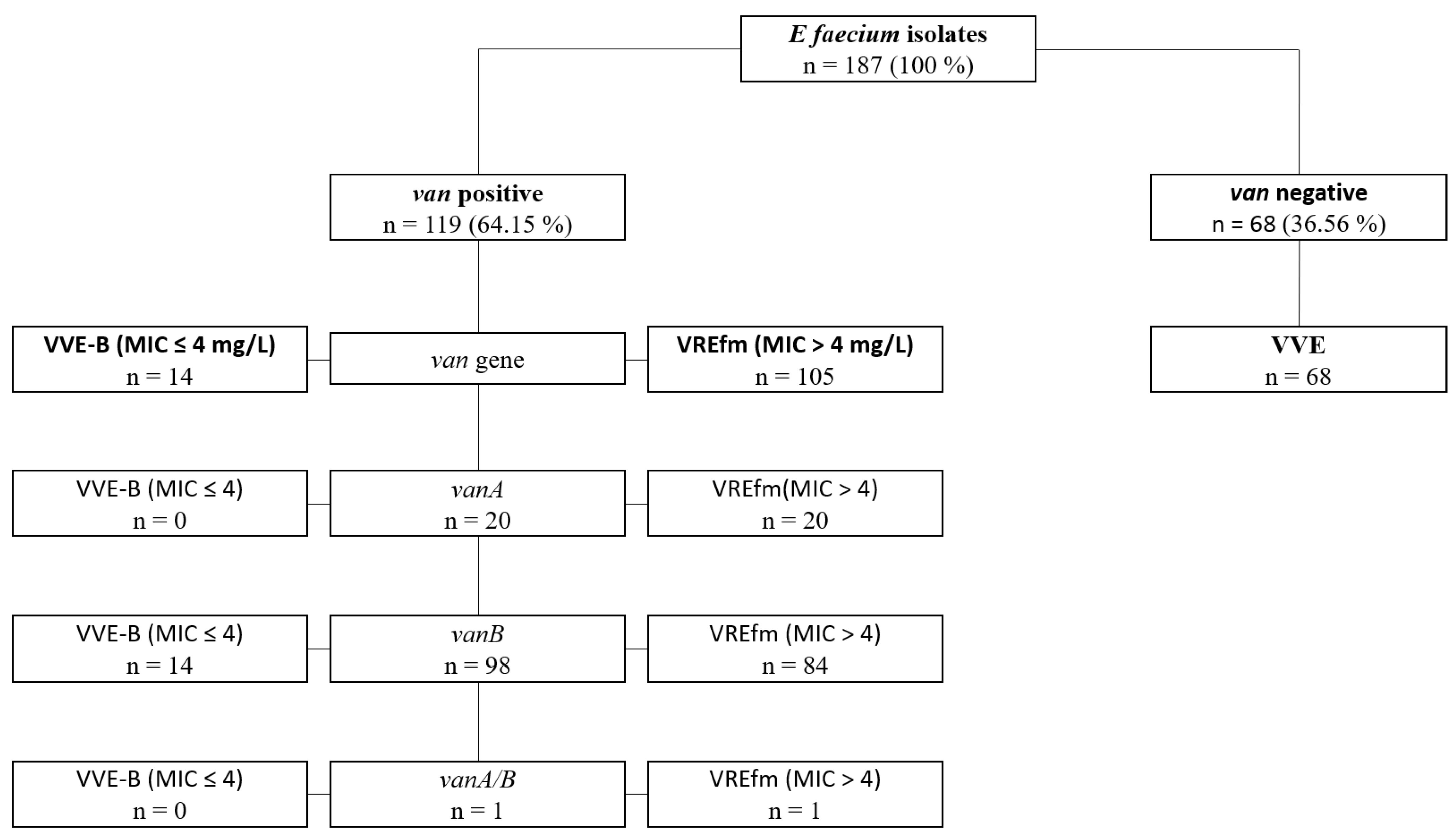
2. Results
3. Discussion
4. Materials and Methods
4.1. Selective Agar
4.2. Clinical Isolates
4.3. Statistics
Supplementary Materials
Author Contributions
Funding
Institutional Review Board Statement
Informed Consent Statement
Data Availability Statement
Conflicts of Interest
References
- Boucher, H.W.; Talbot, G.H.; Bradley, J.S.; Edwards, J.E.; Gilbert, D.; Rice, L.B.; Scheld, M.; Spellberg, B.; Bartlett, J. Bad Bugs, No Drugs: No ESKAPE! An Update from the Infectious Diseases Society of America. Clin. Infect. Dis. 2009, 48, 1–12. [Google Scholar] [CrossRef] [Green Version]
- Markwart, R.; Willrich, N.; Haller, S.; Noll, I.; Koppe, U.; Werner, G.; Eckmanns, T.; Reuss, A. The Rise in Vancomycin-Resistant Enterococcus Faecium in Germany: Data from the German Antimicrobial Resistance Surveillance (ARS). Antimicrob. Resist. Infect. Control 2019, 8, 1–11. [Google Scholar] [CrossRef] [Green Version]
- Werner, G.; Neumann, B.; Weber, R.E.; Kresken, M.; Wendt, C.; Bender, J.K.; Becker, K.; Borgmann, S.; Diefenbach, A.; Hamprecht, A. Thirty Years of VRE in Germany—“Expect the Unexpected”: The View from the National Reference Centre for Staphylococci and Enterococci. Drug Resist. Updates 2020, 53, 100732. [Google Scholar] [CrossRef]
- Surveillance Atlas of Infectious Diseases. Available online: https://www.ecdc.europa.eu/en/surveillance-atlas-infectious-diseases (accessed on 21 January 2023).
- VRE in Healthcare Settings|HAI|CDC. Available online: https://www.cdc.gov/hai/organisms/vre/vre.html (accessed on 21 January 2023).
- WHO Publishes List of Bacteria for Which New Antibiotics Are Urgently Needed. Available online: https://www.who.int/news-room/detail/27-02-2017-who-publishes-list-of-bacteria-for-which-new-antibiotics-are-urgently-needed (accessed on 30 July 2019).
- Sakka, V.; Tsiodras, S.; Galani, L.; Antoniadou, A.; Souli, M.; Galani, I.; Pantelaki, M.; Siafakas, N.; Zerva, L.; Giamarellou, H. Risk-Factors and Predictors of Mortality in Patients Colonised with Vancomycin-Resistant Enterococci. Clin. Microbiol. Infect. 2008, 14, 14–21. [Google Scholar] [CrossRef] [Green Version]
- Alevizakos, M.; Gaitanidis, A.; Nasioudis, D.; Tori, K.; Flokas, M.E.; Mylonakis, E. Colonization with Vancomycin-Resistant Enterococci and Risk for Bloodstream Infection Among Patients With Malignancy: A Systematic Review and Meta-Analysis. Open Forum Infect. Dis. 2017, 4, ofw246. [Google Scholar] [CrossRef] [PubMed] [Green Version]
- Flokas, M.E.; Karageorgos, S.A.; Detsis, M.; Alevizakos, M.; Mylonakis, E. Vancomycin-Resistant Enterococci Colonisation, Risk Factors and Risk for Infection among Hospitalised Paediatric Patients: A Systematic Review and Meta-Analysis. Int. J. Antimicrob. Agents 2017, 49, 565–572. [Google Scholar] [CrossRef]
- DiazGranados, C.A.; Zimmer, S.M.; Mitchel, K.; Jernigan, J.A. Comparison of Mortality Associated with Vancomycin-Resistant and Vancomycin-Susceptible Enterococcal Bloodstream Infections: A Meta-Analysis. Clin. Infect. Dis. 2005, 41, 327–333. [Google Scholar] [CrossRef] [Green Version]
- Brinkwirth, S.; Ayobami, O.; Eckmanns, T.; Markwart, R. Hospital-Acquired Infections Caused by Enterococci: A Systematic Review and Meta-Analysis, WHO European Region, 1 January 2010 to 4 February 2020. Eurosurveillance 2021, 26, 2001628. [Google Scholar] [CrossRef]
- CDC The Biggest Antibiotic-Resistant Threats in the U.S. Available online: https://www.cdc.gov/drugresistance/biggest-threats.html (accessed on 21 January 2023).
- Puchter, L.; Chaberny, I.F.; Schwab, F.; Vonberg, R.-P.; Bange, F.-C.; Ebadi, E. Economic Burden of Nosocomial Infections Caused by Vancomycin-Resistant Enterococci. Antimicrob. Resist. Infect. Control 2018, 7, 1–7. [Google Scholar] [CrossRef]
- Mac, S.; Fitzpatrick, T.; Johnstone, J.; Sander, B. Vancomycin-Resistant Enterococci (VRE) Screening and Isolation in the General Medicine Ward: A Cost-Effectiveness Analysis. Antimicrob. Resist. Infect. Control 2019, 8, 168. [Google Scholar] [CrossRef] [PubMed]
- Zhou, X.; Willems, R.J.L.; Friedrich, A.W.; Rossen, J.W.A.; Bathoorn, E. Enterococcus Faecium: From Microbiological Insights to Practical Recommendations for Infection Control and Diagnostics. Antimicrob. Resist. Infect. Control 2020, 9, 130. [Google Scholar] [CrossRef] [PubMed]
- Krankenhaushygiene und Infektionsprävention (KRINKO) beim Robert Koch-Institut. Hygienemaßnahmen Zur Prävention Der Infektion Durch Enterokokken Mit Speziellen Antibiotikaresistenzen. Bundesgesundheitsblatt-Gesundh.-Gesundh. 2018, 10, 1310. [Google Scholar]
- Centers for Disease Control and Prevention Recommendations for Preventing the Spread of Vancomycin Resistance Recommendations of the Hospital Infection Control Practices Advisory Committee (HICPAC). Available online: https://www.cdc.gov/mmwr/preview/mmwrhtml/00039349.htm (accessed on 19 July 2021).
- Toc, D.A.; Butiuc-Keul, A.L.; Iordache, D.; Botan, A.; Mihaila, R.M.; Costache, C.A.; Colosi, I.A.; Chiorean, C.; Neagoe, D.S.; Gheorghiu, L.; et al. Descriptive Analysis of Circulating Antimicrobial Resistance Genes in Vancomycin-Resistant Enterococcus (VRE) during the COVID-19 Pandemic. Biomedicines 2022, 10, 1122. [Google Scholar] [CrossRef] [PubMed]
- Gorrie, C.; Higgs, C.; Carter, G.; Stinear, T.P.; Howden, B. Genomics of Vancomycin-Resistant Enterococcus Faecium. Microb. Genom. 2019, 5, e000283. [Google Scholar] [CrossRef] [PubMed]
- Gagnon, S.; Lévesque, S.; Lefebvre, B.; Bourgault, A.-M.; Labbé, A.-C.; Roger, M. VanA-Containing Enterococcus Faecium Susceptible to Vancomycin and Teicoplanin Because of Major Nucleotide Deletions in Tn 1546. J. Antimicrob. Chemother. 2011, 66, 2758–2762. [Google Scholar] [CrossRef]
- Kohler, P.; Eshaghi, A.; Kim, H.C.; Plevneshi, A.; Green, K.; Willey, B.M.; McGeer, A.; Patel, S.N.; Network (TIBDN), T.I.B.D. Prevalence of Vancomycin-Variable Enterococcus Faecium (VVE) among VanA-Positive Sterile Site Isolates and Patient Factors Associated with VVE Bacteremia. PLoS ONE 2018, 13, e0193926. [Google Scholar] [CrossRef]
- Merlino, J.; Gray, T. Vancomycin Variable Enterococcus (VVE), E. faecium, Harbouring the VanA Gene Complex. Pathology 2021, 53, 680–682. [Google Scholar] [CrossRef]
- Walker, S.V.; Wolke, M.; Plum, G.; Weber, R.E.; Werner, G.; Hamprecht, A. Failure of Vitek2 to Reliably Detect VanB-Mediated Vancomycin Resistance in Enterococcus Faecium. J. Antimicrob. Chemother. 2021, 76, 1698–1702. [Google Scholar] [CrossRef]
- Viswanath, L.S.; Sugumar, M.; Peela, S.C.M.; Walia, K.; Sistla, S. Detection of Vancomycin Variable Enterococci (VVE) among Clinical Isolates of Enterococcus Faecium Collected across India-First Report from the Subcontinent. Indian J. Med. Microbiol. 2022, 40, 285–288. [Google Scholar] [CrossRef]
- Savini, V.; Marrollo, R.; Coclite, E.; Fusilli, P.; D’Incecco, C.; Fazii, P.; Gherardi, G. Liofilchem® Chromatic VRE and Vancomycin MIC Test Strip Detected Glycopeptide Resistance in a VanB Neonatal Enterococcus Faecium Isolate Showing Alternate Vancomycin Susceptibility and Resistance with BioMérieux Vitek2. Int. J. Clin. Exp. Pathol. 2014, 7, 6274. [Google Scholar]
- Hashimoto, Y.; Kurushima, J.; Nomura, T.; Tanimoto, K.; Tamai, K.; Yanagisawa, H.; Shirabe, K.; Ike, Y.; Tomita, H. Dissemination and Genetic Analysis of the Stealthy VanB Gene Clusters of Enterococcus Faecium Clinical Isolates in Japan. BMC Microbiol. 2018, 18, 1–12. [Google Scholar] [CrossRef] [Green Version]
- Ahmed, M.O.; Baptiste, K.E. Vancomycin-Resistant Enterococci: A Review of Antimicrobial Resistance Mechanisms and Perspectives of Human and Animal Health. Microb. Drug Resist. 2018, 24, 590–606. [Google Scholar] [CrossRef] [Green Version]
- Arthur, M.; Quintiliani, R., Jr. Regulation of VanA-and VanB-Type Glycopeptide Resistance in Enterococci. Antimicrob. Agents Chemother. 2001, 45, 375–381. [Google Scholar] [CrossRef] [Green Version]
- Sadowy, E.; Gawryszewska, I.; Kuch, A.; Żabicka, D.; Hryniewicz, W. The Changing Epidemiology of VanB Enterococcus Faecium in Poland. Eur. J. Clin. Microbiol. Infect. Dis. 2018, 37, 927–936. [Google Scholar] [CrossRef] [Green Version]
- Dahl, K.H.; Simonsen, G.S.; Olsvik, Ø.; Sundsfjord, A. Heterogeneity in the VanB Gene Cluster of Genomically Diverse Clinical Strains of Vancomycin-Resistant Enterococci. Antimicrob. Agents Chemother. 1999, 43, 1105–1110. [Google Scholar] [CrossRef] [Green Version]
- Suwantarat, N.; Roberts, A.; Prestridge, J.; Seeley, R.; Speser, S.; Harmon, C.; Zhang, C.; Henciak, S.; Stamper, P.D.; Ross, T. Comparison of Five Chromogenic Media for Recovery of Vancomycin-Resistant Enterococci from Fecal Samples. J. Clin. Microbiol. 2014, 52, 4039–4042. [Google Scholar] [CrossRef] [Green Version]
- Willey, B.M.; Louie, L.; Gnanasuntharam, P.; Fung, T.; Watt, C.; Gnanasuntharam, P.; Vermeiren, C.; Ricci, G.; Lo, P.; Wong, K.; et al. Prospective Evaluation of Brilliance VRE and VRESelect Chromogenic Agars for Detection of Vancomycin-Resistant Enterococci from Surveillance Specimens; European Society of Clinical Microbiology and Infectious Diseases (ESCMID): Milan, Italy, 2011; p. 1036. Available online: https://www.thermoscientific.com/content/dam/tfs/SDG/MBD/MBD%20Documents/Third-Party%20Papers/Microbiology/Prospective-Evaluation-of-Brilliance-VRE-From-Surveillance-Specimens-EN.pdf (accessed on 4 March 2023).
- Anderson, N.W.; Buchan, B.W.; Young, C.L.; Newton, D.W.; Brenke, C.; Lapsley, L.; Granato, P.A.; Ledeboer, N.A. Multicenter Clinical Evaluation of VRE Select Agar for Identification of Vancomycin-Resistant Enterococcus Faecalis and Enterococcus Faecium. J. Clin. Microbiol. 2013, 51, 2758–2760. [Google Scholar] [CrossRef] [Green Version]
- Wijesuriya, T.M.; Perry, P.; Pryce, T.; Boehm, J.; Kay, I.; Flexman, J.; Coombs, G.W.; Ingram, P.R. Low Vancomycin MICs and Fecal Densities Reduce the Sensitivity of Screening Methods for Vancomycin Resistance in Enterococci. J. Clin. Microbiol. 2014, 52, 2829–2833. [Google Scholar] [CrossRef] [Green Version]
- Delmas, J.; Robin, F.; Schweitzer, C.; Lesens, O.; Bonnet, R. Evaluation of a New Chromogenic Medium, ChromID VRE, for Detection of Vancomycin-Resistant Enterococci in Stool Samples and Rectal Swabs. J. Clin. Microbiol. 2007, 45, 2731–2733. [Google Scholar] [CrossRef] [Green Version]
- Ongut, G.; Kilinckaya, H.; Baysan, B.O.; Ogunc, D.; Colak, D.; Inan, D.; Kasaroglu, K.; Gunseren, F. Evaluation of Brilliance VRE Agar for the Detection of Vancomycin-Resistant Enterococci in Rectal Swab Specimens. J. Med. Microbiol. 2013, 62, 661–662. [Google Scholar] [CrossRef] [Green Version]
- Cuzon, G.; Naas, T.; Fortineau, N.; Nordmann, P. Novel Chromogenic Medium for Detection of Vancomycin-Resistant Enterococcus Faecium and Enterococcus Faecalis. J. Clin. Microbiol. 2008, 46, 2442–2444. [Google Scholar] [CrossRef] [PubMed] [Green Version]
- Peterson, J.F.; Doern, C.D.; Kallstrom, G.; Riebe, K.M.; Sander, T.; Dunne, W.M., Jr.; Ledeboer, N.A. Evaluation of Spectra VRE, a New Chromogenic Agar Medium Designed to Screen for Vancomycin-Resistant Enterococcus Faecalis and Enterococcus Faecium. J. Clin. Microbiol. 2010, 48, 4627–4629. [Google Scholar] [CrossRef] [PubMed] [Green Version]
- Klare, I.; Fleige, C.; Geringer, U.; Witte, W.; Werner, G. Performance of Three Chromogenic VRE Screening Agars, Two Etest(®) Vancomycin Protocols, and Different Microdilution Methods in Detecting VanB Genotype Enterococcus Faecium with Varying Vancomycin MICs. Diagn. Microbiol. Infect. Dis. 2012, 74, 171–176. [Google Scholar] [CrossRef] [PubMed] [Green Version]


| Total no. of Strains of Each Category | ChromID VRE BioMérieux | CHROMagar VRE CHROMagar | Brilliance VRE ThermoFisher | VRESelect Bio-Rad | Chromatic VRE Liofilchem | ||||||
|---|---|---|---|---|---|---|---|---|---|---|---|
| Incubation [Hours] | 24 | 48 | 24 | 48 | 24 | 48 | 24 | 48 | 24 | 48 | |
| Growth | |||||||||||
| No. of VREfm + VVE-B grown | 119 | 113 | 113 | 113 | 113 | 113 | 113 | 112 | 113 | 109 | 111 |
| No. of VREfm grown | 105 | 105 | 105 | 105 | 105 | 105 | 105 | 104 | 105 | 102 | 104 |
| No. of VVE-B grown | 14 | 8 | 8 | 8 | 8 | 8 | 8 | 8 | 8 | 7 | 7 |
| No. of VSE grown | 68 | 1 | 1 | 1 | 3 | 3 | 4 | 2 | 3 | 0 | 1 |
| No. of E. coli grown | 10 | 0 | 0 | 0 | 0 | 0 | 0 | 0 | 0 | 0 | 0 |
| Positive control (DSMZ 17050) | 1 | 1 | 1 | 1 | 1 | 1 | 1 | 1 | 1 | 1 | 1 |
| Negative control (DSMZ 20477) | 1 | 0 | 0 | 0 | 0 | 0 | 0 | 0 | 0 | 0 | 0 |
| Growth score (GS) | |||||||||||
| GS VREfm + VVE-B | 119 | 315 | 336 | 328 | 334 | 329 | 333 | 288 | 324 | 228 | 284 |
| GS VREfm | 105 | 297 | 312 | 304 | 310 | 306 | 310 | 268 | 300 | 215 | 268 |
| GS VVE-B | 14 | 18 | 24 | 24 | 24 | 23 | 23 | 20 | 24 | 13 | 16 |
| GS VSE | 68 | 1 | 1 | 1 | 3 | 5 | 6 | 2 | 3 | 0 | 1 |
| ChromID VRE (BioMérieux) | CHROMagar VRE (CHROMagar) | Brilliance VRE (ThermoFisher) | VRESelect (Bio-Rad) | Chromatic VRE (Liofilchem) | ||||||
|---|---|---|---|---|---|---|---|---|---|---|
| Incubation [Hours] | 24 | 48 | 24 | 48 | 24 | 48 | 24 | 48 | 24 | 48 |
| Sensitivity [%] (95% CI) | 95.0 (88.9–97.9) | 95.0 (88.9–97.9) | 95.0 (88.9–97.9) | 95.0 (88.9–97.9) | 95.0 (88.9–97.9) | 95.0 (88.9–97.9) | 94.1 (88.9–97.9) | 95.0 (88.9–97.9) | 91.6 (84.7–95.7) | 93.3 (86.8–96.8) |
| Specificity [%] (95% CI) | 98.5 (91.0–99.9) | 98.5 (91.0–99.9) | 98.5 (91.0–99.9) | 95.6 (86.8–98.9) | 95.6 (86.8–98.6) | 94.1 (84.7–98.1) | 97.1 (88.8–99.5) | 95.6 (86.8–98.9) | 100.0 (93.3–100.0) | 98.5 (91.0–99.9) |
| Youden-Index | 0.94 | 0.94 | 0.94 | 0.91 | 0.91 | 0.89 | 0.91 | 0.91 | 0.92 | 0.92 |
| VREfm sensitivity [%] | 100.0 | 100.0 | 100.0 | 100.0 | 100.0 | 100.0 | 99.1 | 100.0 | 97.1 | 99.1 |
| VVE-B sensitivity [%] | 57.1 | 57.1 | 57.1 | 57.1 | 57.1 | 57.1 | 57.1 | 57.1 | 50.0 | 50.0 |
| PPV | 0.99 | 0.99 | 0.99 | 0.97 | 0.97 | 0.97 | 0.98 | 0.97 | 1.00 | 0.99 |
| NPV | 0.92 | 0.92 | 0.92 | 0.92 | 0.92 | 0.91 | 0.90 | 0.92 | 0.87 | 0.89 |
| LR+ | 64.57 | 64.57 | 64.57 | 21.52 | 21.52 | 16.14 | 32.00 | 21.52 | n.c. | 63.43 |
| LR− | 0.05 | 0.05 | 0.05 | 0.05 | 0.05 | 0.05 | 0.06 | 0.05 | 0.08 | 0.07 |
| Limit of Detection (CFU/mL) | ||||||
|---|---|---|---|---|---|---|
| 24 h | 48 h | |||||
| Pure Culture | Stool Suspension | Artificial Rectal Swabs | Pure Culture | Stool Suspension | Artificial Rectal Swabs | |
| CHROMID VRE (bioMérieux) | 1.5 × 10³ | 1.5 × 10² | 1.5 × 10³ | 1.5 × 101 | 1.5 × 101 | 1.5 × 10² |
| CHROMagar VRE (CHROMagar) | 1.5 × 101 | 1.5 × 101 | 1.5 × 10² | 1.5 × 101 | 1.5 × 101 | 1.5 × 10² |
| Brilliance VRE (ThermoFisher) | 1.5 × 101 | 1.5 × 101 | 1.5 × 10³ | 1.5 × 101 | 1.5 × 101 | 1.5 × 10³ |
| VRESelect (Bio-Rad) | 1.5 × 10³ | 1.5 × 10² | 1.5 × 10³ | 1.5 × 101 | 1.5 × 10² | 1.5 × 10² |
| Chromatic™ VRE (Liofilchem) | 1.5 × 10⁴ | - | 1.5 × 10² | 1.5 × 10² | - | 1.5 × 10² |
Disclaimer/Publisher’s Note: The statements, opinions and data contained in all publications are solely those of the individual author(s) and contributor(s) and not of MDPI and/or the editor(s). MDPI and/or the editor(s) disclaim responsibility for any injury to people or property resulting from any ideas, methods, instructions or products referred to in the content. |
© 2023 by the authors. Licensee MDPI, Basel, Switzerland. This article is an open access article distributed under the terms and conditions of the Creative Commons Attribution (CC BY) license (https://creativecommons.org/licenses/by/4.0/).
Share and Cite
Boschert, A.L.; Arndt, F.; Hamprecht, A.; Wolke, M.; Walker, S.V. Comparison of Five Different Selective Agar for the Detection of Vancomycin-Resistant Enterococcus faecium. Antibiotics 2023, 12, 666. https://doi.org/10.3390/antibiotics12040666
Boschert AL, Arndt F, Hamprecht A, Wolke M, Walker SV. Comparison of Five Different Selective Agar for the Detection of Vancomycin-Resistant Enterococcus faecium. Antibiotics. 2023; 12(4):666. https://doi.org/10.3390/antibiotics12040666
Chicago/Turabian StyleBoschert, Alessa L., Franca Arndt, Axel Hamprecht, Martina Wolke, and Sarah V. Walker. 2023. "Comparison of Five Different Selective Agar for the Detection of Vancomycin-Resistant Enterococcus faecium" Antibiotics 12, no. 4: 666. https://doi.org/10.3390/antibiotics12040666