Article

Shark Variable New Antigen Receptor (VNAR) Single Domain Antibody Fragments: Stability and Diagnostic Applications

1
The Department of Biochemistry, La Trobe Institute for Molecular Science, La Trobe University, Plenty Road, Bundoora, VIC., 3086, Australia
2
AdAlta Pty. Ltd., 15/2 Park Drive, Bundoora, VIC., 3086, Australia
3
The CSIRO Materials Science and Engineering, 343 Royal Parade, Parkville, VIC., 3052, Australia
4
The Foundation for Innovative New Diagnostics (FIND), Avenue de Budé 16, 1202 Geneva, Switzerland
*
Author to whom correspondence should be addressed.
Antibodies 2013, 2(1), 66-81; https://doi.org/10.3390/antib2010066
Submission received: 29 November 2012 / Revised: 9 January 2013 / Accepted: 21 January 2013 / Published: 25 January 2013
(This article belongs to the Special Issue Single-Domain Antibody)

Abstract

:
The single variable new antigen receptor domain antibody fragments (VNARs) derived from shark immunoglobulin new antigen receptor antibodies (IgNARs) represent some of the smallest known immunoglobulin-based protein scaffolds. As single domains, they demonstrate favorable size and cryptic epitope recognition properties, making them attractive in diagnosis and therapy of numerous disease states. Here, we examine the stability of VNAR domains with a focus on a family of VNARs specific for apical membrane antigen 1 (AMA-1) from Plasmodium falciparum. The VNARs are compared to traditional monoclonal antibodies (mAbs) in liquid, lyophilized and immobilized nitrocellulose formats. When maintained in various formats at 45 °C, VNARs have improved stability compared to mAbs for periods of up to four weeks. Using circular dichroism spectroscopy we demonstrate that VNAR domains are able to refold following heating to 80 °C. We also demonstrate that VNAR domains are stable during incubation under potential in vivo conditions such as stomach acid, but not to the protease rich environment of murine stomach scrapings. Taken together, our results demonstrate the suitability of shark VNAR domains for various diagnostic platforms and related applications.

1. Introduction

The development of rapid, accurate and low cost diagnostics for point-of-care applications in developing countries is a major challenge that needs to be overcome if the morbidity and mortality from diseases is to be reduced in these countries. Such diagnostic tests must be easy to use and sufficiently robust to survive exposure to extremes of temperature and humidity. Many of the rapid diagnostic tests currently in use rely on conventional murine-derived antibodies for the detection of pathogens or molecules derived from them. Monoclonal antibodies (mAbs) have a variety of features that make them particularly well suited to this task, such as high specificity and high affinity for the target antigen. However, despite these favorable characteristics, diagnostic tests based upon such antibodies may be adversely affected by high temperatures or by storage for extended periods under conditions of high humidity, particularly when the test must be conducted in developing countries where constant access to refrigeration is not always practicable [1].
Over the last 15 years, technologies based upon the immunoglobulin new antigen receptor (IgNAR) class of antibodies from the shark immune repertoire have been developed that can potentially overcome mAb limitations. Mature IgNAR antibodies consist of homodimers of one variable new antigen receptor (VNAR) domain and five constant new antigen receptor (CNAR) domains [2]. The evolution of IgNARs to a single domain conformation over significant periods of evolutionary time has resulted in molecules that are highly stable and possess efficient binding characteristics. The inherent stability can be attributed to both (i) the underlying Ig scaffold, which presents a considerable number of charged and hydrophilic surface exposed residues compared to the conventional antibody VH and VL domains found in murine antibodies; and (ii) stabilizing structural features in the complementary determining region (CDR) loops including inter-loop disulphide bridges, and patterns of intra-loop hydrogen bonds [3,4].
As a consequence, we hypothesized that individual VNAR domains would display high stability under conditions that would otherwise compromise the integrity of traditional monoclonal antibodies. Such stability, if demonstrable as a consistent feature of these binding reagents, would be advantageous for use in diagnostic assays where the reagents are likely to be exposed to extreme conditions.
Over recent years, we have developed in particular a model system based around the 12Y-2 family of VNARs that target the apical membrane antigen 1 (AMA-1) of malarial (Plasmodium falciparum) parasites [5]. Studies have included VNAR affinity maturation [6], structural studies [3], multimerisation strategies [7], library design and selection to enhance cross-strain binding [8]. Here, we utilize several of these AMA-1 VNARs along with a model VNAR to illustrate stability properties of the scaffold in conditions including extreme temperature and pH and in the presence of proteases. We have consequently identified a set of conditions which allows one VNAR to retain significant activity for up to 4 weeks at 50 °C.

2. Results and Discussion

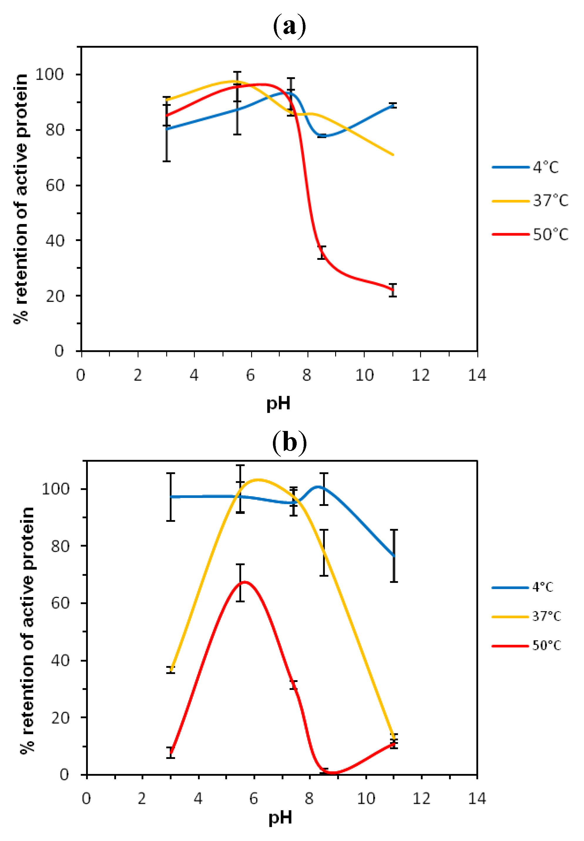
2.1. Thermostability of VNARs at Extreme pH

The VNAR4A-1 binds to a mAb antigen, 5G8, with an affinity of 1 µM (Appendix Figure A1). The paratope of the antibody is readily targeted by our single domain VNAR variants via the motif S/AYP [9]. The 4A-1 variant is hence regarded as a model VNAR. Thermostability experiments were conducted to examine the stability of this VNAR in liquid format at various pH values and temperatures. ProteOn XPR36 SPR biosensor was utilized for the determination of residual binding activities of 4A-1. After 1 day incubation at 4, 37 or 50 °C there was a trend for greater retention of activity at pHs ≤7.4 (Figure 1a). At 4 °C or 37 °C retention of active protein was 90% and 70%, respectively, whereas after incubation at 50 °C, only 20% active protein was retained. After 4 weeks incubation at 4 °C retention of active protein was ≥70% at all pH values investigated (Figure 1b). At 37 °C, retention of active protein was 100% at pH 5.5 and 97% at pH 7.4, but decreased significantly outside this pH range. At 50 °C, maximum retention of active protein was at pH 5.5, with 67% active protein remaining after 4 weeks. At all other pH values, retention of activity was significantly lower, in particular at pH 3.0, 5.5 and 11 only ≤10% active protein remained. A similar stability profile was observed for another VNAR (clone 27P-1) in short-term (1 day) trials at pHs 3, 7.4 and 9 at 37 and 60 °C (data not shown).
Figure 1. Retention of active variable new antigen receptor Variable New Antigen Receptor (VNAR) 4A-1 protein following heating at various pHs and temperatures (a) for 1 day and (b) for 4 weeks.
Antibodies 02 00066 g001

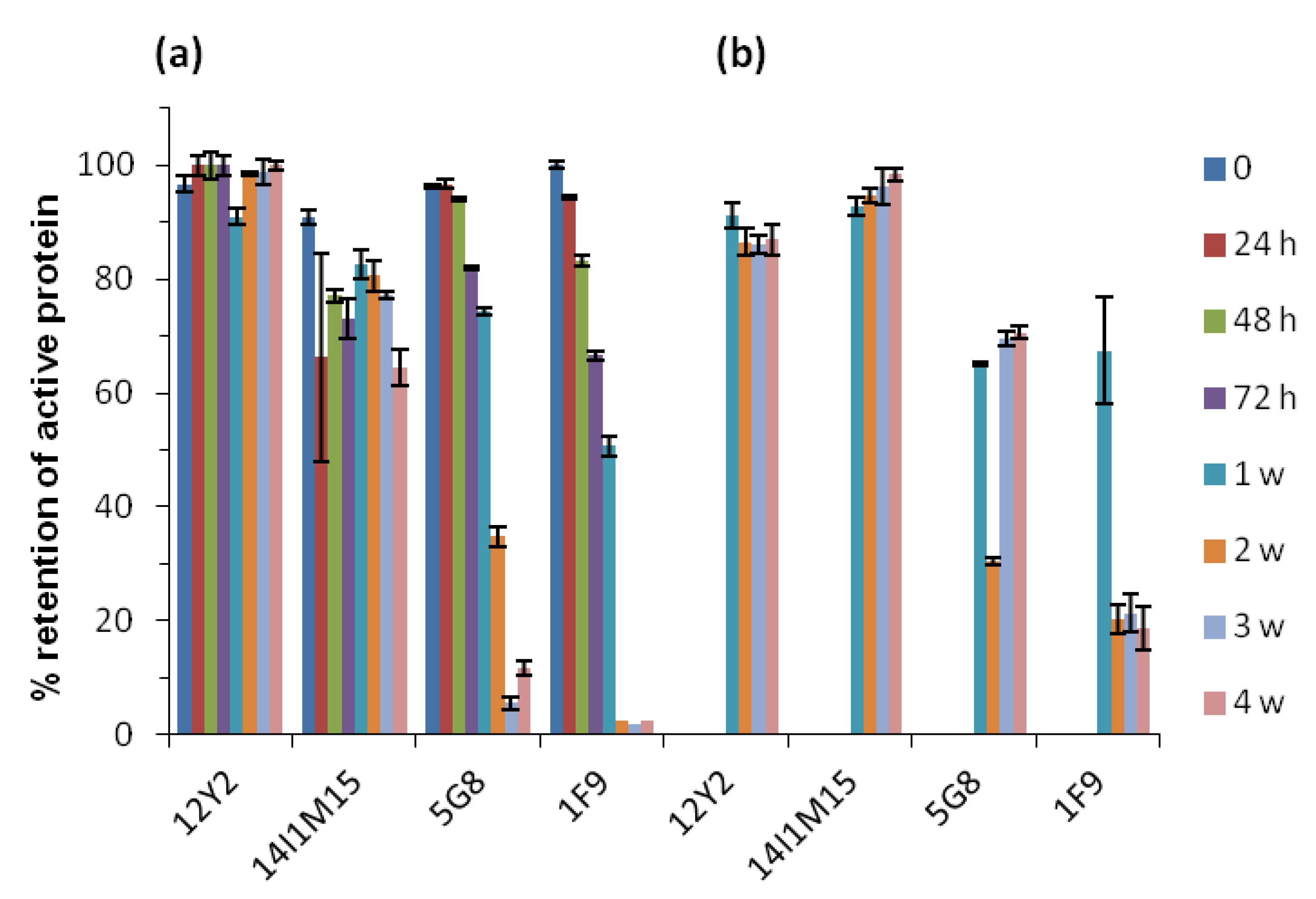
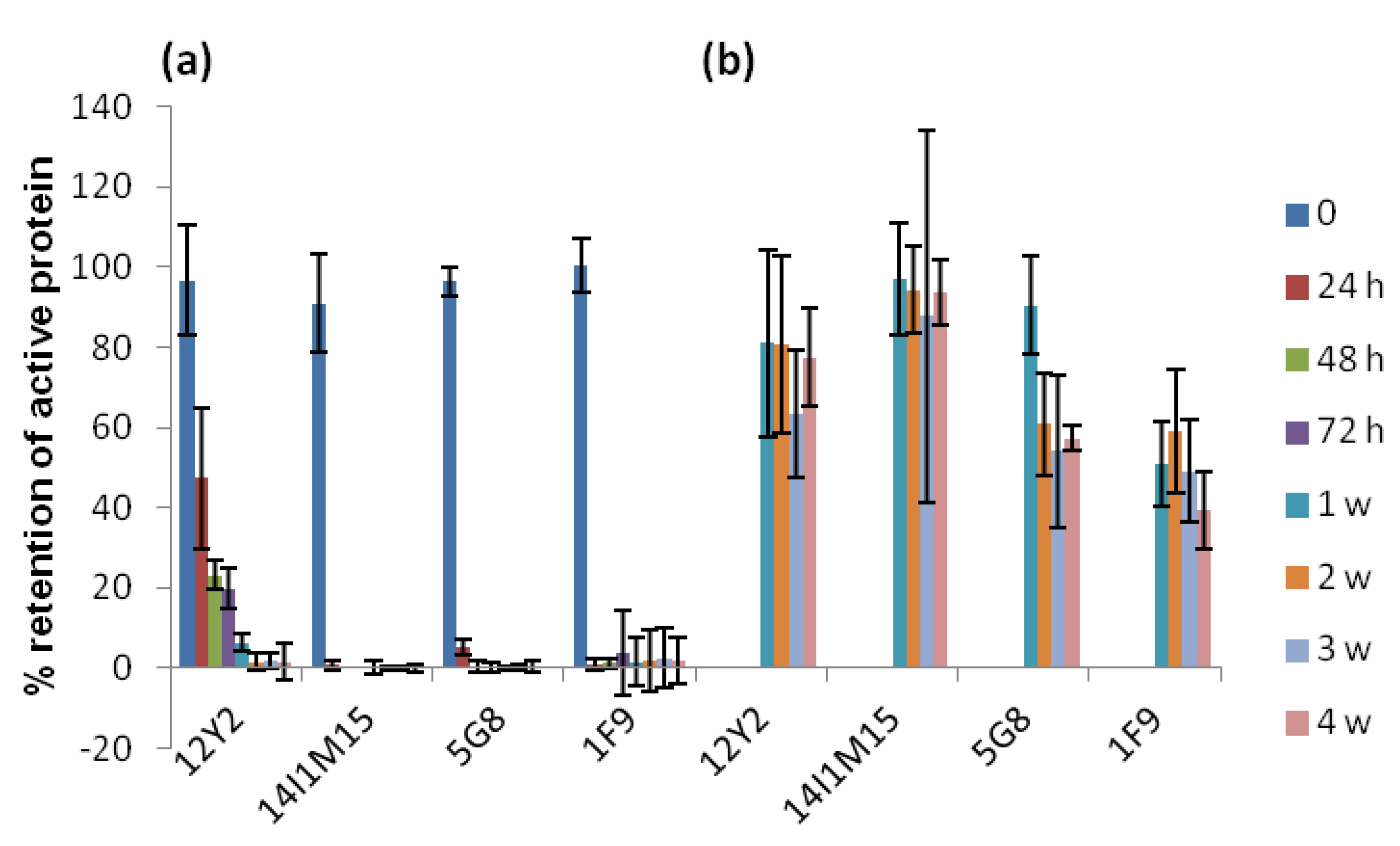
2.2. Thermostability of VNARs and Mabs in Liquid and Lyophilized Formats

Given the very good stability of this model VNAR, we decided to further examine a family of VNARs (12Y-2, 14M-15 and 14I1M-15) which target AMA-1 from Plasmodium falciparum (strain 3D7) using a variety of stability assays. VNARs 14M-15 and 14I1M-15 are affinity matured variants of 12Y-2, and have binding affinities of 31, 5 and 708 nM, respectively for AMA-1 [8].
In the first instance, VNARs which had been semi-purified as periplasmic extracts were heated to 100 °C for 15 min. Using SDS-PAGE, soluble protein content was then compared between untreated and heat treated samples (Figure 2). It was apparent that whilst a reasonable proportion of host cell proteins were heat labile, the VNARs were unaffected by the heat treatment.
Figure 2. SDS-PAGE analysis of VNARs in Escherichia coli periplasmic extracts before and after heat treatment at 100 °C for 15 min. Lane 1: untreated 12Y-2, Lane 2: heat treated 12Y-2, Lane 3: untreated 14M-15, Lane 4: heat treated 14M-15, Lane 5: untreated 14I1M-15, Lane 6: heat treated 14I1M-15.
Antibodies 02 00066 g002
Given these promising results, purified VNARs 12Y-2 and 14I1M-15 were then compared to two mAbs 5G8 and 1F9 at 45 °C in liquid format in short term (0–72 h) and long term (1–4 weeks) stability trials (Figure 3a), and in lyophilized format in long term trials only (Figure 3b). The mAbs 5G8 and 1F9 likewise target AMA-1 [10]. The affinity of 5G8 for AMA-1 is 175 nM [9] and the affinity of 1F9 for AMA-1 is 950 pM (Appendix Figure A2).
The concentration of active protein (VNAR or mAb) remaining after each treatment was evaluated using SPR biosensor (Biacore T200) analyses. This method relied upon determination of residual binding activity to AMA-1 protein of treated samples by comparing their binding responses to those obtained with the starting untreated sample. By extrapolating against a calibration curve, generated using the starting material, the residual active concentration of treated samples was determined.
In short term trials in liquid at 45 °C, VNAR 12Y-2 retained 100% activity after 72 h, whereas 14I1M-15 retained 73% activity. By comparison, under the same conditions, mAbs 5G8 and 1F9 retained 82% and 67% activity, respectively. Additional analyses showed that after 72 h at 60 °C, the only binder to retain activity was 12Y-2, with 20% activity remaining (Appendix Figure A3).
In long term trials VNARs were generally more stable and performed consistently well in both formats, by comparison with mAbs (Figure 3). In liquid format after heating at 45 °C for 4 weeks, 100 and 65% activity was retained by 12Y-2 and 14I1M-15, respectively. By comparison, 12 and 2% activity was retained by 5G8 and 1F9, respectively, under these conditions. Heating at 60 °C abolished most of the activity of all antibodies in liquid format after 1 week (Appendix Figure A3).
Retention of activity of lyophilized VNARs was excellent (Figure 3b). Greater than 86% activity was retained by 12Y-2 and 14I1M-15, regardless of temperature or incubation period. For incubation periods longer than 72 h, both mAbs performed better in lyophilized format rather than in liquid format. The 5G8 and 1F9 retained 71% and 19% activity, respectively, after 4 weeks at 45 °C.
Figure 3. Retention of active VNARs 12Y-2 and 14I1M-15, and monoclonal antibodies (mAbs) 5G8 and 1F9 following heating at 45 °C in (a) liquid and (b) lyophilized formats, as determined by biosensor analysis. The standard deviation for 1F9 time points 2, 3 and 4 weeks was ≤30%, error bars are shown for all other values.
Antibodies 02 00066 g003
Next, we extended these results to include VNARs and mAbs immobilized on nitrocellulose strips that had been incubated at 25, 45 and 60 °C for 4 weeks (Figure 4). To determine retention of activity the heat-treated immobilized antigens were probed with the cognate malarial protein, AMA-1. After incubation for 4 weeks, both VNARs retained almost complete activity at all temperatures, evidenced by excellent binding to AMA-1 protein. By comparison, both mAbs lost significant activity after heating at 45 °C for 4 weeks, and no activity was detectable after heating at 60 °C for 4 weeks.
Most of the current rapid diagnostic tests (RDTs) for malaria are based on antigen detection by monoclonal antibodies immobilized onto nitrocellulose membranes. Although in use for more than 10 years, issues about their thermostability have affected confidence with their performance [11,12,13]. Our results are clear evidence of how the activity of mAbs onto nitrocellulose membranes can be impaired by temperatures commonly found in endemic areas (>40 °C), and that VNARs are an attractive option for the improvement of RDTs for malaria.
Figure 4. Retention of activity of VNARs and monoclonal antibodies immobilized on nitrocellulose, following heating at various temperatures and time periods (W = week).
Antibodies 02 00066 g004

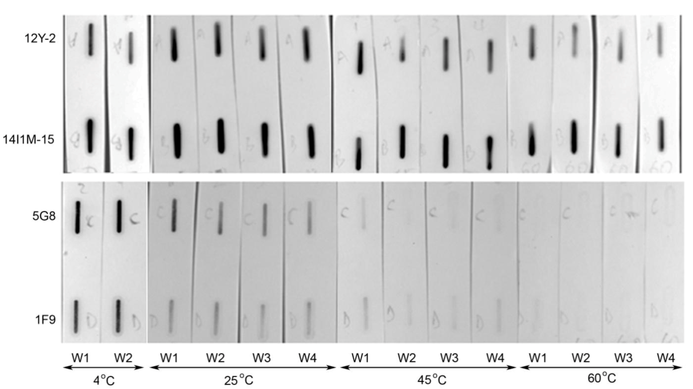
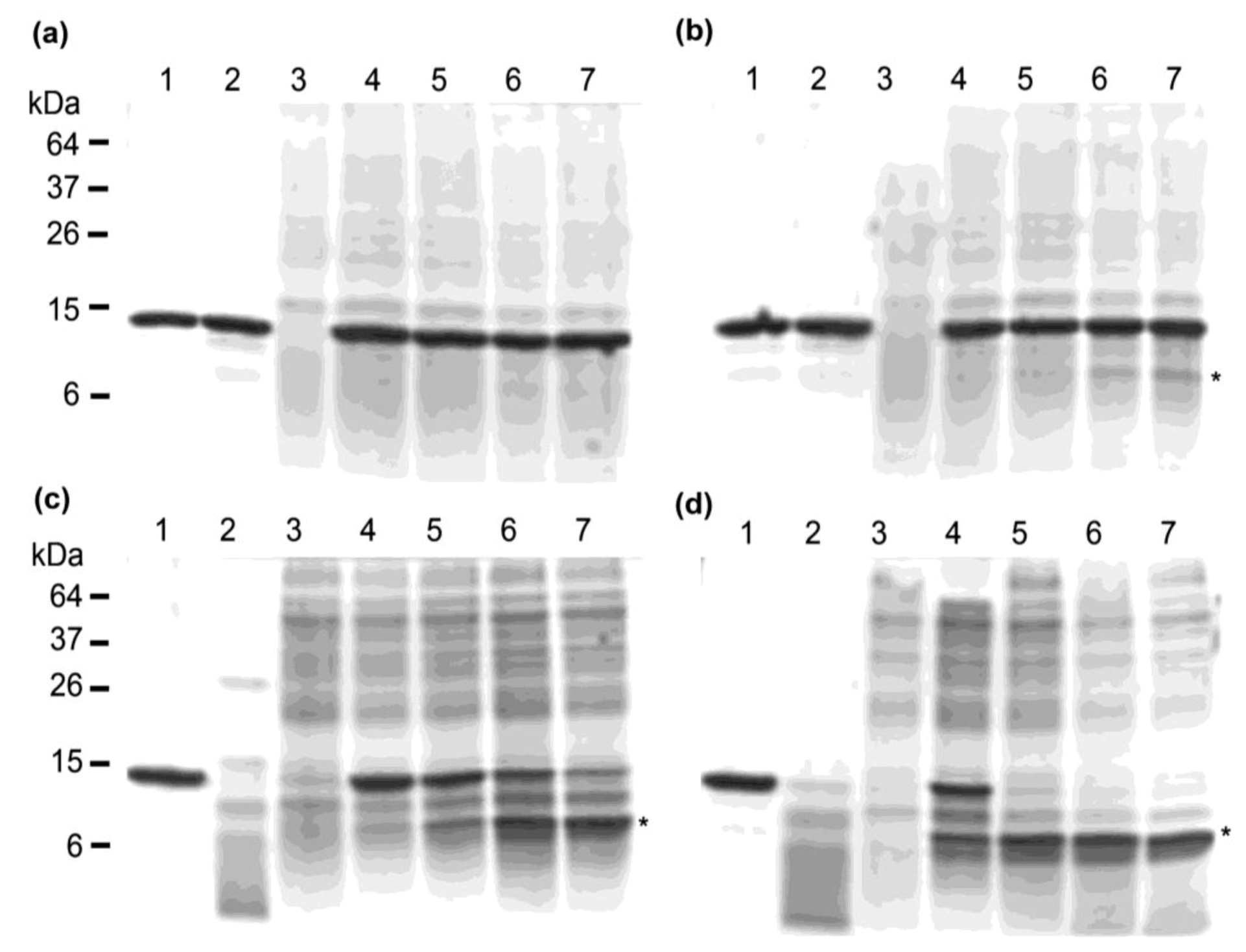
2.3. Thermostability of VNARs in Biological Samples

To further extend these results to possible in vitro applications involving contact with biological samples, or for possible future in vivo administration for gut-specific infectious diseases or cancers, the VNAR antibody fragment 14M-15 was exposed to murine stomach scrapings (acidic, pH 5) and intestinal samples (a protease-rich environment). After 1 hour exposure to stomach scrapings at 4 °C, there was no evidence of VNAR degradation whereas at 37 °C there was a small amount of degradation evidenced by the release of a ~12 kDa fragment (Figure 5). In the control sample incubated at pH 3, 14M-15 remained intact. Incubation in the presence of intestinal scrapings resulted in time- and temperature dependent partial-degradation of 14M-15 to a ~11 kDa fragment. At 4 °C, degradation was initiated at ≤10 min and intensified with incubation time. After 60 min approximately 75% of 14M-15 was degraded. Degradation at 37 °C was initiated immediately (at ≤1 min) and by 30 min was complete, with a single 11 kDa fragment of 14M-15 remaining. An additional VNAR, Help6, which binds Hepatitis B precore protein responded in exactly the same manner to incubation in these murine samples (data not shown). N-terminal sequencing of the VNAR fragment confirmed the N-terminus was intact, thus cleavage occurred approximately 3 kDa from the C-terminus of 14M-15 which is in a conserved framework region at the framework CDR3 junction. Given the framework sequence is largely conserved, this same stability response to gastrointestinal proteases would likely be universal among our VNAR clones.
Figure 5. SDS-PAGE analysis of VNARs during incubation with murine stomach and intestinal scrapings. (a) Incubation at 4 °C, Lane 1: untreated 14M-15, Lane 2: 14M-15 with pH 3 glycine, Lane 3: Murine stomach sample, Lane 4: Murine stomach sample with 14M-15, 1 min incubation, Lane 5: Murine stomach sample with 14M-15, 10 min incubation, Lane 6: Murine stomach sample with 14M-15, 30 min incubation, Lane 7: Murine stomach sample with 14M-15, 60 min incubation. (b) Incubation at 37 °C, loading same as (a). (c) Incubation at 4 °C, Lane 1: untreated 14M-15, Lane 2: 14M-15 with proteinase K, Lane 3: Murine intestinal sample, Lane 4: Murine intestinal sample with 14M-15, 1 min incubation, Lane 5: Murine intestinal sample with 14M-15, 10 min incubation, Lane 6: Murine intestinal sample with 14M-15, 30 min incubation, Lane 7: Murine intestinal sample with 14M-15, 60 min incubation. (d) Incubation at 37 °C, loading same as (c). *14M-15 degradation products resulting from acid or protease activity.
Antibodies 02 00066 g005

2.4. Analysis of Secondary Structure by Circular Dichroism Spectroscopy

In order to investigate the secondary structure and thermal stability of the VNAR 12Y-2 scaffold, we conducted circular dichroism (CD) spectroscopy at low and high temperatures. At 20 °C the secondary structure of the VNAR was predominantly β-strand as evidenced by an absorbance minimum at 218 nm and maximum at 195 nm [14] (Figure 6, blue trace). When heated to 80 °C, a significant decrease in the ellipticity at 205 nm was observed, indicating that 12Y-2 adopts a predominantly random coil structure at the high temperature consistent with unfolding of the scaffold (Figure 6, red trace). However, upon re-cooling to 20 °C (Figure 6, black trace) the CD spectrum adopted a similar profile to that obtained prior to heating. Repeated heating-cooling cycles resulted in reproducible results (Figure 6, gray traces), suggesting the protein is very stable at high temperatures due to an ability to refold reversibly upon thermal denaturation.
Figure 6. Secondary structure and thermostability of 12Y-2 measured by circular dichroism (CD) spectroscopy. Wavelength scans of 12Y-2 were taken at 20 °C and 80 °C, and repeated four times following cycles of heating to 80 °C and cooling to 20 °C (Recool 1–4).
Antibodies 02 00066 g006
Immunoglobulin new antigen receptors (IgNARs) have evolved to function in antigen recognition via single variable domains (VNARs). Single Ig binding domains engineered from paired VH-VL origins typically show a high degree of aggregation due to exposure of hydrophobic faces to solvent, although emerging methods may overcome this tendency [15]. Immunoglobulin domains are inherently stable by virtue of their secondary and tertiary structures. The arrangement of antiparallel β-strands is optimized for H-bond formation and van der Waals interactions between the strands to form two β-sheets. The size and tertiary structure of shark VNARs parallel nanobodies engineered from camelid VHHs, which akin to IgNARs comprise paired heavy chains and no associated light chains [16]. As such, nanobodies and VNARs share comparable physicochemical properties including high thermal stability and nM–pM antigen binding affinities [17]. The finding that by CD analysis the VNAR scaffold refolds following repeated heating-cooling cycles (Figure 6), suggests that the loss of activity observed in liquid formats for prolonged periods and at extreme pH (Figure 1 and Figure 3) is due to irreversible damage to the scaffold such as reduction of the framework disulfide bond or loss of local structure which effects CDR3 conformation.
Particular applications of biologic diagnostic reagents include (i) disease detection and discrimination in hand held point of care devices (with major demand in developing countries or regions or where refrigeration is unreliable or unavailable [18]); (ii) molecular in vivo imaging [19] and (iii) drug or radionuclide delivery via antibody drug conjugates [20]. The stability of several VNARs at elevated temperatures in liquid, lyophilized and immobilized formats suggests this scaffold has excellent suitability for diagnostic markets. Our findings demonstrate that it would be feasible to customize storage parameters for individual diagnostic VNARs, given that 100% activity of VNAR 4A-1 was retained at pH 5.5 and 37 °C (Figure 1). Furthermore, large-scale production of VNARs could be expedited using heating and subsequent cooling as a simple and inexpensive purification step as has been explored for recombinant camelid antibodies [21], and as we have shown in preliminary studies (Figure 2).
Beyond protein engineering for affinity, there exists the possibility of modification to enhance VNAR pharmacokinetic (PK) properties such as stability and half-life. For in vivo applications the target tissue and mode of delivery will dictate specific traits required of potential VNAR candidates. One approach to improve PK properties was recently reported by Müller et al. [22] whereby two- and three-domain VNAR constructs showed increased half-life in mouse, rat and monkey models. It is conceivable that residues that are particularly important for stability may be pinpointed and appropriately modified, in much the same manner that affinity-enhancing and hallmark mutations have been comprehensively mapped for VNAR domains in the study of Fennel et al. [23]. Our analysis of VNAR stability in pH and temperature extremes provides a ready model system for such experimentation.

3. Experimental Section

3.1. Nucleic Acid Isolation and Cloning

VNAR 4A-1 was isolated from a VNAR phagemid library in which a 6-residue CDR1 region was fully randomized and a CDR3 region varied in length from 10–20 residues and was fully randomized (“VNAR 10-20 library”). The VNAR 10–20 library was constructed and biopanned as described elsewhere [5,24] with the following exceptions: mAb 5G8 was coated onto Maxisorb Immuno-plates (Nunc, Germany) at a concentration of 10 µg/mL, plates were rinsed with phosphate buffered saline (PBS, 137 mM NaCl, 2.7 mM KCl, 10 mM Na2HPO4, 2 mM KH2PO4, pH 7.4) then blocked with PBS/5% (w/v) skim milk powder (Diploma, Australia) for 3 h at room temperature (RT) and then incubated with freshly prepared phage particles in PBS/0.5% (w/v) skim milk powder for 1 h at RT, after incubation plates were washed (round 1: 2 × PBS; round 2: 4 × PBS/0.1% Tween20, 2 × PBS; round 3: 6 × PBS/0.1% (v/v) Tween20, 2 × PBS) then phage were eluted using 0.1 M glycine, pH 2.2 and neutralized with 1.5 M Tris-HCl, pH 8.0. Construction of VNAR error prone PCR mutagenised libraries, panning against AMA-1 (type 3D7) [25] and isolation of clones 12Y-2, 14M-15 and 14I1M-15 have been described previously [5,8,24].
VNARs 4A-1, 12Y-2, 14M-15 and 14I1M-15 were expressed from pGC [26] using the cell line E. coli TG1 [supE thi-1 ∆(lac-proAB) ∆(mcrB-hsdSM)5 (rK mK) (F′ traD36 proAB lacIqZM15)] (Stratagene, USA). E. coli transformants were maintained in 2YT medium supplemented with 100 μg/mL ampicillin and 2.0% (w/v) glucose. Solid medium contained 2.0% (w/v) Bacto-agar. Transformation of E. coli was by standard procedures using electro-competent cells. DNA clones were sequenced on both strands using a BigDye terminator cycle sequencing kit (Applied Biosystems, Foster City, CA., USA) and analyzed on an a 3730 × l 96-capillary automated DNA Sequencer (Applied Biosystems, Foster City, CA., USA).

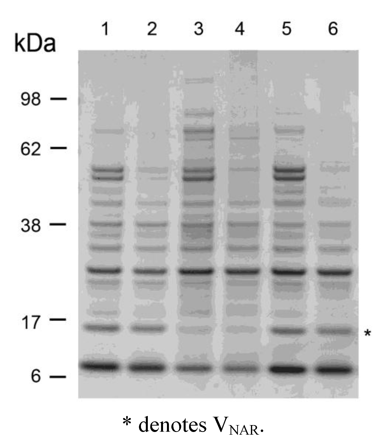
3.2. Soluble Expression of VNAR Constructs from Expression Vector pGC

Recombinant proteins were expressed in the bacterial periplasm as described previously [27,28]. Proteins were purified from the periplasm by the method of Minsky [29] and used either as crude fractions or purified further by affinity chromatography using ANTI-FLAG M2 Affinity gel resin (2–10 × 1 cm) (Sigma, St. Louis, MO, USA). Protein elution was with 0.1 M glycine (pH 3) or ImmunoPureTM gentle elution buffer (Pierce, USA). Neutralized proteins were dialyzed extensively against PBS, pH 7.4, and then concentrated over a YM-3 (3 kDa cut-off) membrane (Amicon, Japan). Apical membrane antigen 1 (AMA-1) from Plasmodium falciparum strain 3D7 was expressed recombinantly and purified as previously described [30].

3.3. Thermal and pH Stability of VNARs and mAbs

3.3.1. pH Stability of VNAR 4A-1

Separate 50 µL aliquots of purified recombinant VNAR 4A-1 (specific for mAb 5G8) at 0.12 mg/mL were adjusted to pHs 3, 5.5 (using 0.2 M Na2HPO4 and 0.1 M citric acid), pH 7.4 (using PBS), and pHs 8.5, 11.0 (using 25 mM borax buffer) then incubated at 4, 37 and 50 °C for 24 h and 1, 2, 3, 4 weeks. Unheated control samples were stored at −20 °C throughout. Following incubation, samples were neutralized with 1 M Tris at pH 11.5 (for samples at pH 3.0 and 5.5) or with 1 M HCl (for samples at pH 8.5 and 11.0).

3.3.2. Heating VNARs in Periplasmic Extracts at 100 °C

Separate 100 µL aliquots of VNARs 12Y-2, 14M-15, 14I1M-15 and 22A-2 in periplasmic extracts were incubated at RT or 100 °C for 15 min then clarified by centrifugation for 5 min. The resulting soluble proteins were analyzed by SDS-PAGE.

3.3.3. Heating VNARs and mAbs at 45 °C and 60 °C for up to 4 Weeks

Separate 40 µL aliquots of purified recombinant VNARs (12Y-2, 14I1M-15) and monoclonal antibodies (1F9, 5G8, Monoclonal Antibody Facility, Walter & Eliza Hall Institute Biotechnology Centre, Melbourne, Australia) all at 1 mg/mL were incubated in liquid and lyophilized formats at 45 °C and 60 °C, for 24, 48 and 72 h and 1, 2, 3 and 4 weeks and then stored at 4 °C until analysis. Unheated control samples were stored at 4 °C throughout.

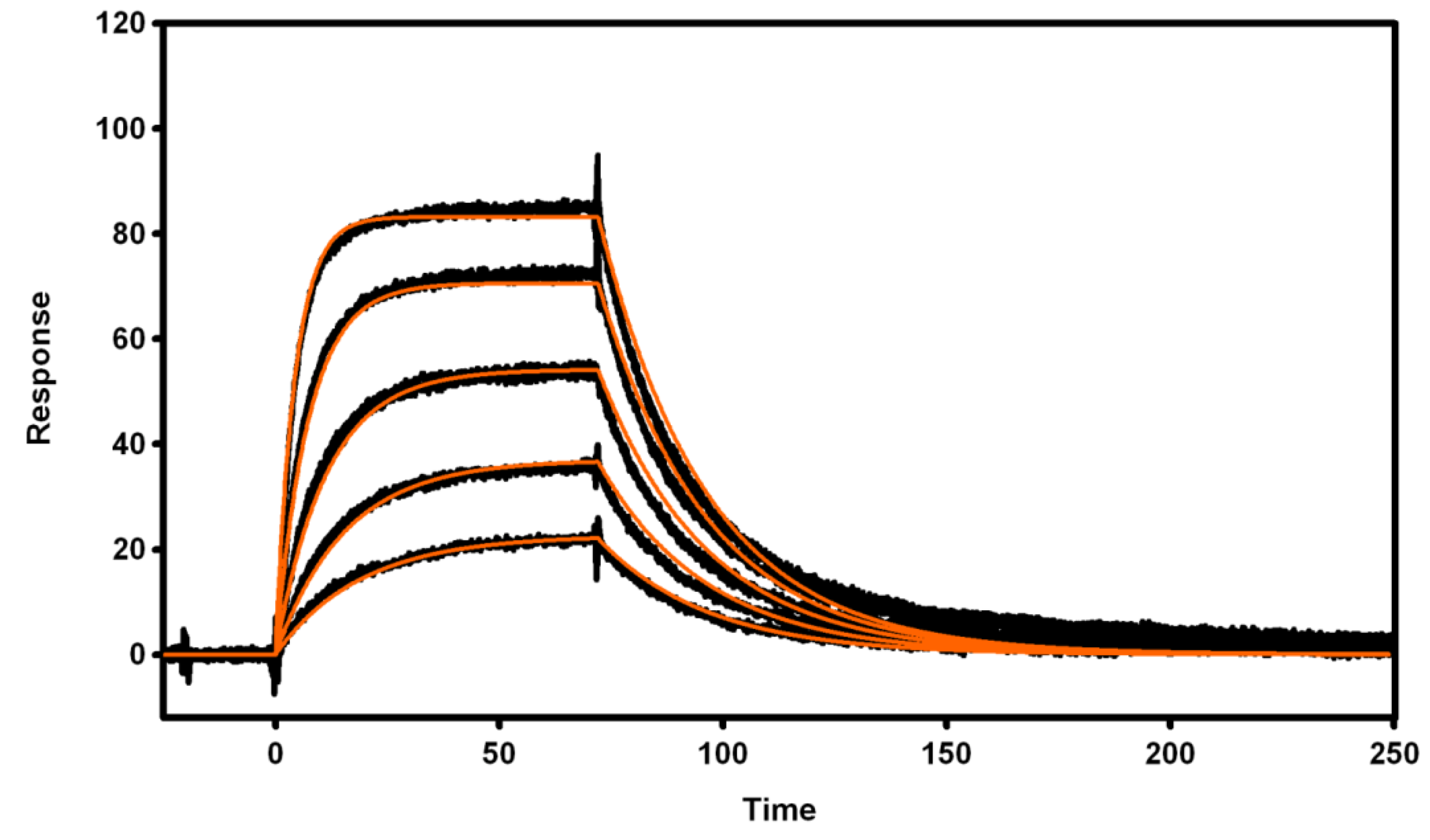
3.4. Biosensor Analysis of VNAR Proteins

All Surface Plasmon Resonance (SPR) biosensor analyses were performed at 25 °C using Biacore 1000, Biacore T100 (GE Healthcare, Uppsala, Sweden), SensiQ Pioneer (SensiQ Technologies, Oklahoma City, OK) and ProteOn XPR36 (Bio-Rad, Hercules, CA) platforms with instrumental fluidics primed in 1 × HBS-EP + [10 mM HEPES, 150 mM NaCl, 3 mM EDTA 0.05% (v/v) surfactant P20, pH 7.4].

3.4.1. Immobilization of Recombinant Proteins

Coupling of AMA-1 on CM5 sensor chips (Biacore) was achieved essentially as described previously [5]. Briefly, the chip surface was activated with a 7 min injection of a freshly prepared 1:1 mixture of 50 mM NHS : 200 mM EDC. Protein coupling was achieved by a single 7 min injection solution (23 μg/mL in 10 mM sodium acetate, pH 4.0). To deactivate residual reactive sites, AMA-1 coupling was followed by a 7 min injection of 1 M ethanolamine (pH 8.5). Immobilization of AMA-1 protein on the surface of a GLM chip (ProteOn) was achieved in a similar manner with the following differences: (1) chip activation 50:50 mixture consisted of a sulfo-NHS at 5 mM and EDC at 20 mM and (2) injections of activation mixture, ligand solution and deactivation solution were all performed at 30 μL/min for 5 min. Both coupling procedures typically resulted in >8,000 Response Units (1 RU; 1000 RU = 1 ng of protein/mm2) of AMA-1 protein being coupled to either of the two chips. Coupling of 5G8 IgG (target antigen for 4A-1 IgNAR) on a GLM chip was performed using a similar method to that described above for AMA-1 except that the 5G8 IgG protein solution was diluted to 20 μg/mL in 10 mM sodium acetate, pH 4.5. Approximately 7,600 RU of IgG protein was coupled to the chip surface.

3.4.2. Active Concentration Determinations

Fully automated SPR-based concentration assay [31] was utilized for the determination of the residual AMA-1 binding activity in diversely treated VNAR and IgG antibody samples The method was configured as a direct detection assay with quantification against a calibration standard, that being an untreated sample stored at 4 °C (t = 0). In case of Biacore T100 assay, a standard curve was established by serially injecting ‘t = 0’ calibration samples at 0, 250, 500, 1,000, 2,000, 4,000, 8,000 and 1,600 ng/mL over the AMA-1 immobilized sensor chip. Assay conditions were: flow rate, 10 μL/min, contact time, 1 min; regeneration: 100 μL/min for 10 s of 10 mM glycine pH 2.2. Binding responses, taken 10 s after sample injection was stopped, were plotted against the injected concentration. Residual active concentrations of heated samples were quantified by interpolation of binding responses against the standard curve. A similar approach was adopted for the ProteOn XPR36. Standard curve was established by a parallel injection of six different ‘t = 0’ sample concentrations (500, 1,000, 2,000, 4,000, 8,000 ng/mL) across the immobilized AMA-1 or 5G8 IgG protein at 25 μL/min for 60 s. Residual active concentrations of unknown samples were determined by injecting six samples simultaneously and comparing their initial binding rates (at t = 5–20 s) to those generated by the calibration standards. Bound samples were regenerated from the surface with a single injection of 10 mM glycine pH 2.2 at 100 μL/min for 15 s.

3.5. Nitrocellulose Dot Blot and Western Analysis

Varying amounts of VNARs (1 µg of 12Y-2, 0.32 µg of 14I1M-15) and mAbs (0.05 µg of 5G8, 0.01 µg of 1F9) were dot blotted onto nitrocellulose membrane (Osmonics, Minnetonka, MN, USA) in 100 μL PBS using a slot blot manifold. These amounts were pre-determined in earlier titration experiments to provide equivalent signal intensity (data not shown). Twenty two strips of nitrocellulose containing each protein were incubated at 4, 25, 45 and 60 °C. Strips were removed weekly from all incubation temperatures and stored at 4 °C until testing. The nitrocellulose strips were cut into two, separating VNARs from mAbs and blocked in 10% (w/v) skim milk powder for 1 h. Between each step the membranes were washed 3 times with PBS containing 0.1% (v/v) Tween20. Antibodies were diluted in PBS containing 0.1% (v/v) Tween20. AMA-1 was added at 5 μg/mL in 5% (w/v) milk powder and incubated for 2 h at room temperature. Rabbit anti-AMA-1 serum was then added at 1:3,000 dilution and incubated for 1 h. Anti-rabbit-HRP (GE Healthcare, WI, USA) was added at 1:1,000 dilution and incubated for 1 h then signal was developed using SuperSignal West Pico Chemiluminescent Signal (ThermoScientific, Rockford, IL, USA).

3.6. Exposure of VNAR 14M-15 to Murine stomach and Intestinal Scrapings

Gastric and intestinal samples were obtained from mice (P. Sutton, School of Veterinary Science, University of Melbourne, Australia) as described previously [32,33]. The samples were homogenised and sonicated in PBS containing 1% (v/v) Triton X 100 and then the total protein concentration was determined via BCA assay (BioRad, Hercules, CA, USA). Aliquots of VNAR 14M-15 (15 μg) were incubated with stomach or intestinal samples (33 μg), unbuffered, in a total volume of 45 μL for 1, 10, 30 or 60 min at 0 and 37 °C. Negative controls were water, glycine (pH 3) or protease K in place of stomach and intestinal samples. Following incubation, samples were analyzed by SDS-PAGE and Western blotting in order to visualize VNAR fragmentation or degradation.

3.7. Circular Dichroism Spectroscopy

CD spectra were recorded using an AVIV Model 420 CD spectrometer using similar methods to those described previously [34]. Wavelength scans were performed using a 1 mm quartz cuvette at 20 °C and 80 °C from 190 and 250 nm. Proteins were solubilized in PBS at a concentration of 0.15 mg/mL. Spectra were baseline-corrected by subtracting corresponding buffer scans collected under identical conditions.

4. Conclusions

Single domain shark VNARs can be readily selected against antigens of diagnostic value. When expressed recombinantly they show excellent stability at elevated temperatures over prolonged periods in liquid, lyophilized and immobilized formats, particularly in comparison with mAbs. Furthermore, the VNAR domains show very good stability properties at pH extremes, with resistance to proteolytic cleavage. Together these data demonstrate the versatility of these unique single domains and their amenability to more complex and varied diagnostic applications.

Acknowledgments

This work was supported by the National Health and Medical Research Council of Australia (ID 487339), a grant from the National Institutes of Health (NIH RO1AI59229) and through funding from The Foundation for Innovative New Diagnostics (FIND). Matthew A. Perugini acknowledges the Australian Research Council for Future Fellowship support (FT0991245). The authors thank Robin Anders for provision of recombinant AMA-1, Denison Chang and Meghan Hattarki for technical assistance and Phil Sutton for supplying the murine stomach and intestine samples. Michael Foley declares a competing financial interest.

References and Notes

  1. Murray, C.K.; Gasser, R.A., Jr.; Magill, A.J.; Miller, R.S. Update on rapid diagnostic testing for malaria. Clin. Microbiol. Rev. 2008, 21, 97–110. [Google Scholar] [CrossRef]
  2. Roux, K.H.; Greenberg, A.S.; Greene, L.; Strelets, L.; Avila, D.; McKinney, E.C.; Flajnik, M.F. Structural analysis of the nurse shark (new) antigen receptor (nar): Molecular convergence of nar and unusual mammalian immunoglobulins. Proc. Natl. Acad. Sci. USA 1998, 95, 11804–11809. [Google Scholar]
  3. Streltsov, V.A.; Varghese, J.N.; Carmichael, J.A.; Irving, R.A.; Hudson, P.J.; Nuttall, S.D. Structural evidence for evolution of shark ig new antigen receptor variable domain antibodies from a cell-surface receptor. Proc. Natl. Acad. Sci. USA 2004, 101, 12444–12449. [Google Scholar]
  4. Stanfield, R.L.; Dooley, H.; Flajnik, M.F.; Wilson, I.A. Crystal structure of a shark single-domain antibody V region in complex with lysozyme. Science 2004, 305, 1770–1773. [Google Scholar] [CrossRef]
  5. Nuttall, S.D.; Humberstone, K.S.; Krishnan, U.V.; Carmichael, J.A.; Doughty, L.; Hattarki, M.; Coley, A.M.; Casey, J.L.; Anders, R.F.; Foley, M.; et al. Selection and affinity maturation of IgNAR variable domains targeting Plasmodium falciparum AMA-1. Proteins 2004, 55, 187–197. [Google Scholar] [CrossRef]
  6. Kopsidas, G.; Roberts, A.S.; Coia, G.; Streltsov, V.A.; Nuttall, S.D. In vitro improvement of a shark IgNAR antibody by Qβ replicase mutation and ribosome display mimics in vivo affinity maturation. Immunol. Lett. 2006, 107, 163–168. [Google Scholar] [CrossRef]
  7. Simmons, D.P.; Abregu, F.A.; Krishnan, U.V.; Proll, D.F.; Streltsov, V.A.; Doughty, L.; Hattarki, M.K.; Nuttall, S.D. Dimerisation strategies for shark IgNAR single domain antibody fragments. J. Immunol. Methods 2006, 315, 171–184. [Google Scholar] [CrossRef]
  8. Henderson, K.A.; Streltsov, V.A.; Coley, A.M.; Dolezal, O.; Hudson, P.J.; Batchelor, A.H.; Gupta, A.; Bai, T.; Murphy, V.J.; Anders, R.F.; et al. Structure of an IgNAR-AMA1 complex: Targeting a conserved hydrophobic cleft broadens malarial strain recognition. Structure 2007, 15, 1452–1466. [Google Scholar] [CrossRef]
  9. Simmons, D.P.; Streltsov, V.A.; Dolezal, O.; Hudson, P.J.; Coley, A.M.; Foley, M.; Proll, D.F.; Nuttall, S.D. Shark IgNAR antibody mimotopes target a murine immunoglobulin through extended CDR3 loop structures. Proteins 2008, 71, 119–130. [Google Scholar] [CrossRef]
  10. Coley, A.M.; Campanale, N.V.; Casey, J.L.; Hodder, A.N.; Crewther, P.E.; Anders, R.F.; Tilley, L.M.; Foley, M. Rapid and precise epitope mapping of monoclonal antibodies against Plasmodium falciparum AMA-1 by combined phage display of fragments and random peptides. Protein Eng. 2001, 14, 691–698. [Google Scholar] [CrossRef]
  11. Bell, D.; Wongsrichanalai, C.; Barnwell, J.W. Ensuring quality and access for malaria diagnosis: How can it be achieved? Nat. Rev. Microbiol. 2006, 4, S7–S20. [Google Scholar]
  12. Chiodini, P.L.; Bowers, K.; Jorgensen, P.; Barnwell, J.W.; Grady, K.K.; Luchavez, J.; Moody, A.H.; Cenizal, A.; Bell, D. The heat stability of Plasmodium lactate dehydrogenase-based and histidine-rich protein 2-based malaria rapid diagnostic tests. Trans. R. Soc. Trop. Med. Hyg. 2007, 101, 331–337. [Google Scholar] [CrossRef]
  13. Ashley, E.A.; Touabi, M.; Ahrer, M.; Hutagalung, R.; Htun, K.; Luchavez, J.; Dureza, C.; Proux, S.; Leimanis, M.; Lwin, M.M.; et al. Evaluation of three parasite lactate dehydrogenase-based rapid diagnostic tests for the diagnosis of falciparum and vivax malaria. Malar. J. 2009, 8, 241. [Google Scholar] [CrossRef] [Green Version]
  14. Greenfield, N.J. Using circular dichroism spectra to estimate protein secondary structure. Nat. Protoc. 2006, 1, 2876–2890. [Google Scholar] [CrossRef]
  15. Dudgeon, K.; Rouet, R.; Kokmeijer, I.; Schofield, P.; Stolp, J.; Langley, D.; Stock, D.; Christ, D. General strategy for the generation of human antibody variable domains with increased aggregation resistance. Proc. Natl. Acad. Sci. USA 2012, 109, 10879–10884. [Google Scholar]
  16. Hamers-Casterman, C.; Atarhouch, T.; Muyldermans, S.; Robinson, G.; Hamers, C.; Songa, E.B.; Bendahman, N.; Hamers, R. Naturally occurring antibodies devoid of light chains. Nature 1993, 363, 446–448. [Google Scholar] [CrossRef]
  17. Hussack, G.; Arbabi-Ghahroudi, M.; van Faassen, H.; Songer, J.G.; Ng, K.K.; MacKenzie, R.; Tanha, J. Neutralization of Clostridium difficile toxin a with single-domain antibodies targeting the cell receptor binding domain. J. Biol. Chem. 2011, 286, 8961–8976. [Google Scholar]
  18. Urdea, M.; Penny, L.A.; Olmsted, S.S.; Giovanni, M.Y.; Kaspar, P.; Shepherd, A.; Wilson, P.; Dahl, C.A.; Buchsbaum, S.; Moeller, G.; et al. Requirements for high impact diagnostics in the developing world. Nature 2006, 444, 73–79. [Google Scholar]
  19. Romer, T.; Leonhardt, H.; Rothbauer, U. Engineering antibodies and proteins for molecular in vivo imaging. Curr. Opin. Biotechnol. 2011, 22, 882–887. [Google Scholar] [CrossRef]
  20. Adair, J.R.; Howard, P.W.; Hartley, J.A.; Williams, D.G.; Chester, K.A. Antibody-drug conjugates—A perfect synergy. Expert Opin. Biol. Ther. 2012, 12, 1191–1206. [Google Scholar] [CrossRef]
  21. Olichon, A.; Schweizer, D.; Muyldermans, S.; de Marco, A. Heating as a rapid purification method for recovering correctly-folded thermotolerant VH and VHH domains. BMC Biotechnol. 2007, 7, 7. [Google Scholar]
  22. Muller, R.M.; Saunders, K.; Grace, C.; Jin, M.; Piche-Nicholas, N.; Steven, J.; O'Dwyer, R.; Wu, L.; Khetemenee, L.; Vugmeyster, Y.; et al. Improving the pharmacokinetic properties of biologics by fusion to an anti-HSA shark VNAR domain. mAbs 2012, 4, 673–685. [Google Scholar] [CrossRef]
  23. Fennell, B.J.; Darmanin-Sheehan, A.; Hufton, S.E.; Calabro, V.; Wu, L.; Muller, M.R.; Cao, W.; Gill, D.; Cunningham, O.; Finlay, W.J. Dissection of the IgNAR V domain: Molecular scanning and orthologue database mining define novel ignar hallmarks and affinity maturation mechanisms. J. Mol. Biol. 2010, 400, 155–170. [Google Scholar] [CrossRef]
  24. Nuttall, S.D.; Krishnan, U.V.; Doughty, L.; Pearson, K.; Ryan, M.T.; Hoogenraad, N.J.; Hattarki, M.; Carmichael, J.A.; Irving, R.A.; Hudson, P.J. Isolation and characterization of an IgNAR variable domain specific for the human mitochondrial translocase receptor tom70. Eur. J. Biochem. 2003, 270, 3543–3554. [Google Scholar] [CrossRef]
  25. Hodder, A.N.; Crewther, P.E.; Anders, R.F. Specificity of the protective antibody response to apical membrane antigen 1. Infect. Immun. 2001, 69, 3286–3294. [Google Scholar] [CrossRef]
  26. Coia, G.; Ayres, A.; Lilley, G.G.; Hudson, P.J.; Irving, R.A. Use of mutator cells as a means for increasing production levels of a recombinant antibody directed against Hepatitis B. Gene 1997, 201, 203–209. [Google Scholar] [CrossRef]
  27. Nuttall, S.D.; Krishnan, U.V.; Doughty, L.; Nathanielsz, A.; Ally, N.; Pike, R.N.; Hudson, P.J.; Kortt, A.A.; Irving, R.A. A naturally occurring NAR variable domain binds the Kgp protease from Porphyromonas gingivalis. FEBS Lett. 2002, 516, 80–86. [Google Scholar] [CrossRef]
  28. Nuttall, S.D.; Rousch, M.J.; Irving, R.A.; Hufton, S.E.; Hoogenboom, H.R.; Hudson, P.J. Design and expression of soluble CTLA-4 variable domain as a scaffold for the display of functional polypeptides. Proteins 1999, 36, 217–227. [Google Scholar] [CrossRef]
  29. Minsky, A.; Summers, R.G.; Knowles, J.R. Secretion of beta-lactamase into the periplasm of Escherichia coli: Evidence for a distinct release step associated with a conformational change. Proc. Natl. Acad. Sci. USA 1986, 83, 4180–4184. [Google Scholar] [CrossRef]
  30. Gupta, A.; Bai, T.; Murphy, V.; Strike, P.; Anders, R.F.; Batchelor, A.H. Refolding, purification, and crystallization of apical membrane antigen 1 from Plasmodium falciparum. Protein Expr. Purif. 2005, 41, 186–198. [Google Scholar] [CrossRef]
  31. Gapper, L.W.; Copestake, D.E.; Otter, D.E.; Indyk, H.E. Analysis of bovine immunoglobulin g in milk, colostrum and dietary supplements: A review. Anal. Bioanal. Chem. 2007, 389, 93–109. [Google Scholar] [CrossRef]
  32. Every, A.L.; Ng, G.Z.; Skene, C.D.; Harbour, S.N.; Walduck, A.K.; McGuckin, M.A.; Sutton, P. Localized suppression of inflammation at sites of helicobacter pylori colonization. Infect. Immun. 2011, 79, 4186–4192. [Google Scholar] [CrossRef]
  33. Chionh, Y.T.; Walduck, A.K.; Mitchell, H.M.; Sutton, P. A comparison of glycan expression and adhesion of mouse-adapted strains and clinical isolates of Helicobacter pylori. FEMS Immunol. Med. Microbiol. 2009, 57, 25–31. [Google Scholar] [CrossRef]
  34. Atkinson, S.C.; Dogovski, C.; Downton, M.T.; Pearce, F.G.; Reboul, C.F.; Buckle, A.M.; Gerrard, J.A.; Dobson, R.C.; Wagner, J.; Perugini, M.A. Crystal, solution and in silico structural studies of dihydrodipicolinate synthase from the common grapevine. PLoS One 2012, 7, e38318. [Google Scholar]

Appendix

Figure A1. SPR analysis of 4A-1 VNAR. Binding of 4A-1 VNAR protein to immobilized 5G8 IgG (1450 RU) was analyzed at a constant flow rate of 50 µL/min with an injection volume of 50 µL. Injected concentrations were 8, 4, 2, 1 and 0.5 µM. No regeneration was required between injection cycles as the bound 4A-1 VNAR completely dissociated within 300 s in the running buffer. Binding data (black lines, in triplicate) were fitted globally with a 1:1 kinetic interaction model (red lines) yielding a ka of 2.4 × 10⁴ M−1s−1, a kd of 0.04 s−1 and an equilibrium dissociation constant (KD) of 1.7 μM. SensiQ Pioneer instrument docked with COOH1 chip was utilized for this measurement.
Antibodies 02 00066 g007
Figure A2. SPR analysis of the interaction between AMA-1 3D7 protein and 1F9 IgG. Approximately 185 RU of 1F9 IgG was captured onto an SPR chip via rabbit anti-mouse IgG at the start of each binding cycle. AMA-1 protein was injected at 50 μL/min for 2 min and the dissociation monitored for either 300 s or 900 s. Injected AMA-1 concentrations were 729, 243, 81, 9 and 3 nM. Following dissociation phase, chip surface was regenerated with 10 mM Glycine pH 1.7 resulting in a complete removal of both AMA-1 and 1F9 proteins. Binding data (black lines, in duplicate) were fitted globally with a 1:1 kinetic interaction model (red lines) yielding a ka of 3.8 × 10⁴ M−1s−1, a kd of 3.5 × 10⁻⁵ M s−1 and an equilibrium dissociation constant (KD) of 0.95 nM. SensiQ Pioneer instrument docked with COOH5 chip was utilized for this experiment.
Antibodies 02 00066 g008
Figure A3. Retention of active VNARs 12Y-2 and 14I1M-15, and mAbs 5G8 and 1F9 following heating at 60 °C in (a) liquid and (b) lyophilized formats, as determined by biosensor analysis. Method described in Section 3.3.3.
Antibodies 02 00066 g009

