Article

Effect of Brown Marmorated Stink Bug (Halyomorpha halys Stål.) Infestation on the Phenolic Response and Quality of Olive Fruits (Olea europaea L.)

Department of Agronomy, Biotechnical Faculty, University of Ljubljana, Jamnikarjeva 101, SI-1000 Ljubljana, Slovenia
*
Author to whom correspondence should be addressed.
Agronomy 2022, 12(9), 2200; https://doi.org/10.3390/agronomy12092200
Submission received: 29 July 2022 / Revised: 9 September 2022 / Accepted: 14 September 2022 / Published: 15 September 2022
(This article belongs to the Section Plant-Crop Biology and Biochemistry)

Abstract

:
Olives ripen in the late autumn and represent a good source of nutrients that Halyomorpha halys uses to prepare for diapause. This is the first study to investigate the impact of H. halys infestation on the phenolic response and olive fruit quality in the pierced tissue of damaged fruits and in the non-pierced part of damaged fruits of ‘Istrska belica’ and ‘Pendolino’ cultivars. Both total phenolic content and antioxidant capacity contents significantly increased in the infested fruits of the cultivar ‘Istrska belica’. Total phenolic content in the pierced tissue of damaged fruits increased by 10.7%, while the content of AC in the non-pierced tissue of damaged fruits increased by 7.11% and in the pierced tissue of damaged fruits by 6.1% compared to control. A total of 44 individual phenolic compounds were identified, 21 of them increased in at least one cultivar after infestation. Huge increases in phenolic content were observed in both cultivars, particularly for flavones, secoiridoids, anthocyanins, and flavonols in the pierced tissue of damaged fruits. The most responsive individual phenolic compound in both cultivars was oleuropein. Its content in the pierced tissue of damaged fruits increased by 44.7% in the cultivar ‘Pendolino’ and for 82.6% in the cultivar ‘Istrska belica’.

1. Introduction

The olive (Olea europaea L.) is the most abundant and economically important fruit species in Mediterranean countries. Most of the total production is used for oil extraction, while a smaller portion is consumed as table olives. The world consumption of olive oil increased by 92.9% between 1990 and 2021 [1]. This is likely due to the recognition of its beneficial effects on human health, as it may reduce the incidence of cardiovascular disease, prevent colorectal cancer, and have anti-inflammatory effects [2]. These beneficial effects have proven to be related to the higher content of phenolic and antioxidant compounds in olive fruits and consequently in olive products [3]. The main groups of phenolic compounds in olive fruits are phenolic acids, flavonoids, other simple phenols, and secoiridoids [4].
One of the major problems for olive growers, which affects the quality of the products, namely that olive fruits are often severely damaged by diseases and insect infestations, such as the brown marmorated stink bug (Halyomorpha halys Stål.), a polyphagous insect that can feed on various ornamental fruit and vegetable plants [5]. H. halys originated in China, Japan, Korea, and Taiwan, but is now present in at least 120 countries and is on the invasive species list [6]. Several factors have contributed to the spread and invasive nature of this pest, including climate change, lack of natural predators, and global trade [7]. Adults of H. halys have a 12–17 mm long, shield-shaped body with a mottled brown-gray color [8]. They are recognized by white and black banding on the antennae, white underside with gray or black markings, and brown legs with faint white banding [9]. These pests release a stinging alarm signal that repels predators [10]. The major components of the predator-repellent secretions are trans-2-octenal and trans-2-decenal, which can contaminate the fruit with an unpleasant odor [11]. Infested fruits by H. halys can result in morphological deformations of fruits known as cat’s facing, localized wilting and necrosis, abscission of fruit forms, altered vegetative growth, and tissue malformations. Other potential impacts of the brown marmorated stink bug on fruit production include crop contamination with other pathogens causing secondary infection by plant pathogens, and fruit contamination by nymphs and adults at harvest [12]. In most cases, infestation results in lower fruit quality, yield loss, and economic damage [13]. In apple orchards in the mid-Atlantic region alone, damage from stink bug infestation is estimated at $37 million [14].
Higher plants have several mechanisms to defend themselves against pest infestations [15]. A key component of induced plant defense is the recognition of the invading pest by the presence of its specific compounds—elicitors [16]. Resistance strategies include physical or chemical barriers that repel other insects [17]. Most plants produce a broad range of secondary metabolites (terpenoids, alkaloids, anthocyanins, phenols, and quinones) that have toxic, repellent, and/or anti-nutrient effects on pests [18]. Plant defense chemicals can be divided into two groups: preformed phenolics, which are present in plants during normal plant tissue development, and induced phenolics, which are synthesized in response to damage by an insect [15]. The synthesis of these compounds may occur in the damaged tissue or may be transferred to other plant organs [17].
Understanding the plant response to H. haly infestation plays an important role in further pest management strategies to control infestations and help producers achieve high-quality yields. However, the metabolic response of olive fruit to disease and pest attack is poorly studied. In this study, for the first time, the chemical response of secondary metabolites in olive fruit of cultivars ‘Istrska belica’ and ‘Pendolino’ damaged by the brown marmorated stink bug infestation was investigated. The aim is to determine the difference in the change in weight, firmness, color, total phenolic content (TPC), individual phenolic compounds, and antioxidant capacity (AC) in the damaged and controlled fruits. We aimed to test the difference between the H. Halys infested tissue of olive fruit and the non-pierced part of the damaged fruit compared to the control. It is expected that the metabolic response in damaged fruit within the pierced tissue will have a higher response to selected parameters compared to the non-pierced tissue in both cultivars. The results of this study may better explain how H. halys infestation affects the metabolic response in damaged and undamaged parts of the olive fruit.

2. Materials and Methods

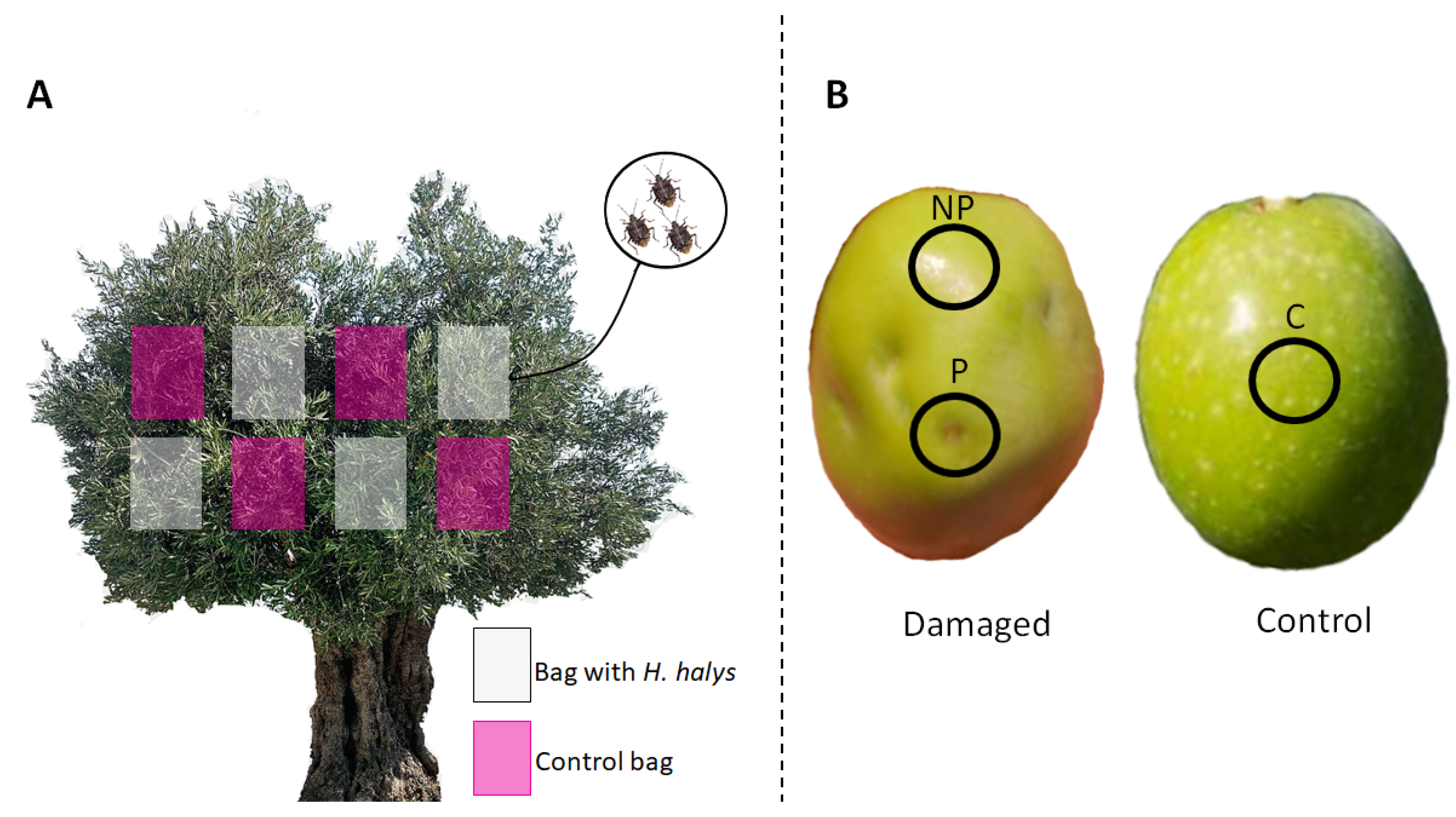
2.1. Plant Materials and Treatments

The study was carried out in Izola (45°32′12.98″ N 13°39′42.98″ E), Slovenia, during the 2021 growing season in an organic orchard. The study included 20-year-old olive trees of cultivars ‘Istrska belica’ and ‘Pendolino’. The olives were harvested at maturity. The study was conducted on six olive trees, three for each cultivar. In mid-September, on each tree, eight net bags were placed on the branches that already had fruits (Figure 1A). The bags were made of a 0.8 mm × 0.8 mm dense insect net and were closed with a rubber cord to prevent the H. halys from escaping. Three adult marmorated stink bugs were enclosed in four net bags per tree (Figure 1A). Four bags per tree without bugs in them represented the control.
Fruits were collected during the harvest period in late October. Immediately after harvest, weight, firmness (measured using a penetrometer with a 3 mm diameter cylindrical probe), and color were measured on 30 fruits from damaged and control bags. From the damaged fruits pierced tissue (P) and non-pierced (NP) tissue were cut out with a cork borer (5 mm). The tissue from control fruits (C) was cut out in the same way (Figure 1B). After cutting, the pieces were immediately frozen with liquid nitrogen and stored in centrifuge tubes at −20 °C until further analysis. Six replicates were performed for each treatment.

2.2. Color of the Fruits

The color of the fruit was measured using the colorimeter CR 300 Chroma (Minolta Co., Osaka, Japan). The results were recorded using the CIE (Commission Internationale d’Eclairage) parameters, where the L content corresponds to the dark-bright scale (0 black, 100 white), the a content represents color on the red-green axis, and the b content represents color on the yellow-blue axis. Hue angle (h°) and chroma (C) were calculated from the a and b content. Hue angle shows the color change from green to yellow and chroma color saturation or intensity.

2.3. Extraction of TPC, AC, and Individual Phenolic Compounds

The frozen material was ground to fine powder using liquid nitrogen and an analytical mill (IKA A11 basic, Staufen, Germany). A total of 0.5 g of the sample was extracted with 7.5 mL of 70% (v/v) MeOH solution containing 3% formic acid and sonicated in ice water (Sonis 4 ultrasonic bath; Iskra pio, Sentjernej, Slovenia). After 30 min of sonication, samples were centrifuged (Eppendorf Centrifuge 5810 R, Hamburg, Germany) at 8000× g rpm for 5 min at 4 °C. The supernatant was filtered through a 0.2 µm polyamide filter (Chromafil AO 20/25; Macherey-Nagel, Düren, Germany) into a vial and stored at −20 °C until further analysis. The extract was used for the identification of individual phenolic compounds, TPC and AC.

2.4. Analysis of Individual Phenolic Compounds

Individual phenolic compounds were determined using the HPLC system (Dionex UltiMate 3000; Thermo Scientific, Waltham, MA, USA). Measurements were made at 280 nm for flavones, flavonols, hydroxycinnamic acid, and secoiridoids and at 530 nm for anthocyanins. Conditions were as previously described by Diarte et al. [12]. Phenolic compounds were identified using a mass spectrometer (Thermo Scientific LCQ Deca XP MAX, San Jose, CA, USA) with heated electrospray ionization (HESI) in the negative and positive ion ranges. Individual phenolic compounds were identified using literature data and MS fragmentation and quantified using a similar standard (Table S1).
Compounds for which standards were not available were expressed as follows: Hydroxytyrosol glucoside and 6′deoxyhexopyranosyl oleoside as 3-hydroxytyrosol, acyclodihydroelenolic acid hexoside isomers, elenolic acid glucoside isomers, elenolic acid, dehydro derivate of tometic acid and caffeoil-6′-secologanoside as caffeic acid, oleoside, oleoside isomers, oleuropein aglycone derivate, demethyloleuropein, oleuropein glucoside isomer, hydroxyoleuropein, oleuropein aglycone isomers, oleuropein aglycone derivative, oleuropein isomer, oleacein and methoxyoleuropein isomer as oleuropein, p-coumaric acid glucoside as p-coumaric acid, β-hydroxy-verbascoside as verbascoside, luteoline 7 O-rutinoside as luteoline 7-O-glucoside, kaempferol-7-O-(6″rhamnosyl) hexoside as kaempferol 3-O-glucoside, tyrosol glucoside as tyrosol and chryptochlorogenic acid as chlorogenic acid. Retention times, molecular weights, and negative and positive ion fragmentation are listed in Table S1. Individual phenolic contents are given in mg kg1 FW.

2.5. Analysis of the Total Phenolic Content (TPC)

The total phenol content was determined using the Folin-Ciocalteu phenol reagent according to the method described by Singleton et al. [19] with some modifications. A total of 6 mL of double-distilled water and 500 µL of Folin-Ciocalteu reagent were added to 100 µL of the sample extract. After resting at room temperature for 5 min, 1.5 mL of sodium carbonate (20% w/v) was added. The mixture was vortexed and left in the oven at 40 °C for 30 min before absorbance was measured at 765 nm using a spectrophotometer (Thermo Scientific Genesys 10S UV-VIS Spectrophotometer, San Jose, CA, USA). The same mixture with 100 µL methanol was used as a blank. A calibration curve with Y = 0.0009x (R2 = 0.9952) was constructed using a gallic acid solution in the range of 5000 mg L−1. TPC was expressed as gallic acid equivalents (GAE) in mg 100 g−1 FW.

2.6. Analysis of Antioxidant Capacity (AC)

The free radical scavenging activity of the extract was measured using DPPH (1,1-diphenil-2-picrylhydrazyl). A total of 50 µL of the fruit extract was placed in a quivette and 1950 µL of a 0.1 mmol L1 solution of DPPH in 80% MeOH (v/v) was added and allowed to react in the dark at room temperature for 30 min. The absorbance of DPPH was measured at 520 nm using a spectrophotometer (Thermo Scientific Genesys 10S UV-VIS Spectrophotometer, San Jose, CA, USA). As a blank solution, 80% MeOH (v/v) was used, and a DPPH solution without test samples served as a control. A calibration curve (y = 0.0005x R2 = 0.9922) was constructed using Trolox in the 2000 µM range. AC was expressed as mmol Trolox kg1 FW.

2.7. Chemicals and Standards

The standards used in our study were: trolox, gallic acid, 3-hydroxytyrosol, oleuropein, luteoline 7-O-glucoside, chlorogenic acid, cyanidin 3-O-glucoside, cyanidin 3-O-rutinoside and 1,1-diphenil-2-picrylhydrazyl (Sigma-Aldrich Chemi GmbH, Steinheim, Germany); caffeic acid, p-coumaric acid, kaempferol 3-O-glucoside, rutin, quercetin 3-O-ramnoside and apigenin-7-O-glucoside (Fluka Chemie GmbH, Buchs, Switzerland); verbascoside (HWI group, Rülzheim, Germany) and tyrosol (PhytoLab, Vestenbergsgreuth, Germany). Double-distilled water was obtained from a Milli-Q water purificantion system (Millipore, Bedford, MA, USA). For extractions, 99.9% HPLC MeOH and formic acid (Fluka Chemie GmbH, Buchs, Switzerland) were used.

2.8. Statistical Analysis

Results were analyzed using Microsoft Excel 2020 and processed using R commander (version 4.0.5, 31 March 2021). Normality and homoscedasticity were tested with Shapiro-Willk and Bartlett tests, respectively. Statistical differences between treatments were determined using Student’s t-test for independent samples, with significance level set at p ≤ 0.05 and multiple comparison test (Tukey) at 95% confidence level. Means and standard deviations are presented as mean ± SD and statistical differences between ripening stages are indicated by different letters.

3. Results

3.1. Visual Appearance, Color, Weight, and Firmness of Olive Fruits

At harvest, an unpleasant odor was detected on the infested fruits. Visual changes were noted between the control fruit and fruit that had been exposed to H. halys in both cultivars. Pierced and corky tissue was observed on the damaged fruits. However, morphological deformities were mainly observed in the ‘Istrska belica’ cultivar, with local necrosis in the cultivar ‘Pendolino’.
At harvest time, the olives of the ‘Istrska belica’ cultivar were still green, while the fruits of the ‘Pendolino’ cultivar turned completely black (Table 1). For ‘Istrska belica’ and ‘Pendolino’ cultivars no significant differences in a, b, C and h° parameters were found between the control and damaged fruits. In the cultivar ‘Istrska belica’, the only significant differences between treatments were found for the parameter L, indicating that the damaged fruits were darker than the control ones. Its value decreased by 2.8%.
A significant difference in fruit weight between damaged and control fruits was found in both cultivars (Table 1). In the ‘Istrska belica’ cultivar, the weight decreased from 2.56 g to 2.01 g, while in ‘Pendolino’ it decreased from 1.63 g to 1.47 g. There was also a significant difference in fruit firmness between the control and damaged fruits of the cultivar ‘Pendolino’ (Table 1). In the damaged fruits, the firmness was 26.6%higher. The same trend is observed in the cultivar ‘Istrska belica’, where the firmness of the control fruits was 6.02 N and that of the damaged fruits was 6.54 N.

3.2. Total Phenolic Content and Antioxidant Capacity

The total phenolic content of the pierced tissue of the damaged fruit, the non-pierced tissue of the damaged fruit, and the control tissue of the cultivars ‘Istrska belica’ and ‘Pendolino’ is shown in Table 2. The highest phenolic response was observed in the pierced tissue of damaged fruits infested by H. halys of the cultivar ‘Istrska belica’. The content was 10.8% higher than in the other treatments. In the ‘Pendolino’ cultivar, no changes were observed between treatments. No significant difference was found between the control and the non-pierced tissue of the damaged fruits.
In the antioxidant capacity (AC), a significant result was found between the control fruits and the other treatments of the cultivar ‘Istrska belica’ (Table 2). In the control fruits the content was 27.83 umol Trolox kg1 FW. In the pierced tissue the content increased by 6.1% and by 7.1% in the non-pierced tissue, both compared to the control fruits. No significant difference was observed between damaged and control fruits in the cultivar ‘Pendolino’ at AC.

3.3. Individual Phenolic Compounds

3.3.1. ‘Istrska belica’

The contents of individual phenolic compounds of the pierced and non-pierced tissue of the damaged fruits and the control fruits for ‘Istrska belica’ cultivar are shown in Table 3. Thirty-five individual phenolic compounds from four phenolic groups were identified in the olive fruits of the ‘Istrska belica’ cultivar. The total content of flavones, flavonols and secoiridoids was significantly higher in the pierced tissue of the damaged fruits compared to the control fruits by 21.2%, 8.8%, and 43.4%, respectively. However, no significant differences in the content of these phenolic groups were noticed between the non-pierced tissue of the damaged fruits and the control fruits. Hydroxycinnamic acid content was not significantly different between treatments.
Flavones. Luteoline 7-O-rutinoside was the most abundant phenolic compound among total flavones, quercetin 3-O-rutinoside among flavonols, verbascoside and β-hydroxy-verbascoside among hydroxycinnamic acids, and oleuropein among secoiridoids in olive fruit of the cultivar ‘Istrska belica’. Among flavones, the content of luteoline 7-O-glucoside was 57.5% higher in the pierced tissue of damaged fruit than in the control and 23.0% higher than in the non-pierced tissue of damaged fruit.
Flavonols. The content of kaempferol 7-O-(6″rhamnosyl) hexoside increased by 81.3% in the pierced tissue of the damaged fruits compared to the control, and by 36.0% compared to the non-pierced tissue of the damaged fruits. An increasing trend in the pierced tissue was also observed for quercetin 3-O-rutinoside, whose content changed by 16.3% compared to the control and by 24.0% compared to the non-pierced tissue of the damaged fruits. Among flavonols, only quercetin 3-O-rhamnoside showed a decrease in the non-pierced tissue of the damaged fruits, by 34.2%, compared to the control and in the pierced tissue of the damaged fruits by 25.2% compared to the control.
Hydroxycinnamic acids. The content of verbascoside decreased by 48.9% in the non-pierced tissue of the damaged fruits and by 40.2% in the pierced tissue compared to the control fruits. p-coumaric acid glucoside and β-hydroxy-verbascoside increased significantly only in the pierced tissue of the damaged fruits by 58.1% and 69% compared to the control fruits.
Secoiridoids. Oleuropein was the most important single phenolic compound from the group of secoiridoids. Its content increased in the pierced (82.6%) and non-pierced tissues (31.0%) of the damaged fruits compared to the control fruits. A significant difference was also observed between the pierced (1712.84 mg kg−1 FW) and non-pierced tissues (1288.32 mg kg−1 FW) of the damaged fruits. The content of oleuropein aglycone derivatives in the pierced and non-pierced tissues of the damaged fruits decreased significantly by 6.1 mg kg−1 FW and by 8.04 mg kg−1 FW, respectively, compared to the control (10.18 mg kg−1 FW). However, the highest content of oleuropein aglycone isomers (528.10 mg kg−1 FW) and oleuropein isomer (19.42 mg kg−1 FW) was present in the control fruits. The content of oleuropein aglycone derivatives showed significant differences between the control fruits and the pierced tissue of the damaged fruits, by 6.7%, while the content of the non-pierced tissue showed no differences compared to the other treatments. The content of oleuropein isomer decreased by 26.1% in the non-pierced tissue of the damaged fruits and by 27.5% in the pierced tissue compared to the control fruits.
Hydroxytyrosol content was highest in the control fruit (5.45 mg kg−1 FW). In the pierced tissue and in the non-pierced tissue of the damaged fruits, the content increased significantly by 46.8% and 67.7%, respectively. There were significant differences between all treatments. However, the content of hydroxyoleuropein was highest in the non-pierced tissue (2.87 mg kg−1 FW). The content in the control tissue and in the pierced tissue of the damaged fruits was 27.5% lower than in the non-pierced tissue.
Elenolic acid increased only in the pierced tissue of the damaged fruits to 4.80 mg kg−1 FW. In the control fruits, the content was 3.03 mg kg−1 FW. In the non-pierced tissue of the damaged fruits, the content of elenolic acid glucoside isomers decreased by 12.4%. These results indicate that the increase in elenolic acid is due to the degradation of oleuropein aglycone.

3.3.2. ‘Pendolino’

Twenty-eight individual phenolic compounds belonging to the group of flavones, flavonols, hydroxycinnamic acids, and anthocyanins were quantified and qualified in the tissue of damaged fruits pierced by H. halys (Table 4). The total content of flavones, flavonols, secoiridoids, and anthocyanins was significantly higher in the pierced and non-pierced tissue of the damaged fruits compared with the control fruits. Only the total content of hydroxycinnamic acids decreased by 3.8% in the pierced tissue of the damaged fruit and by 22.1% in the non-pierced tissue of the damaged fruit compared to the control (145.01 mg kg−1 FW).
Flavones. The content of all individual flavones in the pierced and non-pierced tissues of the damaged fruits of the cultivar ‘Pendolino’ was increased compared to the control fruits. The content of luteoline 7-O-glucoside increased by 109.7% in the pierced tissue and by 49.7% in the non-pierced tissue of the damaged fruits from 171.52 mg kg−1 FW. Apigenin 7-O-glucoside content increased from 71.32 mg kg−1 FW by 89.9% in the pierced tissue and by 45.0% in the non-pierced tissue of the damaged fruits. The last individual flavones with the lowest amount detected in olive fruit was luteoline 7-O-rutinoside. In the control fruit, the content was 55.36 mg kg−1 FW. In the non-pierced tissue of the damaged fruits, it increased by 31.0%, and in the pierced tissue by 43.4%.
Flavonols. Quercetin 3-O-rhamnoside is the most abundant flavonol in olive fruit of the cultivar ‘Pendolino’ and its content increased significantly by 69.94 mg kg−1 FW in the pierced tissue of the damaged fruit compared to the control. In the cultivar ‘Pendolino’, the content of kaempferol 7-O-(6″rhamnosyl) hexoside in the pierced and non-pierced tissues of the damaged fruits also increased by 26.8% and 26.1%, respectively, compared to the control. The situation was different for quercetin 3-O-rutinoside, which had the highest content (46.29 mg kg−1 FW) in the non-pierced tissue of the damaged fruits compared to the other treatments. The content of quercetin 3-O-rutinoside showed no differences between the control fruits and the pierced tissue of the damaged fruits.
Hydroxycinnamic acids. The most abundant hydroxycinnamic acids in olive fruit of the cultivar ‘Pendolino’ are verbascoside and β-Hydroxy-verbascoside. The content of verbascoside significantly decreased by 12.0% and 18.6% in the pierced tissue of the damaged fruit and in the non-pierced tissue of the damaged fruit, respectively, compared to the control. A significant difference was detected between pierced and non-pierced tissues of the damaged fruits for 7.44 mg kg−1 FW. In the cultivar ‘Pendolino’, the content of chlorogenic acid decreased by 45.7% in the pierced tissues of the damaged fruits and by 71.6% in the non-pierced tissues of the damaged fruits. The content of other representatives of this group, cryptochlorogenic acid, and p-coumaric acid glucoside, also showed a significant increase in the pierced tissue of the damaged fruits by 39.1% and 115.6%, respectively, compared to the controls.
Secoiridoids. The most abundant secoiridoid in the cultivar ‘Pendolino’ is oleuropein. As described for the cultivar ‘Istrska belica’, its content in the non-pierced tissue of the damaged fruits increased from 646.96 mg kg−1 FW to 936.26 mg kg−1 FW and in the pierced tissue of the damaged fruits to 784.27 mg kg−1 FW compared to the control. An increasing trend was also observed for the content of tyrosol glucoside, oleuropein glucoside, oleacein, and methoxyoleuropein isomer, the content changed in the pierced and non-pierced tissues of the damaged fruits compared to the control fruits. However, a decrease of 1.21 mg kg−1 FW in the isomers of elenolic acid glucosides was observed in the pierced tissue of olives damaged by H. halys compared to the control. A significant decrease was also detected in the content of hydroxyoleuropein, hidroxytyrosol, and oleuropein aglycone derivatives in the pierced and non-pierced tissues of the damaged fruits compared to the control.
Anthocyanins. Total anthocyanin content increased significantly from 330.93 mg kg−1 FW to 521.04 mg kg−1 FW in the non-pierced tissue of damaged fruit compared to the control, and to 596.38 mg kg−1 FW in the pierced tissue. Both cyanidin 3-O-glucoside and cyanidin 3-O- rutinoside increased by 104.4% and 78.0%, respectively, in the pierced tissue of the damaged fruits and by 96.0% and 53.7%, respectively, in the non-pierced tissue of the damaged fruits compared to the control.

4. Discussion

After initial measurements, weight loss was observed in fruit infested with Halyomorpha halys in both cultivars. This behavior has been previously observed in blueberries [12,20,21]. In addition, damaged fruit weight was found to be dependent on density, as fruits with a higher number of feeding spots had a lower weight [12]. It can be concluded that the damaged olives stop growing at pierced points due to pest infestation, resulting in weight loss. A significant difference was also observed between damaged and control fruit in the cultivar ‘Pendolino’. The changes in firmness could be due to changes in physical structures, such as corky spots as a result of feeding [18,22]. Among color parameters, L was significantly lower in damaged fruit of the cultivar ‘Istrska belica’ compared to the control, with darker skin color than in the control fruit, which could be due to corky and pierced tissue.
All plants have the ability to respond to the environment by activating secondary metabolism as part of the defense mechanisms to protect themselves from attack [15]. Our results show that different olive cultivars do not respond in the same way to H. halys infestation. In the cultivar ‘Istrska belica’, both the pierced and non-pierced tissues of the damaged fruits were affected by an increase in the content of TPC compared to the control. In this way, the plant tries to protect the undamaged tissue and act directly on the pest [17,18]. Results for the cultivar ‘Istrska belica’ agree with those of other authors who reported an increase in TPC content in fruit damaged by H. halys in different varieties [9,23]. However, the effect of H. halys on TPC is cultivar specific, since no effect was observed in cultivar ‘Pendolino’.
It is known that the production of reactive oxygen species (ROS) is strongly associated with a plant response to stress factors that cause cellular damage to its tissues [24]. The antioxidant capacity was higher in the damaged fruits of the cultivar ‘Istrska belica’, which could be a defense strategy of the fruit, as the accumulation of antioxidants helps to prevent or delay oxidative damage to lipids, proteins, and nucleic acids by reactive oxygen species in damaged tissues [25].
Most plants produce a wide range of phenolic compounds as defense molecules that have toxic, repellent, or anti-nutritional effects on insects [26]. By increasing the synthesis of phenolics, plants attempt to prevent pests from feeding on the fruit [23]. Of all 44 individual phenolic compounds determined in the ‘Pendolino’ and ‘Istrska belica’ cultivars, 32 showed changes in at least one cultivar. The strongest phenolic response was detected in the pierced tissue of the damaged fruits by H. halys. These results are consistent with the phenolic responses in strawberries, blueberries, and apples infested by Halyomorpha halys [9,23,27].
The strongest phenolic response was observed for flavones and the group of secoiridoid phenolic compounds in the pierced tissue of damaged fruits in both cultivars. Flavones, together with anthocyanins, flavonols, flavanones, dihydroflavonols, chalcones, aurons, flavan, and proanthocyanidins, belong to the group of flavonoids known as defense molecules in plants, which are synthesized as deterrents against many sucking insects. They can alter the palatability of plants, reduce their nutritional content, decrease digestibility, or have toxic effects on insects [18]. In the cultivar ‘Pendolino’, we observed an increase in all flavones in both tissues of the damaged fruits, except for the content of apigenin 7-O-glucoside in the non-pierced tissue, there were no differences compared with the control fruits. Similar results were reported by Zamljen et al. [28] in pungent Capsicum infested by brown marmorated stink bugs as well as in oats (Avena sativa), as a response to parasitic nematode invasion [29]. In both cultivars, luteoline 7-O-glucoside content increased in the pierced tissue of damaged fruits compared to the control. Our data agree well with those of other authors who reported an increase in luteoline 7-O-glucoside in olive oil damaged by olive fruit flies [30,31,32]. These results suggest that luteoline 7-O-glucoside could be a stress response marker in olives. Apigenin 7-O-glucoside was present only in the ‘Pendolino’ cultivar. An increase in apigenin 7-O-glucoside in the cultivars ‘Aellah’, ‘Chemlal’, ‘Rougette’, and ‘Souidi’ was previously described by Medjkouh et al. [33], who studied the effects of olive fruit fly infestation on the phenolic profiles of eight olive cultivars. An increase in flavonols was detected in both cultivars. The highest increase of individual flavonols was observed in the ‘Pendolino’ cultivar for quercetin 3-O-rhamnoside in the pierced tissue of damaged fruits. However, in cultivar ‘Istrska belica’, an opposite response was observed in the pierced tissue of the damaged fruits, similar to the results in the pericarp of the infested Capsicum cultivar ‘Eris F1′ [28].
Secoiridoids are the main group of phenolic compounds found exclusively in the Oleaceae family [4]. Their contents increased in both cultivars in the pierced tissue of the damaged fruits and also in the non-pierced tissue of the cultivar ‘Istrska belica’. The main individual phenolic compound from the group of secoiridoids was oleuropein in both cultivars. Oleuropein is the most important secoiridoids in olive fruit and is responsible for the bitter taste [4,34]. Its level increased in the pierced and non-pierced tissue of damaged fruits in both cultivars. However, significant differences between the two tissues were also present in both cultivars. A stronger response was seen in the pierced tissue of the damaged fruits. A similar response to oleuropein was also reported by Notario et al. [30] in ‘Manzanilla’ olive oil from fruit damaged by the olive fly.
Olive leaf and fruit extracts are known to contain high content of secoiridoids, particularly oleuropein [35]. They are responsible for insect repellent activity on Dacus olea, Myzus persicae and Phthorimaea operculella [36,37] and could be useful for pest control [38]. The accumulation of oleuropein and consequently of secoiridoids, that we observed, could be the plant’s strategy to repel H. halys.
Oleuropein is degraded to oleuropein aglycone by the endogenous olive enzyme β-glucosidase present in the fruit [39]. In accordance, the content of oleuropein aglycone derivatives decreased in the non-pierced tissue of damaged fruits and even more decreased in the pierced tissue of damaged fruits in both cultivars, included in this study, similar to Gucci et al. [40]. Later in the metabolic pathway, oleuropein aglycone is hydrolyzed to hydroxytyrosol and elenolic acid [41]. Our results show that in the pierced and non-pierced tissue of damaged fruits, hydroxytyrosol decreases in both cultivars, in accordance with the results in other species [31,32,40,42]. These results suggest that oleuropein is not degraded by β-glucosidase after H. halys infestation. Although oxidation of phenolics is a potential defense mechanism of plants against insects [18], it could not affect hydroxytyrosol, since it has the ability to transfer hydrogen from its phenolic hydroxyl group to reactive oxygen species, and therefore minimize damage from oxidative reactions [43]. In accordance, with this study, hydroxytyrosol content decreased in both cultivars in damaged olives compared to control, as it was described in other species [32,40].
Hydroxycinnamic acids contribute to many developmental processes as well as plant adaptation to biotic and abiotic stress responses, such as antioxidant properties, resistance to viruses, and others [44]. The content of p-coumaric acid glucoside increased in the pierced tissue of damaged fruits in both cultivars. Chlorogenic and cryptochlorogenic acid were present only in the cultivar ‘Istrska belica’. Verbascoside has an antioxidant protective effect on phospholipid membranes [45]. Our results in ‘Istrska belica’ cultivars shows that the content of verbascoside decreased in the pierced tissue of the damaged fruits and in the non-pierced part of the damaged fruits compared to the control. At the same time, in the pierced tissue, the content of its oxidized form (β-hydroxy-verbascoside) increased in the same amount, which strongly supports its antioxidant role in plant metabolism.
Anthocyanins were present only in the ‘Pendolino’ cultivar. They are colored water-soluble pigments that serve as attractants for seed dispersers and pollinators, visual repellents for pests, or camouflage [46]. Cyanidin 3-O-glucoside and cyanidin 3-O-rutinoside that we described are responsible for the reddish-purple color of the fruit. The content of anthocyanins in the damaged fruits is higher compared to the control fruits, which was also reported in strawberries and blueberries [23,28]. Schaefer et al. [47] described that the risk of fruit rot in grape varieties infested with Botrytis cinerea decreases with increasing anthocyanin content. Increasing anthocyanin synthesis in the pierced tissue of damaged fruits by H. halys could be a method of repelling or confusing pests or reducing the risk of secondary infection by other microorganisms.

5. Conclusions

H. halys is an invasive pest in many species, which causes reduced fruit quality and consequent economic loss. Our study is the first detailed report on the phenolic response and quality changes in olive fruit of ‘Istrska belica’ and ‘Pendolino’ cultivars infested with H. halys. There were multiple signs of pest feeding at harvest, including corky pierced tissue on olive fruit. Infested fruit lost 9.8% to 22.1% of their weight, which directly affects yield. A strong metabolic response was found in infected olive fruits. Of the 44 individual phenolic compounds determined in both cultivars together, 21 increased and 17 decreased in at least one cultivar. We found that the phenolic response was more localized in the pierced tissue and was strongest in the cultivar ‘Istrska belica’. A cultivar-specific response was also seen in the TPC and AC content, which was higher in the infested olives. We confirmed that both cultivars responded to H. halys infestation with an increase in secondary metabolites, especially flavones, secoiridoids, anthocyanins, and flavonols. The phenolic compound that increased the most in fruit was oleuropein, which increased by 44.7% and 82.6% in the pierced tissue of damaged fruits and by 21.2% and 37.8% in the non-pierced tissue of damaged fruits in ‘Pendolino’ and ‘Istrska belica’, respectively. The content of verbascoside and hydroxytyrosol decreased, probably to minimize the damage caused by oxidative reactions, as part of the plant antioxidant strategy response to stress. At harvest time, an unpleasant odor was detected, which could contaminate olive fruit and derived products. Determining the profile of volatile compounds of infested olives would be an interesting area of investigation for future experiments.

Supplementary Materials

The following supporting information can be downloaded at: https://www.mdpi.com/article/10.3390/agronomy12092200/s1, Table S1: Retention times, cultivars, references and MS data of phenolic compounds in negative and positive mode for cultivars ‘Istrska belica’ (IB) and ‘Pendolino’ (PE) [48,49,50,51,52,53,54]; Table S2: Individual phenolic isomers (mg kg−1 FW) of pierced tissue of damaged fruits, non-pierced tissue of damaged fruits and control fruits of ‘Pendolino’ and ‘Istrska belica’ cultivars.

Author Contributions

Conceptualization, T.I., M.H. and R.V.; methodology, J.J., T.I. and M.C.G.; software, T.I.; validation, M.H.; formal analysis, T.I., J.J. and M.C.G.; investigation, T.I.; resources, M.H.; data curation, T.I.; writing—original draft preparation, T.I.; writing—review and editing, T.I.; visualization, T.I. and M.H.; supervision M.H. and R.V.; project administration, T.I.; funding acquisition, T.I. All authors have read and agreed to the published version of the manuscript.

Funding

This research was funded by Slovenian Research Agency (ARRS) and is a part of program Horticulture P4-0013-0481.

Data Availability Statement

All data are present in the manuscript and Supplementary Materials.

Conflicts of Interest

The authors declare no conflict of interest.

References

  1. IOC-International Olive Council. Huiles D’Olive—Olive Oils Consumption. Sensory Analysis of Table Olives. 2021. Available online: https://www.internationaloliveoil.org/wp-content/uploads/2021/12/HO-W901-17-12-2021-C.pdf?fbclid=IwAR3g3QZFnXNUMnHzeJXWUBfvZWk_5e7Oi1iVHeqtvPrNEt739aKhCEDoaPs (accessed on 8 April 2022).
  2. Spadafora, A.; Mazzuca, S.; Fiorella Chiappetta, F.; Parise, A.; Perri, E.; Innocenti, A.M. Oleuropein Specific Glucosidase Activity Marks the Early Response of Olive Fruits (Olea europaea L.) to Mimed Insect Attack. Agric. Sci. China 2008, 7, 703–712. [Google Scholar] [CrossRef]
  3. Malheiro, R.; Sousa, A.; Casal, S.; Bento, A.; Pereira, J.A. Cultivar Effect on the Phenolic Composition and Antioxidant Potential of Stoned Table Olives. Food Chem. Toxicol. 2011, 49, 450–457. [Google Scholar] [CrossRef] [PubMed]
  4. Fernández-Poyatos, M.D.P.; Llorent-Martínez, E.J.; Ruiz-Medina, A. Effect of Ripening on the Phenolic Composition and Mineral Content of Three Varieties of Olive Fruits. Foods 2021, 10, 380. [Google Scholar] [CrossRef] [PubMed]
  5. Damos, P.; Soulopoulou, P.; Thomidis, T. First Record and Current Status of the Brown Marmorated Sting Bug Halyomorpha Halys Damaging Peaches and Olives in Northern Greece. J. Plant Prot. Res. 2020, 60, 323–326. [Google Scholar] [CrossRef]
  6. CABI. Invasive Species Compendium. Halyomorpha halys (Brown Marmorated Stink Bug). 2022. Available online: https://www.cabi.org/isc/datasheet/27377 (accessed on 8 April 2022).
  7. Rice, K.B.; Bergh, C.J.; Bergmann, E.J.; Biddinger, D.J.; Dieckhoff, C.; Dively, G.; Fraser, H.; Gariepy, T.; Hamilton, G.; Haye, T.; et al. Biology, Ecology, and Management of Brown Marmorated Stink Bug (Hemiptera: Pentatomidae). J. Integr. Pest. Manag. 2014, 5, A1–A13. [Google Scholar] [CrossRef]
  8. Rot, M.; Devetak, M.; Carlevaris, B.; Žežlina, J.; Žežlina, I. First Record of Brown Marmorated Stink Bug (Halyomorpha Halys (Stål, 1855)) (Hemiptera: Pentatomidae) in Slovenia. Acta Entomol. Sloven. 2018, 26, 5–12. [Google Scholar]
  9. Zamljen, T.; Medič, A.; Veberič, R.; Hudina, M.; Štampar, F.; Slatnar, A. Apple Fruit (Malus Domestica Borkh.) Metabolic Response to Infestation by Invasive Brown Marmorated Stink Bug (Halyomorpha Halys Stal.). Horticulturae 2021, 7, 212. [Google Scholar] [CrossRef]
  10. Kehrli, P.; Rösti, J.; Lorenzini, F.; Deneulin, P.; Linder, C. Influence of Processed Halyomorpha Halys Bugs on the Aroma and Taste of “Chardonnay” and “Merlot” Musts and Wines. Vitis-J. Grapevine Res. 2021, 60, 43–50. [Google Scholar] [CrossRef]
  11. Sagun, S.; Collins, E.; Martin, C.; Nolan, J.; Horzempa, J. Alarm Odor Compounds of the Brown Marmorated Stink Bug Exhibit Antibacterial Activity. J. Pharmacogn. Nat. Prod. 2016, 2, 119. [Google Scholar] [CrossRef]
  12. Wiman, N.G.; Parker, J.E.; Rodriguez-Saona, C.; Walton, V.M. Characterizing Damage of Brown Marmorated Stink Bug (Hemiptera: Pentatomidae) in Blueberries. J. Econ. Entomol. 2015, 108, 1156–1163. [Google Scholar] [CrossRef]
  13. Schumm, Z.R.; Alston, D.G.; Spears, L.R.; Manlove, K. Impact of Brown Marmorated Stink Bug (Hemiptera: Pentatomidae) Feeding on Tart Cherry (Rosales: Rosaceae) Quality and Yield in Utah. J. Econ. Entomol. 2020, 113, 2328–2334. [Google Scholar] [CrossRef] [PubMed]
  14. American/Apple Association. Brown Marmorated Stink Bug Causes $37 Million in Losses to Mid-Atlantic Apple Growers. 2011. Available online: http://www.growingproduce.com/news/afg/index.php?storyid=5372 (accessed on 5 March 2022).
  15. Yactayo-Chang, J.P.; Tang, H.V.; Mendoza, J.; Christensen, S.A.; Block, A.K. Plant Defense Chemicals against Insect Pests. Agronomy 2020, 10, 1156. [Google Scholar] [CrossRef]
  16. Korth, K.L. Profiling the Response of Plants to Herbivorous Insects. Genome Biol. 2003, 4, 221. [Google Scholar] [CrossRef] [PubMed]
  17. Lattanzio, V.; Lattanzio, V.M.; Cardinali, A. Role of Phenolics in the Resistance Mechanisms of Plants against Fungal Pathogens and Insects. Phytochem. Adv. Res. 2006, 661, 23–67. [Google Scholar]
  18. War, A.R.; Paulraj, M.G.; Ahmad, T.; Buhroo, A.A.; Hussain, B.; Ignacimuthu, S.; Sharma, H.C. Mechanisms of Plant Defense against Insect Herbivores. Plant Signal. Behav. 2012, 7, 1306–1320. [Google Scholar] [CrossRef]
  19. Singleton, V.L.; Orthofer, R.; Lamuela-Raventós, R.M. Analysis of Total Phenols and Other Oxidation Substrates and Antioxidants by Means of Folin-Ciocalteu Reagent. Methods Enzymol. 1999, 299, 152–178. [Google Scholar] [CrossRef]
  20. Aghaee, M.A.; Lee Rice, S.; Milnes, J.M.; Goding, K.M.; Godfrey, L.D. Is Halyomorpha Halys (Hemiptera: Pentatomidae) a Threat to California Rice? Crop. Prot. 2018, 111, 1–5. [Google Scholar] [CrossRef]
  21. Patel, D.T.; Stout, M.J.; Fuxa, J.R. Effects of Rice Panicle Age on Quantitative and Qualitative Injury by the Rice Stink Bug (Hemiptera: Pentatomidae). Fla. Entomol. 2006, 89, 321–327. [Google Scholar] [CrossRef]
  22. Cody Holthouse, M.; Alston, D.G.; Spears, L.R.; Petrizzo, E. Brown Marmorated Stink Bug [Halyomorpha Halys (Stal)]. Utah Pest Fact Sheet 2017, 1, 1–8. [Google Scholar]
  23. Weber, N.C.; Razinger, J.; Jakopič, J.; Schmitzer, V.; Hudina, M.; Slatnar, A.; Veberič, R.; Štampar, F.; Zamljen, T. Brown Marmorated Stink Bug (Halyomorpha Halys Stål.) Attack Induces a Metabolic Response in Strawberry (Fragaria × Ananassa Duch.) Fruit. Horticulturae 2021, 7, 561. [Google Scholar] [CrossRef]
  24. Fernando Reyes, L.; Emilio Villarreal, J.; Cisneros-Zevallos, L. The Increase in Antioxidant Capacity after Wounding Depends on the Type of Fruit or Vegetable Tissue. Food Chem. 2007, 101, 1254–1262. [Google Scholar] [CrossRef]
  25. Arshiya, S. The Antioxidant Effect of Certain Fruits—A Review. J. Pharm. Sci. Res. 2013, 5, 265–268. [Google Scholar]
  26. Rashid War, A.; Sharma, H.C.; Paulraj, M.G.; War, Y.; Ignacimuthu, S. Plant Signaling & Behavior Herbivore Induced Plant Volatiles: Their Role in Plant Defense for Pest Management. Plant Signal. Behav. 2011, 6, 1973–1978. [Google Scholar] [CrossRef]
  27. Zhou, Y.; Giusti, M.M.; Parker, J.; Salamanca, J.; Rodriguez-Saona, C. Frugivory by Brown Marmorated Stink Bug (Hemiptera: Pentatomidae) Alters Blueberry Fruit Chemistry and Preference by Conspecifics. Environ. Entomol. 2016, 45, 1227–1234. [Google Scholar] [CrossRef] [Green Version]
  28. Zamljen, T.; Veberic, R.; Hudina, M.; Slatnar, A. The Brown Marmorated Stink Bug (Halyomorpha Halys Stål.) Influences Pungent and Non-Pungent Capsicum Cultivars’ Pre-and Post-Harvest Quality. Agronomy 2021, 11, 2252. [Google Scholar] [CrossRef]
  29. Soriano, I.R.; Asenstorfer, R.E.; Schmidt, O.; Riley, I.T. Inducible Flavone in Oats (Avena Sativa) Is a Novel Defense against Plant-Parasitic Nematodes. Phytopathology 2004, 94, 1207–1214. [Google Scholar] [CrossRef]
  30. Notario, A.; Sánchez, R.; Luaces, P.; Sanz, C.; Pérez, A.G. The Infestation of Olive Fruits by Bactrocera Oleae (Rossi) Modifies the Expression of Key Genes in the Biosynthesis of Volatile and Phenolic Compounds and Alters the Composition of Virgin Olive Oil. Molecules 2022, 27, 1650. [Google Scholar] [CrossRef]
  31. Medjkouh, L.; Tamendjari, A.; Alves, R.C.; Araújo, M.; Oliveira, M.B.P.P. Effect of: Bactrocera Oleae on Phenolic Compounds and Antioxidant and Antibacterial Activities of Two Algerian Olive Cultivars. Food Funct. 2016, 7, 4372–4378. [Google Scholar] [CrossRef]
  32. Valenčič, V.; Butinar, B.; Podgornik, M.; Bučar-Miklavčič, M. The Effect of Olive Fruit Fly Bactrocera Oleae (Rossi) Infestation on Certain Chemical Parameters of Produced Olive Oils. Molecules 2021, 26, 95. [Google Scholar] [CrossRef]
  33. Medjkouh, L.; Tamendjari, A.; Alves, R.C.; Laribi, R.; Oliveira, M.B.P.P. Phenolic Profiles of Eight Olive Cultivars from Algeria: Effect of: Bactrocera Oleae Attack. Food Funct. 2018, 9, 890–897. [Google Scholar] [CrossRef]
  34. Cui, M.; Chen, B.; Xu, K.; Rigakou, A.; Diamantakos, P.; Melliou, E.; Logothetis, D.E.; Magiatis, P. Activation of Specific Bitter Taste Receptors by Olive Oil Phenolics and Secoiridoids. Sci. Rep. 2021, 11, 22340. [Google Scholar] [CrossRef] [PubMed]
  35. Xie, P.J.; Huang, L.X.; Zhang, C.H.; Zhang, Y.L. Phenolic Compositions, and Antioxidant Performance of Olive Leaf and Fruit (Olea Europaea L.) Extracts and Their Structure-Activity Relationships. J. Funct. Foods 2015, 16, 460–471. [Google Scholar] [CrossRef]
  36. Hamouda, A.; Boussadia, O.; Khaoula, B.; Laarif, A.; Braham, M. Studies on Insecticidal and Deterrent Effects of Olive Leaf Extracts on Myzus Persicae and Phthorimaea Operculella. Artic. J. Entomol. Zool. Stud. 2015, 3, 294–297. [Google Scholar]
  37. Lo Scalzo, R.; Scarpati, M.L.; Verzegnassi, B.; Vita, G. Olea Europaea Chemicals Repellent to Dacus Oleae Females. J. Chem. Ecol. 1994, 20, 1813–1823. [Google Scholar] [CrossRef]
  38. Goławska, S.; Kapusta, I.; Łukasik, I. Effect of Phenolics on the Pea Aphid, Acyrthosiphon Pisum (Harris) Population on Pisum Sativum L. (Fabaceae). Pestycydy 2008, 3, 71–77. [Google Scholar]
  39. Guggenheim, K.G.; Crawford, L.M.; Paradisi, F.; Wang, S.C.; Siegel, J.B. β-Glucosidase Discovery and Design for the Degradation of Oleuropein. ACS Omega 2018, 3, 15754–15762. [Google Scholar] [CrossRef]
  40. Gucci, R.; Caruso, G.; Canale, A.; Loni, A.; Raspi, A.; Urbani, S.; Taticchi, A.; Esposto, S.; Servili, M. Qualitative Changes of Olive Oils Obtained from Fruits Damaged by Bactrocera Oleae (Rossi). HortScience 2012, 47, 301–306. [Google Scholar] [CrossRef]
  41. Ramírez, E.; Brenes, M.; García, P.; Medina, E.; Romero, C. Oleuropein Hydrolysis in Natural Green Olives: Importance of the Endogenous Enzymes. Food Chem. 2016, 206, 204–209. [Google Scholar] [CrossRef]
  42. Tamendjari, A.; Angerosa, F.; Mettouchi, S.; Bellal, M.M. The Effect of Fly Attack (Bactrocera Oleae) on the Quality and Phenolic Content of Chemlal Olive Oil. Grasas Aceites. 2009, 60, 507–513. [Google Scholar] [CrossRef]
  43. Britton, J.; Davis, R.; O’Connor, K.E. Chemical, Physical and Biotechnological Approaches to the Production of the Potent Antioxidant Hydroxytyrosol. Appl. Microbiol. Biotechnol. 2019, 103, 5957–5974. [Google Scholar] [CrossRef]
  44. Macoy, D.M.; Kim, W.Y.; Lee, S.Y.; Kim, M.G. Biosynthesis, Physiology, and Functions of Hydroxycinnamic Acid Amides in Plants. Plant Biotechnol. Rep. 2015, 9, 269–278. [Google Scholar] [CrossRef]
  45. Cardinali, A.; Pati, S.; Minervini, F.; D’Antuono, I.; Linsalata, V.; Lattanzio, V. Verbascoside, Isoverbascoside, and Their Derivatives Recovered from Olive Mill Wastewater as Possible Food Antioxidants. J. Agric. Food Chem. 2012, 60, 1822–1829. [Google Scholar] [CrossRef] [PubMed]
  46. Khoo, H.E.; Azlan, A.; Tang, S.T.; Lim, S.M. Anthocyanidins and Anthocyanins: Colored Pigments as Food, Pharmaceutical Ingredients, and the Potential Health Benefits. Food Nutr. Res. 2017, 61, 1361779. [Google Scholar] [CrossRef] [PubMed]
  47. Schaefer, H.M.; Rentzsch, M.; Breuer, M. Anthocyanins Reduce Fungal Growth in Fruits. Nat. Prod. Commun. 2008, 3, 1267–1272. [Google Scholar] [CrossRef]
  48. Garcıa-Villalba, R.; Larrosa, M.; Possemiers, S.; Tomas-Barberan, F.A.; Espin, J.C. Bioavailability of phenolics from an oleuropein-rich olive (Olea europaea) leaf extract and its acute effect on plasma antioxidant status: Comparison between pre- and postmenopausal women. Eur. J. Nutr. 2014, 53, 1015–1027. [Google Scholar] [CrossRef]
  49. Fayek, N.M.; Farag, M.A.; Saber, F.R. Metabolome classification via GC/MS and UHPLC/MS of olive fruit varieties grown in Egypt reveal pickling process impact on their composition. Food Chem. 2021, 339, 127861. [Google Scholar] [CrossRef]
  50. Alañón, M.E.; Ivanović, M.; Gómez-Caravaca, A.M.; Arráez-Román, D.; Segura-Carretero, A. Choline chloride derivative-based deep eutectic liquids as novel green alternative solvents for extraction of phenolic compounds from olive leaf. Arab. J. Chem. 2020, 13, 1685–1701. [Google Scholar] [CrossRef]
  51. Michel, T.; Khlifc, I.; Kanakis, P.; Termentzi, A.; Allouche, N.; Halabalaki, M.; Skaltsounis, A.L. UHPLC-DAD-FLD and UHPLC-HRMS/MS based metabolic profiling and characterization of different Olea europaea organs of Koroneiki and Chetoui varieties. Phytol. J. 2015, 11, 424–439. [Google Scholar] [CrossRef]
  52. Li, H.; Yao, W.; Liu, Q.; Xu, J.; Bao, B.; Shan, M.; Cao, Y.; Cheng, F.; Ding, A.; Zhang, L. Application of UHPLC-ESI-Q-TOF-MS to identify multiple constituents in processed products of the herbal medicine ligustri lucidi fructus. Molecules 2017, 22, 689. [Google Scholar] [CrossRef] [PubMed]
  53. Šuković, D.; Knežević, B.; Gašić, U.; Todić, S.; Mutić, J.; Tešić, Ž. Phenolic profiles of leaves, grapes and wine of grapevine variety Vranac (Vitis vinifera L.) from Montenegro. Foods 2020, 9, 138. [Google Scholar] [CrossRef]
  54. Ivancic, T.; Jakopic, J.; Veberic, R.; Vesel, V.; Hudina, M. Effect of ripening on the phenolic and sugar contents in the meso- and epicarp of olive fruits (Olea europaea L.) cultivar ‘Leccino’. Agriculture 2022, 12, 1347. [Google Scholar] [CrossRef]
Figure 1. Visual concept of the study. On each tree, four net bags with three adult H. halys individuals and four net bags without insects were placed (A). Method of fruit tissues cutting from control (right) and damaged fruits (left) (B).
Agronomy 12 02200 g001
Table 1. Color, weight, and firmness of damaged and control olive fruit cv. ‘Istrska belica’ and ‘Pendolino’.
ColorWeight (g)Firmness (N)
LabC
‘Istrska belica’
Control51.86 ± 2.80−4.86 ± 0.7938.32 ± 2.4138.42 ± 2.5297.30 ± 1.362.58 ± 0.366.02 ± 2.06
Damaged50.41 ± 2.35−5.09 ± 0.7637.14 ± 3.3338.42 ± 2.5297.76 ± 1.092.01 ± 0.316.54 ± 3.19
Significance*nsnsnsns***ns
‘Pendolino’
Control23.26 ± 2.174.68 ± 2.922.37 ± 2.505.34 ± 3.7423.66 ± 10.641.63 ± 0.213.70 ± 1.53
Damaged22.56 ± 2.383.90 ± 2.202.96 ± 4.204.52 ± 2.8328.55 ± 12.151.47 ± 0.244.69 ± 1.14
Significancensnsnsnsns****
Data are means ± SD of 30 replicates. Significance between control and damaged fruit within each cultivar (Student’s t-test; α ≤ 0.05); * p ≤ 0.05; ** p ≤ 0.01; *** p ≤ 0.001; ns, not significant.
Table 2. Total phenolic content (mg GAE kg−1 FW) and antioxidant capacity (umol Trolox kg−1 FW) of control fruits, pierced and non-pierced tissue of damaged olive fruits of cultivars ‘Istrska belica’ and ‘Pendolino’.
TPCAC
(mg GAE kg−1 FW)(umol Trolox kg−1 FW)
‘Istrska belica
C2212.28 ± 175.87 a27.83 ± 0.54 a
NP2210.66 ± 49.76 a29.81 ± 1.34 b
P2449.98 ± 71.60 b29.53 ± 0.12 b
‘Pendolino’
C1734.98 ± 128.93 a26.36 ± 0.92 a
NP1928.85 ± 44.98 a27.59 ± 1.81 a
P1873.70 ± 102.32 a27.42 ± 0.71 a
Data are means ± SD of 6 replicates. Different letters indicate statistically significant differences between pierced tissue of damaged fruits (P), non-pierced tissue of damaged fruits (NP) and control (C) fruits (p ≤ 0.05).
Table 3. Individual phenolic compounds (mg kg−1 FW) of control fruits, pierced tissue of damaged fruits, and non-pierced tissue of damaged fruits in cultivar ‘Istrska belica’.
Phenolic CompoundCNPP
Total flavones16.57 ± 2.64 a17.26 ± 1.32 a20.08 ± 1.69 b
Luteoline 7-O-glucoside3.91 ± 0.81 a4.47 ± 0.97 a6.16 ± 0.55 b
Luteoline 7-O-rutinoside12.66 ± 3.00 a12.79 ± 3.48 a13.92 ± 1.50 a
Total flavonols14.64 ± 1.48 a12.95 ± 2.56 a15.93 ± 1.04 b
Kaempferol 7-O-(6″rhamnosyl) hexoside1.23 ± 0.25 a1.64 ± 0.26 a2.23 ± 0.31 b
Quercetin 3-O-rhamnoside4.56 ± 0.32 b3.00 ± 0.90 a3.41 ± 0.31 a
Quercetin 3-O-rutinoside8.85 ± 1.35 a8.30 ± 1.56 a10.29 ± 0.77 b
Total hidroxycinnamic acids19.57 ± 2.53 a17.36 ± 0.93 a18.17 ± 1.88 a
Caffeoil-6′-secologanoside6.37 ± 1.04 a6.04 ± 0.61 a5.47 ± 0.80 a
p-Coumaric acid glucoside0.31 ± 0.058 a0.31 ± 0.02 a0.49 ± 0.08 b
Verbascoside8.12 ± 0.98 b4.86 ± 0.66 a4.15 ± 0.50 a
β-Hydroxy-verbascoside4.77 ± 1.42 a5.14 ± 2.49 a8.06 ± 0.96 b
Total secoiridoids1681.49 ± 62.10 a1970.80 ± 207.82 a2410.56 ± 105.23 b
2″Methoxyoleuropein isomer67.58 ± 8.60 a85.60 ± 16.83 a71.39 ± 5.32 a
6-Deoxyhexopyranosyl-oleoside54.93 ± 10.16 a50.53 ± 10.61 a49.61 ± 10.59 a
Acyclodihydroelenolic acid hexoside isomers ˹0.37 ± 0.08 b0.27 ± 0.05 a0.22 ± 0.05 a
Demethyloleuropein12.21 ± 2.64 a24.91 ± 13.49 a12.07 ± 0.85 a
Elenolic acid3.03 ± 0.57 a3.095 ± 0.21 a4.80 ± 0.33 b
Elenolic acid glucoside isomers ˹2.18 ± 0.25 b1.91 ± 0.17 a2.18 ± 0.14 b
Hydroxyoleuropein2.09 ± 0.15 a2.87 ± 0.46 b2.08 ± 0.14 a
hydroxytyrosol5.45 ± 0.25 c1.76 ± 0.24 a2.90 ± 0.64 b
Hydroxytyrosol glucoside2.66 ± 0.20 a3.54 ± 0.69 a3.47 ± 0.90 a
Secologanoside isomers ˹10.73 ± 1.11 b9.03 ± 0.54 a9.77 ± 0.73 ab
Oleuropein937.78 ± 74.84 a1288.32 ± 219.86 b1712.84 ± 123.69 c
Oleuropein aglycone derivatives10.18 ± 2.80 c8.04 ± 1.37 b6.91 ± 0.64 a
Oleuropein aglycone isomers ˹528.10 ± 77.30 b449.29 ± 68.10 ab492.71 ± 68.73 a
Oleuropein glucoside isomer24.78 ± 2.55 a27.25 ± 2.28 a27.20 ± 1.12 a
Oleuropein isomer19.42 ± 0.96 b14.35 ± 1.51 a14.07 ± 0.87 a
Other
Tormentic acid derivatives0.61 ± 0.22 a0.55 ± 0.17 a0.54 ± 0.14 a
Data are means ± SD of 6 replicates. Different letters indicate statistically significant differences between pierced tissue of damaged fruits (P), non-pierced tissue of damaged fruits (NP) and control (C) fruits (p ≤ 0.05). ˹ are means of the same isomers. Individual contents are presented in Table S2.
Table 4. Individual phenolic compounds (mg kg−1 FW) of control fruits, pierced tissue of damaged fruits, and non-pierced tissue of damaged fruits in cultivar ‘Pendolino’.
Phenolic CompoundCNPP
Total flavones298.2 ± 32.74 a462.61 ± 41.24 b574.61 ± 55.32 c
Apigenin 7-O-glucoside71.32 ± 6.10 a103.44 ± 8.28 b135.46 ± 38.21 b
Luteoline 7-O-glucoside171.52 ± 25.39 a286.77 ± 33.84 b359.74 ± 36.58 c
Luteoline 7-O-rutinoside55.36 ± 6.85 a72.41 ± 3.54 b79.40 ± 11.21 b
Total flavonols131.02 ± 7.43 a180.94 ± 13.57 b207.33 ± 41.65 b
Kaempferol 7-O-(6″rhamnosyl) hexoside17.33 ± 2.49 a21.86 ± 2.07 b21.98 ± 1.39 b
Quercetin 3-O-rhamnoside77.77 ± 7.11 a112.79 ± 13.17 ab147.71 ± 41.66 b
Quercetin 3-O-rutinoside35.91 ± 3.93 a46.29 ± 2.55 b37.64 ± 2.91 a
Total hidroxycinnamic acids145.01 ± 7.62 c113.02 ± 11.27 a139.55 ± 8.62 b
Chlorogenic acid12.56 ± 0.97 c3.57 ± 0.45 a6.82 ± 0.95 b
Cryptochlorogenic acid3.84 ± 0.40 a2.30 ± 0.64 a5.34 ± 0.33 b
p-Coumaric acid glucoside0.64 ± 0.08 a0.47 ± 0.08 a1.38 ± 0.21 b
β-Hydroxy-verbascoside60.58 ± 2.86 ab54.84 ± 4.43 a66.73 ± 4.53 b
Verbascoside67.37 ± 2.33 b51.84 ± 5.67 a59.28 ± 3.62 a
Total secoiridoids4498.62 ± 191.67 a4668.03 ± 144.17 b4650.74 ± 99.84 b
6-Deoxyhexopyranosyl-oleoside557.99 ± 45.89 a583.77 ± 60.17 a586.92 ± 59.60 a
Elenolic acid glucoside isomer ˹5.64 ± 0.32 b6.11 ± 0.79 b4.43 ± 0.48 a
Hydroxyoleuropein224.23 ± 16.46 b197.05 ± 7.98 a196.37 ± 25.58 a
Hydroxytyrosol54.47 ± 4.18 b49.09 ± 9.88 ab40.50 ± 2.54 a
Hydroxytyrosol glucoside60.79 ± 6.99 b98.42 ± 10.95 c43.24 ± 6.75 a
Methoxyoleuropein isomer29.54 ± 3.85 a38.38 ± 4.38 b33.32 ± 6.32 b
Oleacein246.14 ± 32.07 a319.79 ± 36.47 b277.68 ± 52.64 b
Oleoside248.10 ± 16.95 a232.56 ± 41.41 a209.93 ± 35.55 a
Oleuropein646.96 ± 39.62 a784.27 ± 164.36 b936.26 ± 122.95 c
Oleuropein aglycone derivatives63.61 ± 4.42 c42.30 ± 6.69 b32.88 ± 2.68 a
Oleuropein aglycone isomers ˹1709.08 ± 163.74 a1628.76 ± 253.70 a1645.28 ± 161.71 a
Oleuropein glucoside isomer346.32 ± 39.02 a348.13 ± 34.81 a327.55 ± 60.06 a
Oleuropein isomer272.40 ± 41.16 a334.62 ± 18.74 b314.19 ± 43.01 b
Tyrosol glucoside1.51 ± 0.33 a4.78 ± 2.13 b2.18 ± 0.42 b
Total anthocyanins330.93 ± 64.03 a521.04 ± 91.40 b596.38 ± 76.94 c
Cyanidin 3-O-glucoside29.41 ± 6.53 a57.65 ± 10.53 b59.62 ± 3.77 b
Cyanidin 3-O-rutinoside301.45 ± 59.39 a463.39 ± 81.46 b537.86 ± 77.85 c
Other
Tormentic acid derivatives15.00 ± 1.59 a16.23 ± 2.79 a13.03 ± 1.87 a
Data are means ± SD of 6 replicates. Different letters indicate statistically significant differences between pierced tissue of damaged fruits (P), non-pierced tissue of damaged fruits (NP) and control (C) fruits (p ≤ 0.05). ˹ are means of the same isomers. Individual contents are presented in Table S2.
Publisher’s Note: MDPI stays neutral with regard to jurisdictional claims in published maps and institutional affiliations.

