Article

Additive Manufacturing of Astragaloside-Containing Polyurethane Nerve Conduits Influenced Schwann Cell Inflammation and Regeneration

1
Lab of Biomaterials, School of Chinese Medicine, China Medical University, Taichung 40447, Taiwan
2
Department of Bioinformatics and Medical Engineering, Asia University, Taichung 40447, Taiwan
3
Biomaterials Translational Research Center, China Medical University Hospital, Taichung 40447, Taiwan
4
School of Medicine, College of Medicine, China Medical University, Taichung 40447, Taiwan
5
Department of Internal Medicine, Division of Cardiovascular Medicine, China Medical University Hospital, Taichung 40447, Taiwan
6
x-Dimension Center for Medical Research and Translation, China Medical University Hospital, Taichung 40447, Taiwan
7
School of Dentistry, China Medical University, Taichung 40447, Taiwan
*
Author to whom correspondence should be addressed.
These authors contributed equally to this work.
Processes 2021, 9(2), 353; https://doi.org/10.3390/pr9020353
Submission received: 18 January 2021 / Revised: 3 February 2021 / Accepted: 12 February 2021 / Published: 14 February 2021
(This article belongs to the Special Issue Biofabrication Scaffold in Regenerative Medicine)

Abstract

:
The peripheral nervous system is the bridge of communication between the central nervous system and other body systems. Autologous nerve grafting is the mainstream method for repair of nerve lesions greater than 20 mm. However, there are several disadvantages and limitations of autologous nerve grafting, thus prompting the need for fabrication of nerve conduits for clinical use. In this study, we successfully fabricated astragaloside (Ast)-containing polyurethane (PU) nerve guidance conduits via digital light processing, and it was noted that the addition of Ast improved the hydrophilicity of traditional PU conduits by at least 23%. The improved hydrophilicity not only led to enhanced cellular proliferation of rat Schwann cells, we also noted that levels of inflammatory markers tumor necrosis factor-alpha (TNF-α) and cyclooxygenase-2 (COX-2) significantly decreased with increasing concentrations of Ast. Furthermore, the levels of neural regeneration markers were significantly enhanced with the addition of Ast. This study demonstrated that Ast-containing PU nerve conduits can be potentially used as an alternative solution to regenerate peripheral nerve injuries.

1. Introduction

The peripheral nervous system (PNS) is the bridge of communication between the central nervous system (CNS) and other body systems [1,2]. Depending on their degree and extent, peripheral nerve injuries often result in various kinds of sensory and motor dysfunctions that have a dramatic impact on one’s quality of life. The global incidence rate of a peripheral nerve injuries (PNI) is estimated to be over 5 million annually and is set to increase gradually due to an aging population [2]. Peripheral nerves are known to exert regenerative capabilities in response to injuries through axonal regeneration and re-myelination. However, the rate of regeneration is slow, being only 1 to 2 mm per day, and is greatly restricted by the length of the lesion [3,4]. A PNI less than 30 mm is typically treated by tension-free direct suturing between two severed neural ends in the human body. However, for lesions larger than 30 mm, autologous nerve grafting is the mainstream method as there is less risk of rejection due to human leukocyte antigen mismatch [5]. However, donor site morbidity, the formation of neuroma and scar tissue, and limited graft sources are some of the several downsides that are commonly associated with autologous nerve grafting. Thus, nerve guidance conduits have been developed to counteract this problem [6,7].
There are various factors that must be considered while designing nerve conduits for neuronal grafting [8,9]. Biomimicry is a critical component in determining the feasibility of a nerve conduit and is determined by geometrical architecture and its selected biomaterial [10,11]. For the past few decades, three-dimensional (3D) printing technology has gained a lot of interest in the field of tissue engineering due to its ability to fabricate complex designs with high precision [12]. Some studies have demonstrated that 3D-fabricated nerve conduits can provide a suitable microenvironment for nerve cells to retain their native behaviors as we can specifically mimic the micro-environment of native tissues [13,14,15]. Polyurethane (PU), a type of biodegradable photopolymer made up of isocyanate and polyol, is a material commonly used for fabrication of nerve conduits [16]. Furthermore, by controlling factors such as the concentration of polymers and the extent of UV exposure, we can fabricate nerve conduits with varying mechanical properties to suit different needs [17]. We previously fabricated PU nerve conduits, and results demonstrated that, indeed, these novel scaffolds with variable mechanical properties have better effects on cell viability and nerve-related protein expression than traditional scaffolds [6]. Recent advancements in technologies have allowed us to combine biomaterials in order to improve the effects of biomaterials [6,18].
In recent years, scientists have attempted to combine Chinese medicine with the biomedical aspects of Western medicine in order to explore better treatment alternatives [19,20]. Cheng et al. conducted an 8-week study on the effect of astragaloside (Ast) on the recovery of injured rat sciatic nerves. Results revealed enhanced regeneration, with significantly higher axon densities, endoneurial areas, and numbers of myelinated axons in the astragaloside groups [21]. Astragaloside (Ast; 3-O-beta-D-xylopyranosyl-6-O-beta-D-glucopyranosyl-cycloastragenol) is a lanolin alcohol-shaped tetracyclic triterpenoid saponin with high polarity, and its molecular formula is C14H68O14. Ast has been widely used in Chinese medicine for thousands of years and is commonly used for cardiovascular diseases, skin renewal and collagen synthesis, liver hepatitis, liver reperfusion injuries, the endocrine system, the immune system, the hematopoietic system, and the nervous system [22]. For the nervous system, Ast is commonly used to protect against ischemic reperfusion injuries and has recently been found to have strong anti-oxidative capabilities [21]. Furthermore, intravenous AST had been shown to reduce dopamine levels in the substantia nigra of patients with Parkinson disease; thus, it is also used in relieving symptoms of Parkinson disease [23]. Another study performed by Zhang et al. demonstrated an increased number and diameter of myelinated nerves in injured rat sciatic nerves after Ast injection. In addition, several mechanisms had been reported regarding the neuroprotective effects of Ast. These include immunoregulation and the inhibition of oxidation, inflammation, and apoptosis. Besides, studies have also discovered that Ast upregulates the expression level of growth-associated protein-43, which is a critical factor involved in neuronal growth during the process of neural development and regeneration [24].
In this study, we attempted to fabricate UV-cured PU nerve conduits with Ast using digital light processing (DLP) in order to further enhance the anti-inflammatory and neural regenerative behaviors of PU. Physical, chemical, and biological characteristics were assessed, and results showed that addition of Ast enhanced hydrophilicity (by 7.9% to 23.4%), thus influencing the behaviors of Schwann cells. The cells were noted to have better adhesion and proliferation in the Ast groups, and subsequent levels of tumor necrosis factor-alpha (TNF-α) and cyclooxygenase-2 (COX-2) measured significantly reduced in the Ast groups. However, levels of neural regenerative markers such as brain-derived neurotrophic factor (BDNF) were significantly enhanced in the Ast groups. These results showed that PU can still be modified in order to enhance its effects for future clinical applications.

2. Materials and Methods

2.1. Preparation of Ast-Containing Water-Based PU

Photocurable water-based PU resin (Alberdingk Boley, Krefeld, Germany) was prepared according to our previous published methods [6]. First, PU resin was prepared and stirred vigorously at 180 °C for dehydration. Then, 1.5% 2,4,6-trimethylbenzoyl-diphenyl-phosphineoxide (TPO; Ciba Specialty Chemicals Inc., Basel, Switzerland), 0.1% 2-hydroxy-4-methoxybenzophenone-5-sulfonic acid hydrate (HMBS; Tokyo Chemical Industry Co., Ltd. Tokyo, Japan), and 0.01% 4-hydroxy-2,2,6,6-tetramethyl-piperidinooxy (TEMPO; Sigma-Aldrich, St. Louis, MO, USA) were dissolved into 2-hydroxylethyl methacrylate (HEMA; Sigma-Aldrich) solution. The mixture was then added to the dehydrated PU and stirred for another 10 min to produce photo-crosslinkable PU resin. Finally, astragaloside (Sigma-Aldrich) was added to the PU resin, and different concentrations of astragaloside in the conduits were labeled Ast0 (0 µM), Ast10 (10 µM), and Ast20 (20 µM).

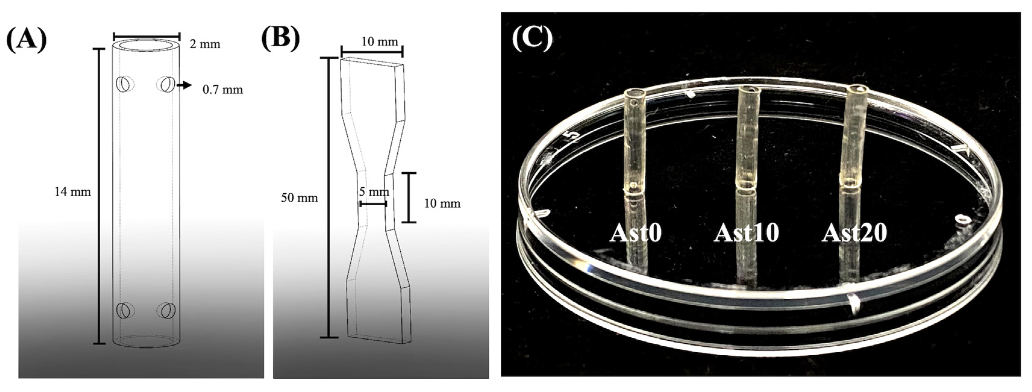
2.2. Nerve Conduit Fabrication

The SolidWorks (Figure 1A, Dassault Systemes SolidWorks Corp., Waltham, MA, USA) program was used to design all conduits, and a MiiCraft high-resolution home DLP 3D printer (Young Optics Inc., Hsinchu, Taiwan) was used for fabrication. The thickness of each layer was designed to be 100 μm, which was then subjected to 20 s of UV exposure for crosslinking. Next, the fabricated conduits were rinsed with ethanol to remove excess PU, and 20 s of UV light exposure was repeated afterward. All conduits were disinfected by soaking in 75% ethanol for 30 min and sterilized by exposing them to UV light for 2 h before conducting subsequent studies.

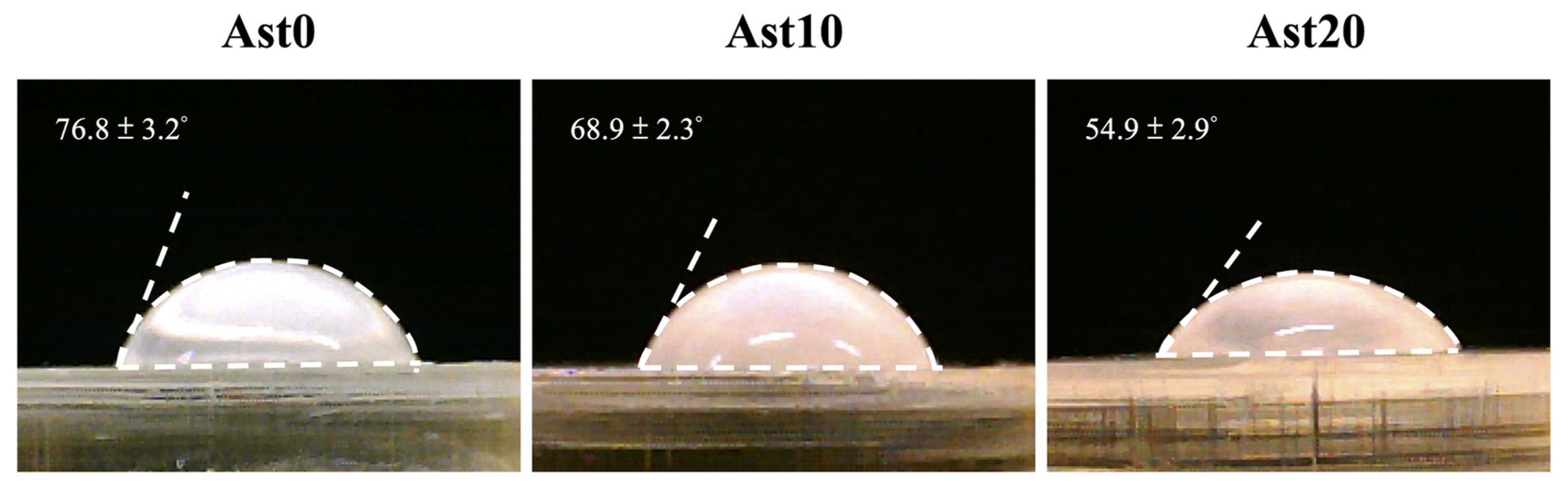
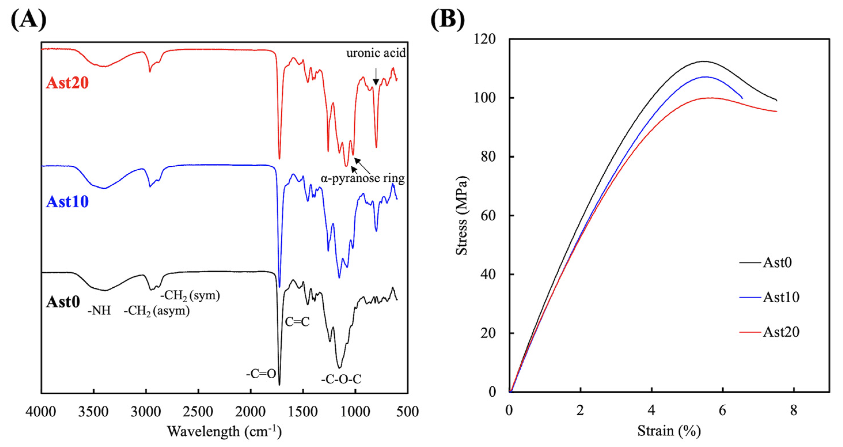
2.3. Physical Properties of Ast-Containing PU Conduits
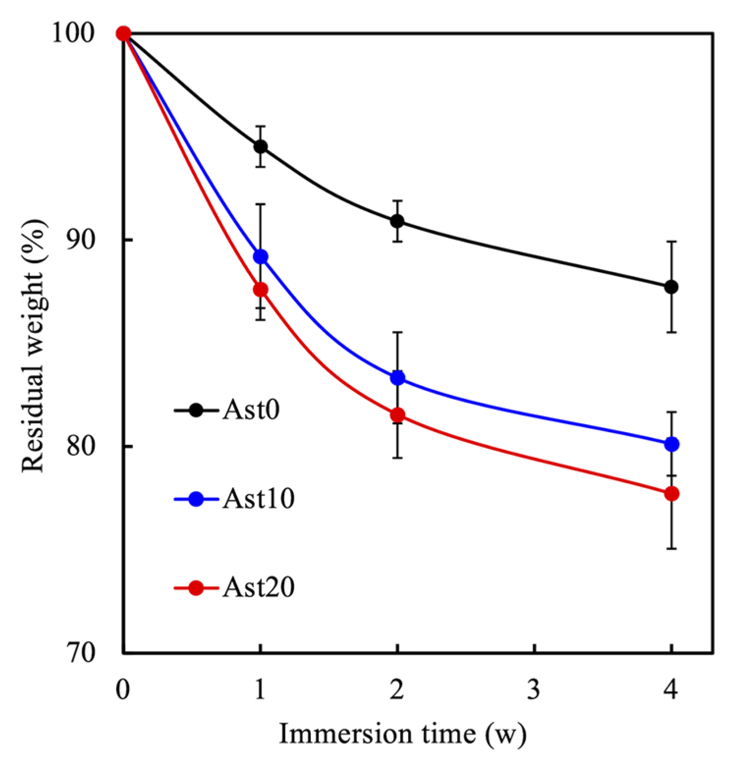
Hydrophilicity of the nerve conduits was assessed using the water contact angle test. In short, all samples were first placed on a stable platform, 5 μL of MilliQ water was dropped on the surface of each samples, and images of the water droplet were captured using a camera after 20 s. Images taken were then evaluated using ImageJ software (National Institutes of Health, Taiwan) to identify the water contact angle. For mechanical property evaluation, a mechanical stress–strain assay was performed using a universal tensile machine. The samples were printed into the shape of a dumbbell (Figure 1B) and stretched from both ends at a rate of 1 mm/min. Six tests were conducted for each group, with average results recorded. In addition, Fourier transform infrared spectroscopic (FTIR) analysis was used to investigate the common compounds and functional groups of the nerve conduits. The test was conducted within the wavelength range of 600–2000 cm−1.

2.4. Biodegradation

To determine the biodegradation rate of different nerve conduits, the specimens were immersed in simulated body fluid (SBF) at 37 °C for 4 weeks. SBF was made with methods according to our previous studies [19]. At every week of immersion, for 4 weeks, the specimens were washed, dehydrated, and weighed on an analytical balance (TE214S; Sartorius, Goettingen, Germany). All data collected were tabulated to determine the biodegradation rate. Six specimens from each group were analyzed, with the average value recorded.

2.5. Cell Proliferation and Morphology

Primary rat Schwann cells (RSCs) used in this study were purchased from ScienCell Research Laboratories (Sciencell, San Diego, CA, USA) and cultured in commercial Schwann cells medium (#1701, Sciencell) to passages 4–8. Each specimen was seeded with 105 cells and placed in an incubator with a pre-determined setting of 37 °C and 5% CO2. At various analyzed time-points (1, 3, and 7 days), Prestoblue® (Invitrogen, Grand Island, NY, USA) was used to assess for levels of proliferation according to protocols stated by the manufacturers. Briefly, PrestoBlue® and fresh medium in a ratio of 1:9 were added to the wells and incubated for 90 min at 37 °C. Absorbance was measured using a Tecan Infinite 200® PRO microplate reader at 570 nm to 600 nm (reference wavelength). RSCs cultured directly on culture plates were used as controls (Ctl). Cellular morphology was observed using F-actin staining according to protocols stated by the manufacturers. After a week of culture, the cells were rinsed with phosphate-buffered saline (PBS, Invitrogen), fixed with 4% paraformaldehyde, and then treated with 0.1% Triton X-100 to allow cell permeation. Next, the conduits were reacted with Alexa-Fluor-488-conjugated phalloidin for 1 h to allow staining, after which images of F-actin were taken using a white light laser confocal microscope (Leica Microsystems GmbH, Wetzlar, Germany).

2.6. Western Blot Analysis

RSCs were seeded on nerve conduits for different time points and washed three times with PBS. Then, the cells were lysed with NP40 buffer (Invitrogen) to assess the protein concentrations using a bicinchoninic acid protein assay kit (Invitrogen). SDS-PAGE was used to separate the cell lysates (40 μg protein) according to the manufacturer’s instructions and shifted onto polyvinylidene difluoride membranes. Target primary antibodies, anti-HuC/HuD (1:500 dilution; Thermo Fisher, Waltham, MA, USA), anti-tumor necrosis factor-alpha (TNF-α, 1:1500 dilution; Abcam, Cambridge, MA, USA), anti-cyclooxygenase-2 (COX-2, 1:1000 dilution; Abcam), anti-BDNF (1:1000 dilution; Abcam), anti-nerve growth factor (NGF, 1:1000 dilution; Abcam), anti-Sry-related HMg-Box gene-10 (SOX10, 1:200 dilution; Abcam), and anti-β-actin (1:10,000 dilution; Abcam) antibodies, were placed onto the membranes and incubated overnight. Then, the membranes were washed and incubated with either horseradish-peroxidase-conjugated anti-rabbit immunoglobulin G (1:2000 dilution; Genetex, Hsinchu, Taiwan) or horseradish-peroxidase-conjugated anti-mouse IgG (1:2000 dilution; Genetex) for 1 h at room temperature. Then, the Fusion-Solo chemiluminescence system (Vilber, Paris, France) and enhanced chemiluminescentdetection reagents (Thermo Fisher) were used to detect the signals emitted from the samples.

2.7. Statistical Analyses

One-way statistical analysis of variance (ANOVA) was performed to analyze the significance of the differences between the different experimental groups in each experiment. Scheffe’s multiple-comparison test was used to determine the significant deviations for each sample. A p-value of <0.05 was considered to be statistically significant, as indicated by *.

3. Results and Discussion

3.1. Characterization of Ast-Containing PU Conduits

The 3D-printed Ast-containing PU conduits are transparent with a light-yellow color (Figure 1C). Hydrophilicity is a critical factor taken into consideration while designing 3D-printed nerve conduits. Nerve conduits with adequate hydrophilicity are said to have positive effects on cellular adhesion and growth, which would influence their downstream cellular behaviors. This was determined by measuring the water contact angle between the water droplet and the surface of each conduit, with a lower angle suggestive of higher hydrophilicity. Figure 2 shows the water contact angles obtained from different groups. The highest water contact angle of 74.6° ± 3.2° was observed in the control group. In contrast, groups with a higher concentration of Ast were observed to have a lower water contact angle, with the Ast20 group at 68.6° ± 2.3° and the Ast10 group at 57.1° ± 2.9°. The water contact angle reduced by 7.9% in the Ast10 group and 23.4% in the Ast20 group (p < 0.05). The results suggested that the addition of Ast enhances the hydrophilicity of nerve conduits, which is optimal for cellular adhesion and growth [25].
FTIR was used to characterize and determine the presence of Ast in order to determine the successful combination PU and Ast (Figure 3A). In the PU spectrum, characteristic peaks detected at 1167, 1525, and 1727 cm−1 corresponded to stretching vibrations of C-O-C (urethane formation), C=C, and C=O (carbonyl urethane group) bonds, respectively [26]. The broad region from 2809 to 3000 cm−1 consisted of two peaks at 2872 and 2959 cm−1, deriving from the CH2 symmetric stretching and anti-symmetric stretching bands. This result clearly indicated that Ast can be incorporated successfully with PU without affecting its structural integrity. The band at 3300 to 3550 cm−1 was hydrogen-bonded NH that formed at the PU chain. The reflecting content of characteristic peaks of Ast could be observed from 750 to 1100 cm−1, which were connected to polysaccharide-containing uronic acid (762 cm−1) and the α-pyranose ring of the glucosyl residue (1019 and 1122 cm−1) [27]. Furthermore, increasing -C-O-C signals were noted with increasing concentrations of Ast, therefore indicating that Ast was successfully integrated with PU. In addition, results also showed that different concentrations of Ast could be loaded into PU, as evidenced by the difference in the intensity of the -C-O-C peak. This was an important factor to note as the main goal of this study was to retain the superior characteristics of PU and yet add on the benefits of Ast for nerve regeneration. To assess the feasibility of our conduits, a tensile stress–strain test was performed to determine their mechanical properties. As shown in Figure 3B, the highest maximal tensile strength was observed in the Ast0 group (112 ± 8 MPa), and the addition of Ast reduced the mechanical properties, with the lowest observed in the Ast20 group (98 ± 6 MPa). Studies have reported that the stress–strain behavior of human nerves is similar to that of rat nerves, which is approximately 13.8 MPa [28]. Therefore, even though conduits with Ast exhibit reduced tensile strength in comparison to PU conduits, they still possess sufficient mechanical strength for surgical handlings as well as implantations.
An ideal nerve conduit should have adequate degradability to allow the efficient nerve repair process to take place. As mentioned, nerves regenerate at a rate of 1-2 mm/day, and regeneration usually starts after a period of rest [23]. Therefore, the degradation rate of all conduits immersed in SBF were analyzed and recorded at 1, 2, 3, and 4 weeks, as shown in Figure 4. Complete regeneration takes about 6 to 12 weeks; thus, an ideal conduit should have the majority of its mass at the end of 4 weeks. An increased degradation rate was observed in all conduits with longer immersion times, with the fastest degradation rate seen in the first week of immersion in SBF. The maximal weight loss in the Ast0 group was approximately 5%, with increased weight loss observed in Ast-containing conduits during the first week of SBF immersion. After immersion for 4 weeks, the weight loss in the Ast0, Ast10, and Ast20 groups was 12%, 19%, and 21%, respectively. The results shown above displayed the highest degradation rate in the Ast20 group, followed by the Ast10 group, and the lowest in the Ast0 group. In a previous study, the time taken for nerve regeneration was about 6 to 12 weeks, depending on the severity of injury. Therefore, logically, an ideal nerve conduit should be able to last at least 12 weeks to provide complete regeneration. Magnetic resonance imaging evaluation can also confirm nerve regeneration and recovery of function that are fully recovered about 12 weeks after the onset of nerve injury [29]. Thus, we can conclude that the degradability of nerve conduits is related and can be greatly enhanced with the addition of Ast, which is ideal for optimal nerve regeneration.

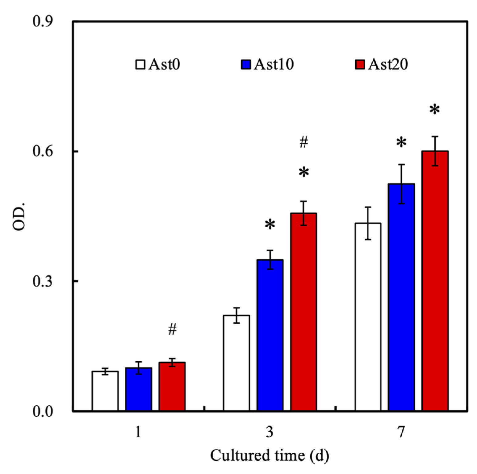
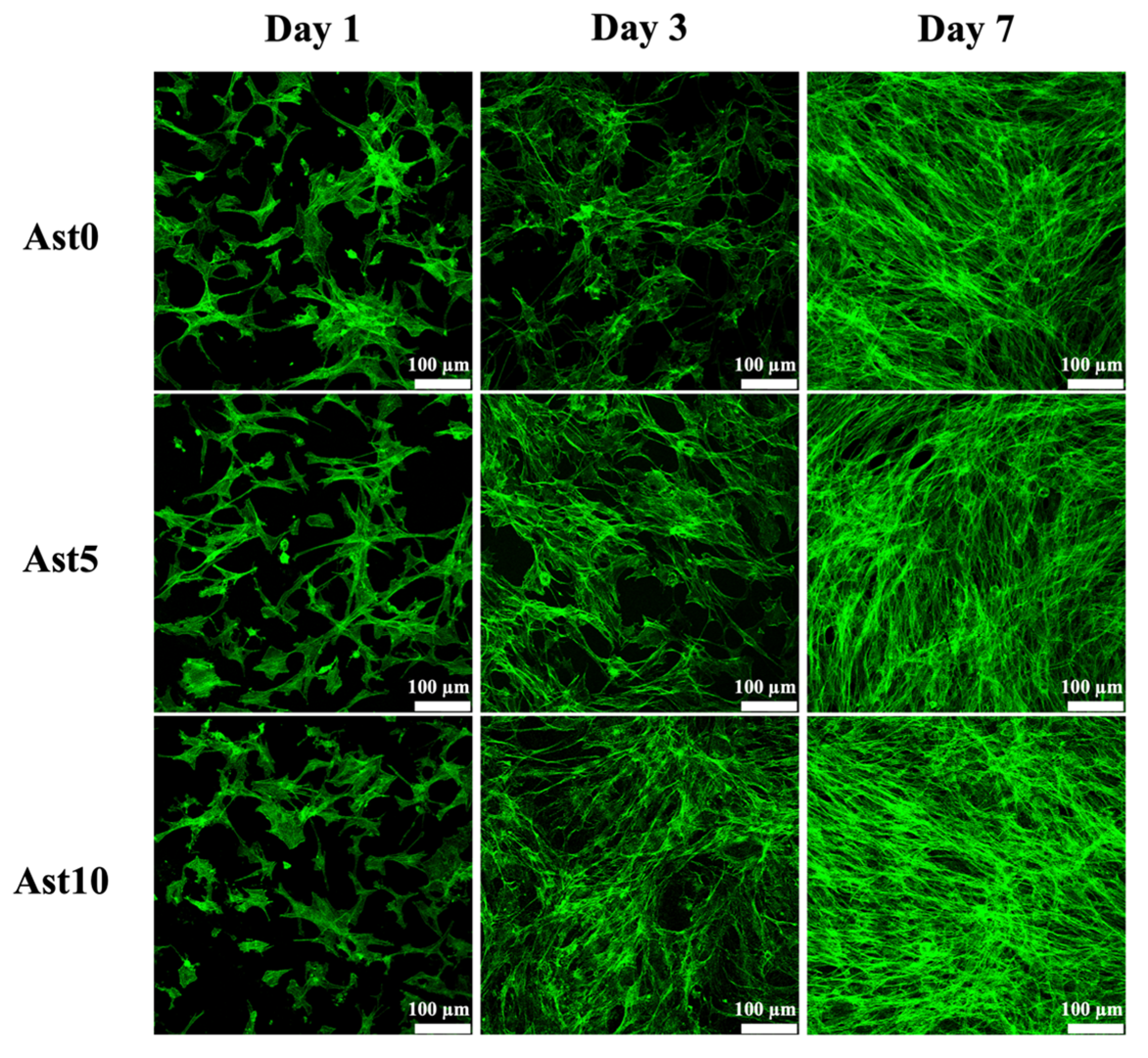
3.2. Cell Proliferation and Morphology

Figure 5 shows differences in the proliferation of rat Schwann cells (RSCs) cultured on different conduits. According to the results shown, the highest proliferation rate was observed in the Ast20 group, followed by the Ast10 group, with the lowest in the Ast0 group at all time points of 1, 3, and 7 days. The differences were obvious since day 1 of cell culture, with as ignificantly higher proliferation rate observed in the Ast20 group as compared to the Ast0 group. In addition, the differences in the cell proliferation rate were even greater in the Ast20 group in comparison to both Ast0 and Ast10 groups at days 3 and 7. In contrast, the proliferation rate in the Ast20 group after 3 days of culture was approximately 1.6- and 1.3-times higher than that in Ast10 and Ast0 groups, respectively. Besides, the Ast10 group also showed higher proliferation as compared to the Ast0 group at both days 3 and 7. These results indicated that the proliferation of RSCs is directly related to Ast concentrations, which is in good agreement with in vitro and in vivo studies conducted by Chen et al. [30]. According to their latest studies, in vitro results revealed that the addition of Ast upregulated the proliferation of neuronal stem cells but did not influence differentiation. In addition, 2 µg/kg of Ast was administered via the IV route into experimental stroke rat models, and results proved that Ast was able to upregulate the expression of nestin, p-epidermal growth factor receptor (EGFR), and p-mitogen-activated protein kinase proteins (MAPK), thus improving the repair of neurological function in the rats. Furthermore, it was also hypothesized that improved hydrophilicity of Ast leads to enhanced cell adhesion and attachment, therefore allowing for improved cellular behaviors such as proliferation. These results also showed that Ast is non-cytotoxic to cells, thus making it a potential candidate for further neural-related studies. Figure 6 shows the immunofluorescence staining results of RSCs with F-actin. At first glance, Figure 6 is in good agreement with the proliferation results above. There were more cells in the Ast10 group at days 1, 3, and 7 as compared to Ast5 and Ast0 groups, as seen by the area covered by cells. Furthermore, cells in the Ast10 group were flatter and had elongated spindles at day 1 as compared to the rest of the groups, thus indicating that the micro-environment was more favorable to RSCs. In addition, the RSCs cultured on the conduits with Ast stimulation showed longer axons and more spindle cells, which proved the positive influence of Ast stimulation on neuronal cell proliferation [31]. These data indicated that the addition of Ast can promote cellular adhesion and proliferation and can be used as a neurogenic factor in future nerve-regeneration-related studies.

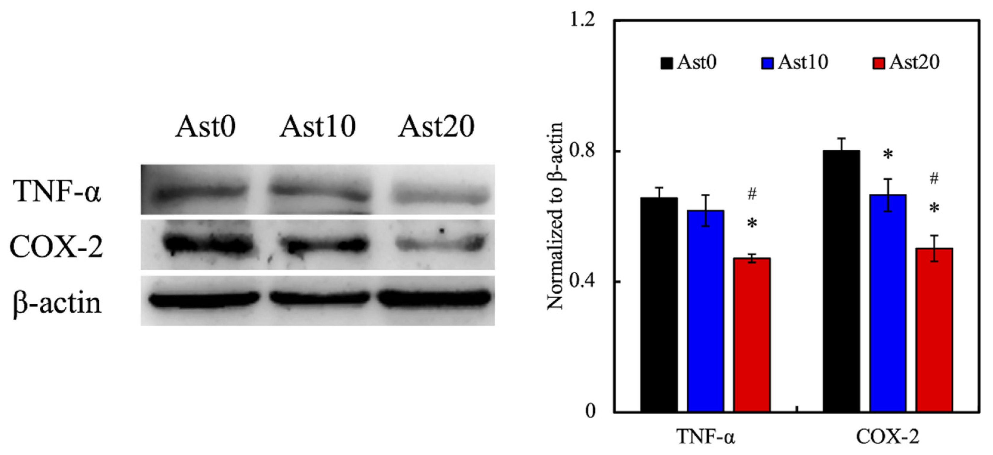
3.3. Immune Responses

In nerve regeneration studies, qualities including good biocompatibility and low immunogenicity are the basic requirements in developing an ideal nerve conduit [32]. The feasibility of a conduit depends largely on the host’s immune response after implantation, as an increased immune response would lead to suboptimal tissue regeneration and graft rejection. A suitable conduit should thus have low immunogenicity so as not to completely suppress nerve regeneration. In fact, adequate inflammatory reactions play an important role in balancing neurological activities between various stakeholders such as Schwann cells, macrophages, and dendritic cells [33]. A study by Buttner et al. demonstrated that excessive levels of macrophage infiltration and inflammatory signals reduce the regenerative capacity of aging nerves [34]. In this study, we used inflammatory markers TNF-α and COX-2 to assess for levels of immune responses to our 3D-printed Ast-containing PU nerve conduits. TNF-α is a cytokine that is involved in both innate and adaptive immune responses [35]. In contrast, COX-2 is an inflammatory exclusive cytokine that induces secretion of downstream prostaglandin E2 to bring about inflammatory responses [36]. Furthermore, levels of COX-2 were found to be significantly enhanced in the spinal cord after traumatic injuries [37]. As seen from the Western blot results in Figure 7, the protein expression levels of both TNF-α and COX-2 were lower in the Ast10 and Ast20 groups as compared to the Ast0 group. Quantification results further showed that increasing Ast concentrations was inversely related to the levels of inflammatory markers. The Ast20 group had approximately 40% lower levels of both TNF-α and COX-2 as compared to the Ast0 group. It is important to note that the addition of Ast did not completely inhibit inflammatory responses; instead, it reduced the levels of inflammation. As mentioned above, adequate levels of inflammation are required for efficient neural regeneration. Therefore, with reference to studies done by others, we hypothesized that the addition of Ast induces certain levels of immunoregulatory effects that might be beneficial for neural regeneration.

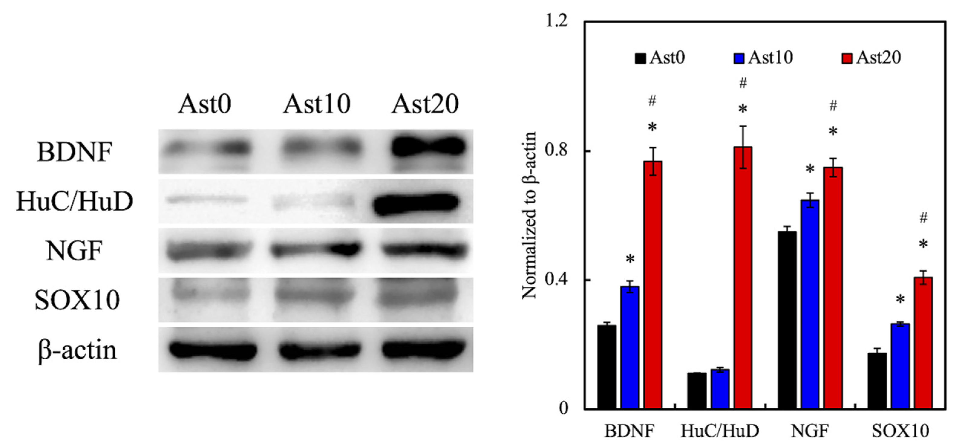
3.4. Nerve-Regeneration-Related Protein Expression

Western blot was used to evaluate the secretion levels of various neurogenic-related proteins. Brain-derived neurotrophic factor (BDNF) is a member of the neurotrophin family and is known to play a role in promoting the growth and differentiation of new neurons and synapses [38]. HuC/HuD proteins are mammalian embryonic lethal abnormal visual system (ELAV)-like neuronal RNA-binding proteins that are involved in neuronal development in both central and peripheral nervous systems and are great markers for determining the presence of developing neurons [39]. Nerve growth factor (NGF), just as its name suggests, is known to promote the proliferation, survival, and maintenance of neurons [40]. Sry-related HMg-Box gene-10 (SOX10), in contrast, is a nucleocytoplasmic shuttle protein that plays a critical role in the development of the neural crest and peripheral nervous system [41]. From the results obtained (as shown in Figure 8), the expression levels of all neurogenic proteins were the highest in the Ast20 group, followed by the Ast10 group, and the lowest in the Ast0 group. The secretion levels of these neurogenic proteins were directly related to the concentration of Ast. BDNF and HuC/HuD expression in the Ast20 group was even more than 2 times higher than that in the Ast0 group. Interestingly, similar expression levels of HuC/HuD were observed in both Ast0 and Ast10 groups. However, the HuC/HuD expression level significantly increased in the Ast20 group, indicating that Ast20 is a better concentration level for promoting nerve growth as compared to the other two concentrations. In addition, a study done by Cheng et al. revealed that the addition of Ast was able to positively regulate the nerve regeneration process through interactions with various receptors on nerve cells, such as tropomyosin receptor kinase A (TRKA) and p75NTR [21]. The TRKA receptor works by enhancing the expression level of B cell lymphoma-2, which leads to better nerve tissue regeneration [42]. Overall, these results suggested that Ast can enhance neural development and regeneration, which is consistent with other related studies.

4. Conclusions

In summary, Ast-containing PU nerve conduits were successfully fabricated using DLP 3D printing technology in this study. The addition of Ast enhanced neural proliferation and regeneration, as demonstrated by increased absorbance levels and F-actin immunofluorescence staining results of RSCs, with direct correlation to the concentrations of Ast. The neurogenic properties of Ast might be attributed to its role in immunoregulation, as evidenced by the decreased expression levels of inflammatory markers TNF-α and COX-2, which was also in good agreement with data obtained from other related studies. Besides, the hydrophilicity of Ast was hypothesized to be another factor that contributes to the neurogenic properties of Ast, as increased hydrophilicity was associated with better cell adhesion and proliferation. Overall, these results demonstrated that 3D-printed Ast-containing PU nerve conduits not only supply suitable mechanical properties, they also are able to enhance neural cell proliferation and neural regeneration, as noted from the increased expression of neural-related markers. Therefore, we hypothesized that 3D-printed Ast-containing PU nerve conduits may be a promising strategy for future peripheral nerve-regeneration-related applications.

Author Contributions

Data curation, H.Y.N.; formal analysis, C.-C.C.; funding acquisition, Y.-S.C. and M.-Y.S.; investigation, Y.-X.H. and C.-C.C.; methodology, Y.-X.H. and C.-C.C.; writing—original draft, S.-S.C. and H.Y.N.; writing—review and editing, Y.-S.C. and M.-Y.S. All authors have read and agreed to the published version of the manuscript.

Funding

The authors acknowledge receipt grants from the Ministry of Science and Technology (MOST 109-2224-E-080-001 and 109-2221-E-039-007) of Taiwan and the China Medical University (CMU109-S-25) of Taiwan.

Institutional Review Board Statement

Not applicable.

Informed Consent Statement

Not applicable.

Data Availability Statement

Data available in a publicly accessible repository.

Acknowledgments

Experiments and data analysis were performed, in part, through the use of the Medical Research Core Facilities, Office of Research & Development, at China Medical University, Taichung, Taiwan.

Conflicts of Interest

The authors declare no conflict of interest.

References

  1. Zhang, K.; Li, J.; Jin, J.; Dong, J.; Li, L.; Xue, B.; Wang, W.; Jiang, Q.; Cao, Y. Injectable, anti-inflammatory and conductive hydrogels based on graphene oxide and diacerein-terminated four-armed polyethylene glycol for spinal cord injury repair. Mater. Des. 2020, 196, 109092. [Google Scholar] [CrossRef]
  2. Ye, W.; Li, H.; Yu, K.; Xie, C.; Wang, P.; Zheng, Y.; Zhang, P.; Xiu, J.; Yang, Y.; He, Y.; et al. 3D printing of gelatin methacrylate-based nerve guidance conduits with multiple channels. Mater. Des. 2020, 192, 108757. [Google Scholar] [CrossRef]
  3. Abzan, N.; Kharaziha, M.; Labbaf, S. Development of three-dimensional piezoelectric polyvinylidene fluoride-graphene oxide scaffold by non-solvent induced phase separation method for nerve tissue engineering. Mater. Des. 2019, 167, 107636. [Google Scholar] [CrossRef]
  4. Tajdaran, K.; Gordon, T.; Wood, M.D.; Shoichet, M.S.; Borschel, G.H. A glial cell line-derived neurotrophic factor delivery system enhances nerve regeneration across acellular nerve allografts. Acta Biomater. 2016, 29, 62–70. [Google Scholar] [CrossRef]
  5. Grinsell, D.; Keating, C.P. Peripheral Nerve Reconstruction after Injury: A Review of Clinical and Experimental Therapies. BioMed. Res. Int. 2014, 2014, 1–13. [Google Scholar] [CrossRef] [Green Version]
  6. Chen, C.-C.; Ng, H.Y.; Lou, C.-W.; Shie, M.-Y. Additive Manufacturing of Nerve Decellularized Extracellular Matrix-Contained Polyurethane Conduits for Peripheral Nerve Regeneration. Polymers 2019, 11, 1612. [Google Scholar] [CrossRef] [Green Version]
  7. Xu, X.; Tao, J.; Wang, S.; Yang, L.; Zhang, J.; Zhang, J.; Liu, H.; Cheng, H.; Xu, J.; Gou, M.; et al. 3D printing of nerve conduits with nanoparticle-encapsulated RGFP966. Appl. Mater. Today 2019, 16, 247–256. [Google Scholar] [CrossRef]
  8. Elnaggar, M.A.; El-Fawal, H.A.; Allam, N.K. Biocompatible PCL-nanofibers scaffold with immobilized fibronectin and laminin for neuronal tissue regeneration. Mater. Sci. Eng. C 2021, 119, 111550. [Google Scholar] [CrossRef]
  9. Niu, Y.; Stadler, F.J.; Fu, M. Biomimetic electrospun tubular PLLA/gelatin nanofiber scaffold promoting regeneration of sciatic nerve transection in SD rat. Mater. Sci. Eng. C 2021, 121, 111858. [Google Scholar] [CrossRef] [PubMed]
  10. VijayaVenkataRaman, S.; Kuan, L.Y.; Lu, W.F. 3D-printed ceramic triply periodic minimal surface structures for design of functionally graded bone implants. Mater. Des. 2020, 191, 108602. [Google Scholar] [CrossRef]
  11. Wu, Z.; Li, Q.; Xie, S.; Shan, X.; Cai, Z. In vitro and in vivo biocompatibility evaluation of a 3D bioprinted gelatin-sodium alginate/rat Schwann-cell scaffold. Mater. Sci. Eng. C 2020, 109, 110530. [Google Scholar] [CrossRef]
  12. Kao, C.-T.; Chen, Y.-J.; Huang, T.-H.; Lin, Y.-H.; Hsu, T.-T.; Ho, C.-C. Assessment of the Release Profile of Fibroblast Growth Factor-2-Load Mesoporous Calcium Silicate/Poly-ε-caprolactone 3D Scaffold for Regulate Bone Regeneration. Processes 2020, 8, 1249. [Google Scholar] [CrossRef]
  13. Li, W.-D.; Wang, C.; Jiang, Z.-H.; Chen, L.-J.; Wei, Y.-H.; Zhang, L.-Y.; Chen, M.-Y.; Yang, X.; Zhang, G.-J. Stereolithography based additive manufacturing of high-k polymer matrix composites facilitated by thermal plasma processed barium titanate microspheres. Mater. Des. 2020, 192, 108733. [Google Scholar] [CrossRef]
  14. Wu, H.; Chen, P.; Yan, C.; Cai, C.; Shi, Y. Four-dimensional printing of a novel acrylate-based shape memory polymer using digital light processing. Mater. Des. 2019, 171, 107704. [Google Scholar] [CrossRef]
  15. Chen, Z.; Yang, M.; Ji, M.; Kuang, X.; Qi, H.J.; Wang, T. Recyclable thermosetting polymers for digital light processing 3D printing. Mater. Des. 2021, 197, 109189. [Google Scholar] [CrossRef]
  16. Naureen, B.; Haseeb, A.; Basirun, W.; Muhamad, F. Recent advances in tissue engineering scaffolds based on polyurethane and modified polyurethane. Mater. Sci. Eng. C 2021, 118, 111228. [Google Scholar] [CrossRef] [PubMed]
  17. Huang, C.-T.; Shrestha, L.K.; Ariga, K.; Hsu, S.-H. A graphene–polyurethane composite hydrogel as a potential bioink for 3D bioprinting and differentiation of neural stem cells. J. Mater. Chem. B 2017, 5, 8854–8864. [Google Scholar] [CrossRef] [PubMed]
  18. Chen, Y.-W.; Shen, Y.-F.; Ho, C.-C.; Yu, J.; Wu, Y.-H.A.; Wang, K.; Shih, C.-T.; Shie, M.-Y. Osteogenic and angiogenic potentials of the cell-laden hydrogel/mussel-inspired calcium silicate complex hierarchical porous scaffold fabricated by 3D bioprinting. Mater. Sci. Eng. C 2018, 91, 679–687. [Google Scholar] [CrossRef]
  19. Kao, C.-T.; Chiu, Y.-C.; Lee, A.K.-X.; Lin, Y.-H.; Huang, T.-H.; Liu, Y.-C.; Shie, M.-Y. The synergistic effects of Xu Duan combined Sr-contained calcium silicate/poly-ε-caprolactone scaffolds for the promotion of osteogenesis marker expression and the induction of bone regeneration in osteoporosis. Mater. Sci. Eng. C 2021, 119, 111629. [Google Scholar] [CrossRef]
  20. Huang, M.-H.; Kao, C.-T.; Chen, Y.-W.; Hsu, T.-T.; Shieh, D.-E.; Huang, T.-H.; Shie, M.-Y. The synergistic effects of chinese herb and injectable calcium silicate/β-tricalcium phosphate composite on an osteogenic accelerator in vitro. J. Mater. Sci. Mater. Electron. 2015, 26, 1–12. [Google Scholar] [CrossRef]
  21. Cheng, C.-Y.; Yao, C.-H.; Liu, B.-S.; Liu, C.-J.; Chen, G.-W.; Chen, Y.-S. The role of astragaloside in regeneration of the peripheral nerve system. J. Biomed. Mater. Res. Part A 2006, 76, 463–469. [Google Scholar] [CrossRef]
  22. Ren, S.; Zhang, H.; Mu, Y.; Sun, M.; Liu, P. Pharmacological effects of Astragaloside IV: A literature review. J. Tradit. Chin. Med. 2013, 33, 413–416. [Google Scholar] [CrossRef]
  23. Chan, W.-S.; Durairajan, S.S.K.; Lu, J.-H.; Wang, Y.; Xie, L.-X.; Kum, W.-F.; Koo, I.; Yung, K.K.L.; Li, M. Neuroprotective effects of Astragaloside IV in 6-hydroxydopamine-treated primary nigral cell culture. Neurochem. Int. 2009, 55, 414–422. [Google Scholar] [CrossRef] [PubMed]
  24. Zhang, X.; Chen, J. The mechanism of astragaloside IV promoting sciatic nerve regeneration. Neural Regen. Res. 2013, 8, 2256–2265. [Google Scholar]
  25. Chen, C.-C.; Yu, J.; Ng, H.-Y.; Lee, A.K.-X.; Chen, C.-C.; Chen, Y.-S.; Shie, M.-Y. The Physicochemical Properties of Decellularized Extracellular Matrix-Coated 3D Printed Poly(ε-caprolactone) Nerve Conduits for Promoting Schwann Cells Proliferation and Differentiation. Materials 2018, 11, 1665. [Google Scholar] [CrossRef] [PubMed] [Green Version]
  26. Saad, N.M.; Malaysia, U.S.; Zubir, S.A. Palm Kernel Oil Polyol-based Polyurethane as Shape Memory Material: Effect of Polyol Molar Ratio. J. Phys. Sci. 2019, 30, 77–89. [Google Scholar] [CrossRef] [Green Version]
  27. Yin, J.-Y.; Chan, B.C.-L.; Yu, H.; Lau, I.Y.-K.; Han, X.-Q.; Cheng, S.-W.; Wong, C.-K.; Lau, C.B.-S.; Xie, M.-Y.; Fung, K.-P.; et al. Separation, structure characterization, conformation and immunomodulating effect of a hyperbranched heteroglycan from Radix Astragali. Carbohydr. Polym. 2012, 87, 667–675. [Google Scholar] [CrossRef]
  28. Topp, K.S.; Boyd, B.S. Structure and Biomechanics of Peripheral Nerves: Nerve Responses to Physical Stresses and Implications for Physical Therapist Practice. Phys. Ther. 2006, 86, 92–109. [Google Scholar] [CrossRef]
  29. Bendszus, M.; Wessig, C.; Solymosi, L.; Reiners, K.; Koltzenburg, M. MRI of peripheral nerve degeneration and regeneration: Correlation with electrophysiology and histology. Exp. Neurol. 2004, 188, 171–177. [Google Scholar] [CrossRef]
  30. Chen, X.; Wu, H.; Chen, H.; Wang, Q.; Xie, X.-J.; Shen, J. Astragaloside VI Promotes Neural Stem Cell Proliferation and Enhances Neurological Function Recovery in Transient Cerebral Ischemic Injury via Activating EGFR/MAPK Signaling Cascades. Mol. Neurobiol. 2018, 56, 3053–3067. [Google Scholar] [CrossRef] [PubMed]
  31. Wang, Y.; Zhou, J.; Tang, C.; Yu, J.; Zhu, W.; Guo, J.; Wang, Y. Positive effect of Astragaloside IV on neurite outgrowth via talin-dependent integrin signaling and microfilament force. J. Cell. Physiol. 2021, 236, 2156–2168. [Google Scholar] [CrossRef]
  32. Hollister, S.J. Scaffold Design and Manufacturing: From Concept to Clinic. Adv. Mater. 2009, 21, 3330–3342. [Google Scholar] [CrossRef]
  33. McLean, N.A.; Verge, V.M.K. Dynamic impact of brief electrical nerve stimulation on the neural immune axis-polarization of macrophages toward a pro-repair phenotype in demyelinated peripheral nerve. Glia 2016, 64, 1546–1561. [Google Scholar] [CrossRef]
  34. Büttner, R.; Schulz, A.; Reuter, M.; Akula, A.K.; Mindos, T.; Carlstedt, A.; Riecken, L.B.; Baader, S.L.; Bauer, R.; Morrison, H. Inflammaging impairs peripheral nerve maintenance and regeneration. Aging Cell 2018, 17, e12833. [Google Scholar] [CrossRef] [PubMed]
  35. Cicuéndez, M.; Portolés, P.; Montes-Casado, M.; Izquierdo-Barba, I.; Vallet-Regí, M.; Portolés, M.T. Effects of 3D nanocomposite bioceramic scaffolds on the immune response. J. Mater. Chem. B 2014, 2, 3469–3479. [Google Scholar] [CrossRef] [PubMed] [Green Version]
  36. Lee, H.-P.; Wu, Y.-C.; Chen, B.-C.; Liu, S.-C.; Li, T.-M.; Huang, W.-C.; Hsu, C.-J.; Tang, C.-H. Soya-cerebroside reduces interleukin production in human rheumatoid arthritis synovial fibroblasts by inhibiting the ERK, NF-kappa B and AP-1 signalling pathways. Food Agric. Immunol. 2020, 31, 740–750. [Google Scholar] [CrossRef]
  37. Zhao, Z.; Chen, S.; Eisenach, J.; Busija, D.; Pan, H. Spinal cyclooxygenase-2 is involved in development of allodynia after nerve injury in rats. Neuroscience 2000, 97, 743–748. [Google Scholar] [CrossRef]
  38. Blesch, A.; Tuszynski, M.H. GDNF gene delivery to injured adult CNS motor neurons promotes axonal growth, expression of the trophic neuropeptide CGRP, and cellular protection. J. Comp. Neurol. 2001, 436, 399–410. [Google Scholar] [CrossRef]
  39. Zhang, X.-Y.; Yanagi, Y.; Sheng, Z.; Nagata, K.; Nakayama, K.; Taguchi, T. Regeneration of diaphragm with bio-3D cellular patch. Biomaterials 2018, 167, 1–14. [Google Scholar] [CrossRef]
  40. Zhang, D.; Yao, Y.; Duan, Y.; Yu, X.; Shi, H.; Nakkala, J.R.; Zuo, X.; Hong, L.; Mao, Z.; Gao, C. Surface-Anchored Graphene Oxide Nanosheets on Cell-Scale Micropatterned Poly(d,l-lactide-co-caprolactone) Conduits Promote Peripheral Nerve Regeneration. ACS Appl. Mater. Interfaces 2020, 12, 7915–7930. [Google Scholar] [CrossRef]
  41. Chang, H.-T.; Heuer, R.A.; Oleksijew, A.M.; Coots, K.S.; Roque, C.B.; Nella, K.T.; McGuire, T.L.; Matsuoka, A.J. An engineered three-dimensional stem cell niche in the inner ear by applying a nanofibrillar cellulose hydrogel with a sustained-release neurotrophic factor delivery system. Acta Biomater. 2020, 108, 111–127. [Google Scholar] [CrossRef] [PubMed]
  42. Vishnoi, T.; Singh, A.; Teotia, A.K.; Kumar, A. Chitosan-Gelatin-Polypyrrole Cryogel Matrix for Stem Cell Differentiation into Neural Lineage and Sciatic Nerve Regeneration in Peripheral Nerve Injury Model. ACS Biomater. Sci. Eng. 2019, 5, 3007–3021. [Google Scholar] [CrossRef] [PubMed]
Figure 1. Schematic drawing of (A) the conduit and (B) the dumbbell-shaped sample used in mechanical testing. (C) A photograph of 3D-printed astragaloside (Ast)-containing polyurethane (PU) conduits.
Processes 09 00353 g001
Figure 2. The water contact angle in Ast0 (0 µM), Ast10 (10 µM), and Ast20 (20 µM) groups.
Processes 09 00353 g002
Figure 3. (A) Fourier transform infrared spectroscopic (FTIR) spectra and (B) tensile stress–strain curves of the various 3D-printed Ast-containing PU nerve conduits.
Processes 09 00353 g003
Figure 4. The weight loss of 3D-printed Ast-containing PU nerve conduits after immersion in simulated body fluid (SBF).
Processes 09 00353 g004
Figure 5. Proliferation of rat Schwann cells (RSCs) on 3D-printed Ast-containing conduits for 1, 3, and 7 days. * Significant difference (p < 0.05) from the Ast0 group. # Significant difference (p < 0.05) from the Ast10 group.
Processes 09 00353 g005
Figure 6. The morphology of RSCs cultured on 3D-printed Ast-containing nerve conduits for different time points.
Processes 09 00353 g006
Figure 7. The expression levels of tumor necrosis factor-alpha (TNF-α) and cyclooxygenase-2 (COX-2) that were inflammation-related proteins in RSCs cultured on 3D-printed Ast-containing PU nerve conduits with different Ast concentrations. * Significant difference (p < 0.05) from the Ast0 group. # Significant difference (p < 0.05) from the Ast10 group.
Processes 09 00353 g007
Figure 8. Western blot and quantification results of neural-regeneration-related proteins in RSCs cultured on 3D-printed Ast-containing PU nerve conduits with different Ast concentrations. * Significant difference (p < 0.05) from the Ast0 group. # Significant difference (p < 0.05) from the Ast10 group.
Processes 09 00353 g008
Publisher’s Note: MDPI stays neutral with regard to jurisdictional claims in published maps and institutional affiliations.

