Article

First-Row Transition 7-Oxo-5-phenyl-1,2,4-triazolo[1,5-a]pyrimidine Metal Complexes: Antiparasitic Activity and Release Studies

by
Álvaro Martín-Montes
1,†,
Sandra Jimenez-Falcao
2,†,
Santiago Gómez-Ruiz
3,
Clotilde Marín
1,* and
José M. Mendez-Arriaga
3,*
1
Departamento De Parasitología, Universidad De Granada, Avenida Fuentenueva, 18071 Granada, Spain
2
Organic Nanotechnology Lab, Departamento De Materiales Y Producción Aeroespacial E.T.S.I Aeronáutica Y Del Espacio, Universidad Politécnica De Madrid, 28040 Madrid, Spain
3
COMET-NANO Group, Departamento De Biología y Geología, Física Y Química Inorgánica, E.S.C.E.T., Universidad Rey Juan Carlos, Calle Tulipán s/n, 28933 Móstoles, Spain
*
Authors to whom correspondence should be addressed.
These authors contributed equally to this work.
Pharmaceuticals 2023, 16(10), 1380; https://doi.org/10.3390/ph16101380
Submission received: 30 August 2023 / Revised: 21 September 2023 / Accepted: 26 September 2023 / Published: 28 September 2023
(This article belongs to the Special Issue Pyrimidine and Purine Derivatives in Medicinal Chemistry)

Abstract

:
Leishmaniasis and Chagas disease are still considered neglected illnesses due to the lack of investment in research, despite the fact that almost one million new cases are reported every year. Four 7-oxo-5-phenyl-1,2,4-triazolo[1,5-a]pyrimidine (HftpO) first-row transition complexes (Cu, Co, Ni, Zn) have been studied for the first time in vitro against five different species of Leishmania spp. (L. infantum, L. braziliensis, L. donovani, L. peruviana and L. mexicana) as well as Trypanosoma cruzi, showing higher efficacy than the reference commercial drugs. UV and luminescence properties were also evaluated. As a proof of concept, anchoring of a model high-effective-metal complex as an antiparasitic agent on silica nanoparticles was carried out for the first time, and drug-release behaviour was evaluated, assessing this new approach for drug vehiculation.

Graphical Abstract

1. Introduction

Triazolopyrimidine derivatives have been known since the beginning of the last century, when Bülow and Haas published for the first time the synthesis of some of them [1]. Nevertheless, their complexation properties were not studied until 1952, by Birr [2], who used this organic family to stabilize photographic silver emulsions. Since then, applications, including antipyretic, analgesic, anti-inflammatory, herbicidal, antifungal, antimicrobial, antitumor and antiparasitic properties [3,4], have been displayed for triazolopyrimidine ligands, as well as for their metal complexes, because of their remarkable pharmacology [5]. In the specific case of 1,2,4-triazolo[1,5-a]pyrimidines ligands, their structural similarity with purines makes them excellent candidates for the study of reactivity with different metal ions as biomimetic models [6,7,8,9,10], due to their structural similarity with these nucleobases. Triazolopyrimidines exhibit great versatility as ligands, owing to the presence of accessible electron pairs on their skeleton, which are located in at least three nitrogen atoms. Their main coordination mode is via N3-monodentate or N3,N4-bridging, but many other bonding routes could also be possible when the number of exocyclic substituents in the aromatic ring increases. The interaction between metal ions and 1,2,4-triazolo[1,5-a]pyrimidines ligands shows a great capability to act as building blocks with great versatility, with applications for the synthesis of metal–organic frameworks (MOFs) or multidimensional systems with useful properties such as magnetism, luminescence or biological activity [11,12,13,14,15].
The triazolopyrimidine family mentioned before includes a wide variety of derived structures, such as the 1,2,4-triazolo[1,5-a]pyrimidine itself (tp) in its non-substituted form [16] and the 5,7-dimethyl-1,2,4-triazolo[1,5-a]pyrimidine (dmtp) [17] and 7-amine-1,2,4-triazolo[1,5-a]pyrimidine (7-atp) [18] structures. Additionally, among the most reactive groups of triazolopyrimidines are those containing exocyclic oxygen atoms. These additional oxygen coordination positions spread out the range of metallic centres that can interact with these ligands (i.e., lanthanide ions). Some examples of oxygenated triazolopyrimidine derivatives are dihydro-5-oxo-[1,2,4]triazolo-[1,5-a]pyrimidine (5HtpO and 7HtpO), whose structures were described by Abul Haj et al. [19], or the commercial 4,7-dihydro-5-methyl-7-oxo[1,2,4]-triazolo[1,5-a]pyrimidine (HmtpO) [20]. One of the last-synthesized and characterized oxo triazolopyrimidine derivatives, 7-oxo-5-phenyl-1,2,4-triazolo[1,5-a]pyrimidine (HftpO) [21] (Scheme 1), has been employed in the present study.
Leishmaniasis is one of the seven primary illnesses that are present in all continents. According to the World Health Organisation (WHO), it is endemic nowadays in almost 100 countries, with more than 350 million people on the planet at risk to be infected. Millions of new cases appear every year, and the mortality rate is higher than 60,000 deaths in a year, a value among parasitic diseases which is only surpassed by malaria. Although it is usually located in poor and unhealthy areas of the Third World, it is also present in Europe and Asia due to migration phenomena, making it a global problem. This fact is enhanced when large population groups coexist, as happens in crowded refugee camps or in war and conflict areas, before the populations dissemination to their host countries [22]. Leishmaniasis can manifest in humans mainly in three different clinical conditions: visceral, the most aggressive and mortal form; cutaneous, leading to nodules and ulcers in the skin; and mucocutaneous, which causes permanent lesions in the mucosa (mouth, nose or genital) [23,24]. In all cases, the transmission vectors are dipteral insects of the genera Lutzomyia in the New World and Phlebotomus in the Old World (Table 1) [25]. Leishmaniasis is considered to be a neglected disease because of the low investment in the search for new, efficient drugs. Glucantime is the only drug available to fight these diseases nowadays [26,27], since the use of Pentostam is deprecated [28]. On the other hand, Chagas disease (or American trypanosomiasis) [29] is a chronic neglected illness caused by a Trypanosoma cruzi parasite, and affecting more than 10 million people worldwide. Its main incidence region comprises poor South and Central America areas, but the infection is present in many other continents through migration patterns and blood transfusions from latent donors. The transmission mechanism runs through contact with urine and faeces of infected blood-sucking triatomine bugs [30,31]. The most commonly used commercial drugs to fight against these infections are nifurtimox and benznidazole, two nitro heterocyclic derivatives with contrasted trypanozidal effects [32,33,34]. In the past three decades, many biochemical studies have pointed to different possible targets to stop the infections, like the inhibition of ADP phosphorylation or the beta-oxidation of fatty acids [35,36]. Despite the obvious advantages of these commercial drugs and the fact that they are still the first-line antiparasitic treatments, they exhibit several limitations, like serious side effects [37]. Thus, the development of novel and more efficient therapies to fight Chagas disease, minimizing its impact in society, is also an urgent need [38,39,40].
The molecular structures of antimonial drugs like meglumine antimoniate (Glucantime), as well as their modes of action and metabolic natures, are still being explored [41,42,43]. Alternatives with non-antimonial antiparasitic prodrugs such as anfotericine B are also being used as leishmaniasis treatments [44,45], but their prices restrict their availability and implementation in underdeveloped countries, which are the most affected areas. Moreover, these non-antimonial prodrugs present adverse side effects, especially kidney failure, in addition to the beginning of resistance, as reported last year [46]. The antimonial drugs also present serious side effects, like allergic dermopathy, peripheral polyneuropathy or vomiting [47], the need for daily parenteral administration, and the development of drug resistance [48,49]. Many research groups are making advances in this field by using transition-metal complexes to combat parasitic diseases [50,51], mainly with ruthenium compounds [52,53,54,55,56]. Antiparasitic evaluation studies and intra- and extracellular assays have been carried out by our research group using 1,2,4-triazolo[1,5-a]pyrimidine derivatives and their metal complexes, studies which have shown great results against tropical diseases currently without effective treatments, and particularly against leishmaniasis and Chagas disease [57,58,59,60,61]. Many other authors have also pointed to a metal-complexes strategy as a promising alternative for fighting tropical parasitic diseases caused by members of the Trypanosomatidae family, such as leishmaniasis, as well as other tropical illnesses like Chagas disease or malaria. A synergetic effect is observed in antiparasitic efficacy when the triazolopyrimidine derivatives are combined with a variety of metal ions in coordination complexes [62,63]. This renders triazolopyrimidine metal complexes potential chemotherapeutic agents for curing diseases related to parasites. The interest in this topic has led to some reviews being published in the last decade [64,65,66].
The use of and interest in nanomaterials in biomedicine is increasing every year due to their characteristic mechanical, optical and electrical properties [67,68], especially as carriers for drug delivery [69]. Focusing on that, nanoparticles are some of the most widely used transporters. The possibility of size and porosity control in their synthesis, as well as their versatility of functionalization make them one of the most useful types of carriers to distribute drugs in an efficient way [70,71]. Their nature can range from polymer- [72] or protein-based [73] to metallic gold [74], iron [75], or copper nanoparticles [76]. Mesoporous silica nanoparticles are one of the most-used nanosystems in biomedicine for drug delivery [77,78,79], despite the fact that their use in parasitology has just bloomed in the last five years [80,81,82].
In the present work, luminescence properties, solution stability and biological assays of four 7-oxo-5-phenyl-1,2,4-triazolo[1,5-a]pyrimidine (HftpO) first-transition complexes with the general formula [M(ftpO)2(H2O)4] with M = Cu (1), Co (2), Ni (3), and Zn (4) are discussed for the first time. The antiparasitic activity of HftpO complexes was evaluated against an Arequipa strain of T. cruzi and five Leishmania spp. species (the European type L. infantum; the South American parasites L. braziliensis present in Brazil, L. peruviana indigenous to Peru, and L. mexicana, which acts mainly in the Yucatán Peninsula; and the mostly-reported-in-the-Indian-subcontinent L. donovani), as well as evaluated for toxicity against macrophage J774.2 and Vero host cells. The results reveal a better selectivity index in most cases, compared with the isolated ligand and the commercial drugs Glucantime and benznidazole. Thus, a proof-of-concept of drug release for these kinds of complexes using silica nanoparticles as carriers was performed using the copper one as model, confirming that the complex antiparasitic capacity continues to be active against different Leishmania spp. species.

2. Results

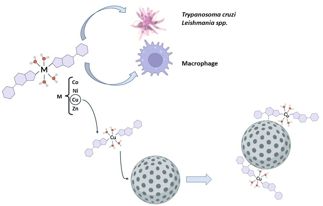
2.1. Synthesis and Characterization of Triazolopyrimidine Complexes

The four metal complexes of 7-oxo-5-phenyl-1,2,4-triazolo[1,5-a]pyrimidine (HftpO) were synthesized following the method previously reported by our group [21]. The general procedure can be found in the Materials and Methods section of this work.

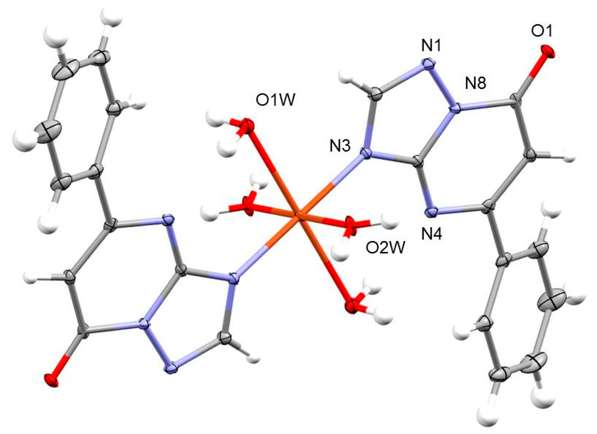
2.1.1. Crystal Structures of [M(ftpO)2(H2O)4] (14)

Compound 1 crystallizes in the monoclinic P21/c space group. A perspective view of this complex is shown in Figure 1. The structure consists of one mononuclear [Cu(ftpO)2(H2O)4] coordination complex, in which each Cu (II) ion has an octahedral CuN2O4 stereochemistry. This geometry is constituted by four coordination water molecules and two nitrogen atoms belonging to two different anionic ftpO ligands. A strong hydrogen-bond interaction between O1W and N4 is also present, leading both coordinated ftpO moieties up to an almost coplanar position. The crystallographic data of compound 1 were previously reported by our group [21], and the whole dataset can be obtained free of charge from The Cambridge Crystallographic Data Centre via “www.ccdc.cam.ac.uk/data_request/cif accessed on 25 September 2023” linked to identification number 1536813. X-ray powder diffraction (XRD) data confirm the isostructural nature of the four complexes, assigning the general formula [M(ftpO)2(H2O)4] to the whole series.

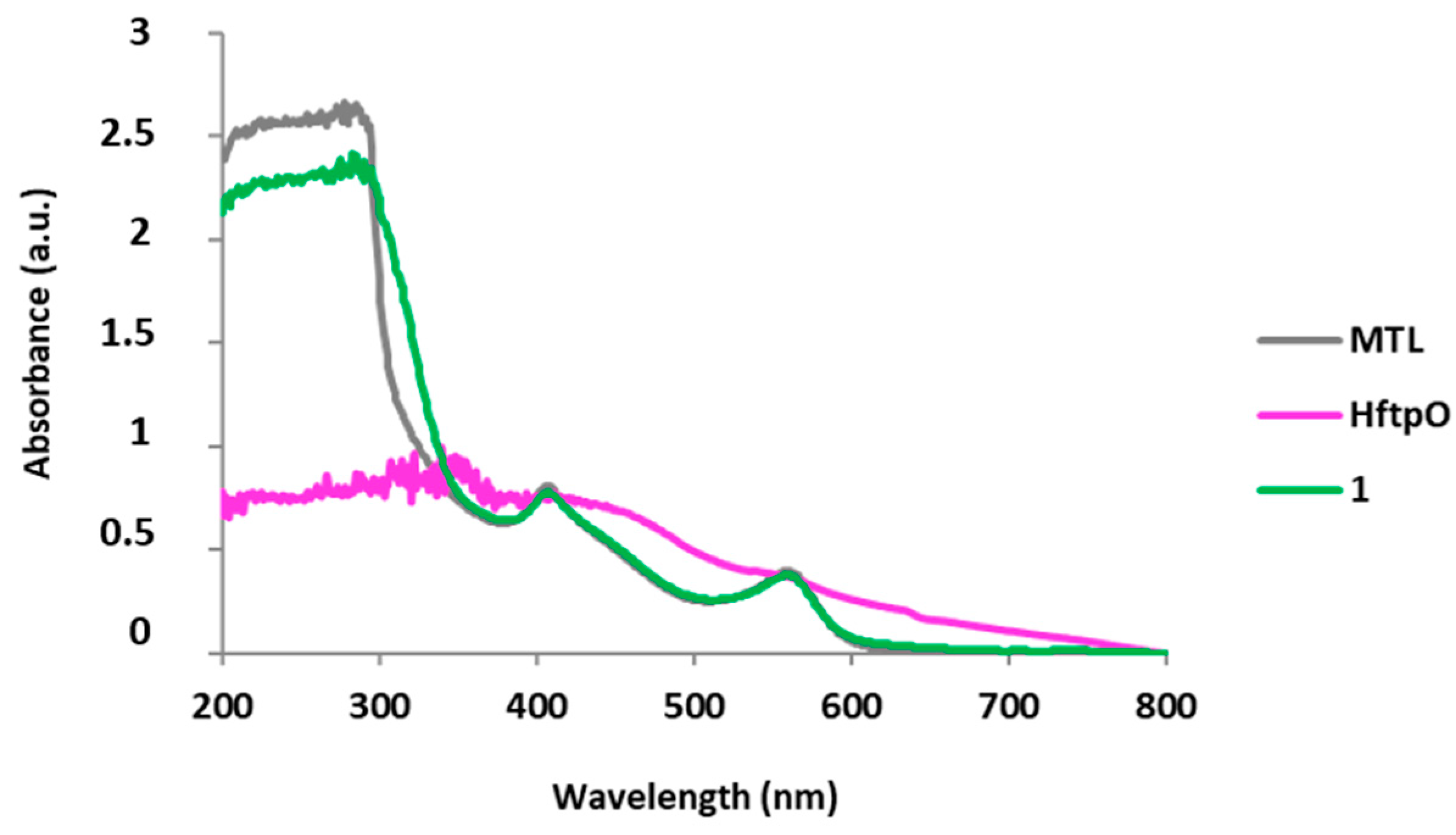
2.1.2. Spectroscopic Properties

In order to complete the spectroscopic studies of these complexes, stability over time was measured by UV using 10−3 M ethanol absolute solutions of the four complexes. The spectra were recorded across 3 days at different moments, maintaining the curves in the same shape after 72 h in solution (Figure 2). Absorption maximums for each complex were distorted from the HftpO maximum, indicating that complex integrity remains unaltered in the ethanol solution (Table 2).
UV behaviour was also studied for the samples in vitro, in trypanosomal liquid medium (MTL) solution after two days of heating at 37 °C, via ultrasounds in sealed Eppendorf tubes. As can be noticed in Figure 3, the curves corresponding to MTL without sample, the sample containing HftpO and the sample containing the [Cu(ftpO)2(H2O)4] complex exhibit different shapes. The four metal compounds have identical spectrum shapes, a result which supports the isostructural nature of the samples (a description can be found in the Supplementary Material, Figures S1–S3). The HftpO ligand presents a maximum of around 280 cm−1 in MTL, while for the metal compounds, this value shifts to 300 cm−1, supporting the stability-in-solution of the complexes in a parasite culture medium.
Complementary IR spectra information, as well as a complete thermoanalytical study of the complexes 14, can be found in Mendez-Arriaga et al. [21].

2.1.3. Luminescent Properties

The luminescent emission spectra of free HftpO and its metal complexes were recorded in ethanol solution (1 × 10⁻³ M) with an excitation wavelength of 348 nm at room temperature. The maximum UV-absorption wavelength previously obtained for each compound was tested, but no emission signals were obtained. These emission spectra are depicted in Figure 4. Free-ligand HftpO exhibits a strong luminescent emission between 400–440 nm, in contrast to the partial luminescence-quenching phenomena observed in the nickel complex (3). In the cases of complexes 1,2, with Cu(II) and Co(II) centres, the luminescence signal fully disappears. The zinc complex (4) exhibits a different emission pattern in comparison with the cobalt one and HftpO, at 348 nm. The decreases in emission intensity were in the order of Co(II) = Cu(II) > Zn(II) > Ni(II). The decrease of the luminescence signal is a rather common phenomenon attributed to the interaction between metal ions and strongly luminescent ligands, such as Schiff bases or aromatic heterocycles, during the complexation process [83].

2.2. Synthesis and Characterization of Functionalized Silica Nanoparticles

The synthesis of MCM-41 silica nanoparticles (MSN) was carried out following the procedure reported by Zhao et al. [84], with slight modifications. To enhance the coordination capacity of the proposed nanosystems, MSN nanoparticles were covered with bromide groups from bromopropyl triethoxysilane ligand to obtain MSN-Br systems. Finally, as a proof of concept, HftpO ligand and complex 1 were selected for immobilisation on the MSN surface for further release in order to study kinetics and evaluate their efficacy as leishmanicidal agents. A standard procedure of stir-and-heat in toluene in a Schlenk tube with a nitrogen atmosphere were performed to obtain MSN-ftpO and MSN-CuftpO nanosystems (Scheme 2). The functionalization of silica nanoparticles was performed following the methodology previously reported by our group [70], which can be found in the Section 4.

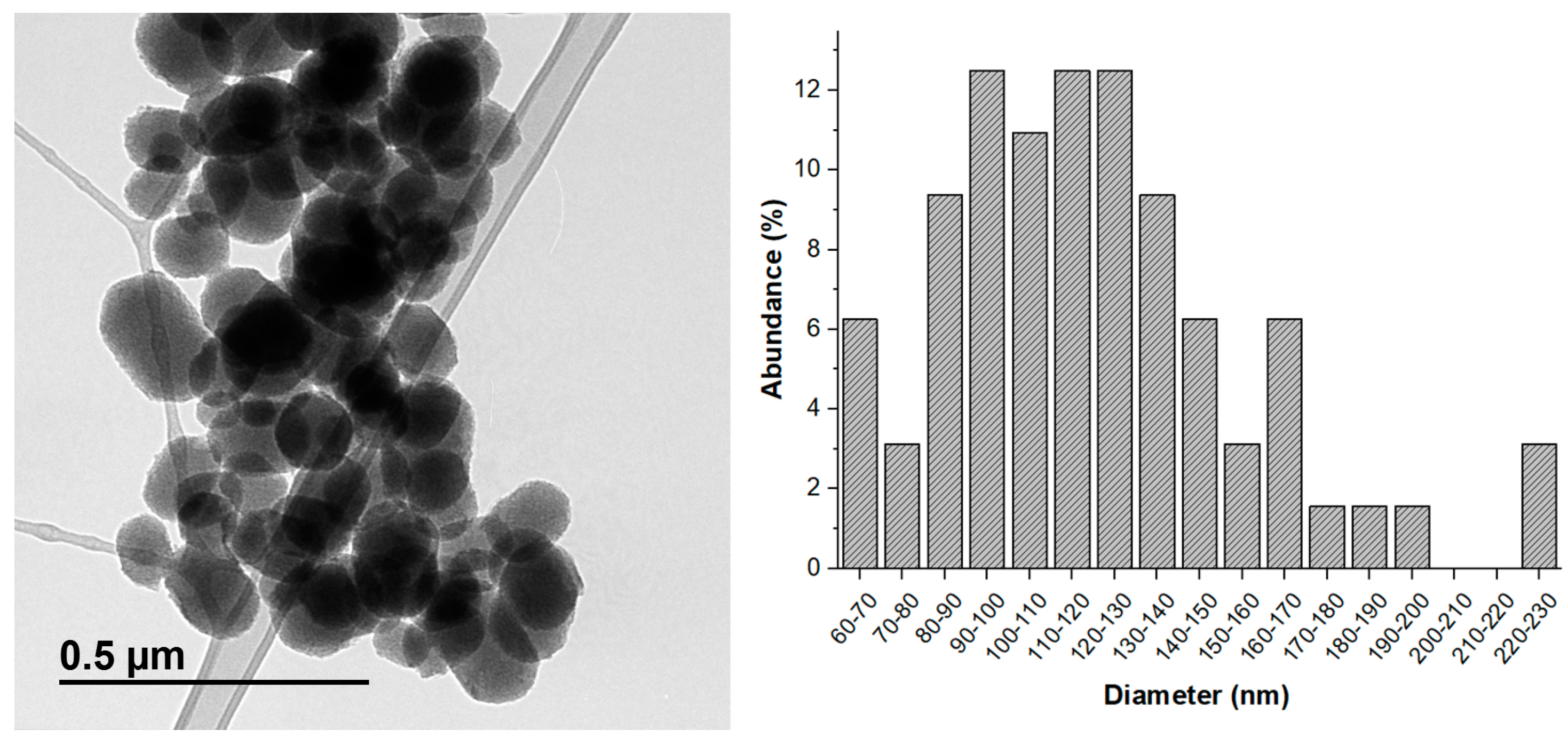
2.2.1. Transmission Electron Microscopy (TEM)

To evaluate the nanoparticles’ appearance, mesoporous silica nanoparticle (MSN) starting material was characterized by TEM. As shown in Figure 5, MSN presents a semi-spherical morphology with a wide size distribution, with a diameter prevalence between 110 and 130 nm. A few, larger, 220–230 nm nanoparticles can be noticed in the histogram, but these data can be attributed to the fusion of small silica nanoparticles. Well-defined pores in a hexagonal arrangement can be observed, a common characteristic of MCM-41 MSN.

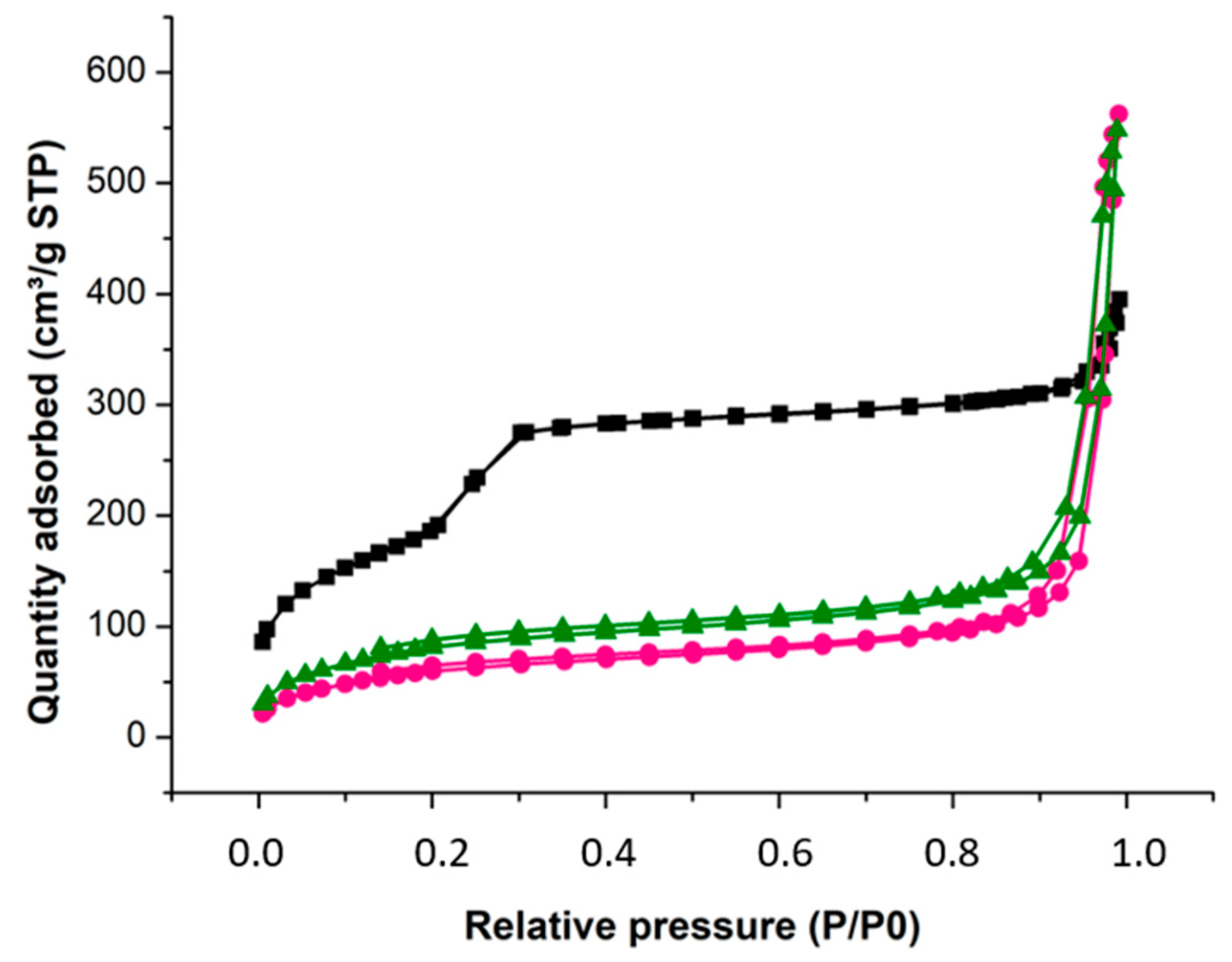
2.2.2. Nitrogen Adsorption/Desorption Isotherms

The porous properties of the starting material (MSN) and both functionalized carriers (MSN-ftpO and MSN-CuftpO) were evaluated with nitrogen adsorption/desorption isotherms at 77 K, as shown in Figure 6. Experimental results show a decrease in the surface area of the modified MSN compared to the starting material, as expected. MSN exhibits the classical type IV isotherm (characteristic of mesoporous silica supports), while both functionalized nanosystems exhibit the characteristic appearance of materials with filled pores [85,86]. Data-fitting of the nitrogen adsorption/desorption results to Brunauer–Emmett–Teller (BET) isotherms permitted the determination of the most relevant surface parameters (Table 3). Raw MSN presented a BET surface of 682 m2g−1, a pore volume of 0.52 cm3g−1 and a 3.2 nm pore diameter. The functionalization of MSN surface with Br and the subsequent anchoring of the HftpO ligand and complex 1 resulted in a decrease of the three parameters in both studied nanomaterials. Surface area and total pore volume presented slightly higher values in MSN-CuftpO due to the steric hindrance of the larger size of the metallic complex occupying the available pores of the silica.

2.3. Release Studies

The release behaviour of copper complex (1) was studied by a specific assay of MSN-CuftpO. Thus, three different samples containing 0,4 mg copper complex nanomaterial were suspended in 4 mL of PBS buffer (0.1 mg/mL, pH 7.4). This buffer was chosen as a model to mimic the cell culture media that was subsequently used, which was coherent with human pH, and to avoid contamination using MTL. After an initial sonication, the sample was incubated at 37 °C and centrifuged at specific time intervals (Figure 7), in order to analyse the absorption via UV spectroscopy at the maximum absorbance wavelength of complex 1. The experiment was conducted for 3 days, but the stabilization was reached at 3 h, verifying the complete release of the loaded compound.

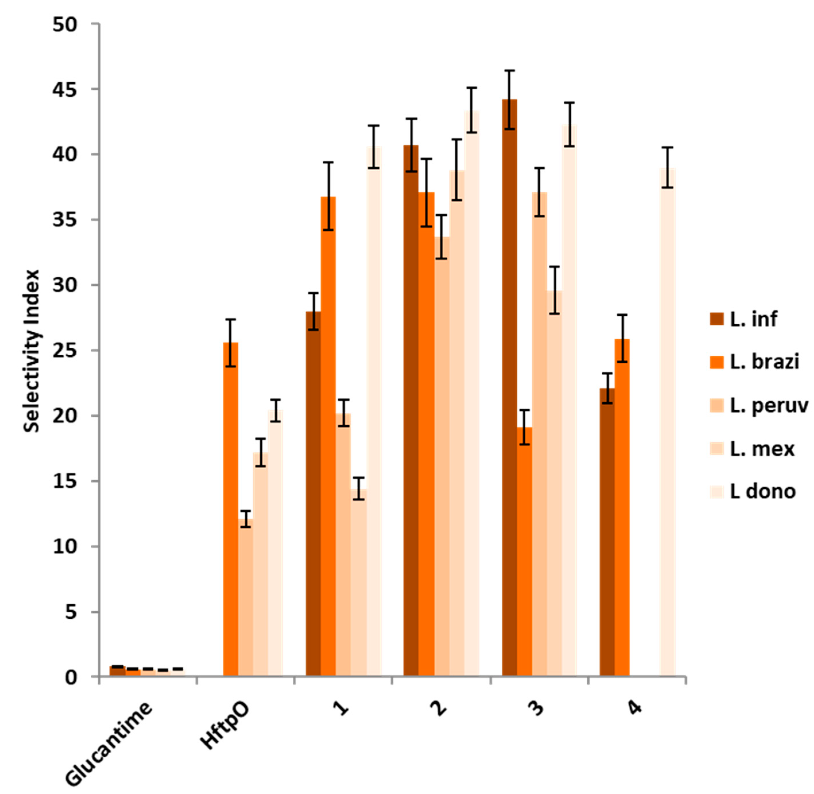
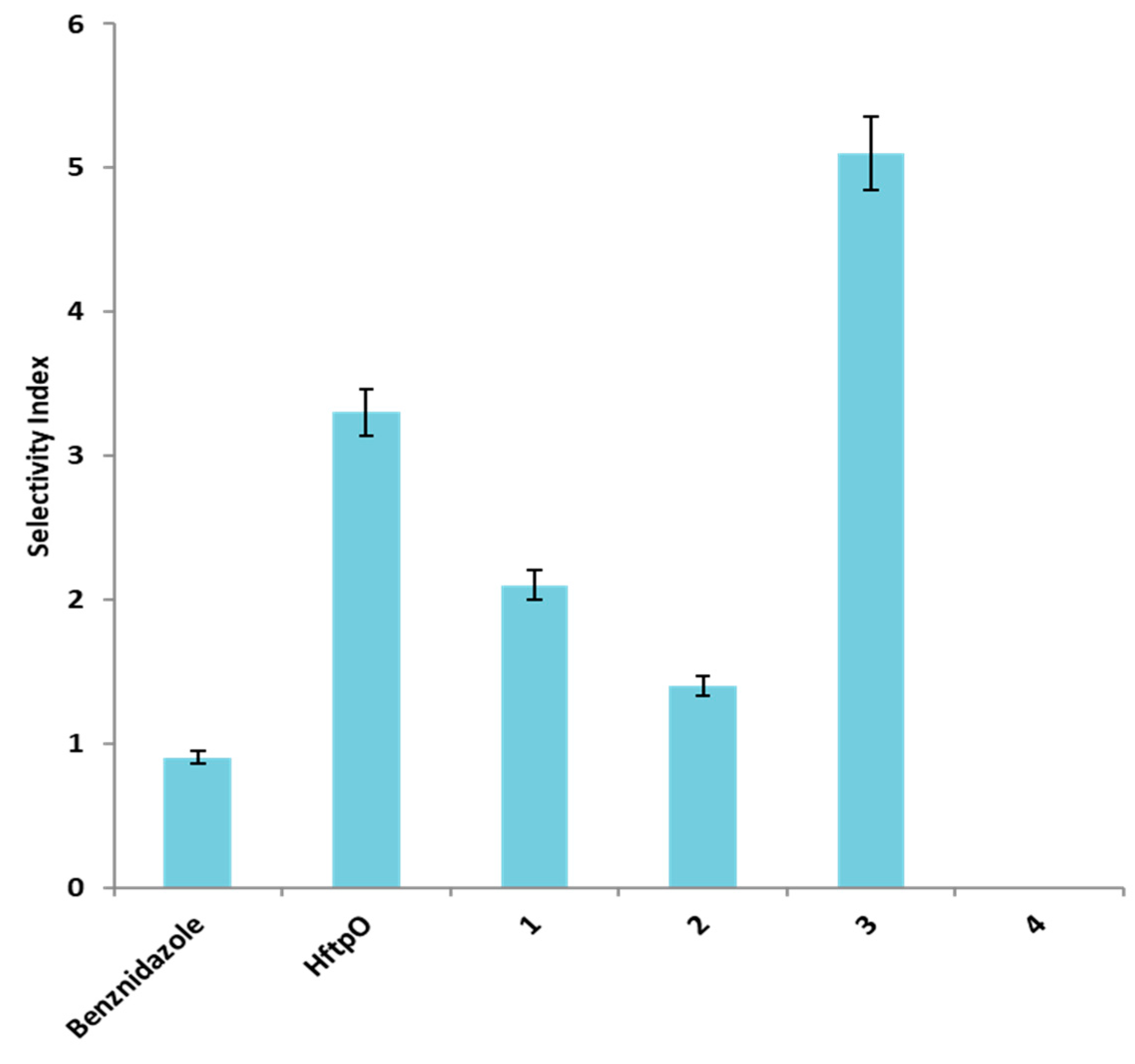
2.4. In Vitro Antiparasitic Activity and Toxicity

2.4.1. In Vitro Efficacy of HftpO and Its Complexes (14)

Taking into account the synergic properties of the metallic complexes with triazolopyrimidine ligands, as previously described by several authors [64,65,66], and given that currently used treatments against leishmaniasis usually cause serious problems, we decided to check the antiproliferative activity and cytotoxicity of the four mentioned complexes against five different species of Leishmania spp. (L. infantum, L. braziliensis, L. peruviana, L. mexicana and L. donovani) and Trypanosoma cruzi. The results were compared with commercial drugs Glucantime and benznidazole, isolated HftpO derivative, and inorganic salts employed in the synthesis (Table 4 and Table 5, Figure 8 and Figure 9) in order to study the potential efficacy improvement provoked by the metal coordination. According to the promastigote growth inhibition, no significant effect was noticed for the inorganic salts, as expected, so the data are not included in the tables.

2.4.2. In Vitro Screening of MSN-ftpO and MSN-CuftpO

Cytotoxicity assays and promastigote screenings were performed for the silica nanoparticles and their derived nanomaterials to check their capability to inhibit leishmania spp. growth. L. infantum and braziliensis were selected as parasite models of two different clinical behaviour infections, and were incubated with HftpO and copper complex 1 silica nanomaterials. Compound 1 was selected due to its promising behaviour against both studied Leishmania spp. species and a significant efficacy against Chagas disease. Table 6 exhibits the results of these biological assays. The inhibition tendency of the studied materials is represented in Figure 10.

3. Discussion

The HftpO ligand showed a significant effect on the extracellular parasite forms, but its IC50 was higher than those of the reference drugs. The metallic complexes presented similar acceptable values for IC50 in the promotion-of-cytotoxicity assays, but they still did not exceed the performance of commercial drugs in some cases, like that of L. braziliensis. Nevertheless, for the HftpO ligand as well as its metal complex, the cytotoxicity results were better than those of the reference drugs. Comparing Glucantime and HftpO, the concentration needed to consider the triazolopyrimidine toxic for the host cells was more than 60-fold higher than that of the commercial leishmanicidal drug (Table 4, IC50 against J774.2 macrophages column). This improvement of the toxicity effect was incrementally achieved in the complexes due to the synergetic effect derived from the coordination to a metal ion centre. For all of the metallic compounds, the cytotoxicity IC50 values for macrophages were extraordinarily high. On the other hand, the Vero cells assay resulted in very strong cytotoxicity values which surpass the Benznidazole ones, but the free HftpO still exhibits the lowest toxic activity for Vero cells. The selectivity index (SI) is obtained by the combination of promastigote inhibition growth and cytotoxicity in host cells and is used to evaluate the efficacy of a prodrug [87]. The mentioned coefficient illustrates the great efficacy of these HftpO complexes as anti-leishmania agents, improving upon values in comparison to the commercial drug and free ligand in all cases. Figure 8 and Figure 9 depict and compare the SI values for all the compounds assayed against different Leishmania spp. species and Trypanosoma cruzi, respectively.
As shown in Figure 8, nickel and cobalt complexes (2 and 3) exhibit the best SI values of the series, being more than 50-fold better than Glucantime values for every single Leishmania spp.-assayed species, with only the exception of 3 for L. braziliensis. Nevertheless, focusing on the L. braziliensis inhibition, the Cu(II) coordination complex (1) possesses the best ratio of efficacy for fighting this illness. The only compound with an apparently worse activity is 4, which is in good agreement with the previously reported data for zinc triazolopyrimidine complexes [65]. Nevertheless, if we compare the published data of the only two zinc 1,2,4-triazolo[1,5-a]pyrimidine complexes already assayed against Leishmania spp. species, [Zn(dmtp)2(H2O)4](ClO4)2·2dmtp·2H2O [11] and [Zn2(7atp)4(µ-bypym)(H2O)4](ClO4)·2(7atp) [88], this new zinc compound improves the SI result for L. infantum, L. braziliensis and L. donovani, thus becoming the best zinc triazolopyrimidine compound with antiparasitic effectiveness yet reported, as far as we know. The T. cruzi SI values show that complex 3 is the best antiparasitic prodrug, followed by the free ligand. Only the zinc compound under discussion in this work turns out to be an exception, with no significant antiproliferative efficacy shown against T. cruzi, L. peruviana or L. mexicana. This phenomenon can be attributed to the general tendency towards a low solubility in Zn complexes in comparison with other transition-metal compounds [89,90], which could prevent a correct interaction in solution with the parasite species studied.
In general, HftpO coordination compounds display better SI coefficients against Leishmania spp. strains, not only compared to Glucantime, but also surpassing the free triazolopyrimidine derivative. In the case of Chagas disease, only the nickel complex improves upon the ligand results, but the commercial reference is always surpassed by Cu, Co and Ni. This fact supports the theory of a synergetic effect caused by the coordinated combination between metal centres and organic ligands, improving the effectiveness against parasite infections by associating it with a decrease in the toxicity levels tolerated by the host cells.
The results for the antiparasitic activity of the nanomaterials evaluated indicate a great efficacy of the copper complex, with higher values of SI in comparison with free Glucantime, while keeping a similar efficacy range, despite the fact that silica nanoparticles were modified with only 20% of the ligand or metal complex, relative to the weight percentage. The release profile shows a complete liberation of the drug inside of two hours, confirming the complete administration of the complex during the screening experiment. A prevailing trend at low concentrations for both proposed systems can be noticed by looking at the inhibition graphics (Figure 10), which are especially pronounced for L. braziliensis. This behaviour can be attributed to an agglomeration tendency for silica nanomaterials in the MTL medium in the experiment wells in scalation concentrations.

4. Materials and Methods

4.1. General

All reagents were purchased from commercial sources and used as received without further purification. The synthesis of HftpO ligand and its metal complexes followed the methodology described in Méndez-Arriaga et al. [21], which is briefly described below.

4.2. Synthesis of HftpO and [M(ftpO)2(H2O)4] where M(II) = Cu (1), Co (2), Ni (3) and Zn (4)

Following the procedure previously reported by our research group [21], a mixture containing ethyl benzoylacetate (0.02 mol) and 3-amino-1,2,4-triazole (0.02 mol) was refluxed in 10 mL of acetic acid for 12 h. A white solid appeared when cooling, which was washed with abundant water and CH2Cl2 to remove all the acid excess. Then, the white solid was dried in a vacuum desiccator. For the synthesis of the metal complexes, a hot solution of HftpO (1 mmol) in 20 mL of ethanol was added to another warm solution of 5 mL of ethanol containing 1 mmol of the respective nitrate salt (1:1). First precipitate, which was formed immediately, was removed from the solution. After one week at room temperature, a microcrystalline product was obtained.

4.3. Synthesis of Mesoporous Silica Nanoparticles

The synthesis of MCM-41 silica nanoparticles (MSN) was carried out following the procedure reported by Zhao et al. [84], with slight modifications. A total of 3.5 mL of a 2 M aqueous sodium hydroxide solution was added to an aqueous solution of CTAB (1.0 g, 2.74 mmol in 480 mL of Milli-Q water). After that, the silica precursor TEOS (5 mL, 22.4 mmol) was added dropwise under vigorous stirring, allowing the mixture to react for 2 h at 80 °C. Once a white precipitate appeared, the product was isolated by filtration, washed with abundant Milli-Q water and methanol (2 × 20 mL) and dried for 24 h at 80 °C on a stove. Finally, the MSN were calcined at 550 °C for 24 h with an increasing temperature ramp of 1 °C/min.

4.4. Functionalization of Mesoporous Silica Nanoparticles with (3-Bromopropyl)triethoxysilane (MSN-Br)

MSN nanoparticles were functionalized with bromide groups. This process was performed following the procedures reported previously by Bollu et al. [91]. A quantity of 500 mg of MSN in a Schlenk tube was dehydrated at 90 °C under vacuum overnight. The activated silica was dispersed in dry toluene (40 mL), adding 525 µL of bromopropyl triethoxysilane ligand (BrP) (100% w/w BrP/SiO2) to the solution. The mixture was heated to 110 °C and stirred for two days. After that, the dispersion was centrifuged and the solid was washed with toluene (×2) and with diethyl ether (×1). The obtained white solid, MSN-Br, was dried overnight at 70 °C on a stove.

4.5. Ligand and Complex Coordination (MSN-ftpO and MSN-CuftpO)

HftpO ligand and copper complex 1 were incorporated into the silica nanomaterial MSN by coordination through HBr liberation. Initially, 100 mg of MSN-Br was activated at 90 °C and under vacuum in a Schlenk tube overnight. After that, 20 mL of dry toluene and 20 mg of ligand or complex, respectively, were added and heated for 48 h at 110 °C. Finally, the suspension was centrifuged, and the isolated solid was washed twice with toluene and once with diethylether. Finally, the solids were dried at room temperature, obtaining a 20% in weight functionalization.

4.6. Physical Measurements

Luminescence and UV spectra for ethanol solutions were recorded with Varian Cary Eclipse equipment at University of Granada. Adsorption−desorption isotherms of nitrogen were measured using a Micromeritics ASAP 2020 at University Rey Juan Carlos. Surface areas and pore size were calculated using Brunauer–Emmett–Teller (BET) and Barret–Joyner–Halenda (BJH) methods, respectively. Transmission electron microscopy (TEM) images were obtained with a JEOL JEM 1010 at a 100 kV operating voltage. The samples were dispersed using ethanol solvent, followed by an ultrasonic bath for 30 min before being spread onto a TEM copper grid (300 mesh) covered with a holey carbon film. The grid was then air-dried at room temperature. The images were acquired with the equipment’s software and managed with ImageJ, version 1.53t [92].

4.7. Parasite Strain Culture

Extracellular forms of the studied parasites (known as promastigotes in Leishmania and epimastigotes in T. cruzi) were cultured in a liquid culture medium following the methodology described in González et al. [93]. Studied strains were obtained from: T. cruzi (MHOM/Pe/2011/Arequipa, DTU V), L. peruviana (MHOM/PE/84/LC26), L. mexicana (MHOM/BZ/82/Bel21), L. braziliensis (MHOM/BR/1975/M2904), L. donovani (MHOM/PE/84/LC26) and L. infantum (MCAN/ES/2001/UCM-10). All of these strains were transferred to our research group by Laboratorio de Leishmaniosis y Chagas, Instituto Nacional de Salud, Lima (Peru).

4.8. In Vitro Activity Assays

The screening of extracellular forms of parasites was carried out using 24-well plates with MTL medium and 5 × 10⁴ parasites per well. HftpO complexes present serious solubility problems due to stacking interactions between the phenyl groups of the ligand, so the dilution was not possible in water or biologically compatible mixtures of ethanol, DMF or DMSO. The samples were finally suspended in culture medium after two days of moderate heating at 37 °C and ultrasounds in sealed Eppendorf tubes. Afterwards, they were tested at 1, 10, 25 and 50 μM, leaving some wells without drugs as control, and were incubated at 28 °C during 72 h before the parasite final count by haemocytometric Neubauer chamber.

4.9. Cell Culture and Cytotoxicity Test

Macrophages were used as cellular models to simulate host cells, due to the fact that those are the usual cells Leishmania parasites infect. J774.2 macrophages from the European Collection of Cell Culture (ECACC, number 85011428) were cultured in RPMI medium supplemented with 10% inactivated calf serum and maintained at 37 °C with a 5% CO2 atmosphere. Vero cells (EACC, number 84113001), chosen with the same purpose as that of the T. cruzi studies, underwent the same culture conditions. All the mammal cells used in the cytotoxicity test were purchased from Centro de Instrumentación Científica, University of Granada (Spain) (https://cic.ugr.es/, accessed on 25 September 2023).
Cytotoxicity tests were performed in 96-well microplates using compound concentrations of 50, 100, 200 and 400 μM. Resazurin blue was added to each well in order to measure colour intensity using an ELISA reader.
The half maximal inhibitory concentration (IC50) is a quantitative measure of the potential of a compound to inhibit a specific biological process by 50%, and is primarily defined through in vitro test models [93]. Selectivity index (SI) can be defined as the ratio of the toxic concentration of a sample against its effective bioactive concentration [94,95].

5. Conclusions

Four triazolopyrimidine metal complexes have been characterized by spectroscopic techniques, verifying the coordination of the triazolopyrimidine ligand to the metal centres, and their biological activity was studied. The tested coordination complexes improved upon the previously reported leishmanicidal effect of commercial drugs and free HftpO ligand in most of the species analysed, showing much less toxicity against hosting macrophage and Vero cells, despite the fact that they present lower antipromastigote activity, upholding the proposed synergetic effect of the combination of a metallic centre with tryazolopyrimidine derivatives. Cobalt complex 2 seems to be the most versatile compound for different Leishmania spp. species, together with copper complex 1, due to the homogeneity of results between the two strains. Nanoparticles with an HftpO coordinated copper complex have shown an interesting profile of high efficacy at very low concentrations and a similar profile to those of commercial drugs, even with only 20% of the active compound in their surfaces. The assayed materials have turned out to be great candidates for further biological assays in the search for side-effect-free drugs, as well as multifunctional materials which can incorporate key molecules to facilitate the interaction between the drug candidates and biological targets.

Supplementary Materials

The following supporting information can be downloaded at: https://www.mdpi.com/article/10.3390/ph16101380/s1, Figure S1. UV spectra complex 2 in MTL medium; Figure S2. UV spectra complex 3 in MTL medium; Figure S3. UV spectra complex 4 in MTL medium.

Author Contributions

Conceptualization, J.M.M.-A. and C.M.; methodology, S.J.-F., C.M. and J.M.M.-A.; validation, S.G.-R., C.M. and J.M.M.-A.; formal analysis, Á.M.-M., S.J.-F. and J.M.M.-A.; investigation, Á.M.-M., S.J.-F. and J.M.M.-A.; resources, S.G.-R. and C.M.; data curation, Á.M.-M., S.J.-F. and J.M.M.-A.; writing—original draft preparation, S.J.-F. and J.M.M.-A.; supervision, S.G.-R. and C.M.; funding acquisition, S.G-R. and C.M. All authors have read and agreed to the published version of the manuscript.

Funding

We are thankful for the funding from the research project RTI2018-094322-B-I00, financed by MCIN/AEI/10.13039/501100011033/; “ERDF A way of making Europe”; and the Research Thematic Network RED2022-134091-T, financed by MCIN/AEI/10.13039/501100011033. Á.M.-M. is grateful to the European Union and Junta de Andalucía for a post-doc grant (Ayuda a la contratación de personal investigador doctor).

Institutional Review Board Statement

Not applicable.

Informed Consent Statement

Not applicable.

Data Availability Statement

Data is contained within the article and supplementary material.

Acknowledgments

In memoriam of Manuel Sánchez-Moreno, a cherished mentor who shared their guidance and wisdom.

Conflicts of Interest

The authors declare no conflict of interest. The funders had no role in the design of the study; in the collection, analyses, or interpretation of data; in the writing of the manuscript; or in the decision to publish the results.

References

  1. Bülow, C.; Haas, K. Synthetische Versuche Zur Darstellung von Derivaten Des Heterokondensierten, Heterocyclischen 1.3-Triazo-7.0′-Pyrimidins. Berichte Dtsch. Chem. Ges. 1909, 42, 4638–4644. [Google Scholar] [CrossRef]
  2. Birr, E.J. Azo-indolizine als photochemisch interessante Substanzen. Z. Wiss. Phot. 1952, 47, 1755–1764. [Google Scholar]
  3. Rodríguez Arce, E.; Sarniguet, C.; Moraes, T.S.; Vieites, M.; Tomaz, A.I.; Medeiros, A.; Comini, M.A.; Varela, J.; Cerecetto, H.; González, M.; et al. A New Ruthenium Cyclopentadienyl Azole Compound with Activity on Tumor Cell Lines and Trypanosomatid Parasites. J. Coord. Chem. 2015, 68, 2923–2937. [Google Scholar] [CrossRef]
  4. Ruiz, J.; Villa, M.D.; Cutillas, N.; López, G.; de Haro, C.; Bautista, D.; Moreno, V.; Valencia, L. Palladium(II) and Platinum(II) Organometallic Complexes with 4,7-Dihydro-5-Methyl-7-Oxo[1,2,4]Triazolo[1,5-a]Pyrimidine. Antitumor Activity of the Platinum Compounds. Inorg. Chem. 2008, 47, 4490–4505. [Google Scholar] [CrossRef] [PubMed]
  5. Fischer, G. Recent Progress in 1,2,4-Triazolo[1,5-a]Pyrimidine Chemistry. In Advances in Heterocyclic Chemistry; Katritzky, A.R., Ed.; Academic Press: Cambridge, MA, USA, 2007; Volume 95, pp. 143–219. [Google Scholar]
  6. Romero, M.A.; Salas, J.M.; Quiros, M.; Sanchez, M.P.; Romero, J.; Martin, D. Structural and Magnetic Studies on a Bromine-Bridged Copper(II) Dimer with 5,7-Dimethyl[1,2,4]Triazolo[1,5-a]Pyrimidine. Inorg. Chem. 1994, 33, 5477–5481. [Google Scholar] [CrossRef]
  7. Salas, J.M.; Angustias Romero, M.; Purificación Sánchez, M.; Quirós, M. Metal Complexes of [1,2,4]Triazolo-[1,5-a]Pyrimidine Derivatives. Coord. Chem. Rev. 1999, 193–195, 1119–1142. [Google Scholar] [CrossRef]
  8. Hoffmann, K.; Wiśniewska, J.; Wojtczak, A.; Sitkowski, J.; Denslow, A.; Wietrzyk, J.; Jakubowski, M.; Łakomska, I. Rational Design of Dicarboxylato Platinum(II) Complexes with Purine-Mimetic Ligands as Novel Anticancer Agents. J. Inorg. Biochem. 2017, 172, 34–45. [Google Scholar] [CrossRef]
  9. Pinheiro, S.; Pinheiro, E.M.C.; Muri, E.M.F.; Pessôa, J.C.; Cadorini, M.A.; Greco, S.J. Biological Activities of [1,2,4]Triazolo[1,5-a]Pyrimidines and Analogs. Med. Chem. Res. 2020, 29, 1751–1776. [Google Scholar] [CrossRef]
  10. Hibot, A.; Oumessaoud, A.; Hafid, A.; Khouili, M.; Pujol, M.D. Different Synthetic Methods for the Preparation of Triazolopyrimidines and Their Biological Profile. ChemistrySelect 2023, 8, e202301654. [Google Scholar] [CrossRef]
  11. Caballero, A.B.; Rodríguez-Diéguez, A.; Quirós, M.; Salas, J.M.; Huertas, Ó.; Ramírez-Macías, I.; Olmo, F.; Marín, C.; Chaves-Lemaur, G.; Gutierrez-Sánchez, R.; et al. Triazolopyrimidine Compounds Containing First-Row Transition Metals and Their Activity against the Neglected Infectious Chagas Disease and Leishmaniasis. Eur. J. Med. Chem. 2014, 85, 526–534. [Google Scholar] [CrossRef]
  12. Astakhov, A.V.; Sokolov, A.N.; Pyatakov, D.A.; Shishkina, S.V.; Shishkin, O.V.; Chernyshev, V.M. Reactivity of 2-Amino[1,2,4]Triazolo[1,5-a]-Pyrimidines with Various Saturation of the Pyrimidine Ring towards Electrophiles. Chem. Heterocycl. Compd. 2015, 51, 1039–1047. [Google Scholar] [CrossRef]
  13. Wang, H.; Lee, M.; Peng, Z.; Blázquez, B.; Lastochkin, E.; Kumarasiri, M.; Bouley, R.; Chang, M.; Mobashery, S. Synthesis and Evaluation of 1,2,4-Triazolo[1,5-a]Pyrimidines as Antibacterial Agents Against Enterococcus Faecium. J. Med. Chem. 2015, 58, 4194–4203. [Google Scholar] [CrossRef] [PubMed]
  14. Méndez-Arriaga, J.M.; Rubio-Mirallas, E.; Quirós, M.; Sánchez-Moreno, M. Zinc 1,2,4-Triazolo[1,5-a]Pyrimidine Complexes: Synthesis, Structural Characterization and Their Effect Against Chagas Disease. Med. Chem. 2022, 18, 444–451. [Google Scholar] [CrossRef]
  15. Fandzloch, M.; Dobrzańska, L.; Jędrzejewski, T.; Jezierska, J.; Wiśniewska, J.; Łakomska, I. Synthesis, Structure and Biological Evaluation of Ruthenium(III) Complexes of Triazolopyrimidines with Anticancer Properties. JBIC J. Biol. Inorg. Chem. 2020, 25, 109–124. [Google Scholar] [CrossRef]
  16. Maldonado, C.R.; Quirós, M.; Salas, J.M. 1,2,4-Triazolo[1,5-a]Pyrimidin-3-Ium Chloride. Acta Crystallogr. Sect. E 2007, 63, o1509–o1510. [Google Scholar] [CrossRef]
  17. Méndez-Arriaga, J.M.; Rodríguez-Diéguez, A.; Sánchez-Moreno, M. In Vitro Leishmanicidal Activity of Copper (II) 5,7-Dimethyl-1,2,4-Triazolo[1,5-a]Pyrimidine Complex and Analogous Transition Metal Series. Polyhedron 2020, 176, 114272. [Google Scholar] [CrossRef]
  18. Méndez-Arriaga, J.M.; Oyarzabal, I.; Escolano, G.; Rodríguez-Diéguez, A.; Sánchez-Moreno, M.; Salas, J.M. In Vitro Leishmanicidal and Trypanocidal Evaluation and Magnetic Properties of 7-Amino-1,2,4-Triazolo[1,5-a]Pyrimidine Cu(II) Complexes. J. Inorg. Biochem. 2018, 180, 26–32. [Google Scholar] [CrossRef] [PubMed]
  19. Abul Haj, M.; Salas, J.M.; Quirós, M.; Molina, J.; Faure, R. 5-Oxo and 7-Oxo Derivatives of [1,2,4]Triazolo-[1,5-a]Pyrimidine: Characterization and Theoretical Study. J. Mol. Struct. 2000, 519, 165–172. [Google Scholar] [CrossRef]
  20. Navarro, J.A.R.; Romero, M.A.; Salas, J.M.; Faure, R.; Solans, X. Polymeric Silver(I) Complexes of the Multinucleating Ligand4,7-Dihydro-5-Methyl-7-Oxo[1,2,4]Triazolo[1,5-a]Pyrimidine.Analogous Hydrogen-Bonded Structures in the Crystal and Vapour Phases Ofthe Ligand. J. Chem. Soc. Dalton Trans. 1997, 1, 2321–2326. [Google Scholar] [CrossRef]
  21. Méndez-Arriaga, J.M.; Esteban-Parra, G.M.; Juárez, M.J.; Rodríguez-Diéguez, A.; Sánchez-Moreno, M.; Isac-García, J.; Salas, J.M. Antiparasitic Activity against Trypanosomatid Diseases and Novel Metal Complexes Derived from the First Time Characterized 5-Phenyl-1,2,4-Triazolo[1,5-a]Pyrimidi-7(4H)-One. J. Inorg. Biochem. 2017, 175, 217–224. [Google Scholar] [CrossRef]
  22. Steverding, D. The History of Leishmaniasis. Parasit. Vectors 2017, 10, 82. [Google Scholar] [CrossRef] [PubMed]
  23. Croft, S.L.; Coombs, G.H. Leishmaniasis Current Chemotherapy and Recent Advances in the Search for Novel Drugs. Trends Parasitol. 2003, 19, 502–508. [Google Scholar] [CrossRef]
  24. Uliana, S.R.B.; Trinconi, C.T.; Coelho, A.C. Chemotherapy of Leishmaniasis: Present Challenges. Parasitology 2018, 145, 464–480. [Google Scholar] [CrossRef] [PubMed]
  25. Oryan, A. Plant-Derived Compounds in Treatment of Leishmaniasis. Iran. J. Vet. Res. 2015, 16, 1–19. [Google Scholar] [CrossRef]
  26. Mohammadzadeh, M.; Behnaz, F.; Golshan, Z. Efficacy of Glucantime for Treatment of Cutaneous Leishmaniasis in Central Iran. J. Infect. Public Health 2013, 6, 120–124. [Google Scholar] [CrossRef]
  27. Alborzi, A.; Pouladfar, G.; Attar, A.; Falahi, F.; Jafarpour, Z.; Karimi, A.; Kadivar, M.R. Effectiveness of Short-Course Meglumine Antimoniate (Glucantime®) for Treatment of Visceral Leishmaniasis: A 13-Year, Multistage, Non-Inferiority Study in Iran. Am. J. Trop. Med. Hyg. 2017, 96, 182–189. [Google Scholar] [CrossRef] [PubMed]
  28. Zhou, Y.; Messier, N.; Ouellette, M.; Rosen, B.P.; Mukhopadhyay, R. Leishmania major LmACR2 Is a Pentavalent Antimony Reductase That Confers Sensitivity to the Drug Pentostam. J. Biol. Chem. 2004, 279, 37445–37451. [Google Scholar] [CrossRef]
  29. Lidani, K.C.F.; Andrade, F.A.; Bavia, L.; Damasceno, F.S.; Beltrame, M.H.; Messias-Reason, I.J.; Sandri, T.L. Chagas Disease: From Discovery to a Worldwide Health Problem. Front. Public Health 2019, 7, 166. [Google Scholar] [CrossRef]
  30. Hotez, P.J.; Molyneux, D.H.; Fenwick, A.; Kumaresan, J.; Sachs, S.E.; Sachs, J.D.; Savioli, L. Control of Neglected Tropical Diseases. N. Engl. J. Med. 2007, 357, 1018–1027. [Google Scholar] [CrossRef]
  31. Córdoba-Aguilar, A. Chagas Bugs and Trypanosoma cruzi: Puppets and Puppeteer? Acta Trop. 2020, 211, 105600. [Google Scholar] [CrossRef]
  32. Abbott, A. Characteristics and Adverse Events of Patients for Whom Nifurtimox Was Released Through CDC-Sponsored Investigational New Drug Program for Treatment of Chagas Disease—United States, 2001–2021. MMWR Morb. Mortal. Wkly. Rep. 2022, 71, 371–374. [Google Scholar] [CrossRef] [PubMed]
  33. Navarro, M.; Norman, F.F.; Pérez-Molina, J.A.; López-Vélez, R. Benznidazole Shortage Makes Chagas Disease a Neglected Tropical Disease in Developed Countries: Data from Spain. Am. J. Trop. Med. Hyg. 2012, 87, 489–490. [Google Scholar] [CrossRef] [PubMed]
  34. Crespillo-Andújar, C.; Comeche, B.; Hamer, D.H.; Arevalo-Rodriguez, I.; Alvarez-Díaz, N.; Zamora, J.; Pérez-Molina, J.A. Use of Benznidazole to Treat Chronic Chagas Disease: An Updated Systematic Review with a Meta-Analysis. PLoS Negl. Trop. Dis. 2022, 16, e0010386. [Google Scholar] [CrossRef] [PubMed]
  35. Herman, J.D.; Gallalee, J.V.; Best, J.M. Sodium Stibogluconate (Pentostam) Inhibition of Glucose Catabolism via the Glycolytic Pathway, and Fatty Acid β-Oxidation in Leishmania mexicana Amastigotes. Biochem. Pharmacol. 1987, 36, 197–201. [Google Scholar] [CrossRef]
  36. Baneth, G.; Shaw, S.E. Chemotherapy of Canine Leishmaniosis. Vet. Parasitol. 2002, 106, 315–324. [Google Scholar] [CrossRef]
  37. Oliveira, L.F.; Schubach, A.O.; Martins, M.M.; Passos, S.L.; Oliveira, R.V.; Marzochi, M.C.; Andrade, C.A. Systematic Review of the Adverse Effects of Cutaneous Leishmaniasis Treatment in the New World. Acta Trop. 2011, 118, 87–96. [Google Scholar] [CrossRef]
  38. Moreno-Viguri, E.; Jiménez-Montes, C.; Martín-Escolano, R.; Santivañez-Veliz, M.; Martin-Montes, A.; Azqueta, A.; Jimenez-Lopez, M.; Zamora Ledesma, S.; Cirauqui, N.; López de Ceráin, A.; et al. In Vitro and in Vivo Anti-Trypanosoma cruzi Activity of New Arylamine Mannich Base-Type Derivatives. J. Med. Chem. 2016, 59, 10929–10945. [Google Scholar] [CrossRef]
  39. Baquedano, Y.; Alcolea, V.; Toro, M.Á.; Gutiérrez, K.J.; Nguewa, P.; Font, M.; Moreno, E.; Espuelas, S.; Jiménez-Ruiz, A.; Palop, J.A.; et al. Novel Heteroaryl Selenocyanates and Diselenides as Potent Antileishmanial Agents. Antimicrob. Agents Chemother. 2016, 60, 3802–3812. [Google Scholar] [CrossRef]
  40. Beltran-Hortelano, I.; Perez-Silanes, S.; Galiano, S. Trypanothione Reductase and Superoxide Dismutase as Current Drug Targets for Trypanosoma cruzi: An Overview of Compounds with Activity against Chagas Disease. Curr. Med. Chem. 2017, 24, 1066–1138. [Google Scholar] [CrossRef]
  41. Frézard, F.; Demicheli, C.; Ribeiro, R.R. Pentavalent Antimonials: New Perspectives for Old Drugs. Molecules 2009, 14, 2317–2336. [Google Scholar] [CrossRef]
  42. Marques, S.A.; Merlotto, M.R.; Ramos, P.M.; Marques, M.E.A. American Tegumentary Leishmaniasis: Severe Side Effects of Pentavalent Antimonial in a Patient with Chronic Renal Failure. An. Bras. Dermatol. 2019, 94, 355–357. [Google Scholar] [CrossRef]
  43. An, I.; Harman, M.; Esen, M.; Çelik, H. The Effect of Pentavalent Antimonial Compounds Used in the Treatment of Cutaneous Leishmaniasis on Hemogram and Biochemical Parameters. Cutan. Ocul. Toxicol. 2019, 38, 294–297. [Google Scholar] [CrossRef]
  44. Sundar, S.; Chakravarty, J. Liposomal Amphotericin B and Leishmaniasis: Dose and Response. J. Glob. Infect. Dis. 2010, 2, 159. [Google Scholar] [CrossRef]
  45. Diego, V.L.J.; Cristina, C.C.D. Leishmaniasis cutánea y anfotericina B liposomal: Reporte de caso. Infectio 2013, 17, 201–204. [Google Scholar] [CrossRef]
  46. Alpizar-Sosa, E.A.; Ithnin, N.R.B.; Wei, W.; Pountain, A.W.; Weidt, S.K.; Donachie, A.M.; Ritchie, R.; Dickie, E.A.; Burchmore, R.J.S.; Denny, P.W.; et al. Amphotericin B Resistance in Leishmania mexicana: Alterations to Sterol Metabolism and Oxidative Stress Response. PLoS Negl. Trop. Dis. 2022, 16, e0010779. [Google Scholar] [CrossRef]
  47. Palumbo, E. Current Treatment for Cutaneous Leishmaniasis: A Review. Am. J. Ther. 2009, 16, 178. [Google Scholar] [CrossRef]
  48. Natera, S.; Machuca, C.; Padrón-Nieves, M.; Romero, A.; Díaz, E.; Ponte-Sucre, A. Leishmania spp.: Proficiency of Drug-Resistant Parasites. Int. J. Antimicrob. Agents 2007, 29, 637–642. [Google Scholar] [CrossRef]
  49. Wijnant, G.-J.; Dumetz, F.; Dirkx, L.; Bulté, D.; Cuypers, B.; Van Bocxlaer, K.; Hendrickx, S. Tackling Drug Resistance and Other Causes of Treatment Failure in Leishmaniasis. Front. Trop. Dis. 2022, 3, 837460. [Google Scholar] [CrossRef]
  50. Rice, D.R.; Vacchina, P.; Norris-Mullins, B.; Morales, M.A.; Smith, B.D. Zinc(II)-Dipicolylamine Coordination Complexes as Targeting and Chemotherapeutic Agents for Leishmania Major. Antimicrob. Agents Chemother. 2016, 60, 2932–2940. [Google Scholar] [CrossRef]
  51. Soldera, P.d.F.; Chagas, A.F.d.S.; Brasil, A.M.V.; Comandolli-Wyrepkowski, C.D.; Porchia, M.; Pereira, A.M.R.F. In Vitro and in Vivo Anti-Leishmanial Potential of [Ag(PTA)4]BF4 and [Ag(HBPz3)(PPh3)] Silver Complexes. Rev. Soc. Bras. Med. Trop. 2022, 55, e0478. [Google Scholar] [CrossRef]
  52. Fernández, M.; Arce, E.R.; Sarniguet, C.; Morais, T.S.; Tomaz, A.I.; Azar, C.O.; Figueroa, R.; Diego Maya, J.; Medeiros, A.; Comini, M.; et al. Novel Ruthenium(II) Cyclopentadienyl Thiosemicarbazone Compounds with Antiproliferative Activity on Pathogenic Trypanosomatid Parasites. J. Inorg. Biochem. 2015, 153, 306–314. [Google Scholar] [CrossRef]
  53. Fandzloch, M.; Arriaga, J.M.M.; Sánchez-Moreno, M.; Wojtczak, A.; Jezierska, J.; Sitkowski, J.; Wiśniewska, J.; Salas, J.M.; Łakomska, I. Strategies for Overcoming Tropical Disease by Ruthenium Complexes with Purine Analog: Application against Leishmania Spp. and Trypanosoma cruzi. J. Inorg. Biochem. 2017, 176, 144–155. [Google Scholar] [CrossRef]
  54. Rodríguez Arce, E.; Machado, I.; Rodríguez, B.; Lapier, M.; Zúñiga, M.C.; Maya, J.D.; Olea Azar, C.; Otero, L.; Gambino, D. Rhenium(I) Tricarbonyl Compounds of Bioactive Thiosemicarbazones: Synthesis, Characterization and Activity against Trypanosoma cruzi. J. Inorg. Biochem. 2017, 170, 125–133. [Google Scholar] [CrossRef]
  55. do Nascimento, N.R.F.; de Aguiar, F.L.N.; Santos, C.F.; Costa, A.M.L.; Hardoim, D.d.J.; Calabrese, K.d.S.; Almeida-Souza, F.; de Sousa, E.H.S.; Lopes, L.G.d.F.; Teixeira, M.J.; et al. In Vitro and in Vivo Leishmanicidal Activity of a Ruthenium Nitrosyl Complex against Leishmania (Viannia) braziliensis. Acta Trop. 2019, 192, 61–65. [Google Scholar] [CrossRef]
  56. Braga, S.S. Ruthenium Complexes, an Emerging Class of Leishmanicidal Drug Candidates. Appl. Biosci. 2022, 1, 129–142. [Google Scholar] [CrossRef]
  57. Caballero, A.B.; Rodríguez-Diéguez, A.; Vidal, I.; Dobado, J.A.; Castillo, Ó.; Lezama, L.; Salas, J.M. Insights on the Binding Ability of a New Adenine Analog: 7-Amine-1,2,4-Triazolo[1,5-a]Pyrimidine. Synthesis and Magnetic Study of the First Copper(II) Complexes. Dalton Trans. 2012, 41, 1755–1764. [Google Scholar] [CrossRef]
  58. Caballero, A.B.; Rodríguez-Diéguez, A.; Salas, J.M.; Sánchez-Moreno, M.; Marín, C.; Ramírez-Macías, I.; Santamaría-Díaz, N.; Gutiérrez-Sánchez, R. Lanthanide Complexes Containing 5-Methyl-1,2,4-Triazolo[1,5-a] Pyrimidin-7(4H)-One and Their Therapeutic Potential to Fight Leishmaniasis and Chagas Disease. J. Inorg. Biochem. 2014, 138, 39–46. [Google Scholar] [CrossRef]
  59. Esteban-Parra, G.M.; Méndez-Arriaga, J.M.; Rodríguez-Diéguez, A.; Quirós, M.; Salas, J.M.; Sánchez-Moreno, M. High Antiparasitic Activity of Silver Complexes of 5,7-Dimethyl-1,2,4-Triazolo[1,5 a]Pyrimidine. J. Inorg. Biochem. 2019, 201, 110810. [Google Scholar] [CrossRef]
  60. Méndez-Arriaga, J.M.; Oyarzabal, I.; Martín-Montes, Á.; García-Rodríguez, J.; Quirós, M.; Sánchez-Moreno, M. First Example of Antiparasitic Activity Influenced by Thermochromism: Leishmanicidal Evaluation of 5,7-Dimethyl-1,2,4-Triazolo[1,5-a]Pyrimidine Metal Complexes. Med. Chem. 2020, 16, 422–430. [Google Scholar] [CrossRef]
  61. García-García, A.; Méndez-Arriaga, J.M.; Martín-Escolano, R.; Cepeda, J.; Gómez-Ruiz, S.; Salinas-Castillo, A.; Seco, J.M.; Sánchez-Moreno, M.; Choquesillo-Lazarte, D.; Ruiz-Muelle, A.B.; et al. In Vitro Evaluation of Leishmanicidal Properties of a New Family of Monodimensional Coordination Polymers Based on Diclofenac Ligand. Polyhedron 2020, 184, 114570. [Google Scholar] [CrossRef]
  62. Sumithaa, C.; Ganeshpandian, M. Half-Sandwich Ruthenium Arene Complexes Bearing Clinically Approved Drugs as Ligands: The Importance of Metal–Drug Synergism in Metallodrug Design. Mol. Pharm. 2023, 20, 1453–1479. [Google Scholar] [CrossRef] [PubMed]
  63. de Souza, C.C.; de Azevedo-França, J.A.; Barrias, E.; Cavalcante, S.C.F.; Vieira, E.G.; Ferreira, A.M.D.C.; de Souza, W.; Navarro, M. Silver and Copper-Benznidazole Derivatives as Potential Antiparasitic Metallodrugs: Synthesis, Characterization, and Biological Evaluation. J. Inorg. Biochem. 2023, 239, 112047. [Google Scholar] [CrossRef] [PubMed]
  64. Łakomska, I.; Fandzloch, M. Application of 1,2,4-Triazolo[1,5-a]Pyrimidines for the Design of Coordination Compounds with Interesting Structures and New Biological Properties. Coord. Chem. Rev. 2016, 327–328, 221–241. [Google Scholar] [CrossRef]
  65. Salas, J.M.; Caballero, A.B.; Esteban-Parra, G.M.; Méndez-Arriaga, J.M. Leishmanicidal and Trypanocidal Activity of Metal Complexes with 1,2,4-Triazolo[1,5-a]Pyrimidines: Insights on Their Therapeutic Potential against Leishmaniasis and Chagas Disease. Curr. Med. Chem. 2017, 24, 2796–2806. [Google Scholar] [CrossRef] [PubMed]
  66. Scalese, G.; Kostenkova, K.; Crans, D.C.; Gambino, D. Metallomics and Other Omics Approaches in Antiparasitic Metal-Based Drug Research. Curr. Opin. Chem. Biol. 2022, 67, 102127. [Google Scholar] [CrossRef] [PubMed]
  67. Damodharan, J. Nanomaterials in Medicine—An Overview. Mater. Today Proc. 2021, 37, 383–385. [Google Scholar] [CrossRef]
  68. Yusuf, A.; Almotairy, A.R.Z.; Henidi, H.; Alshehri, O.Y.; Aldughaim, M.S. Nanoparticles as Drug Delivery Systems: A Review of the Implication of Nanoparticles’ Physicochemical Properties on Responses in Biological Systems. Polymers 2023, 15, 1596. [Google Scholar] [CrossRef] [PubMed]
  69. Mitchell, M.J.; Billingsley, M.M.; Haley, R.M.; Wechsler, M.E.; Peppas, N.A.; Langer, R. Engineering Precision Nanoparticles for Drug Delivery. Nat. Rev. Drug Discov. 2021, 20, 101–124. [Google Scholar] [CrossRef] [PubMed]
  70. Díaz-García, D.; Fischer-Fodor, E.; Vlad, C.I.; Méndez-Arriaga, J.M.; Prashar, S.; Gómez-Ruiz, S. Study of Cancer Cell Cytotoxicity, Internalization and Modulation of Growth Factors Induced by Transferrin-Conjugated Formulations of Metallodrug-Functionalized Mesoporous Silica Nanoparticles. Microporous Mesoporous Mater. 2021, 323, 111238. [Google Scholar] [CrossRef]
  71. Jimenez-Falcao, S.; Torres, D.; Martínez-Ruiz, P.; Vilela, D.; Martínez-Máñez, R.; Villalonga, R. Sucrose-Responsive Intercommunicated Janus Nanoparticles Network. Nanomaterials 2021, 11, 2492. [Google Scholar] [CrossRef]
  72. Madej, M.; Kurowska, N.; Strzalka-Mrozik, B. Polymeric Nanoparticles—Tools in a Drug Delivery System in Selected Cancer Therapies. Appl. Sci. 2022, 12, 9479. [Google Scholar] [CrossRef]
  73. Hong, S.; Choi, D.W.; Kim, H.N.; Park, C.G.; Lee, W.; Park, H.H. Protein-Based Nanoparticles as Drug Delivery Systems. Pharmaceutics 2020, 12, 604. [Google Scholar] [CrossRef] [PubMed]
  74. Jimenez-Falcao, S.; Méndez-Arriaga, J.M.; García-Almodóvar, V.; García-Valdivia, A.A.; Gómez-Ruiz, S. Gold Nanozymes: Smart Hybrids with Outstanding Applications. Catalysts 2023, 13, 13. [Google Scholar] [CrossRef]
  75. Montiel Schneider, M.G.; Martín, M.J.; Otarola, J.; Vakarelska, E.; Simeonov, V.; Lassalle, V.; Nedyalkova, M. Biomedical Applications of Iron Oxide Nanoparticles: Current Insights Progress and Perspectives. Pharmaceutics 2022, 14, 204. [Google Scholar] [CrossRef]
  76. Crisan, M.C.; Teodora, M.; Lucian, M. Copper Nanoparticles: Synthesis and Characterization, Physiology, Toxicity and Antimicrobial Applications. Appl. Sci. 2022, 12, 141. [Google Scholar] [CrossRef]
  77. del Hierro, I.; Pérez, Y.; Fajardo, M. Supported Choline Hydroxide (Ionic Liquid) on Mesoporous Silica as Heterogeneous Catalyst for Knoevenagel Condensation Reactions. Microporous Mesoporous Mater. 2018, 263, 173–180. [Google Scholar] [CrossRef]
  78. Carvalho, G.C.; Sábio, R.M.; de Cássia Ribeiro, T.; Monteiro, A.S.; Pereira, D.V.; Ribeiro, S.J.L.; Chorilli, M. Highlights in Mesoporous Silica Nanoparticles as a Multifunctional Controlled Drug Delivery Nanoplatform for Infectious Diseases Treatment. Pharm. Res. 2020, 37, 191. [Google Scholar] [CrossRef]
  79. Díaz-García, D.; Ferrer-Donato, Á.; Méndez-Arriaga, J.M.; Cabrera-Pinto, M.; Díaz-Sánchez, M.; Prashar, S.; Fernandez-Martos, C.M.; Gómez-Ruiz, S. Design of Mesoporous Silica Nanoparticles for the Treatment of Amyotrophic Lateral Sclerosis (ALS) with a Therapeutic Cocktail Based on Leptin and Pioglitazone. ACS Biomater. Sci. Eng. 2022, 8, 4838–4849. [Google Scholar] [CrossRef]
  80. Tessarolo, L.D.; de Menezes, R.R.P.P.B.; Mello, C.P.; Lima, D.B.; Magalhães, E.P.; Bezerra, E.M.; Sales, F.A.M.; Neto, I.L.B.; Oliveira, M.d.F.; dos Santos, R.P.; et al. Nanoencapsulation of Benznidazole in Calcium Carbonate Increases Its Selectivity to Trypanosoma cruzi. Parasitology 2018, 145, 1191–1198. [Google Scholar] [CrossRef]
  81. Arrúa, E.C.; Seremeta, K.P.; Bedogni, G.R.; Okulik, N.B.; Salomon, C.J. Nanocarriers for Effective Delivery of Benznidazole and Nifurtimox in the Treatment of Chagas Disease: A Review. Acta Trop. 2019, 198, 105080. [Google Scholar] [CrossRef]
  82. Arrua, E.C.; Hartwig, O.; Loretz, B.; Goicoechea, H.; Murgia, X.; Lehr, C.-M.; Salomon, C.J. Improving the Oral Delivery of Benznidazole Nanoparticles by Optimizing the Formulation Parameters through a Design of Experiment and Optimization Strategy. Colloids Surf. B Biointerfaces 2022, 217, 112678. [Google Scholar] [CrossRef] [PubMed]
  83. Ray, S.; Konar, S.; Jana, A.; Das, K.; Dhara, A.; Chatterjee, S.; Kar, S.K. Syntheses, Crystal Structure, Spectroscopic and Photoluminescence Studies of Mononuclear Copper(II), Manganese(II), Cadmium(II), and a 1D Polymeric Cu(II) Complexes with a Pyrimidine Derived Schiff Base Ligand. J. Mol. Struct. 2014, 1058, 213–220. [Google Scholar] [CrossRef]
  84. Zhao, Y.; Trewyn, B.G.; Slowing, I.I.; Lin, V.S.-Y. Mesoporous Silica Nanoparticle-Based Double Drug Delivery System for Glucose-Responsive Controlled Release of Insulin and Cyclic AMP. J. Am. Chem. Soc. 2009, 131, 8398–8400. [Google Scholar] [CrossRef] [PubMed]
  85. Vallet-Regi, M.; Balas, F.; Colilla, M.; Manzano, M. Drug Confinement and Delivery in Ceramic Implants. Drug Metab. Lett. 2007, 1, 37–40. [Google Scholar] [CrossRef] [PubMed]
  86. Nastase, S.; Bajenaru, L.; Berger, D.; Matei, C.; Moisescu, M.; Constantin, D.; Savopol, T. Mesostructured Silica Matrix for Irinotecan Delivery Systems. Open Chem. 2014, 12, 813–820. [Google Scholar] [CrossRef]
  87. In Vitro Screening of Antileishmanial Activity of Natural Product Compounds: Determination of IC50, CC50 and SI Values. Available online: https://en.bio-protocol.org/en/bpdetail?id=3410&type=0 (accessed on 18 September 2023).
  88. Caballero, A.B.; Marín, C.; Ramírez-Macías, I.; Rodríguez-Diéguez, A.; Quirós, M.; Salas, J.M.; Sánchez-Moreno, M. Structural Consequences of the Introduction of 2,2′-Bipyrimidine as Auxiliary Ligand in Triazolopyrimidine-Based Transition Metal Complexes. In Vitro Antiparasitic Activity. Polyhedron 2012, 33, 137–144. [Google Scholar] [CrossRef]
  89. Champagne, E.T.; Fisher, M.S. Binding Differences of Zn(II) and Cu(II) Ions with Phytate. J. Inorg. Biochem. 1990, 38, 217–223. [Google Scholar] [CrossRef]
  90. Côrte-Real, L.; Pósa, V.; Martins, M.; Colucas, R.; May, N.V.; Fontrodona, X.; Romero, I.; Mendes, F.; Pinto Reis, C.; Gaspar, M.M.; et al. Cu(II) and Zn(II) Complexes of New 8-Hydroxyquinoline Schiff Bases: Investigating Their Structure, Solution Speciation, and Anticancer Potential. Inorg. Chem. 2023, 62, 11466–11486. [Google Scholar] [CrossRef]
  91. Bollu, V.S.; Barui, A.K.; Mondal, S.K.; Prashar, S.; Fajardo, M.; Briones, D.; Rodríguez-Diéguez, A.; Patra, C.R.; Gómez-Ruiz, S. Curcumin-Loaded Silica-Based Mesoporous Materials: Synthesis, Characterization and Cytotoxic Properties against Cancer Cells. Mater. Sci. Eng. C 2016, 63, 393–410. [Google Scholar] [CrossRef]
  92. Schneider, C.A.; Rasband, W.S.; Eliceiri, K.W. NIH Image to ImageJ: 25 Years of Image Analysis. Nat. Methods 2012, 9, 671–675. [Google Scholar] [CrossRef]
  93. Aykul, S.; Martinez-Hackert, E. Determination of Half-Maximal Inhibitory Concentration Using Biosensor-Based Protein Interaction Analysis. Anal. Biochem. 2016, 508, 97–103. [Google Scholar] [CrossRef] [PubMed]
  94. Pritchett, J.C.; Naesens, L.; Montoya, J. Chapter 19—Treating HHV-6 Infections: The Laboratory Efficacy and Clinical Use of Anti-HHV-6 Agents. In Human Herpesviruses HHV-6A, HHV-6B & HHV-7, 3rd ed.; Flamand, L., Lautenschlager, I., Krueger, G.R.F., Ablashi, D.V., Eds.; Elsevier: Boston, MA, USA, 2014; pp. 311–331. ISBN 978-0-444-62703-2. [Google Scholar]
  95. Indrayanto, G.; Putra, G.S.; Suhud, F. Chapter Six—Validation of in-Vitro Bioassay Methods: Application in Herbal Drug Research. In Profiles of Drug Substances, Excipients and Related Methodology; Al-Majed, A.A., Ed.; Academic Press: Cambridge, MA, USA, 2021; Volume 46, pp. 273–307. [Google Scholar]
Scheme 1. HftpO derivative structure.
Pharmaceuticals 16 01380 sch001
Figure 1. Perspective view of [Cu(ftpO)2(H2O)4] (1). Thermal ellipsoids are drawn at the 50% probability level. Reproduced with permission from [21], Elsevier, 2017.
Pharmaceuticals 16 01380 g001
Figure 2. Comparison between UV spectra of HftpO ligand and [M(ftpO)2(H2O)4] [M = Cu (1), Co (2), Ni (3), and Zn (4)] complexes in ethanol.
Pharmaceuticals 16 01380 g002
Figure 3. Comparison between UV spectra of MTL (grey), free HftpO (pink) and complex 1 (green). Only one complex spectrum is included in this comparison for simplicity, since the shape remains the same for the 4 metal complexes synthesized.
Pharmaceuticals 16 01380 g003
Figure 4. Comparison between the luminescent emission spectrum of free HftpO derivative (red) and its complexes 1,2 (yellow and blue, coincident), 3 (purple) and 4 (green).
Pharmaceuticals 16 01380 g004
Scheme 2. Schematic procedure for the immobilization of HftpO and model CuftpO complex 1 onto the MSN surface.
Pharmaceuticals 16 01380 sch002
Figure 5. MSN TEM micrography (left) and mean diameter size-distribution of MSN (right).
Pharmaceuticals 16 01380 g005
Figure 6. N2 adsorption/desorption isotherms of MSN (black) MSN-ftpO (pink) and MSN-CuftpO (green).
Pharmaceuticals 16 01380 g006
Figure 7. Kinetic release profile of the copper complex (1) from MSN-CuftpO. Data are shown only up to 3 h for simplicity.
Pharmaceuticals 16 01380 g007
Figure 8. Comparative SI values for Leishmania spp. strains between Glucantime, free HftpO ligand, and complexes 14.
Pharmaceuticals 16 01380 g008
Figure 9. Comparative SI values for Trypanosoma cruzi between benznidazole, free HftpO ligand, and complexes 14.
Pharmaceuticals 16 01380 g009
Figure 10. Comparative SI values for Leishmania infantum (left) and Leishmania braziliensis (right) between Glucantime (grey), MSN-ftpO (pink) and MSN-CuftpO (green).
Pharmaceuticals 16 01380 g010
Table 1. Leishmania spp. species used in this work, with their geographical locations, reservoir hosts and vectors.
Leishmania SpeciesInfectionGeographical LocationReservoirVector
L. infantumVL, CL, MLNew World for VL
Old World for CL and ML
Rodents, dogs, foxes, jackalsL. longipalpis
P. pcrniciosufi, P. arias
L. braziliensisCL, MCLNew WorldForest rodents, anteatersLutzomyia spp.
L. peruvianaMCLNew WorldDogsL. verrucarum, L. pvmenis
L. mexicanaCL, DCL, MCLNew WorldForest rodentsL. olmeca
L. donovaniVL, PKDLOld WorldHumans, rodentsP. argentipes, P. orinntalis, P. martini
VL: Visceral leishmaniasis; CL: Cutaneous leishmaniasis; ML: Mucosal leishmaniasis; MCL: Mucocutaneous leishmanisis; DCL: Diffuse cutaneous leishmaniasis; PKDL: Post-kala azar dermal leishmaniasis. Data in Table 1 were obtained from WHO databases and Oryan et al. [25], Shiraz University, 2015.
Table 2. Maximum absorption peaks for HftpO and complexes 14.
CompoundWavelength (nm)
HftpO238
1245
2244
3246
4240
Table 3. Surface parameters of MSN, MSN-ftpO and MSN-CuftpO obtained by N2 adsorption/desorption analysis.
MaterialSBET (m2g−1)Total Pore Volume (cm3g−1)Pore Diameter (nm)
MSN6820.523.2
MSN-ftpO2270.47<2.0
MSN-CuftpO3050.49<2.0
Table 4. In vitro activity of complexes (14) against promastigote forms of Leishmania spp. and J774.2 macrophages after 72 h of incubation at 37 °C.
CompoundIC50 (µM) a ± SDSI b
L. inf.L. brazi.L. peruvL. mex.L. dono.J774.2 macro.L. inf.L. brazi.L. peruv.L. mex.L. dono.
Glucantime18.0 ± 3.125.6 ± 1.725.0 ± 2.031.0 ± 2.326.6 ± 5.415.2 ± 1.00.80.60.60.50.6
HftpO>20037.5 ± 2.979.0 ± 6.7116.6 ± 10.246.9 ± 3.8958.5 ± 74.2-25.6 (42)12.1 (20)17.2 (34)20.4 (34)
171.5 ± 5.854.4 ± 3.699.2 ± 8.7139.3 ± 11.649.3 ± 3.91827.0 ± 138.625.6 (32)33.6 (56)18.4 (31)13.1 (26)37.1 (62)
249.1 ± 4.453.9 ± 4.159.3 ± 6.651.6 ± 4.546.1 ± 3.31200.8 ± 111.424.5 (31)22.3 (37)20.2 (34)23.3 (47)26.0 (43)
345.2 ± 3.8104.8 ± 7.453.9 ± 5.167.6 ± 5.447.2 ± 3.72438.6 ± 156.353.9 (67)23.3 (39)45.2 (57)36.1 (72)51.7 (86)
490.3 ± 6.177.3 ± 4.0>200>20051.3 ± 3.21026.9 ± 89.411.4 (14)13.3 (22)--20.0 (33)
Three different determinations were measured in each case to elaborate the table. a IC50 = Concentration required to obtain a 50% inhibition in parasite or cell population, calculated with a linear regression analysis from the values employed (1, 10, 25 and 100 µM for promastigote forms of Leishmania spp. and 50, 100, 200 and 400 µM for host cells). b Selectivity index = IC50 against J774.2 macrophages/IC50 parasite (promastigote forms). In brackets: number of times that ligand SI surpassed the reference drug SI.
Table 5. In vitro activity of complexes (14) against epimastigote forms of T. cruzi and Vero cells after 72 h of incubation at 37 °C.
CompoundIC50 (µM) a ± SDSI b
T. cruziVero CellT. cruzi
Benznidazole15.5 ± 1.413.6 ± 0.90.9
HftpO173.0 ± 16.7573.0 ± 43.83.3 (4)
1153.1 ± 13.2322.8 ± 26.82.1 (2)
2112.5 ± 9.8156.9 ± 11.41.4 (1)
373.5 ± 6.8375.8 ± 32.75.1 (6)
4>200--
Three different determinations were measured in each case to elaborate the table. a IC50 = Concentration required to obtain a 50% inhibition in parasite or cell population, calculated with a linear regression analysis from the values employed (1, 10, 25 and 100 µM epimastigote forms of T. cruzi and 50, 100, 200 and 400 µM for host cells). b Selectivity index = IC50 against Vero cells/IC50 parasite (epimastigote forms). In brackets: number of times that ligand SI surpassed the reference drug SI.
Table 6. In vitro activity of nanomaterials against promastigote forms of Leishmania spp. and J774.2 macrophages after 72 h of incubation at 37 °C.
CompoundIC50 (µg/mL) a ± SDSI b
L. inf.L. brazi.J774.2 macro.L. inf.L. brazi.
Glucantime14.0 ± 1.46.0 ± 0.717.2 ± 1.01.22.9
MSN-->1000--
MSN-Br--54.6 ± 4.9--
MSN-ftpO25.0 ± 3.133.0 ± 3.855.4 ± 5.12.1 (2)1.7 (0)
MSN-CuftpO16.0 ± 0.819.0 ± 1.0162 ± 18.68.7 (7)8.5 (3)
Three different determinations were measured in each case to elaborate the table. a IC50 = Concentration required to obtain a 50% inhibition in parasite or cell population, calculated with a linear regression analysis from the values employed (1, 10, 25 and 100 µM for Glucantime and 0.025, 0.01, 0.005 mg/mL for nanomaterials). b Selectivity index = IC50 against J774.2 macrophages/IC50 parasite (promastigote forms). In brackets: number of times that ligand SI surpassed the reference drug SI.
Disclaimer/Publisher’s Note: The statements, opinions and data contained in all publications are solely those of the individual author(s) and contributor(s) and not of MDPI and/or the editor(s). MDPI and/or the editor(s) disclaim responsibility for any injury to people or property resulting from any ideas, methods, instructions or products referred to in the content.

