
A Novel and Cost-Effective CsVO3 Quantum Dots for Optoelectronic and Display Applications

 ,
,  , , and
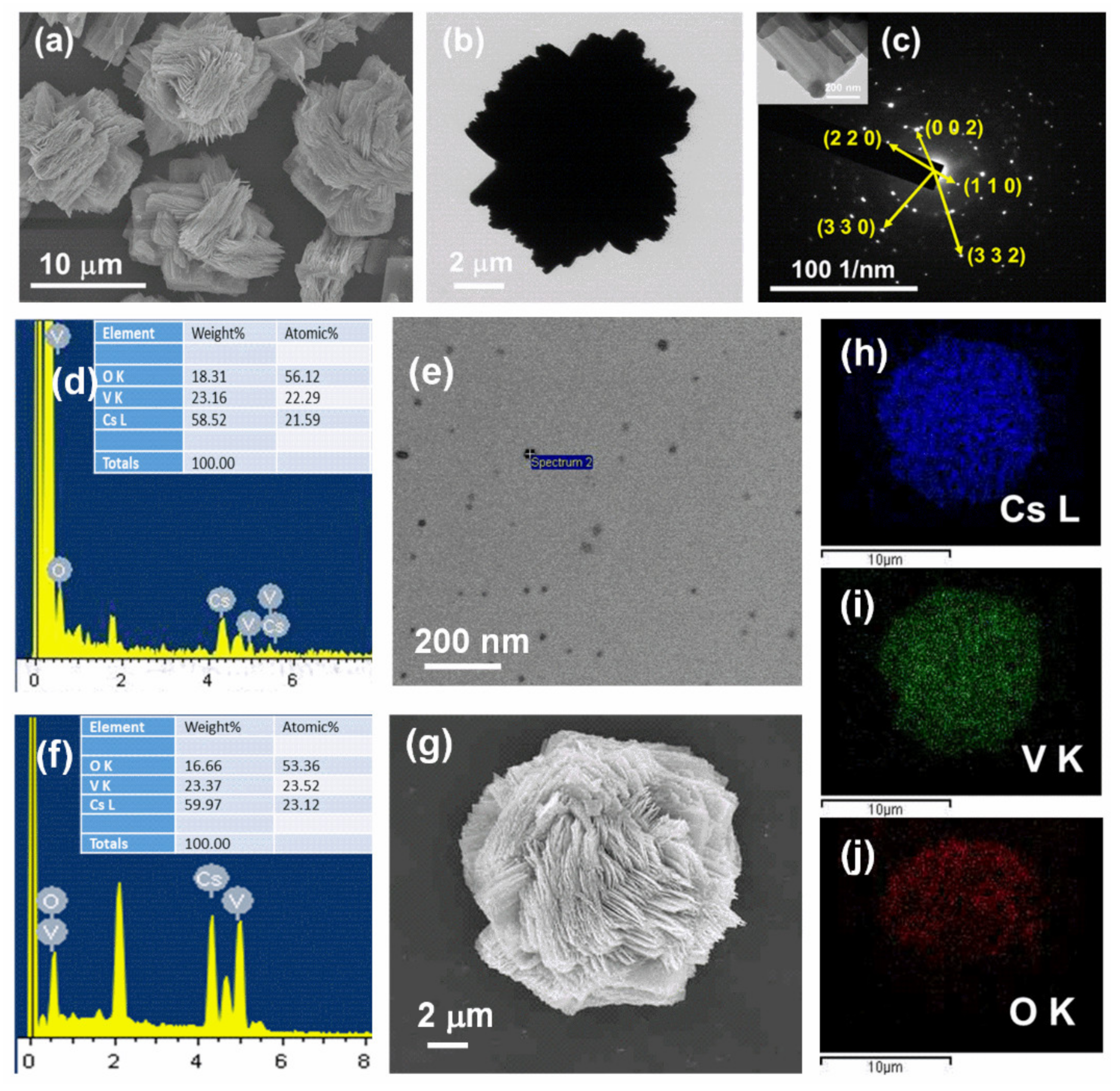
Abstract
:1. Introduction
2. Materials and Methods
2.1. Materials
2.2. Synthesis of CsVO3 QDs
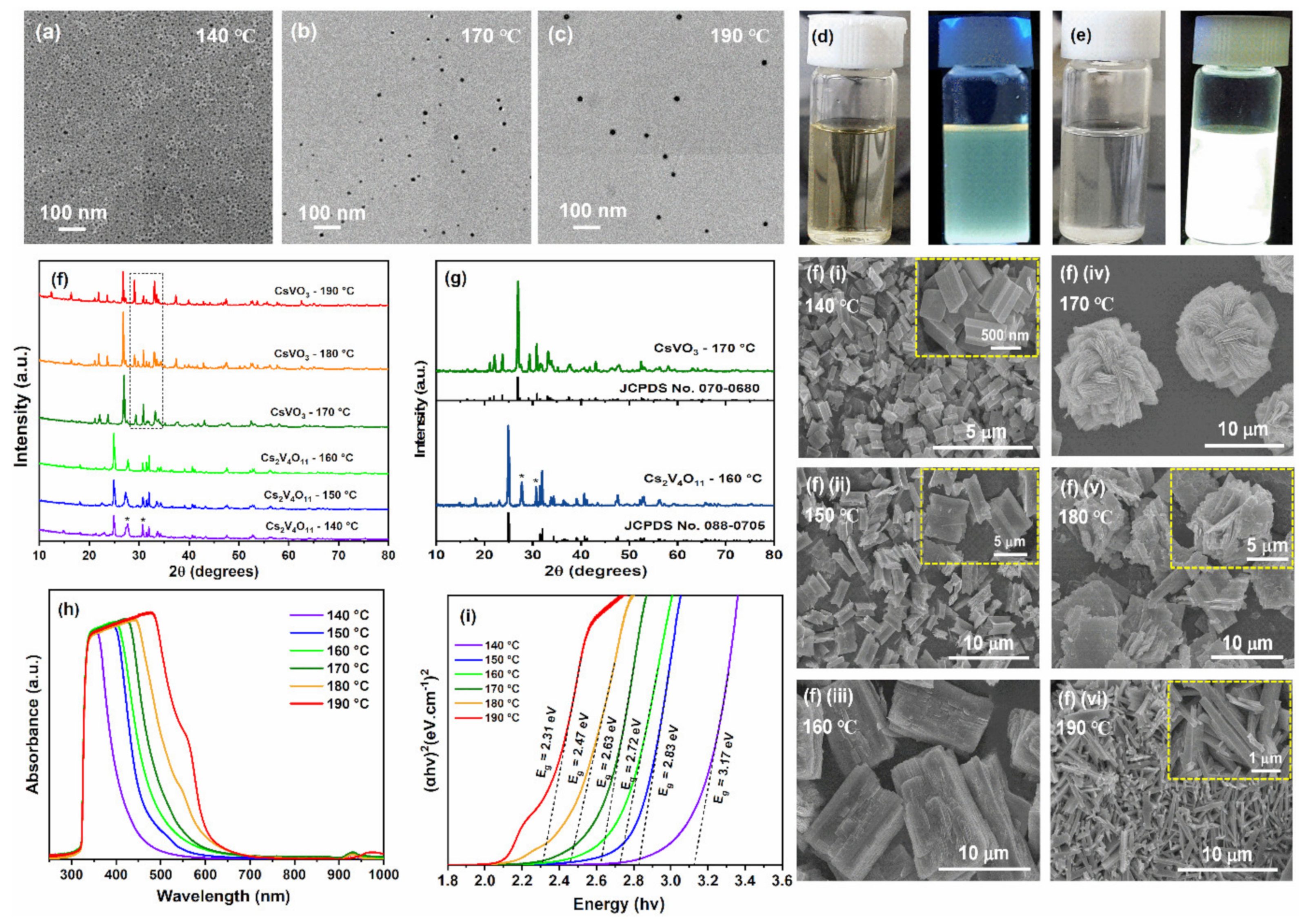
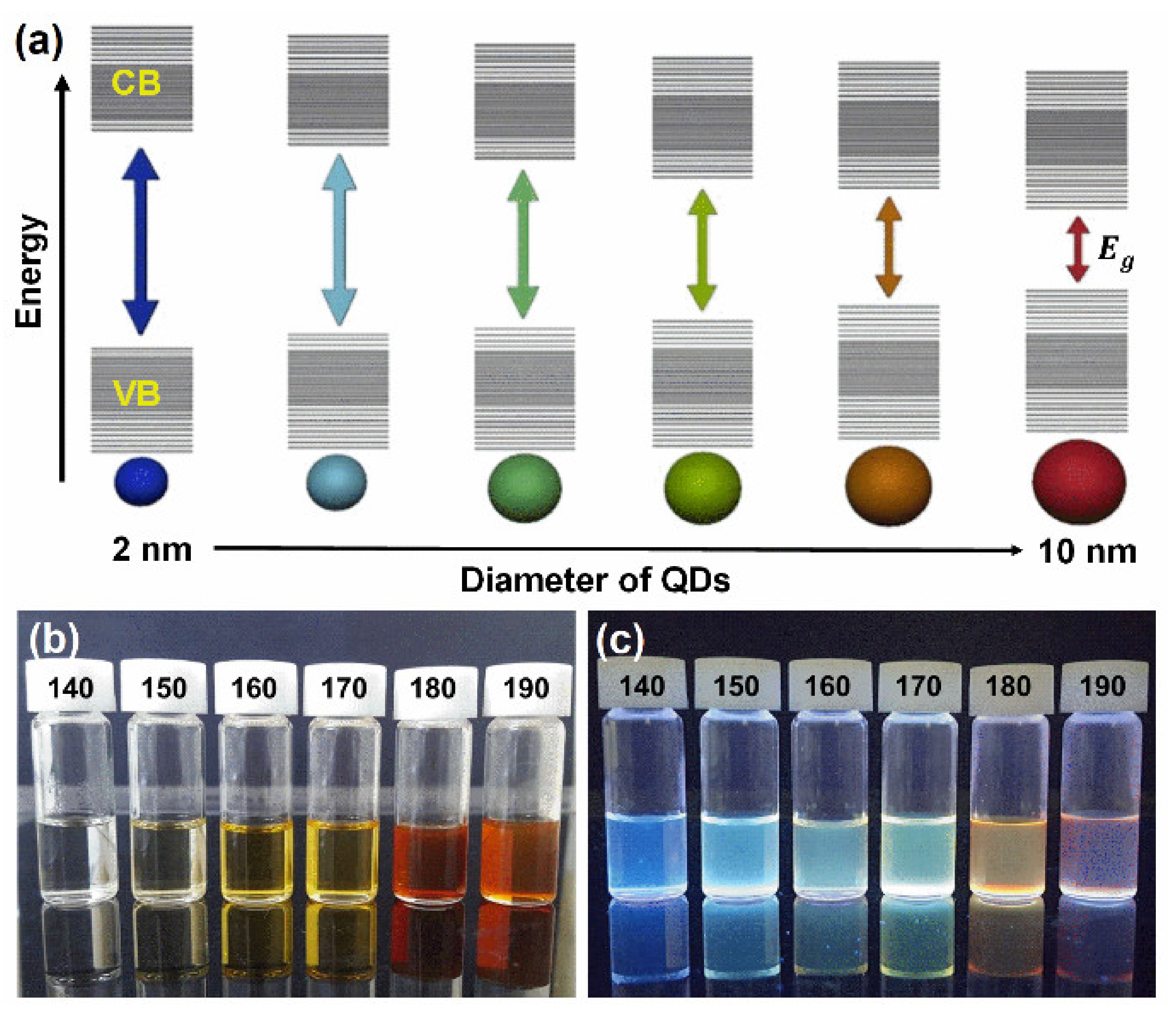
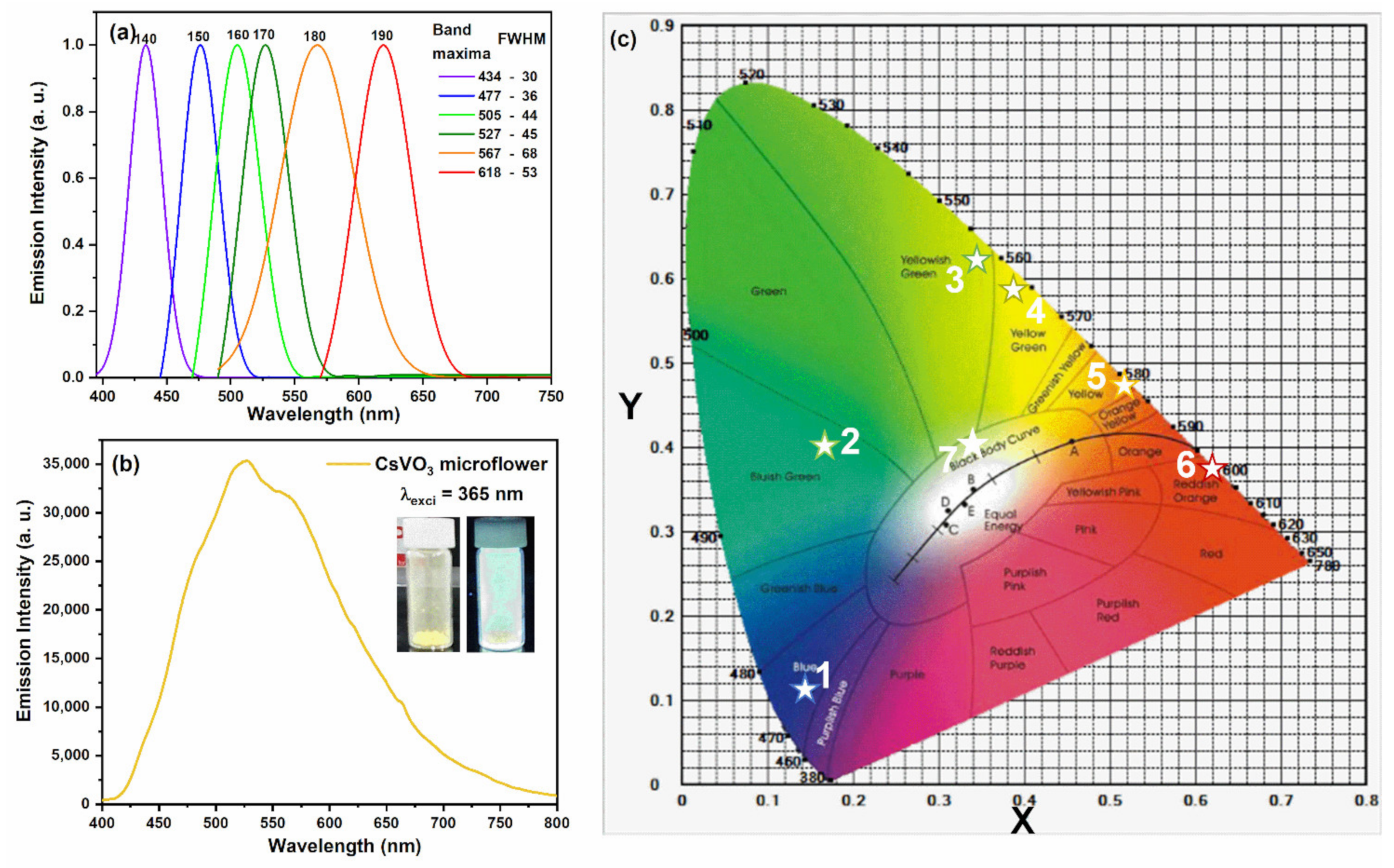
3. Results and Discussion
4. Conclusions
Supplementary Materials
Author Contributions
Funding
Institutional Review Board Statement
Informed Consent Statement
Data Availability Statement
Conflicts of Interest
References
- Mohamed, W.A.A.; El-Gawad, H.A.; Mekkey, S.; Galal, H.; Handal, H.; Mousa, H.; Labib, A. Quantum dots synthetization and future prospect applications. Nanotechnol. Rev. 2021, 10, 1926–1940. [Google Scholar] [CrossRef]
- Kim, T.Y.; Park, S.; Kim, B.J.; Heo, S.B.; Yu, J.H.; Shin, J.S.; Hong, J.-A.; Kim, B.-S.; Kim, Y.D.; Park, Y.; et al. Dual-functional quantum-dots light emitting diodes based on solution processable vanadium oxide hole injection layer. Sci. Rep. 2021, 11, 1700. [Google Scholar] [CrossRef] [PubMed]
- Cao, Z.; Shu, Y.; Qin, H.; Su, B.; Peng, X. Quantum Dots with Highly Efficient, Stable, and Multicolor Electrochemiluminescence. ACS Cent. Sci. 2020, 6, 1129–1137. [Google Scholar] [CrossRef] [PubMed]
- Colvin, V.L.; Schlamp, M.C.; Alivisatos, A.P. Light-emitting diodes made from cadmium selenide nanocrystals and a semiconducting polymer. Nature 1994, 370, 354–357. [Google Scholar] [CrossRef]
- Liu, S.; Xiong, K.; Wang, K.; Liang, G.; Li, M.-Y.; Tang, H.; Yang, X.; Huang, Z.; Lian, L.; Tan, M.; et al. Efficiently Passivated PbSe Quantum Dot Solids for Infrared Photovoltaics. ACS Nano 2021, 15, 3376–3386. [Google Scholar] [CrossRef]
- Rakshit, S.; Piatkowski, P.; Mora-Seró, I.; Douhal, A. Combining Perovskites and Quantum Dots: Synthesis, Characterization, and Applications in Solar Cells, LEDs, and Photodetectors. Adv. Opt. Mater. 2022, 10, 2102566. [Google Scholar] [CrossRef]
- Sukharevska, N.; Bederak, D.; Goossens, V.M.; Momand, J.; Duim, H.; Dirin, D.N.; Kovalenko, M.V.; Kooi, B.J.; Loi, M.A. Scalable PbS Quantum Dot Solar Cell Production by Blade Coating from Stable Inks. ACS Appl. Mater. Interfaces 2021, 13, 5195–5207. [Google Scholar] [CrossRef]
- Ko, J.; Jeong, B.G.; Chang, J.H.; Joung, J.F.; Yoon, S.-Y.; Lee, D.C.; Park, S.; Huh, J.; Yang, H.; Bae, W.K.; et al. Chemically resistant and thermally stable quantum dots prepared by shell encapsulation with cross-linkable block copolymer ligands. NPG Asia Mater. 2020, 12, 19. [Google Scholar] [CrossRef]
- Ye, M.-Y.; Zhao, Z.-H.; Hu, Z.-F.; Liu, L.-Q.; Ji, H.-M.; Shen, Z.-R.; Ma, T.-Y. 0D/2D Heterojunctions of Vanadate Quantum Dots/Graphitic Carbon Nitride Nanosheets for Enhanced Visible-Light-Driven Photocatalysis. Angew. Chem. Int. Ed. 2017, 56, 8407–8411. [Google Scholar] [CrossRef]
- Zhu, C.; Chen, Z.; Gao, S.; Goh, B.L.; Samsudin, I.B.; Lwe, K.W.; Wu, Y.; Wu, C.; Su, X. Recent advances in non-toxic quantum dots and their biomedical applications. Prog. Nat. Sci. Mater. Int. 2019, 29, 628–640. [Google Scholar] [CrossRef]
- Eren, G.O.; Sadeghi, S.; Bahmani Jalali, H.; Ritter, M.; Han, M.; Baylam, I.; Melikov, R.; Onal, A.; Oz, F.; Sahin, M.; et al. Cadmium-Free and Efficient Type-II InP/ZnO/ZnS Quantum Dots and Their Application for LEDs. ACS Appl. Mater. Interfaces 2021, 13, 32022–32030. [Google Scholar] [CrossRef]
- Tsoi, K.M.; Dai, Q.; Alman, B.A.; Chan, W.C.W. Are Quantum Dots Toxic? Exploring the Discrepancy Between Cell Culture and Animal Studies. Acc. Chem. Res. 2013, 46, 662–671. [Google Scholar] [CrossRef]
- Walkey, C.D.; Chan, W.C.W. Chapter 21—Quantum Dots for Traceable Therapeutic Delivery. In Cancer Theranostics; Chen, X., Wong, S., Eds.; Academic Press: Oxford, UK, 2014; pp. 393–417. [Google Scholar]
- Xu, G.; Zeng, S.; Zhang, B.; Swihart, M.T.; Yong, K.-T.; Prasad, P.N. New Generation Cadmium-Free Quantum Dots for Biophotonics and Nanomedicine. Chem. Rev. 2016, 116, 12234–12327. [Google Scholar] [CrossRef]
- Derfus, A.M.; Chan, W.C.W.; Bhatia, S.N. Probing the Cytotoxicity of Semiconductor Quantum Dots. Nano Lett. 2004, 4, 11–18. [Google Scholar] [CrossRef]
- Soenen, S.J.; Manshian, B.B.; Aubert, T.; Himmelreich, U.; Demeester, J.; De Smedt, S.C.; Hens, Z.; Braeckmans, K. Cytotoxicity of Cadmium-Free Quantum Dots and Their Use in Cell Bioimaging. Chem. Res. Toxicol. 2014, 27, 1050–1059. [Google Scholar] [CrossRef]
- Pavitra, E.; Seeta Rama Raju, G.; Krishna Bharat, L.; Park, J.Y.; Kwak, C.H.; Chung, J.W.; Han, Y.-K.; Huh, Y.S. Evolution of highly efficient rare-earth free Cs(1−x)RbxVO3 phosphors as a single emitting component for NUV-based white LEDs. J. Mater. Chem. C 2018, 6, 12746–12757. [Google Scholar] [CrossRef]
- Nakajima, T.; Isobe, M.; Uzawa, Y.; Tsuchiya, T. Rare earth-free high color rendering white light-emitting diodes using CsVO3 with highest quantum efficiency for vanadate phosphors. J. Mater. Chem. C 2015, 3, 10748–10754. [Google Scholar] [CrossRef]
- Luo, J.; Yang, A.; Xie, Z.; Huang, J.; Zuo, X. Preparation, optical properties and first principle calculation of CsVO3. J. Lumin. 2021, 229, 117658. [Google Scholar] [CrossRef]
- Tsiani, E.; Fantus, I.G. Vanadium Compounds: Biological Actions and Potential as Pharmacological Agents. Trends Endocrinol. Metab. 1997, 8, 51–58. [Google Scholar] [CrossRef]
- Rehder, D. Perspectives for vanadium in health issues. Future Med. Chem. 2016, 8, 325–338. [Google Scholar] [CrossRef]
- Holder, C.F.; Schaak, R.E. Tutorial on Powder X-ray Diffraction for Characterizing Nanoscale Materials. ACS Nano 2019, 13, 7359–7365. [Google Scholar] [CrossRef] [PubMed]
- Ren, J.; Stagi, L.; Malfatti, L.; Garroni, S.; Enzo, S.; Innocenzi, P. Boron Nitride–Titania Mesoporous Film Heterostructures. Langmuir 2021, 37, 5348–5355. [Google Scholar] [CrossRef]
- Ren, J.; Stagi, L.; Malfatti, L.; Paolucci, V.; Cantalini, C.; Garroni, S.; Mureddu, M.; Innocenzi, P. Improving the Photocatalytic Activity of Mesoporous Titania Films through the Formation of WS2/TiO2 Nano-Heterostructures. Nanomaterials 2022, 12, 1074. [Google Scholar] [CrossRef] [PubMed]
- Ren, J.; Malfatti, L.; Enzo, S.; Carbonaro, C.M.; Calvillo, L.; Granozzi, G.; Innocenzi, P. Boron oxynitride two-colour fluorescent dots and their incorporation in a hybrid organic-inorganic film. J. Colloid Interface Sci. 2020, 560, 398–406. [Google Scholar] [CrossRef] [PubMed]
- Tauc, J. Optical properties and electronic structure of amorphous Ge and Si. Mater. Res. Bull. 1968, 3, 37–46. [Google Scholar] [CrossRef]
- Pisanic Ii, T.R.; Zhang, Y.; Wang, T.H. Quantum dots in diagnostics and detection: Principles and paradigms. Analyst 2014, 139, 2968–2981. [Google Scholar] [CrossRef] [PubMed]
- Dang, P.; Liu, D.; Wei, Y.; Li, G.; Lian, H.; Shang, M.; Lin, J. Highly Efficient Cyan-Green Emission in Self-Activated Rb3RV2O8 (R = Y, Lu) Vanadate Phosphors for Full-Spectrum White Light-Emitting Diodes (LEDs). Inorg. Chem. 2020, 59, 6026–6038. [Google Scholar] [CrossRef] [PubMed]


Publisher’s Note: MDPI stays neutral with regard to jurisdictional claims in published maps and institutional affiliations. |
© 2022 by the authors. Licensee MDPI, Basel, Switzerland. This article is an open access article distributed under the terms and conditions of the Creative Commons Attribution (CC BY) license (https://creativecommons.org/licenses/by/4.0/).
Share and Cite
Raju, G.S.R.; Varaprasad, G.L.; Lee, J.-H.; Park, J.Y.; Chodankar, N.R.; Ranjith, K.S.; Pavitra, E.; Huh, Y.S.; Han, Y.-K. A Novel and Cost-Effective CsVO3 Quantum Dots for Optoelectronic and Display Applications. Nanomaterials 2022, 12, 2864. https://doi.org/10.3390/nano12162864
Raju GSR, Varaprasad GL, Lee J-H, Park JY, Chodankar NR, Ranjith KS, Pavitra E, Huh YS, Han Y-K. A Novel and Cost-Effective CsVO3 Quantum Dots for Optoelectronic and Display Applications. Nanomaterials. 2022; 12(16):2864. https://doi.org/10.3390/nano12162864
Chicago/Turabian StyleRaju, Ganji Seeta Rama, Ganji Lakshmi Varaprasad, Jeong-Hwan Lee, Jin Young Park, Nilesh R. Chodankar, Kugalur Shanmugam Ranjith, Eluri Pavitra, Yun Suk Huh, and Young-Kyu Han. 2022. "A Novel and Cost-Effective CsVO3 Quantum Dots for Optoelectronic and Display Applications" Nanomaterials 12, no. 16: 2864. https://doi.org/10.3390/nano12162864