
Hydrothermal Synthesis of MnO2/Reduced Graphene Oxide Composite for 4-Nitrophenol Sensing Applications
by
 , and
, and
Praveen Kumar
1,†,
Mohd Quasim Khan
2,
Rais Ahmad Khan
3,
Khursheed Ahmad
4,† and
Haekyoung Kim
4,* 1
Department of Chemistry, Indian Institute of Technology Indore, Khandwa Road, Simrol, Indore 453552, Madhya Pradesh, India
2
Department of Chemistry, M.M.D.C, Moradabad, M.J.P. Rohilkhand University, Bareilly 244001, Uttar Pradesh, India
3
Department of Chemistry, College of Science, King Saud University, Riyadh 11451, Saudi Arabia
4
School of Materials Science and Engineering, Yeungnam University, Gyeongsan 38541, Republic of Korea
*
Author to whom correspondence should be addressed.
†
These authors contributed equally to this work.
Inorganics 2022, 10(12), 219; https://doi.org/10.3390/inorganics10120219
Submission received: 10 October 2022
/
Revised: 18 November 2022
/
Accepted: 22 November 2022
/
Published: 24 November 2022
(This article belongs to the Special Issue Electrochemical Study of Nanocarbon Based Materials)
Abstract
:Recently, the electrochemical sensing approach has attracted materials/electrochemical scientists to design and develop electrode materials for the construction of electrochemical sensors for the detection of para-nitrophenol (4-NP). In the present study, we have prepared a hybrid composite of MnO2 and rGO (MnO2/rGO) using a hydrothermal approach. The morphological features of the prepared MnO2/rGO composite were studied by scanning electron microscopy, whereas the phase purity and formation of the MnO2/rGO composite were authenticated via the powder X-ray diffraction method. Energy-dispersive X-ray spectroscopy was also employed to analyze the elemental composition of the prepared MnO2/rGO composite. In further studies, a glassy carbon electrode (GCE) was modified with MnO2/rGO composite (MnO2/rGO/GCE) and explored as 4-nitrophenol (4-NP) sensor. The fabricated MnO2/rGO/GCE exhibited a reasonably good limit of detection of 0.09 µM with a sensitivity of 0.657 µA/µMcm2. The MnO2/rGO/GCE also demonstrates good selectivity, stability and repeatability in 50 cycles.
1. Introduction
In the past few decades, environmental pollution has become one of the major threats globally. There are a large number of toxic and hazardous compounds which have negative influences on human health and the environment [1]. Nitrophenols are widely used in various industries manufacturing rubber, pesticides, petrochemical products, plastics, dyes, fungicides, and paints [2,3,4,5,6,7,8]. Nitrophenols are the major water pollutants that are directly released to the water reservoir bodies from various industries as discharged products. The presence of nitrophenols in water reservoir bodies and the environment can change the biological and chemical nature of the terrestrial environment and water [1,5]. It has been found that even the presence of nitrophenols with low concentrations could significantly influence the human being and environment [3]. The environment and human health may be at risk from the widespread use of 4-NP [3]. Excessive exposure to 4-NP may damage the kidney, liver, and human central nervous system, including some other health problems such as headaches, skin irritation, cyanosis, methemoglobinemia, sleepiness, and nausea [2,5,8]. The soil, crops, and water bodies can all be impacted by the presence of 4-NP [2]. These are serious issues; hence it is vital to regulate the use of 4-NP in all areas, including food, soil, and water. In this context, the sensitive detection of 4-NP is essential [4]. Numerous analytical techniques, including fluorescence, spectrofluorimetric, optical spectroscopy, enzyme-linked immunosorbent assay, liquid chromatography, spectrophotometry, gas chromatography, capillary zone electrophoresis, and high-performance liquid chromatography, have been explored for the detection of 4-NP [9,10,11,12,13,14,15,16,17]. However, all of these approaches are time-consuming and have their own drawbacks/limitations [18,19]. Recently, the electrochemical technique has gained much attention because of its fast detection response, excellent selectivity, high sensitivity and cost-effectiveness [20,21,22,23]. Electrochemical sensing approach has been employed as a detection technique that records the signal during the oxidation/reduction process [24,25]. The electrochemical sensors need electro-catalysts that catalyze the oxidation/reduction of 4-NP. In previous years, various semiconducting metal oxides (manganese oxide (MnO2), zinc oxide (ZnO), nickel oxide (NiO), copper oxide (CuO), magnesium oxide (MgO)) and perovskite materials, including SrTiO3 have been employed as electro-catalysts for the development of electrochemical sensor [26,27,28,29,30,31]. MnO2 is one of the promising candidates and has been extensively used in supercapacitors, wastewater treatment, catalytic sensors, and solar cells due to its cost-effectiveness, high energy density, natural abundance, and non-toxicity [32]. It has been reported that pristine MnO2 has low conductivity, which can influence the electrochemical performance of MnO2-based devices.
In this regard, recent reports showed that the incorporation of Pd with MnO2 improves the electrochemical performance of MnO2-based devices [33]. Ahmad et al. also introduced copper to the MnO2 to enhance the electrochemical activity of the MnO2-based sensors [26]. In some other works, it has been studied that the incorporation of carbon-based materials such as reduced graphene oxide (rGO) can significantly improve the conductive nature of the MnO2. rGO has excellent mechanical stability, good conductivity, thermal stability, and high surface area [34,35]. rGO has been extensively used as conductive support to improve the conductive nature of poor semiconducting metal oxides [36,37]. It is clear that rGO is one of the most suitable candidates for improving the conductivity of MnO2 [35]. Jaiswal et al. [35] prepared MnO2/rGO composite and used it as electrode material for the construction of nitrite sensors. The fabricated nitrite sensor exhibited good electrocatalytic properties for the detection of nitrite. This can be attributed to the synergistic effects between MnO2 and rGO. This clearly showed that MnO2 has good electro-catalytic properties, whereas rGO has a good surface area and conductivity [35]. This indicated that the combination of MnO2 and rGO may be useful for the development of electrochemical sensors.
Herein, we have fabricated a hybrid composite of MnO2 and rGO using the hydrothermal method. Further, a 4-NP electrochemical sensor was fabricated using the drop-cast method. The developed sensor exhibits a reasonable limit of detection, decent sensitivity, and good selectivity and stability.
2. Experimental Section
All the used materials and chemicals were bought from Loba, SRL, TCI, Alfa-Aesar, Sigma, and Merck and used without any further purification.
2.1. Synthesis of Graphene Oxide (GO)
In accordance with the earlier study, GO was synthesized using the renowned Hummer’s Method [38]. In brief, 23.0 mL H2SO4 was added to 1.0 g of graphite flakes and 0.5 g of NaNO3, which were thoroughly mixed before being kept at room temperature for 30 min. The exothermic reaction was controlled by using ICE (temperature below 20 °C), adding KMnO4 (3.0 g) gradually, which was then continuously stirred for an entire night at room temperature. After adding, 60 mL of distilled water and 1.5 g of KMnO4 were gently added to the reaction mixture, which was then stirred once more for 14 h at room temperature. After completion, the mixture was allowed to cool, and 500 mL of DI water was added, along with 7 mL of 30% H2O2. After brief washing with 1M HCl, the resulting crude was repeatedly rinsed with water. To achieve the final product, the final crude was washed with extra D.I. water and dried at 50 °C for 6 h.
2.2. Synthesis of MnO2/rGO
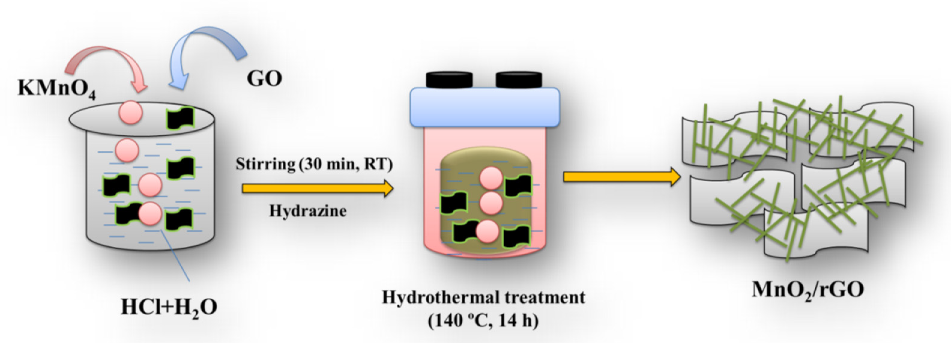
The MnO2/rGO composite and MnO2 were synthesized using a hydrothermal method. First, 3.5 mmol of KMnO4 was added to the 25 mL of distilled water, and a few drops of HCl were added. This reaction mixture was stirred for a few minutes at room temperature. On the other side, GO (50 mg) was dispersed in 10 mL of distilled water (having hydrazine) using a sonication bath. The GO dispersion was added to the KMnO4 solution and stirred for 0.5 h at RT. Further, this solution was added to the Teflon lining hydrothermal autoclave (100 mL capacity) and heated to 140° for 12–14 h. Once the reaction finished, it was allowed to cool down to room temperature before the crude product was separated using a centrifuge. The crude was thoroughly cleaned using DI water and ethanol and dried overnight (70 °C). MnO2 was prepared under similar conditions without adding GO solution [32]. rGO was prepared according to a previous report [38].
2.3. Fabrication of Electrochemical Sensor
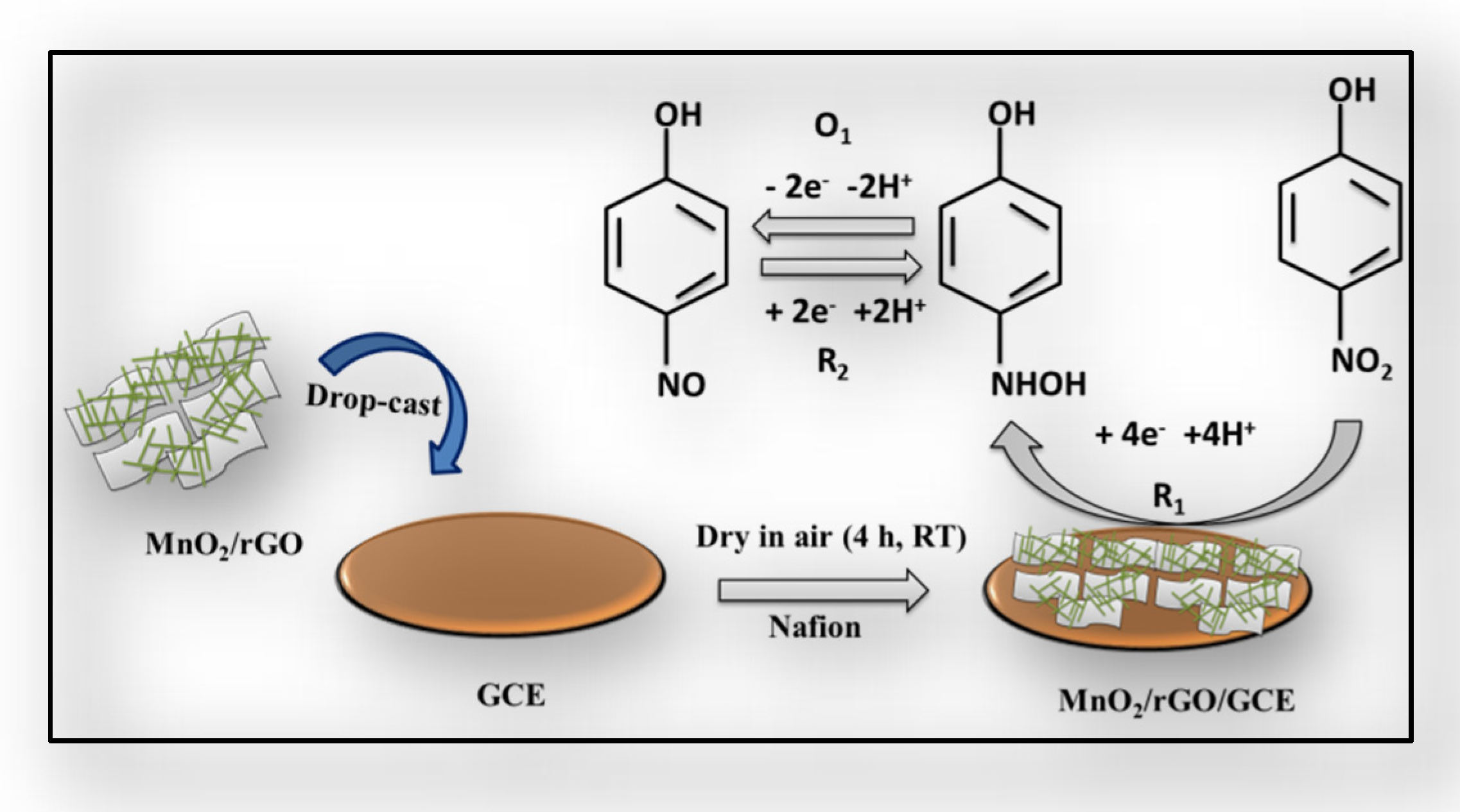
For the electrochemical investigations, three electrodes—counter electrode (platinum; Pt), reference electrode (Ag/AgCl), and working electrode (GCE) were utilized. A glassy carbon electrode (GCE) served as the working electrode for the sensing application. The surface of GCE was modified with prepared materials (Scheme 1). The MnO2, rGO, and MnO2/rGO coated electrodes are labeled as MnO2/GCE, rGO/GCE, and MnO2/rGO/GCE, respectively, which were employed as the working electrode. On the other hand, bare GCE was labeled as GCE. 0.1 M PBS of pH 7.0 was used for all the electrochemical studies. A computer-controlled Potentiostat was used for all of the electrochemical experiments (CH Instrument). The working electrodes (GCE) have a 3 mm-diameter geometrical area.
3. Results and Discussion
3.1. General Characterization
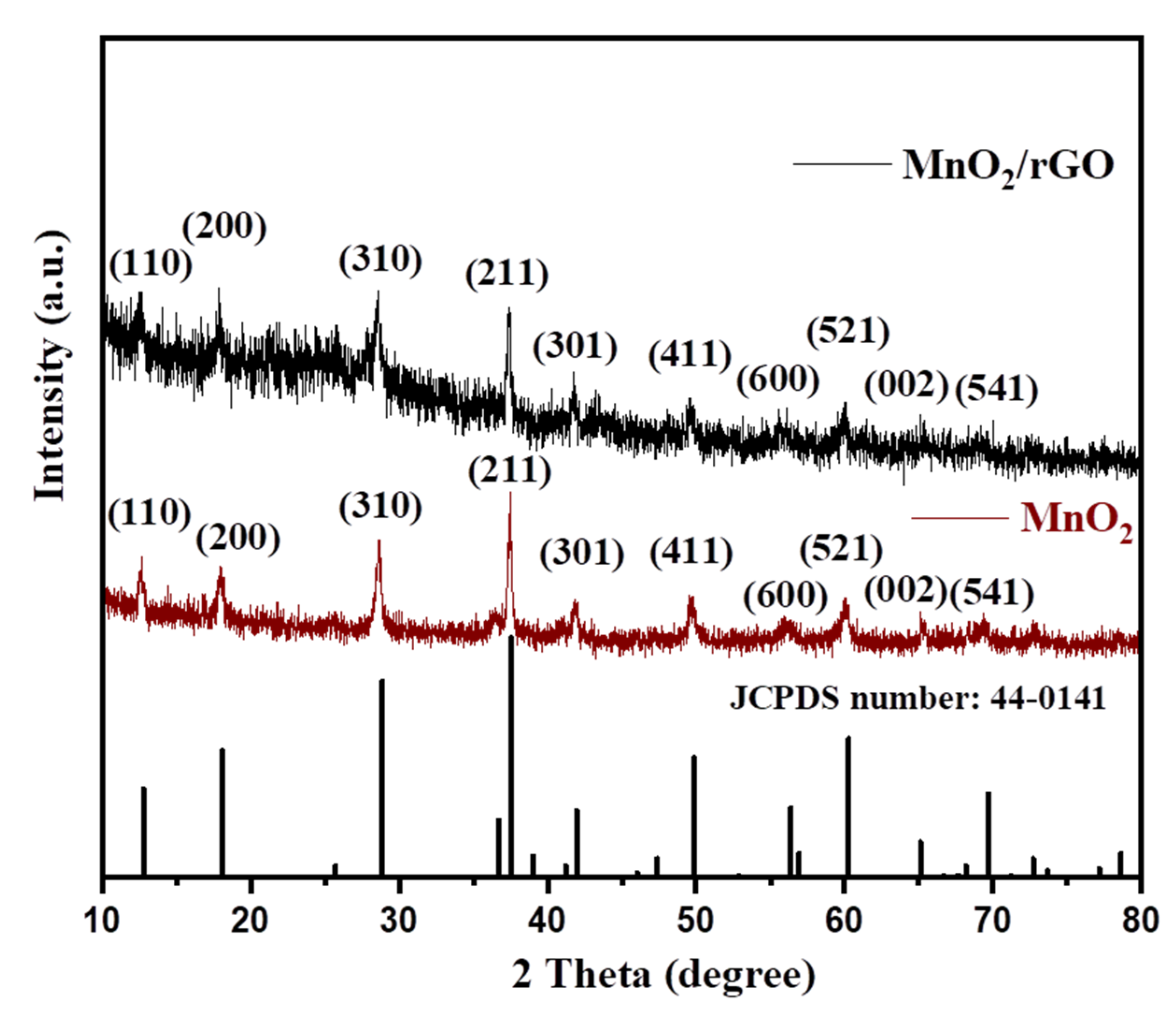
In order to confirm the formation of the MnO2/rGO composite, we have obtained the X-ray diffraction (XRD) of the prepared powder sample of the MnO2/rGO composite (Rigaku, Japan, Instrument). The XRD of the MnO2/rGO composite was collected at the 2θ of 10–80°. The collected XRD pattern of the MnO2/rGO composite has been displayed in Figure 1. Figure 1 demonstrates the presence of various diffraction peaks, which can be assigned to the (110), (200), (310), (211), (301), (411), (600), (521), (002), and (521) diffraction planes of MnO2 (JCPDS number 44-0141). However, no diffraction peak related to the rGO could be observed. The XRD pattern of the pristine MnO2 was also collected and has been incorporated in Figure 1. The XRD of MnO2 exhibited the presence of various diffraction planes of (110), (200), (310), (211), (301), (411), (600), (521), (002), and (521). The XRD studies showed that the incorporation of rGO into MnO2 changes the crystallinity of the prepared MnO2/rGO composite, which may be due to the amorphous nature of rGO. The XRD pattern rGO was also collected to verify the formation of rGO. The XRD data of the rGO has been presented in Figure S1 in the supporting information.
A clearly defined diffraction plane (002) was visible in the rGO XRD pattern. This confirmed the preparation of MnO2, rGO, and MnO2/rGO composite. The Raman spectra (Horiba-Scientific Instrument; laser wavelength = 532 nm; grating = 1800 gr/mm) of the rGO, MnO2 and MnO2/rGO were also obtained. The obtained results showed that MnO2 has two peaks at 569.4 and 640.1 cm−1, which can be assigned to the symmetric stretching vibration of the MnO6 octahedral group and the stretching vibration of the Mn-O bond, respectively (Figure S2). Raman spectrum of rGO exhibits the presence of D and G bands. The Raman spectrum of MnO2/rGO showed Raman peaks for MnO2 along with D and G bands of rGO (Figure S2). This suggested the formation of MnO2/rGO composite.
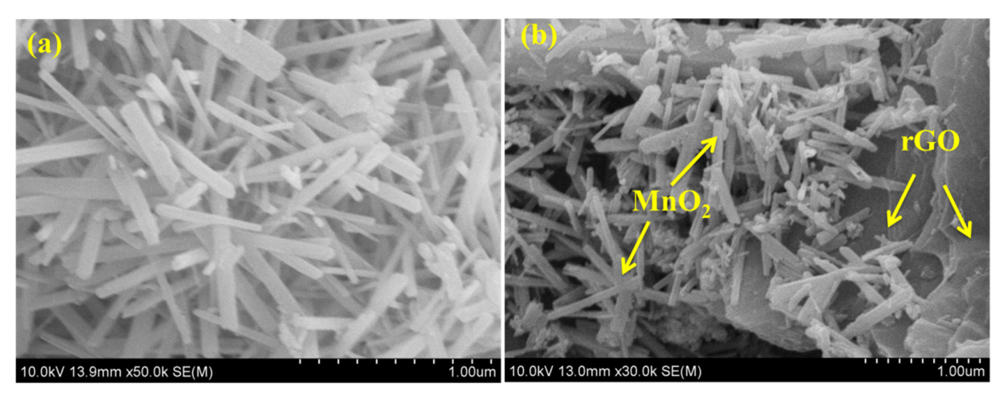
The morphology of electrode materials can significantly alter the performance of the electrochemical devices.
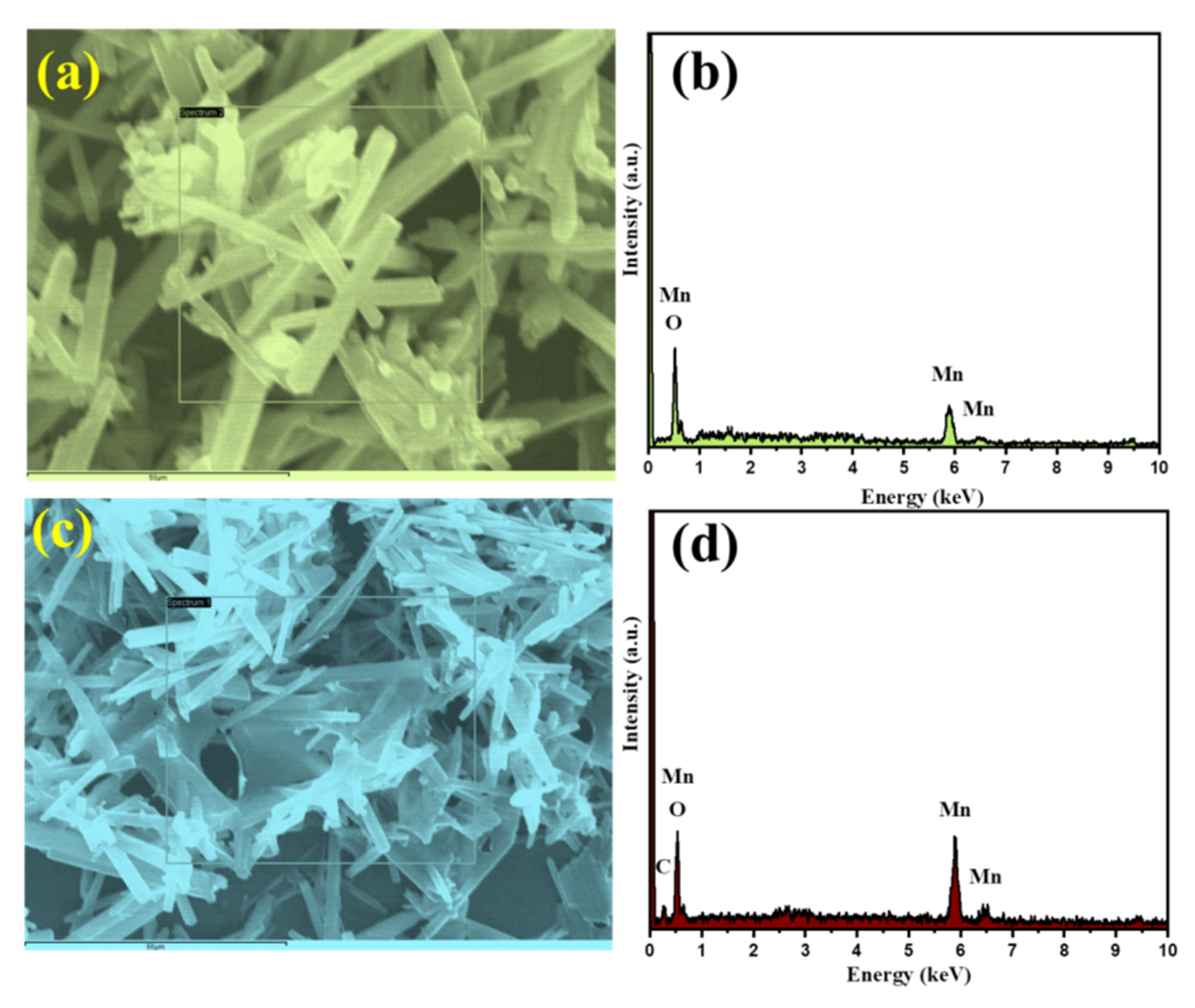
Thus, morphological characteristics of the prepared MnO2 and MnO2/rGO composite were also studied. The morphological features of the MnO2 and MnO2/rGO composite were investigated by recording scanning electron microscopic (SEM) images on Hitachi (S-4800) SEM instrument. The SEM pictures of the MnO2 and MnO2/rGO composite are presented in Figure 2. Figure 2a demonstrates the presence of rod-like surface properties of the prepared MnO2. Similarly, Figure 2b shows that MnO2 rods are present on rGO sheets. Thus, it can be understood that rGO has a sheet-like morphology on which rod-like MnO2 has been grown via the hydrothermal method (Figure 2b). Transmission electron microscopy (TEM) was also used to further characterize the prepared MnO2/rGO composite. The obtained TEM image of the MnO2/rGO composite has been presented in Figure S3, which indicates that MnO2 rods are present on the rGO surface. The determination of the elemental composition of the MnO2 and MnO2/rGO composites is necessary to verify the formation of the MnO2 and MnO2/rGO composites. Hence, energy-dispersive X-ray spectroscopy (EDX; Horiba instrument) was explored to authenticate the formation of MnO2 and MnO2/rGO composite. The obtained EDX results of the MnO2 and MnO2/rGO composite are presented in Figure 3a–d. The EDX spectrum of MnO2 indicated the presence of Mn and O elements (Figure 3b), which suggested the formation of MnO2. On the other side, the EDX spectrum of MnO2/rGO indicated the presence of Mn, O, and C elements and authenticated the formation of the MnO2/rGO composite (Figure 3d). In the MnO2/rGO composite, C, Mn, and O elements have weight percentages of 9.78%, 61.13%, and 29.09%, respectively.
Thus, it can be said that MnO2 and MnO2/rGO composite are prepared successfully via the hydrothermal approach with good phase purity.
3.2. Electrochemical Performance
To calculate the electrochemically active surface area (ECSA) of the GCE, MnO2/GCE, rGO/GCE, and MnO2/rGO/GCE, CVs curves were recorded in 5 mM [Fe(CN)6]3−/4− redox couple scan rate of 100 mVs−1 (Figure S4). The electrochemically active surface area of the GCE, MnO2/GCE, rGO/GCE and MnO2/rGO/GCE were calculated by using the Randles–Sevcik equation given below,
Ip = 2.69 × 10⁵ AD1/2 n3/2 y1/2 C
In Equation (1), Ip is the peak current, A is the ECSA (to be calculated), y is scan rate (V/s), and n = no. of electrons (n = 1) for redox couple [Fe(CN)6]3−/4−, C is the concentration (mol/L), and D is the diffusion coefficient (6.7 × 10⁻⁶ cm2 s−1)). The ECSAs of the bare GCE, MnO2/GCE, rGO/GCE, and MnO2/rGO/GCE were 0.07, 0.078, 0.084, and 0.097 cm2, respectively.
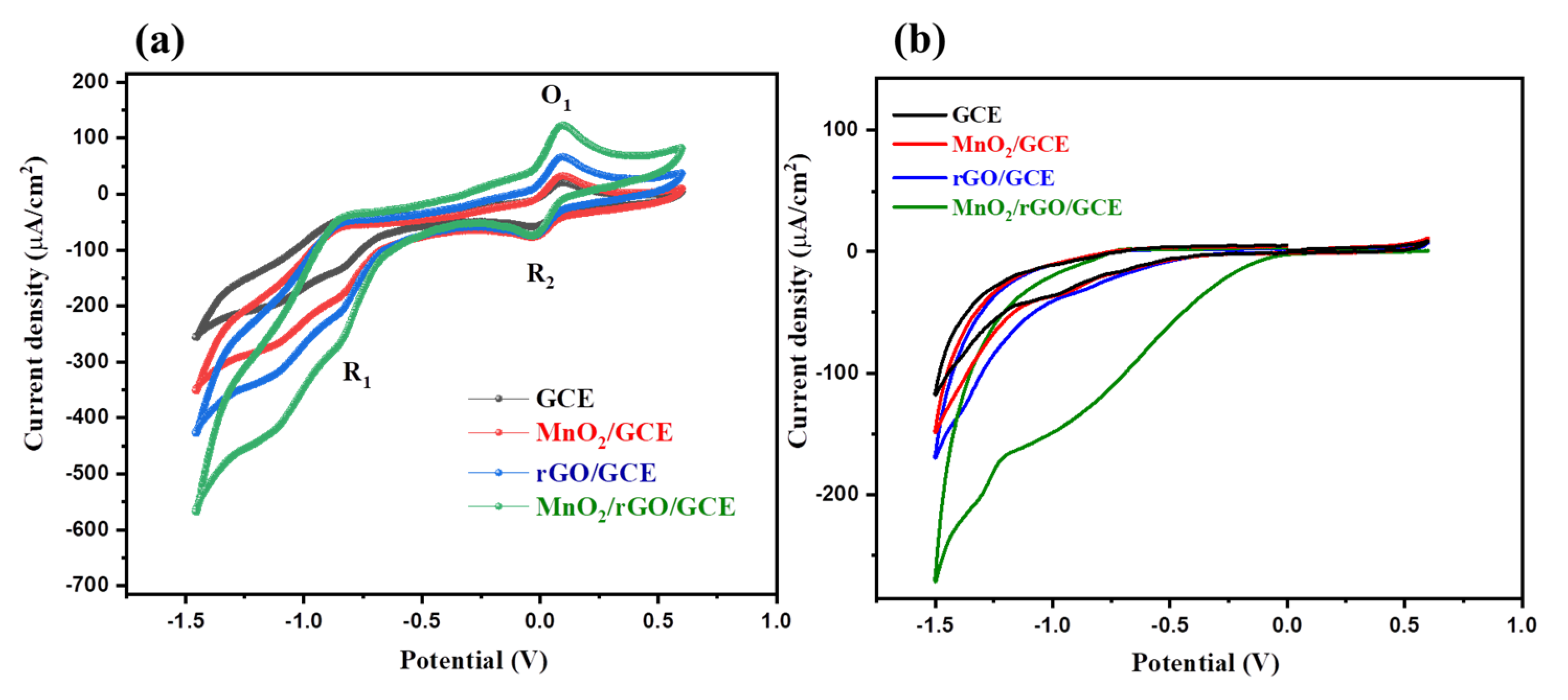
By recording the CVs of the GCE, MnO2/GCE, rGO/GCE, and MnO2/rGO/GCE in 50 µM 4-NP (scan rate = 50 mV/s), the electrochemical sensing activity of the GCE, MnO2/GCE, rGO/GCE, and MnO2/rGO/GCE was assessed. Figure 4a shows the calculated CV curves for the GCE, MnO2/GCE, rGO/GCE, and MnO2/rGO/GCE in 50 µM 4-NP. The CV findings indicated low electro-catalytic current response for GCE whereas MnO2/rGO produced a somewhat better electro-catalytic current. However, rGO/GCE exhibits a better current response compared to the GCE or MnO2/GCE. But the highest electro-catalytic current was observed for MnO2/rGO/GCE, which may be due to the presence of synergistic effects between MnO2 and rGO. It can be clearly seen that MnO2/rGO/GCE possesses the highest sensing property for the sensing of 50 µM 4-NP compared to the GCE, MnO2/GCE or rGO/GCE (Figure 4a).
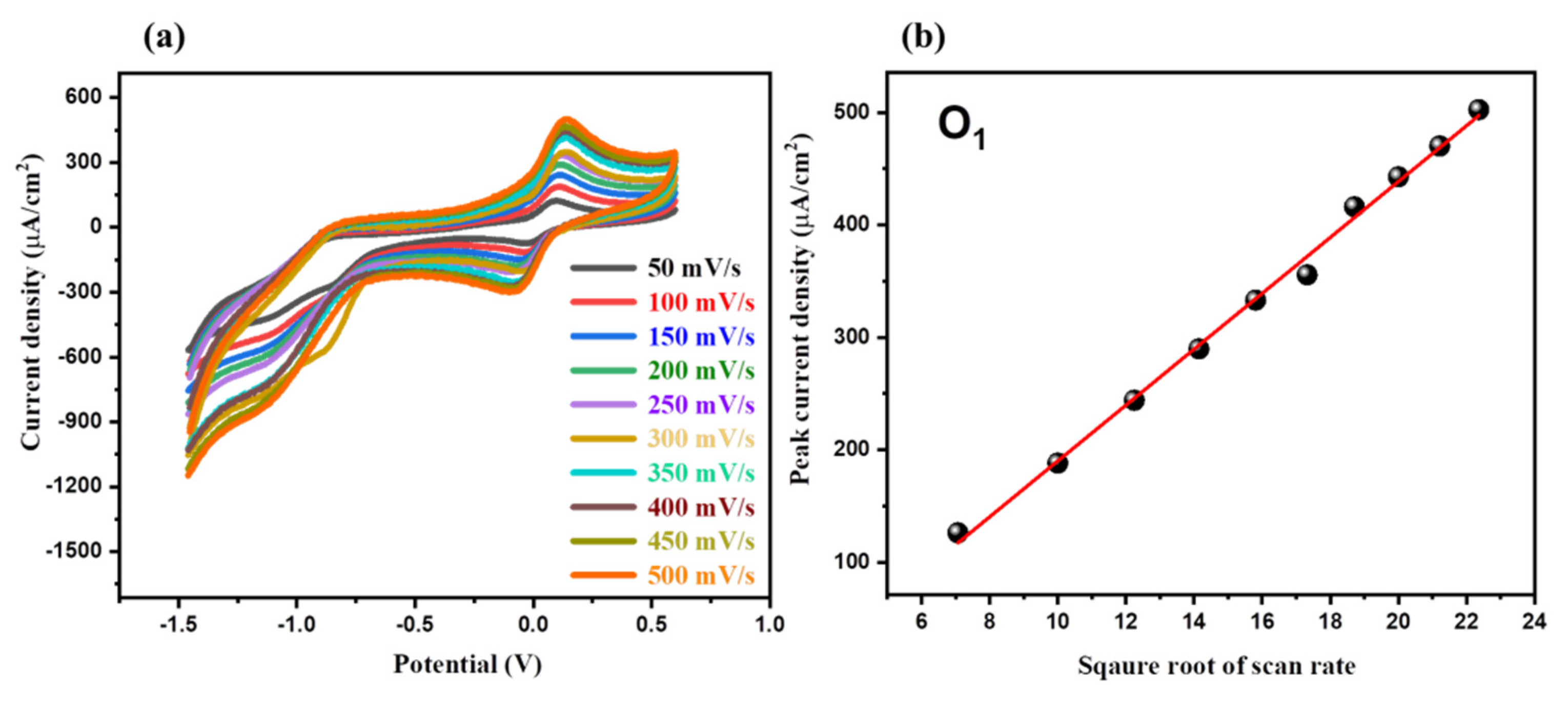
In the absence of 4-NP, the CV responses of the GCE, MnO2/GCE, rGO/GCE, and MnO2/rGO/GCE were taken at a scan rate of 50 mV/s in 0.1 M PBS at pH 7.0. (Figure 4b). The obtained findings demonstrated that, in comparison to the other electrodes, MnO2/rGO/GCE had the highest electro-catalytic activity. As a result, we used MnO2/rGO/GCE as a possible working electrode for more CV research. The CV results showed the presence of one oxidation peak (O1) and two reduction peaks (R1 and R2) for the detection of 4-NP, which may be related to the oxidation and reduction of the 4-NP (Scheme 2) during the electrochemical process/reactions. The applied scan rate can significantly alter the current response of the MnO2/rGO/GCE. Thus, it is necessary to check the influence of different applied scan rates on the electro-catalytic behavior of the MnO2/rGO/GCE. In this regard, CV graphs of the MnO2/rGO/GCE were recorded at various scan rates of 50–500 mV/s in a fixed concentration of 4-NP (50 µM).
The obtained CV results of the MnO2/rGO/GCE at various scan rates (50–500 mV/s) are presented in Figure 5a. With regard to the scan rate, it can be seen that the MnO2/rGO/GCE’s current response grows (Figure 5a) with increasing concentration. According to the calibration plot between the current response and the scan rate’s square root, the current response appears to grow linearly (Figure 5b).
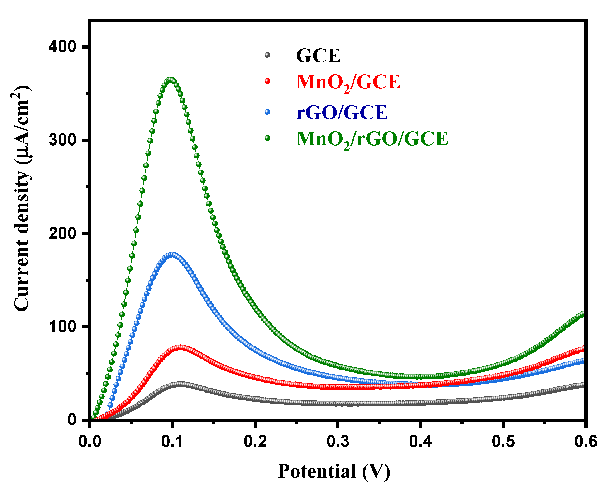
Recently, the LSV method has received enormous attention for electrochemical sensing applications. In our study, we also adopted the LSV technique for the detection of 4-NP. In the presence of 50 µM 4-NP and a scan rate of 50 mV/s, the LSV graphs of the GCE, MnO2/GCE, rGO/GCE, and MnO2/rGO/GCE were obtained. Figure 6 shows the obtained LSV curves of the GCE, MnO2/GCE, rGO/GCE, and MnO2/rGO/GCE. The GCE has the lowest current response, while MnO2/GCE showed an improved current response for the detection of 50 µM 4-NP (Figure 6). Further enhancement in the current response of the rGO/GCE was also observed, which may be due to the conductive nature of rGO. The MnO2/rGO/GCE exhibited the highest current response compared to the GCE, MnO2/GCE or rGO/GCE (Figure 6). The presence of a synergistic interaction between MnO2 and rGO may be responsible for this increased current response (Figure 6). Further LSV research was conducted using the MnO2/rGO/GCE. We have also prepared MnO2/rGO composite using different amounts of GO (5 mg, 15 mg, and 35 mg). Further, GCE was modified with prepared different MnO2/rGO composites, and their electro-catalytic properties were examined using LSV. The obtained results are presented in Figure S5. The observations showed that MnO2/rGO composite prepared with 35 mg GO has good electrocatalytic activity but is still lower than that of the MnO2/rGO composite prepared with 50 mg GO. Furthermore, we have also physically mixed 50 mg of GO with 100 mg MnO2 using mortar and pestle. The GCE was also modified with physically mixed MnO2/rGO, and its electrocatalytic properties were also checked under similar conditions via the LSV method. The obtained LSV result is presented in Figure S6. The observations showed that physically mixed MnO2/rGO has decent performance but is still lower than that of the hydrothermally prepared MnO2/rGO composite.
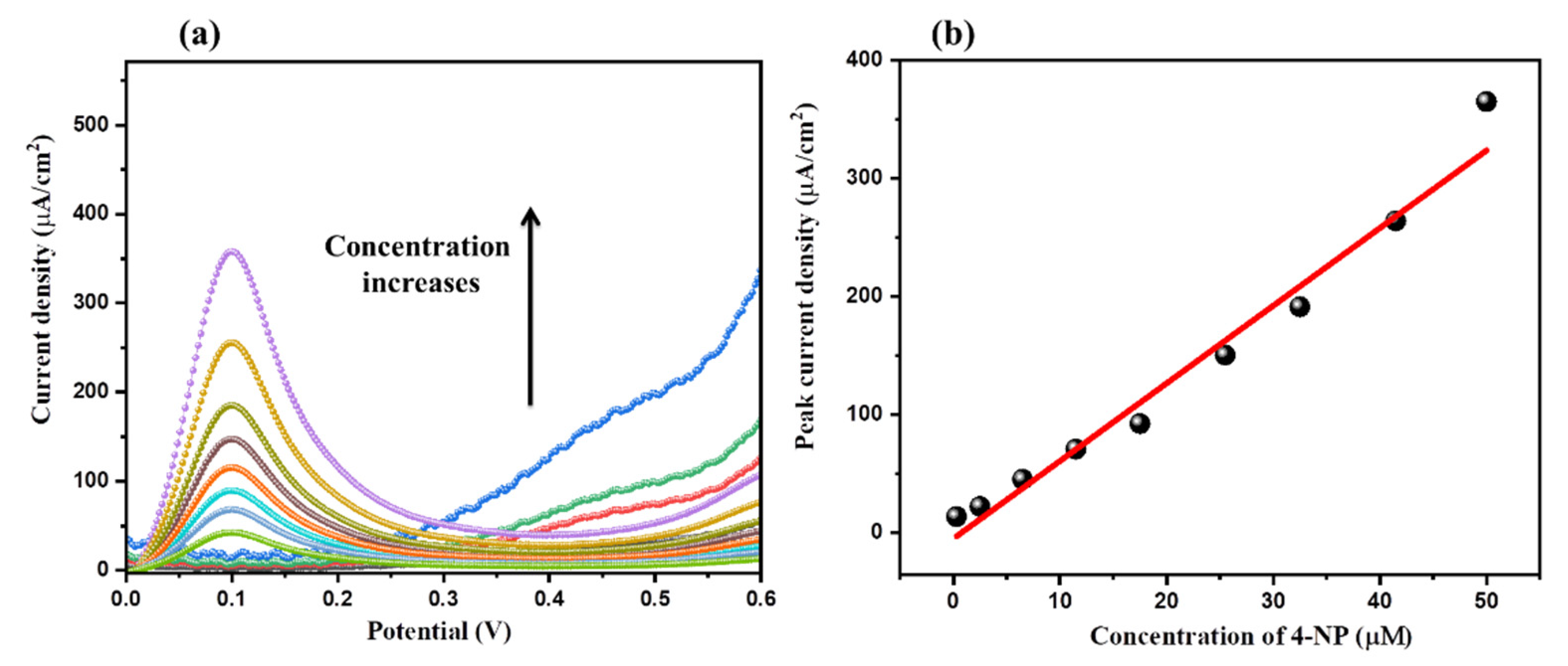
The concentration of 4-NP can influence the current response of the MnO2/rGO/GCE. In this regard, we have obtained LSV graphs of the MnO2/rGO/GCE at various concentrations of 4-NP (0, 0.3 µM, 2.5 µM, 6.5 µM, 11.5 µM, 17.5 µM, 25.5 µM, 32.5 µM, 41.5 µM, and 50 µM (scan rate = 50 mV/s).
The obtained LSV results of the MnO2/rGO/GCE at various concentrations of 4-NP are depicted in Figure 7a. The observations indicated that the electro-current of the MnO2/rGO/GCE increases with respect to the concentration of the 4-NP. Figure 7b is a calibration curve showing the relationship between the peak current density response and 4-NP concentration. The LSV findings indicated that the current response rises linearly with rising 4-NP concentration.
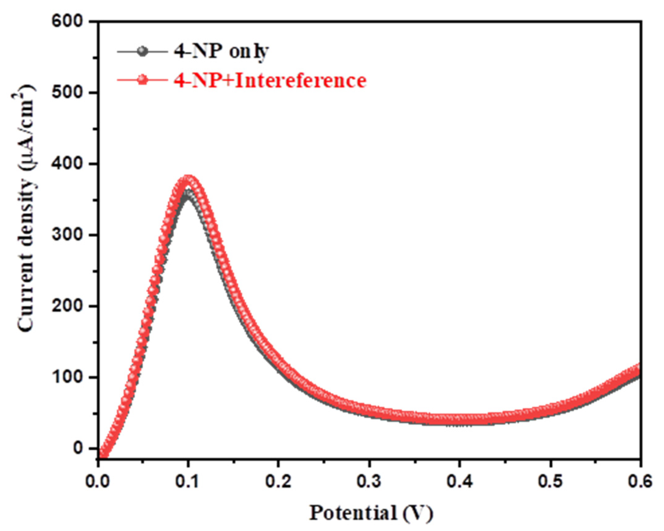
The selectivity of the sensor is an important consideration when evaluating how well an active material can detect toxic materials. In this context, the MnO2/rGO/GCE selectivity test toward the sensing of 4-NP in the presence of several interfering species was conducted using the LSV approach. Figure 8 shows the LSV graphs of the MnO2/rGO/GCE recorded at a scan rate of 50 mV/s in the presence of 50 µM 4-NP and 50 µM 4-NP + 200 µM interfering species (urea, uric acid, glucose, dopamine, catechol, ascorbic acid, hydroquinone, hydrazine, and H2O2).
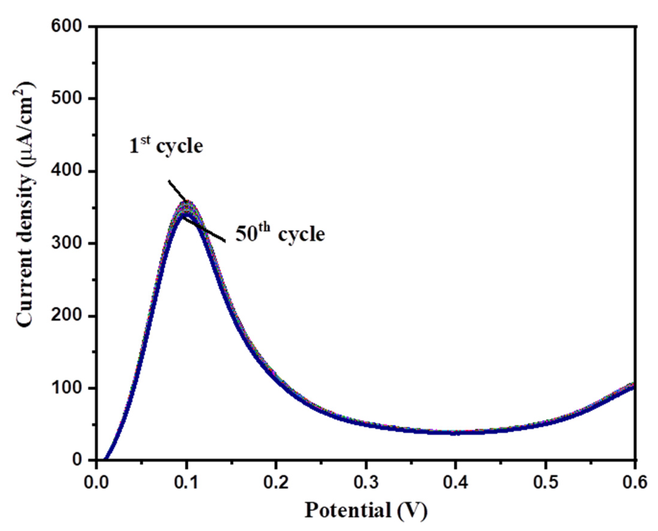
The LSV results showed that the presence of interfering species could not alter the current response or potential, which suggested the good selective nature of MnO2/rGO/GCE for the sensing of 4-NP in the presence of various interfering species (Figure 8). The repeatability and stability of the MnO2/rGO/GCE were also evaluated using the LSV method. Fifty consecutive LSV graphs of the MnO2/rGO/GCE were recorded in the presence of 50 µM 4-NP at a scan rate of 50 mV/s. The obtained LSV graphs are presented in Figure 9.
The acquired findings demonstrated that little fluctuations in current responsiveness were seen, indicating strong cyclic stability and repeatability up to 50 cycles. Under comparable circumstances, the repeatability of the MnO2/rGO/GCE was also investigated. Four freshly prepared MnO2/rGO/GCEs were used to investigate the reproducibility studies. The observations showed decent results, which suggested good reproducibility.
The probable sensing mechanism for the detection of 4-NP using MnO2/rGO/GCE can be explained according to the reported literature [31]. In Scheme 2, the likely detection process for 4-NP is depicted. The oxidation and reduction reactions of the 4-NP are indicated in Scheme 2 by O1 and R1, R2. Firstly, reduction (R1) is responsible for the transformation of 4-NP into p-hydroxyl-amino-phenol (R1).
In a subsequent redox reaction, the p-hydroxyl-amino-phenol is oxidized (O1) and transformed into the p-nitroso-phenol, and reversible reduction (R2) completes the redox reaction (Scheme 2). The performance of MnO2/rGO/GCE was then evaluated in terms of stability, repeatability, and selectivity for sensing 4-NP. Additional calculations of crucial parameters, such as the sensitivity and the detection limit (LOD), were made in order to assess the performance, accuracy, and application of the results.
As shown below, the detection limit (LoD) and Sensitivity were determined using the modified electrode’s Equations (2) and (3).
LoD = 3.3 (σ/S)
Sensitivity = slope/area
(σ is the standard deviation or error; S is the slope, and the area of the electrode is 0.07 cm2).
The calculated LoD and sensitivity of the MnO2/rGO/GCE are summarized in Table 1.
In the past years, various 4-NP sensors have been reported. In this connection, Gao et al. [39] prepared MnO2 nanotubes and investigated the role of MnO2 nanotubes as an electrochemical sensor for the detection of 4-NP. The developed sensor showed an LoD of 0.1 mM. In another report, Zhang et al. [40] also developed a 4-NP sensor using Mg(Ni)FeO as electrode material which demonstrated LoD of 0.2 µM. A flower-shaped zinc oxide-based nanomaterial was also synthesized by Yu et al. [41] and fabricated a 4-NP sensor. This 4-NP sensor displayed an LoD of 13 µM. Chu et al. [42] also coated nano-gold particles on GCE for electrochemical sensing of 4-NP and reported an LoD of 8 µM. Yang et al. [43] also reported a good LoD of 0.4 µM for multi-walled carbon nanotube-modified GCEs. A subsequent study of silver particle-modified GCE showed an LoD of 0.5 µM [44]. In other reports, palladium-graphene composite/poly(N-isopropylacrylamide) based sensor exhibited good LoD of 0.1 µM [45] whereas SnO2@ZIF-8/gC3N4 nanohybrids based sensor showed LoD of 0.565 µM [46]. In another recent report, MWCNTs/MnO2-based sensors also showed LoDs of 0.64 µM [47]. The interesting LoD of 0.16 µM was also reported for the sensing of 4-NP using Ti3C2TX/GR/GCE [48]. P-doped Fe/Fe3O4@C based 4-NP sensor showed LoD of 0.462 µM [49] while Ti3C2Tx MXene-based sensor showed LoD of 0.11 µM [50]. Dighole et al. [51] also explored the potential role of bismuth oxide/MWCNTs as 4-NP sensing material and obtained a good LoD of 0.1 µM. Our obtained LoD is comparable with previous reports, as listed in Table 1. In the present work, the better electro-catalytic properties of MnO2 and the high surface area of rGO with good conductivity improved the detection of 4-NP.

| Material | LoD (µM) | Sensitivity (µA/µMcm2) | References |
|---|---|---|---|
| α-MnO2 nanotube/GCE | 100 | 0.19 | [39] |
| Mg(Ni)FeO/CPE | 0.2 | 0.81 | [40] |
| ZnO/GCE | 13 | 0.404 | [41] |
| Nanogold/GCE | 8 | - | [42] |
| MWCNT/GCE | 0.4 | - | [43] |
| Silver particles/GCE | 0.5 | - | [44] |
| palladium-graphene composite/poly(N-isopropylacrylamide) | 0.1 | - | [45] |
| SnO2@ZIF-8/gC3N4 nanohybrids | 0.565 | 2.63 | [46] |
| MWCNTs/MnO2 | 0.64 | 0.186 | [47] |
| Ti3C2TX/GR/GCE | 0.16 | - | [48] |
| P-doped Fe/Fe3O4@C | 0.462 | - | [49] |
| Ti3C2Tx MXene | 0.11 | 1.22 | [50] |
| Bi2O3@MWCNTs | 0.1 | - | [51] |
| MnO2/rGO/GCE | 0.09 | 0.657 | Praveen et al. |
4. Conclusions
It can be summarized that MnO2/rGO composite has been obtained using a hydrothermal approach. Further, the physiochemical properties of the prepared MnO2/rGO composite were checked by various advanced techniques, including scanning electron microscopy and X-ray diffraction techniques. The para-nitrophenol (4-NP) sensor was constructed by modifying the surface of the glassy carbon electrode with the prepared MnO2/rGO composite as an electrode modifier. Due to the beneficial interactions (synergistic) between MnO2 and rGO, the fabricated 4-NP sensor had an excellent detection limit. The 4-NP sensor that was built demonstrated high stability and selectivity when it came to sensing 4-NP.
Supplementary Materials
The following supporting information can be downloaded at: https://www.mdpi.com/article/10.3390/inorganics10120219/s1, Figure S1: XRD of rGO; Figure S2: Raman of MnO2, rGO and MnO2/rGO; Figure S3: TEM of MnO2/rGO; Figure S4: CV responses; Figure S5: LSV responses; Figure S6: LSV responses.
Author Contributions
Conceptualization, P.K. and K.A.; methodology, K.A. and M.Q.K.; formal analysis, P.K., K.A. and M.Q.K.; investigation, P.K. and K.A.; resources, R.A.K. and H.K.; writing—original draft preparation, P.K., M.Q.K. and K.A.; writing—review and editing, R.A.K. and H.K.; supervision, R.A.K. and H.K.; funding acquisition, R.A.K. and H.K. All authors have read and agreed to the published version of the manuscript.
Funding
This research received no external funding.
Data Availability Statement
Not applicable.
Acknowledgments
R.A.K. acknowledged researchers supporting project number (RSP-2021/400), King Saud University, Riyadh, Saudi Arabia. P.K. thanks DST-Inspire, for the PhD fellowship. This work was supported by the Korea Innovation Foundation (INNOPOLIS) grant funded by the Korean government (MSIT; 2020-DD-UP-0278) and the National Research Foundation of Korea (NRF) grant funded by the Korean government (MSIT; No. 2019R1A5A8080290).
Conflicts of Interest
The authors declare no conflict of interest.
References
- Singh, K.; Ibrahim, A.A.; Umar, A.; Kumar, A.; Chaudhary, G.R.; Singh, S.; Mehta, S.K. Synthesis of CeO2–ZnO Nanoellipsoids as Potential Scaffold for the Efficient Detection of 4-Nitrophenol. Sens. Actuators B Chem. 2014, 202, 1044–1050. [Google Scholar] [CrossRef]
- Ahmad, N.; Al-Fatesh, A.S.; Wahab, R.; Alam, M.; Fakeeha, A.H. Synthesis of Silver Nanoparticles Decorated on Reduced Graphene Oxide Nanosheets and Their Electrochemical Sensing towards Hazardous 4-Nitrophenol. J. Mater. Sci. Mater. Electron. 2020, 31, 11927–11937. [Google Scholar] [CrossRef]
- Al-Kahtani, A.A.; Almuqati, T.; Alhokbany, N.; Ahamad, T.; Naushad, M.; Alshehri, S.M. A Clean Approach for the Reduction of Hazardous 4-Nitrophenol Using Gold Nanoparticles Decorated Multiwalled Carbon Nanotubes. J. Clean. Prod. 2018, 191, 429–435. [Google Scholar] [CrossRef]
- Dai, H.; Deng, Z.; Zeng, Y.; Zhang, J.; Yang, Y.; Ma, Q.; Hu, W.; Guo, L.; Li, L.; Wan, S.; et al. Highly Sensitive Determination of 4-Nitrophenol with Coumarin-Based Fluorescent Molecularly Imprinted Poly (Ionic Liquid). J. Hazard. Mater. 2020, 398, 122854. [Google Scholar] [CrossRef]
- Ding, Q.; Kang, Z.; Cao, L.; Lin, M.; Lin, H.; Yang, D.-P. Conversion of Waste Eggshell into Difunctional Au/CaCO3 Nanocomposite for 4-Nitrophenol Electrochemical Detection and Catalytic Reduction. Appl. Surf. Sci. 2020, 510, 145526. [Google Scholar] [CrossRef]
- Jeyapragasam, T. Molybdenum Disulfide-Based Modifier for Electrochemical Detection of 4-Nitrophenol. Ionics 2018, 24, 4033–4041. [Google Scholar] [CrossRef]
- Lu, W.; Ning, R.; Qin, X.; Zhang, Y.; Chang, G.; Liu, S.; Luo, Y.; Sun, X. Synthesis of Au Nanoparticles Decorated Graphene Oxide Nanosheets: Noncovalent Functionalization by TWEEN 20 in Situ Reduction of Aqueous Chloroaurate Ions for Hydrazine Detection and Catalytic Reduction of 4-Nitrophenol. J. Hazard. Mater. 2011, 197, 320–326. [Google Scholar] [CrossRef] [PubMed]
- Manjula, N.; Chen, S.-M. Simple Strategy Synthesis of Manganese Cobalt Oxide Anchored on Graphene Oxide Composite as an Efficient Electrocatalyst for Hazardous 4-Nitrophenol Detection in Toxic Tannery Waste. Microchem. J. 2021, 168, 106514. [Google Scholar] [CrossRef]
- Padilla-Sánchez, J.A.; Plaza-Bolaños, P.; Romero-González, R.; Garrido-Frenich, A.; Martínez Vidal, J.L. Application of a Quick, Easy, Cheap, Effective, Rugged and Safe-Based Method for the Simultaneous Extraction of Chlorophenols, Alkylphenols, Nitrophenols and Cresols in Agricultural Soils, Analyzed by Using Gas Chromatography–Triple Quadrupole-Mass Spectrometry/Mass Spectrometry. J. Chromatogr. A 2010, 1217, 5724–5731. [Google Scholar]
- El-Shahawi, M.S.; Othman, A.M.; El-Houseini, M.E.; Nashed, B.; Elsofy, M.S. Spectrofluorimetric Method for Measuring the Activity of the Enzyme α-l-Fucosidase Using the Ion Associate of 2-Chloro-4-Nitro Phenol–Rhodamine-B. Talanta 2009, 80, 19–23. [Google Scholar] [CrossRef]
- Salcedo, G.M.; Kupski, L.; Degang, L.; Marube, L.C.; Caldas, S.S.; Primel, E.G. Determination of Fifteen Phenols in Wastewater from Petroleum Refinery Samples Using a Dispersive Liquid—Liquid Microextraction and Liquid Chromatography with a Photodiode Array Detector. Microchem. J. 2019, 146, 722–728. [Google Scholar] [CrossRef]
- Bogireddy, N.K.R.; Cruz Silva, R.; Valenzuela, M.A.; Agarwal, V. 4-Nitrophenol Optical Sensing with N Doped Oxidized Carbon Dots. J. Hazard. Mater. 2020, 386, 121643. [Google Scholar] [CrossRef] [PubMed]
- Konstantinou, G.N. Enzyme-Linked Immunosorbent Assay (ELISA). In Food Allergens: Methods and Protocols; Lin, J., Alcocer, M., Eds.; Methods in Molecular Biology; Springer: New York, NY, USA, 2017; pp. 79–94. ISBN 978-1-4939-6925-8. [Google Scholar]
- Chu, Y.Y.; Qian, Y.; Wang, W.J.; Deng, X.L. A Dual-Cathode Electro-Fenton Oxidation Coupled with Anodic Oxidation System Used for 4-Nitrophenol Degradation. J. Hazard. Mater. 2012, 199–200, 179–185. [Google Scholar] [CrossRef] [PubMed]
- Zhang, H.; Zhang, W.; Zhang, Z.; Shi, Z.; Liu, W. Green analytical method for the sensitive determination of mononitrophenol isomers by dynamic pH junction cappilary electrophoresis. J. Liq. Chromatogr. Relat. 2014, 37, 1145–1162. [Google Scholar] [CrossRef]
- Hao, T.; Wei, X.; Nie, Y.; Xu, Y.; Yan, Y.; Zhou, Z. An Eco-Friendly Molecularly Imprinted Fluorescence Composite Material Based on Carbon Dots for Fluorescent Detection of 4-Nitrophenol. Microchim Acta 2016, 183, 2197–2203. [Google Scholar] [CrossRef]
- Neng, N.R.; Nogueira, J.M.F. Determination of Phenol Compounds In Surface Water Matrices by Bar Adsorptive Microextraction-High Performance Liquid Chromatography-Diode Array Detection. Molecules 2014, 19, 9369–9379. [Google Scholar] [CrossRef] [PubMed] [Green Version]
- Jiang, Y.; Zhao, H.; Liang, J.; Yue, L.; Li, T.; Luo, Y.; Liu, Q.; Lu, S.; Asiri, A.M.; Gong, Z.; et al. Anodic Oxidation for the Degradation of Organic Pollutants: Anode Materials, Operating Conditions and Mechanisms. A Mini Review. Electrochem. Commun. 2021, 123, 106912. [Google Scholar] [CrossRef]
- Chang, G.; Luo, Y.; Lu, W.; Qin, X.; Asiri, A.M.; Al-Youbi, A.O.; Sun, X. Ag Nanoparticles Decorated Polyaniline Nanofibers: Synthesis, Characterization, and Applications toward Catalytic Reduction of 4-Nitrophenol and Electrochemical Detection of H2O2 and Glucose. Catal. Sci. Technol. 2012, 2, 800–806. [Google Scholar] [CrossRef]
- Manjula, N.; Chen, S.-M. Electrochemical Sensors for β-Adrenoceptor Agonist Isoprenaline Analysis in Human Urine and Serum Samples Using Manganese Cobalt Oxide-Modified Glassy Carbon Electrode. New J. Chem. 2021, 45, 9084–9095. [Google Scholar] [CrossRef]
- Karimi-Maleh, H.; Karimi, F.; Fu, L.; Sanati, A.L.; Alizadeh, M.; Karaman, C.; Orooji, Y. Cyanazine Herbicide Monitoring as a Hazardous Substance by a DNA Nanostructure Biosensor. J. Hazard. Mater. 2022, 423, 127058. [Google Scholar] [CrossRef]
- Karimi-Maleh, H.; Khataee, A.; Karimi, F.; Baghayeri, M.; Fu, L.; Rouhi, J.; Karaman, C.; Karaman, O.; Boukherroub, R. A Green and Sensitive Guanine-Based DNA Biosensor for Idarubicin Anticancer Monitoring in Biological Samples: A Simple and Fast Strategy for Control of Health Quality in Chemotherapy Procedure Confirmed by Docking Investigation. Chemosphere 2022, 291, 132928. [Google Scholar] [CrossRef] [PubMed]
- Karimi-Maleh, H.; Orooji, Y.; Karimi, F.; Alizadeh, M.; Baghayeri, M.; Rouhi, J.; Tajik, S.; Beitollahi, H.; Agarwal, S.; Gupta, V.K.; et al. A Critical Review on the Use of Potentiometric Based Biosensors for Biomarkers Detection. Biosens. Bioelectron. 2021, 184, 113252. [Google Scholar] [CrossRef] [PubMed]
- Ahmad, K.; Shinde, M.A.; Kim, H. Molybdenum Disulfide/Reduced Graphene Oxide: Progress in Synthesis and Electro-Catalytic Properties for Electrochemical Sensing and Dye Sensitized Solar Cells. Microchem. J. 2021, 169, 106583. [Google Scholar] [CrossRef]
- Ahmad, K.; Kim, H. Fabrication of Nitrogen-Doped Reduced Graphene Oxide Modified Screen Printed Carbon Electrode (N-RGO/SPCE) as Hydrogen Peroxide Sensor. Nanomaterials 2022, 12, 2443. [Google Scholar] [CrossRef] [PubMed]
- Ahmad, K.; Kumar, P.; Mobin. Hydrothermally grown novel pyramids of the CaTiO3 perovskite as an efficient electrode modifier for sensing applications. Mater. Adv. 2020, 1, 2003–2009. [Google Scholar] [CrossRef]
- Mohammad, A.; Ahmad, K.; Rajak, R.; Mobin, S.M. Binder Free Modification of Glassy Carbon Electrode by Employing Reduced Graphene Oxide/ZnO Composite for Voltammetric Determination of Certain Nitroaromatics. Electroanalysis 2018, 30, 274–282. [Google Scholar] [CrossRef]
- Khan, M.Q.; Ahmad, K.; Alsalme, A.; Kim, H. Hydrothermal Synthesis of Nanostructured NiO for Hydrazine Sensing Application. Mater. Chem. Phys. 2022, 289, 126463. [Google Scholar] [CrossRef]
- Abaker, M.; Dar, G.N.; Umar, A.; Zaidi, S.A.; Ibrahim, A.A.; Baskoutas, S.; Al-Hajry, A. CuO Nanocubes Based Highly-Sensitive 4-Nitrophenol Chemical Sensor. Sci. Adv. Mater. 2012, 4, 893–900. [Google Scholar] [CrossRef]
- Ahmad, K.; Mobin, S.M. Shape Controlled Synthesis of High Surface Area MgO Microstructures for Highly Efficient Congo Red Dye Removal and Peroxide Sensor. J. Environ. Chem. Eng. 2019, 7, 103347. [Google Scholar] [CrossRef]
- Ahmad, K.; Mohammad, A.; Mathur, P.; Mobin, S.M. Preparation of SrTiO3 Perovskite Decorated RGO and Electrochemical Detection of Nitroaromatics. Electrochim. Acta 2016, 215, 435–446. [Google Scholar] [CrossRef]
- Ahmad, K.; Mohammad, A.; Mobin, S.M. Hydrothermally Grown α-MnO2 Nanorods as Highly Efficient Low Cost Counter-Electrode Material for Dye-Sensitized Solar Cells and Electrochemical Sensing Applications. Electrochim. Acta 2017, 252, 549–557. [Google Scholar] [CrossRef]
- Zhang, Y.; Huang, B.; Shao, Q.; Feng, Y.; Xiong, L.; Peng, Y.; Huang, X. Defect Engineering of Palladium–Tin Nanowires Enables Efficient Electrocatalysts for Fuel Cell Reactions. Nano Lett. 2019, 19, 6894–6903. [Google Scholar] [CrossRef] [PubMed]
- Cheemalapati, S.; Palanisamy, S.; Mani, V.; Chen, S.-M. Simultaneous Electrochemical Determination of Dopamine and Paracetamol on Multiwalled Carbon Nanotubes/Graphene Oxide Nanocomposite-Modified Glassy Carbon Electrode. Talanta 2013, 117, 297–304. [Google Scholar] [CrossRef] [PubMed]
- Jaiswal, N.; Tiwari, I.; Foster, C.W.; Banks, C.E. Highly Sensitive Amperometric Sensing of Nitrite Utilizing Bulk-Modified MnO2 Decorated Graphene Oxide Nanocomposite Screen-Printed Electrodes. Electrochim. Acta 2017, 227, 255–266. [Google Scholar] [CrossRef]
- Ahmad, K.; Mobin, S.M. Construction of polyanilne/ITO electrode for electrochemical sensor applications. Mater. Res. Express 2019, 6, 085508. [Google Scholar] [CrossRef]
- Ahmad, K.; Mohammad, A.; Ansari, S.N.; Mobin, S.M. Construction of graphene oxide sheets based modified glassy carbon electrode (GO/GCE) for the highly sensitive detection of nitrobenzene. Mater. Res. Express 2018, 5, 075601. [Google Scholar] [CrossRef]
- Ahmad, K.; Kumar, P.; Mobin, S.M. A Highly Sensitive and Selective Hydroquinone Sensor Based on a Newly Designed N-RGO/SrZrO3 Composite. Nanoscale Adv. 2020, 2, 502–511. [Google Scholar] [CrossRef] [PubMed] [Green Version]
- Wu, J.; Wang, Q.; Umar, A.; Sun, S.; Huang, L.; Wang, J.; Gao, Y. Highly Sensitive p -Nitrophenol Chemical Sensor Based on Crystalline α-MnO2 Nanotubes. New J. Chem. 2014, 38, 4420–4426. [Google Scholar] [CrossRef]
- Xu, Y.; Wang, Y.; Ding, Y.; Luo, L.; Liu, X.; Zhang, Y. Determination of P-Nitrophenol on Carbon Paste Electrode Modified with a Nanoscaled Compound Oxide Mg(Ni)FeO. J. Appl. Electrochem. 2013, 43, 679–687. [Google Scholar] [CrossRef]
- Sinhamahapatra, A.; Bhattacharjya, D.; Yu, J.-S. Green Fabrication of 3-Dimensional Flower-Shaped Zinc Glycerolate and ZnO Microstructures for p -Nitrophenol Sensing. RSC Adv. 2015, 5, 37721–37728. [Google Scholar] [CrossRef]
- Chu, L.; Han, L.; Zhang, X. Electrochemical Simultaneous Determination of Nitrophenol Isomers at Nano-Gold Modified Glassy Carbon Electrode. J. Appl. Electrochem. 2011, 41, 687–694. [Google Scholar] [CrossRef]
- Yang, C. Electrochemical Determination of 4-Nitrophenol Using a Single-Wall Carbon Nanotube Film-Coated Glassy Carbon Electrode. Microchim. Acta 2004, 148, 87–92. [Google Scholar] [CrossRef]
- Casella, I.G.; Contursi, M. The Electrochemical Reduction of Nitrophenols on Silver Globular Particles Electrodeposited under Pulsed Potential Conditions. J. Electrochem. Soc. 2007, 154, D697. [Google Scholar] [CrossRef]
- Dan, X.; Ruiyi, L.; Qinsheng, W.; Yongqiang, T.; Guangli, W.; Zaijun, L. Thermal-switchable sensor based on palladium-graphene composite and poly(N-isopropylacrylamide) for electrochemical detection of 4-nitrophenol. Microchem. J 2022, 172, 106970. [Google Scholar] [CrossRef]
- Mohanta, D.; Mahanta, A.; Mishra, S.R.; Jasimuddin, S.; Ahmaruzzaman, M. Novel SnO2@ZIF-8/gC3N4 nanohybrids for excellent electrochemical performance towards sensing of p-nitrophenol. Environ. Res. 2021, 197, 111077. [Google Scholar] [CrossRef]
- Anbumannan, V.; Dinesh, M.; Kumar, R.T.R.; Suresh, K. Hierarchical α-MnO2 wrapped MWCNTs sensor for low level detection of p-nitrophenol in water. Ceram. Int. 2019, 45, 23097–23103. [Google Scholar] [CrossRef]
- Wang, X.; Li, M.; Yang, S.; Bai, X.; Shan, J. Self-assembled Ti3C2TX MXene/graphene composite for the electrochemical reduction and detection of p-nitrophenol. Microchem. J. 2022, 179, 107473. [Google Scholar] [CrossRef]
- Ren, D.; Wang, X.; Leng, C.; Meng, W.; Zhang, J.; Han, C. A Highly Sensitive Electrochemical Sensing Platform Based on P-doped Fe/Fe3O4@C for the Detection of 4-Nitrophenol. J. Electrochem. Soc. 2022, 169, 097501. [Google Scholar] [CrossRef]
- Lei, L.; Li, C.; Huang, W.; Wu, K. Simultaneous detection of 4-chlorophenol and 4-nitrophenol using a Ti3C2Tx MXene based electrochemical sensor. Analyst 2021, 146, 7593–7600. [Google Scholar] [CrossRef]
- Dighole, R.P.; Munde, A.V.; Mulik, B.B.; Sathe, B.R. Bi2O3 Nanoparticles Decorated Carbon Nanotube: An Effective Nanoelectrode for Enhanced Electrocatalytic 4-Nitrophenol Reduction. Front. Chem. 2020, 8, 325. [Google Scholar] [CrossRef]
Figure 1.
XRD pattern of MnO2 and MnO2/rGO.

Figure 2.
SEM images of MnO2 (a) and MnO2/rGO (b).

Figure 3.
EDX analysis of MnO2 (a,b) and MnO2/rGO (c,d).

Figure 4.
CV of GCE, MnO2/GCE, rGO/GCE and MnO2/rGO/GCE in the presence (a) and absence (b) of 50 µM 4-NP (scan rate = 50 mV/s).
Figure 4.
CV of GCE, MnO2/GCE, rGO/GCE and MnO2/rGO/GCE in the presence (a) and absence (b) of 50 µM 4-NP (scan rate = 50 mV/s).

Figure 5.
CV (a) of MnO2/rGO/GCE in 50 µM 4-NP (PBS = 0.1 M; pH = 7.0) at various scan rates (50–500 mV/s). Calibration plot (b) between the peak current and square root of various scan rates.
Figure 5.
CV (a) of MnO2/rGO/GCE in 50 µM 4-NP (PBS = 0.1 M; pH = 7.0) at various scan rates (50–500 mV/s). Calibration plot (b) between the peak current and square root of various scan rates.

Figure 6.
LSV of GCE, MnO2/GCE, rGO/GCE and MnO2/rGO/GCE in 50 µM 4-NP (PBS = 0.1 M; pH = 7.0; scan rate = 50 mV/s).
Figure 6.
LSV of GCE, MnO2/GCE, rGO/GCE and MnO2/rGO/GCE in 50 µM 4-NP (PBS = 0.1 M; pH = 7.0; scan rate = 50 mV/s).

Figure 7.
LSV (a) of MnO2/rGO/GCE in various concentrations (0, 0.3 µM, 2.5 µM, 6.5 µM, 11.5 µM, 17.5 µM, 25.5 µM, 32.5 µM, 41.5 µM and 50 µM) of 4-NP (scan rate = 50 mV/s). The calibration curve (b) of the peak current density versus the concentration of 4-NP.
Figure 7.
LSV (a) of MnO2/rGO/GCE in various concentrations (0, 0.3 µM, 2.5 µM, 6.5 µM, 11.5 µM, 17.5 µM, 25.5 µM, 32.5 µM, 41.5 µM and 50 µM) of 4-NP (scan rate = 50 mV/s). The calibration curve (b) of the peak current density versus the concentration of 4-NP.

Figure 8.
LSV of MnO2/rGO/GCE in 50 µM 4-NP and 50 µM 4-NP + 200 µM interference species (urea, uric acid, glucose, dopamine, catechol, ascorbic acid, hydroquinone, hydrazine, and H2O2) (scan rate = 50 mV/s).
Figure 8.
LSV of MnO2/rGO/GCE in 50 µM 4-NP and 50 µM 4-NP + 200 µM interference species (urea, uric acid, glucose, dopamine, catechol, ascorbic acid, hydroquinone, hydrazine, and H2O2) (scan rate = 50 mV/s).

Figure 9.
Fifty consecutive LSV curves of MnO2/rGO/GCE in 50 µM 4-NP (scan rate = 50 mV/s).

Scheme 1.
Schematic diagram for the synthesis of MnO2/rGO composite.

Scheme 2.
The schematic diagram shows the construction and working of the 4-NP sensor.

Publisher’s Note: MDPI stays neutral with regard to jurisdictional claims in published maps and institutional affiliations. |
© 2022 by the authors. Licensee MDPI, Basel, Switzerland. This article is an open access article distributed under the terms and conditions of the Creative Commons Attribution (CC BY) license (https://creativecommons.org/licenses/by/4.0/).
Share and Cite
MDPI and ACS Style
Kumar, P.; Khan, M.Q.; Khan, R.A.; Ahmad, K.; Kim, H. Hydrothermal Synthesis of MnO2/Reduced Graphene Oxide Composite for 4-Nitrophenol Sensing Applications. Inorganics 2022, 10, 219. https://doi.org/10.3390/inorganics10120219
AMA Style
Kumar P, Khan MQ, Khan RA, Ahmad K, Kim H. Hydrothermal Synthesis of MnO2/Reduced Graphene Oxide Composite for 4-Nitrophenol Sensing Applications. Inorganics. 2022; 10(12):219. https://doi.org/10.3390/inorganics10120219
Chicago/Turabian StyleKumar, Praveen, Mohd Quasim Khan, Rais Ahmad Khan, Khursheed Ahmad, and Haekyoung Kim. 2022. "Hydrothermal Synthesis of MnO2/Reduced Graphene Oxide Composite for 4-Nitrophenol Sensing Applications" Inorganics 10, no. 12: 219. https://doi.org/10.3390/inorganics10120219
Note that from the first issue of 2016, this journal uses article numbers instead of page numbers. See further details here.