Article

Salicylate UV-Filters in Sunscreen Formulations Compromise the Preservative System Efficacy against Pseudomonas aeruginosa and Burkholderia cepacia

Innovation department, Sharon Laboratories Ltd., Odem St. Industrial zone Ad-Halom, Ashdod 7898800, Israel
*
Author to whom correspondence should be addressed.
Cosmetics 2020, 7(3), 63; https://doi.org/10.3390/cosmetics7030063
Submission received: 15 July 2020 / Revised: 31 July 2020 / Accepted: 1 August 2020 / Published: 3 August 2020
(This article belongs to the Special Issue Feature Papers in Cosmetics in 2020)

Abstract

:
Contamination of personal-care products are a serious health concern and therefore, preservative solutions are necessary for the costumers’ safety. High sun protection factor (SPF) sunscreen formulations are known to be difficult to preserve, due to their high ratio of organic phase containing the UV-filters. Salicylate esters such as octyl salicylate (OS) and homosalate (HS) are among the most common UV-filters currently used in the market, and can undergo hydrolysis by esterase molecules produced by contaminant microorganisms. The hydrolysis product, salicylic acid (SA) can be assimilated by certain bacteria that contain the chorismate pathway, in which its final product is pyochelin, an iron-chelating siderophore. Here, we show that OS and HS can compromise the preservative efficacy against two pathogenic important bacteria, Pseudomonas aeruginosa and Burkholderia cepacia. Challenge tests of formulations containing the UV-filters demonstrated that only bacteria with the chorismate pathway failed to be eradicated by the preservation system. mRNA expression levels of the bacterial pchD gene, which metabolizes SA to produce pyochelin, indicate a significant increase that was in correlation with increasing concentrations of both OS and HS. These data suggest that certain UV-filters can provide a source for bacterial resistance against common preservatives in sunscreen formulations.

1. Introduction

Preservation of non-sterile personal-care products containing water and organic/inorganic compounds is necessary in order to prevent spoilage by contaminants microorganisms, which subsequently can cause severe health risk for the consumers [1]. High sun protection factor (SPF) sunscreen formulations in particular are known to be difficult to preserve, due to the presence of hydrophobic UV-filters in high concentration, thus enhancing the size of their oil phases. As most commercially available preservatives display poor water solubility themselves, they will tend to migrate towards the oily phase, rendering the water phase where the bacteria are present, unprotected.
Salicylate esters were the first UV-filters used in sunscreen formulations and yet remain highly popular in the market [2]. Such esters as octyl salicylate (OS) and homosalate (HS) can be hydrolyzed to salicylic acid (SA) by esterases molecules, possibly produced by the contaminant microorganisms [3,4]. Some bacterial species such as Pseudomonas aeruginosa and Burkholderia cepacia contains genes for the chorismate biochemical pathway, in which SA is a precursor to produce pyochelin (Figure 1). As part of the pathway, SA can be adenylated at the carboxyl group by PchD (2,3-dihydroxybenzoyl adenylate synthase), which is encoded by the pchD gene, found in both above mentioned bacterial species [5,6,7].
Growing evidences for microbial tolerance and resistance towards common preservatives and disinfectants are accumulating in the past two decades, specifically in regard to Pseudomonas and Burkholderia spp., and represent a major health concern [8,9,10,11,12]. Nosocomial outbreaks of contaminated personal-care products have been repeatedly reported in hospitals [13,14,15,16]. A recent report exposed that 11.76% of the products were recalled because of a contamination with a potentially pathogenic microorganism, while P. aeruginosa was the most frequently found microorganism (35%) [17].
In addition to the classical scheme representing the inherent physico-chemical reasons for which high SPF sunscreens are difficult to preserve, we now would like to report an additional difficulty of finding its source in a specific microbiological phenomenon and shedding a new light on sunscreen preservation.

2. Materials and Methods

2.1. Microorganisms Strains and Growth Conditions

Escherichia coli (ATCC 8739), Staphylococcus aureus (ATCC 6538), P. aeruginosa (ATCC 9027), B. cepacia (ATCC 25416), Candida albicans (ATCC 10231), and Aspergillus brasiliensis (ATCC 16404) were obtained from ATCC and cultured according to the manufacturer’s instructions. These microorganisms are routinely used for minimum inhibitory concentration (MIC) and challenge tests according to the US and EU recommendations for the efficacy of preservatives in the cosmetic industry. Bacterial strains were maintained in tryptic soy agar (TSA) (Hy-labs, Rehovot, Israel), while yeast and mold strains were maintained in sabouraud dextrose agar (SDA) (Hy-labs, Israel), supplemented with oxytetracycline at a concentration of 1% (Hy-labs, Israel). Preservative efficacy assays (challenge tests) were performed in order to enumerate the number of microorganisms present in a formulation containing the preservative system, while the diluent solution was supplemented with neutralizers containing 4% Tween 20 and 0.5% lecithin (Hy-labs, Israel).

2.2. Preservative System and Formulations

For the challenge tests, creams having SPF values of 30 and 45 and basic cream with or without OS were used and the constitution of compounds are listed in Table 1. A typical representative preservative system, Sharomix™ Amplify AM-25 was used in this study at a final concentration of 0.5%. The preservative blend contained 80% Phenoxyethanol, 5% Didecyldimethylammonium chloride, 10% Chlorophenesin, and 5% Caprylyl glycol.

2.3. Antimicrobial and Antifungal Activity of the Preservative
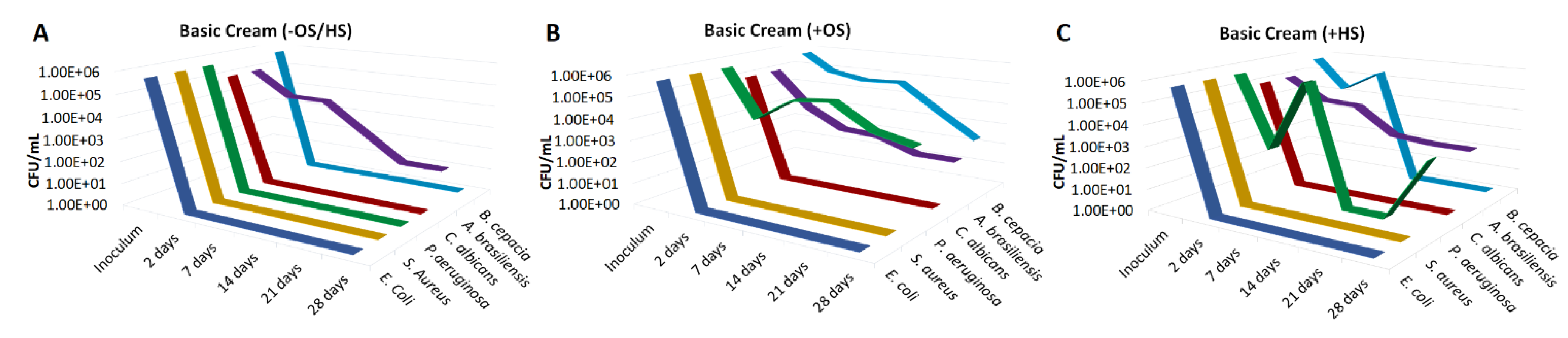
The minimum inhibitory concentration (MIC) of the preservative system, Sharomix™ Amplify AM-25, was evaluated against the five pharmacopeia strains. Briefly, growth of E. coli, S. aureus, P. aeruginosa, C. albicans, and A. brasiliensis was observed during incubation with different concentrations of AM-25, in a 10 mL tryptic soy broth (TSB) growth medium. Increasing concentrations of AM-25 preservative were dissolved in TSB and inoculated with the test culture to a final concentration of 106 CFU/mL of bacteria and 105 CFU/mL of yeast and mold. Bacteria inoculated test vials were incubated overnight (ON) at 32 °C with shaking, while yeast and mold inoculated vials were incubated at 23 °C for three days with shaking. The microorganism growth was evaluated by eye, while vials with clear media (where no growth was detected) were used for determining the minimum biocidal concentration (MBC) via seeding on TSA plates.

2.4. Challenge Test

Challenge tests of preservative efficacy in formulations were performed according to the ISO 11,930 regulations. The challenge test method with preservative Sharomix™ Amplify AM-25 was validated and confirmed for its accuracy. The total count for any prior contamination was performed for all formulations. Specifically, in order to evaluate the antimicrobial and antifungal activity of the preservative system in a basic cream, as well as in sunscreens with a SPF 30 and 45 containing OS as a UV-B filter, samples were inoculated separately with each microorganism at a final concentration of 106 cfu/mL for bacteria and 105 cfu/mL for yeast and mold. Samples were incubated in the dark at 22 °C for 28 days. The preservative efficacy was determined by sampling 1 g from the formulation at each time-point of 2, 7, 14, 21, and 28 days. To enumerate the microorganisms at each time point, serial dilutions were made up to 10−4, and 1 mL were seeded in duplicates onto a petri dish with the appropriate media TSA/SDA (bacteria vs. yeast and mold, respectively), using the pour plate method. Plates were incubated at 32 °C for three days for bacteria while yeast and mold were incubated at 22 °C for five days until the enumeration of viable microorganisms.

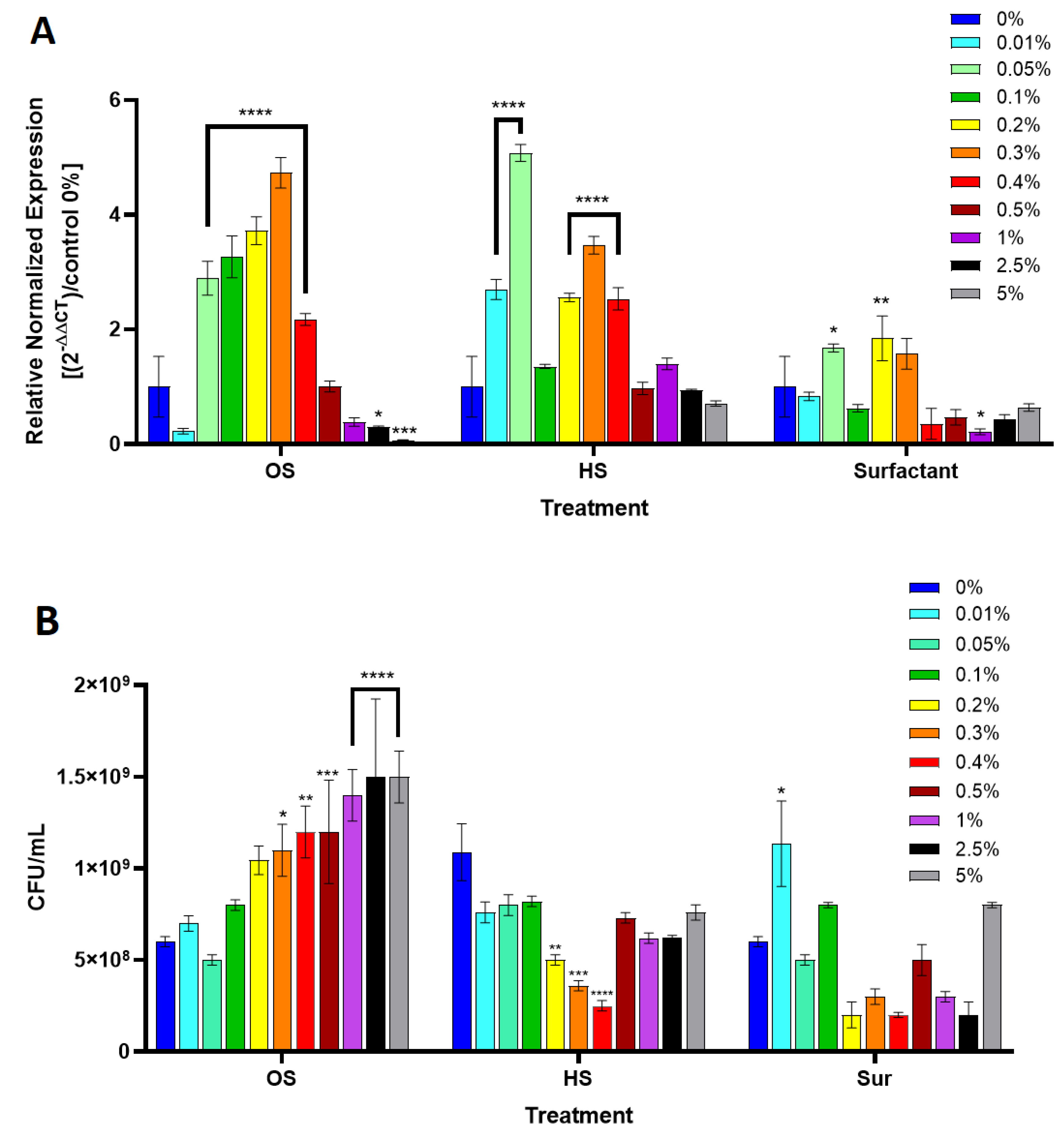
2.5. P. aeruginosa and B. cepacia Growth Culture in the Presence of Octyl Salicylate

Increasing concentrations of OS, 0.01, 0.05, 0.1, 0.2, 0.3, 0.4, 0.5, 1, 2.5, 5%, were added to 10 mL of TSB media. In order to dissolve the OS in the TSB medium, Tween 20 and Span 20 surfactants each at a concentration of 1.5% were used (Table 2) and the stability of the formulation was evaluated using centrifugation and cooling ON at 4 °C. In order to measure the effect of the surfactants alone, a “Surfactant” control was placed using the same concentrations as in the OS/HS solutions. Test vials with OS or HS and surfactant were inoculated separately with P. aeruginosa and B. cepacia at a final concentration of 106 CFU/mL and were incubated ON at 32 °C with shaking. The following day, 1 mL from each sample was used for RNA extraction, and 1 mL was used to measure bacterial viability by seeding on a TSA plate.

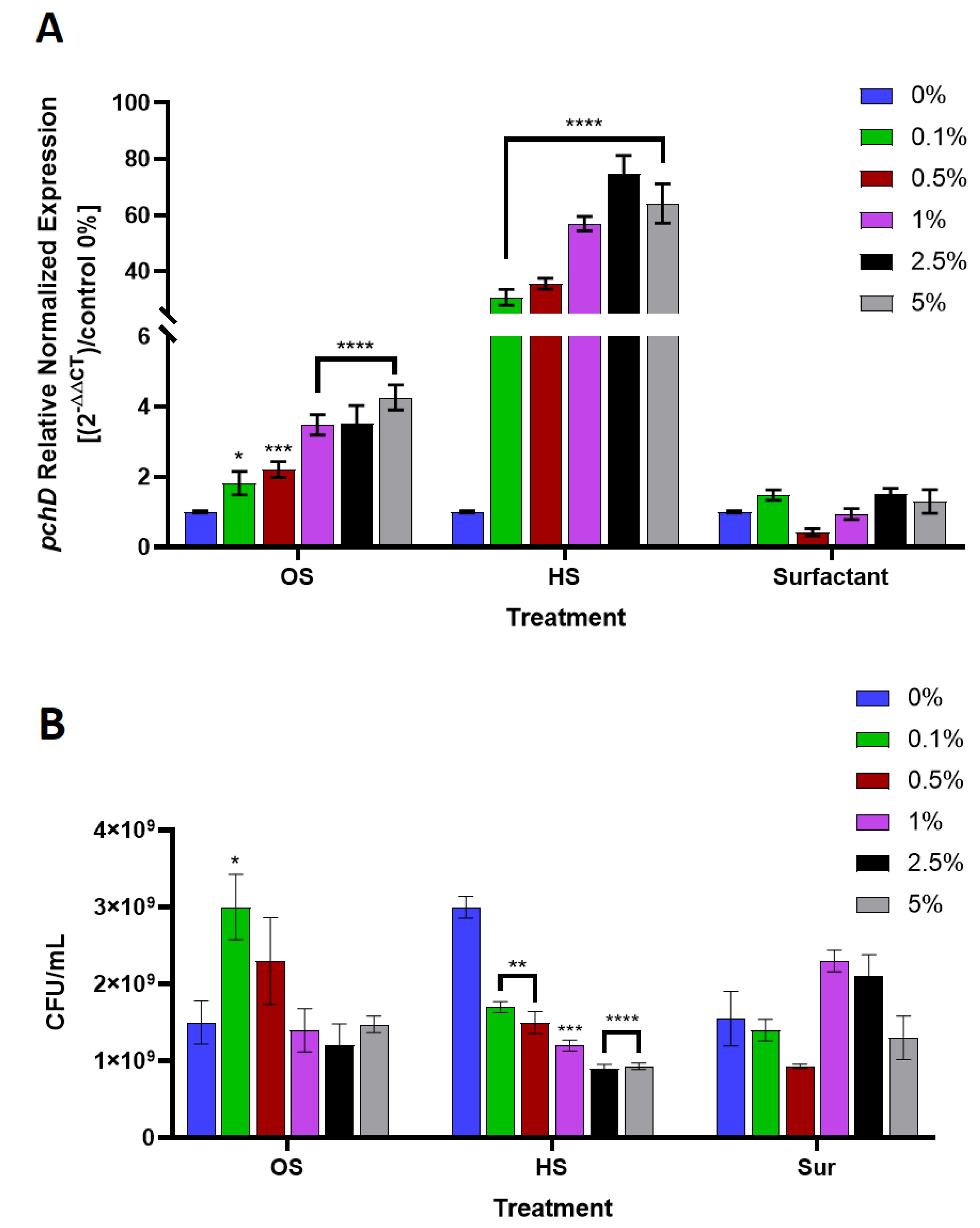
2.6. Quantitative Polymerase Chain Reaction for pchD Gene Expression

RNA was extracted from P. aeruginosa and B. cepacia samples incubated with OS, HS, or the Surfactant control using GeneJet RNA extraction kit (Thermo Fisher, Modi’in, Israel), according to the manufacturer’s instructions. DNase I (Thermo Fisher, Modi’in, Israel) was used to deplete any DNA residues from the sample. RNA concentrations and purity were determined using the Take3 instrument (BioTek, Petah Tikva, Israel). 1 μg of total RNA was reverse transcribed using the High Capacity cDNA Reverse Transcription kit (Thermo Fisher, Modi’in Israel), in accordance with the manufacturer’s instructions. Primers for P. aeruginosa and B. cepacia housekeeping genes (rpoS and recA, respectively) and the target genes, pchD (Hy-labs, Rehovot, Israel) were used and listed in Table 3. Primers efficiency was determined via standard curves quantification using serial dilutions (not shown). qPCR reactions were performed in triplicates using platinum SYBR Green qPCR superMIX-UDG (Bio-Rad, Haifa, Israel) with the CFX 96 instrument (Bio-Rad, Haifa, Israel). Cycling conditions were 95 °C for 10 min, then 35 cycles of 95 °C for 10 s, 60 °C for 30 s, and a final elongation step at 72 °C for 15 s. Results of Cq from target gene pchD were normalized against the mRNA expression of the reference gene rpoS and recA in each cDNA sample. Data are presented as normalized values of 2−ΔΔCT, relative to the control treatment (0%, without OS/HS/Surfactant).

2.7. Statistical Analysis

Data from the qPCR gene expression results statistically analyzed using Prism GraphPad v.8 (GraphPad Software, 2020) and significance was determined using the two-way ANOVA test. Data are presented as mean values ± SD. As for the challenge tests, the method and protocol were validated, while the statistical SD was previously determined at ±½Log CFU/mL. For the enumeration of the microorganisms’ count, cultures were seeded in duplicates using the pour plate method and the results are presented as an average. All tests were repeated twice separately in order to confirm these data.

3. Results

In order to evaluate the efficacy of the preservative system, Sharomix™ Amplify AM-25, in sunscreen formulations having SPF values of 30 and 45, a challenge test was performed (Figure 2A,B). While the preservative efficacy test results from E. coli, S. aureus, C. albicans, and A. brasiliensis, passed the European Pharmacopoeia (EP) acceptance criteria, P. aeruginosa did not. In the challenge test with SPF 30, there was an immediate reduction of P. aeruginosa growth after two days and reemergence at seven days, which continued to persist throughout the rest of the 28 days experiment (Figure 2A). In the case of SPF 45, P. aeruginosa CFU/mL were high throughout the entire experiment (Figure 2B). The Sharomix™ Amplify AM-25 was selected as a standard preservative system and was used during all the challenge tests. It was previously tested to be efficacious against all tested microorganisms via MIC tests, with marked low concentrations. In three bacterial strains E. coli, S. aureus, and P. aeruginosa, the MIC values were <500 ppm, while in yeast and mold MIC values were <300 ppm.
To test the hypothesis that salicylate UV-filters may assist P. aeruginosa to recover from the inhibitory effect of the preservative, three challenge tests with the same basic cream formulation (Table 1) were performed using the AM-25 preservative system. The first formulation did not contain any salicylate UV-filters (Figure 3A), while the second and third ones were, supplemented with 5% of either OS or HS, respectively (Figure 3B,C). The preservative system in the basic cream formulation without UV-filters was efficacious to eradicate both P. aeruginosa and B. cepacia and passed the EP acceptance criteria, along with all other pharmacopeia strains tested (Figure 3A). However, when the formulation was supplemented with OS and HS, the preservative failed to inhibit P. aeruginosa and B. cepacia growth (Figure 3B,C).
To understand the role of OS and HS in the recovery of P. aeruginosa and B. cepacia during the challenge tests, a biochemical pathway search was performed using KEGG (https://www.genome.jp/kegg/). P. aeruginosa and B. cepacia were found to contain the genes for the chorismate biochemical pathway, in which SA is a precursor for the biosynthesis of pyochelin (Figure 1). We then hypothesized that salicylate UV-filters in the sunscreen formulation are going through hydrolysis to produce SA, which is further incorporated in the metabolic cycle of P. aeruginosa and B. cepacia. The chorismate biochemical pathway was confirmed to be absent from the other pharmacopeia strains used in our experiments, E. coli, S. aureus, C. albicans, and A. brasiliensis.
In order to investigate the effect of OS and HS on P. aeruginosa and B. cepacia chorismate cycle and specifically, the enzymatic reaction that metabolizes SA, mRNA expression levels of pchD gene were analyzed (Figure 4 and Figure 5). Both bacteria were incubated overnight in the TSB growth medium, supplemented with increasing concentrations of either OS or HS. Since OS and HS are hydrophobic, a surfactant was used to dissolve these compounds in the growth medium. As a control, bacteria were incubated with increasing concentrations of the surfactant alone, to account for its effect on the gene expression and growth. In both bacteria, gene expression results indicated a concentration dependent increase and upregulation of the pchD gene in correlation with OS and HS increasing concentrations. For P. aeruginosa, this increase in expression level was significantly higher in comparison to the control (0%), with a maximum expression at 0.3% OS (p-value < 0.0001). Following the highest gene expression peak there was a gradual decrease until reaching 5% OS with significantly low expression levels in comparison to the control (Figure 4A; OS) (p-value < 0.0008). As for the HS treatment, P. aeruginosa pchD expression levels increased significantly at 0.01% and 0.05% (p-value < 0.0001), then a slight decrease at a concentration of 0.1% HS. Expression levels increased again gradually until reaching a secondary peak at 0.3% (Figure 4A; HS). After that expression levels decreased gradually in a similar pattern as in the OS treatment. Furthermore, the surfactant treatment did not affect the pchD gene expression and there was no concentration dependency trend correlating with the increasing concentrations of the Surfactant (Figure 4A).
P. aeruginosa viability in the presence of increasing concentrations of OS and HS was monitored in parallel to its pchD expression levels and indicated a concentration dependent increase in viability (Figure 4B), reaching its maximum growth in the presence of 5% OS. The growth pattern of P. aeruginosa incubated in the presence of HS did not show a correlation with HS concentrations. In addition, average CFU/mL of P. aeruginosa treated with HS was lower than in the OS treatment. The Surfactant control treatment did not have a noticeable effect on P. aeruginosa growth pattern and did not show a concentration dependency (Figure 4B; Surfactant).
A similar experimental procedure was performed using B. cepacia (Figure 5), which contains the same chorismate pathway including pchD gene that metabolizes SA. B. cepacia showed a gradual increase in pchD gene expression, consistent with the increasing concentrations of both OS and HS, which was significantly higher compared with the no-treatment control (p-value < 0.0001). The HS treatment caused an upregulation of pchD gene expression in B. cepacia that was a Log larger than in the OS treatment (Figure 5A). This could be explained by the differences in the molecular structure of the two UV-filters, which may exhibit different affinity towards the esterase molecules that hydrolyze them to SA. The rate in which SA is formed could directly influence the pchD expression levels. The pchD expression levels during the Surfactant control treatment at all concentrations were similar to the no-treatment control at 0% OS/HS.
The growth pattern of B. cepacia in the presence of increasing concentrations of OS and HS showed an opposite response to P. aeruginosa growth. As the concentrations of OS and HS increased, CFU/mL of B. cepacia decreased (Figure 5B). This pattern was specifically accurate in the HS treatment at all concentrations.

4. Discussion

The intrinsic physico-chemical properties of sunscreens are known to be a major contributing factor to the difficulty in preservation of such formulations. While a preservative system containing more hydrophilic compounds may overcome these difficulties, additional microbiological factors can play an important role under certain circumstances. Here, we demonstrate that some bacterial species could survive in preserved sunscreen formulations thanks to specific components commonly found in sunscreens.
In this study, challenge tests of sunscreen formulations having SPF of 30 and 45 showed that only P. aeruginosa was able to survive in the presence of a preservative, while in the basic cream formulation, the same preservative was efficacious to eradicate both B. cepacia and P. aeruginosa growth quickly after two days of sampling time-point. These results suggested that certain components in the sunscreen formulations can either interact with the preservative system or directly promote the growth of P. aeruginosa. Specifically, when OS and HS were added to the basic cream formulation, the preservative which was previously effective against P. aeruginosa and B. cepacia, failed to eradicate their growth. All other microorganisms in the basic cream formulation supplemented with OS and HS passed the EP criteria, indicating a caveat related to these two bacterial species.
Both P. aeruginosa and B. cepacia contains a remarkable array for the biosynthesis of secondary metabolites which contributes to their ability to survive and thrive in highly diverse ecological systems, which some are extremely hostile for bacterial growth [10,19]. It was previously shown that some compounds with antimicrobial activity can induce their growth, virulence factors, and adaptive mechanisms for resistance [9,20,21]. One of these pathways for the production of secondary metabolites is the chorismate pathway, which its final product is pyochelin. Pyochelin is a siderophore, i.e., a bacterial chelating molecule that can efficiently capture iron which is often found in very small quantities in the environment. Under iron-limiting conditions, siderophores are produced via non-ribosomal peptide synthesis, excreted by the cell, and then imported via specific transporters once bound to ferric iron (Fe3+), allowing these bacteria to assimilate traces of iron from the environment [22].
Although being produced by the same bacteria via the chorismate pathway, SA was previously shown to inhibit several virulence factors in P. aeruginosa and B. cepacia, when used in sub-inhibitory concentrations [23]. It also reduced biofilm formation and downregulated several membrane proteins which are involved in adhesion and twitching motility [24,25]. On the other hand, the presence of an external stress factor such as preservatives and competitor bacteria, can drive gene expression cascades that are related to increased growth, biofilm formation, antibiotic resistance, and siderophores production [11,26,27].
Thereby, to evaluate the mechanism in which salicylate UV-filters can support the growth of P. aeruginosa and B. cepacia despite preservative presence, gene expression for pchD, which encodes the enzyme that metabolizes SA down the pyochelin biosynthesis pathway, was quantified. Gene expression results clearly indicated that pchD was significantly upregulated in response to increasing concentrations of OS and HS in both P. aeruginosa and B. cepacia in a concentration dependent way. In P. aeruginosa, bacterial viability increased in correlation to the increasing concentrations of OS, pointing out its positive effect on growth. In addition, the gaussian curve resulting from the expression levels of pchD with a peak at 0.3% suggested that OS is likely to play a role as a signaling factor in P. aeruginosa.
In concordance with the previous proteomic study, P. aeruginosa incubated with SA showed an increase in gene resistance against antibiotics carbapenem, along with several translated proteins, such as OprB and other membrane proteins. [25]. While in B. cepacia for example, previous studies show that SA induced its resistance against ciprofloxacin and tetracycline [28]. Furthermore, a study from 2004 showed that both extrinsic SA and iron starvation appear to upregulate antibiotic efflux gene cluster in B. cepacia, [21]. The authors suggested that SA is a natural substrate for the efflux pump in B. cepacia. Thus, the OS hydrolysis product, SA, may trigger a signaling cascade similar to a stress response in bacterial species, that eventually enhances their resistance against preservatives [9]. Our results, supported by previous data, suggest that some bacterial species containing the chorismate pathway may confer a unique resistance mechanism which is triggered by the presence of certain UV-filters in common sunscreen formulations.

5. Conclusions

In this study we have demonstrated the ability of P. aeruginosa and B. cepacia to survive in preserved formulations supplemented with common UV-filters, OS, and HS often found in sunscreen formulations. While in the formulations where OS and HS were absent, the same preservative system displayed a sufficient efficacy to eradicate the bacterial growth. Furthermore, we investigated the gene expression involved in the siderophore metabolism, in which SA is being produced and metabolized to pyochelin, an important metabolic molecule that is involved in the ability of P. aeruginosa and B. cepacia to survive under iron limited conditions and other environmental stress factors. These results indicate the ability of these bacteria to use the salicylate esters hydrolysis product as part of their metabolic cycle. Finally, UV-filters commonly used in sunscreen formulations may induce a signaling response which could result in a resistance against the common preservative used in the cosmetic industry.

Author Contributions

Conceptualization, N.Z., I.T., R.S., and P.S.; methodology, N.Z. and I.T.; validation, N.Z.; formal analysis, N.Z.; investigation, N.Z.; resources, P.S.; data curation, N.Z.; writing—original draft preparation, N.Z.; writing—review and editing, P.S. and N.Z.; supervision, P.S.; project administration, I.T.; funding acquisition, P.S. All authors have read and agreed to the published version of the manuscript.

Funding

This research received no external funding.

Acknowledgments

We would like to thank Nadia Babayain and Ariel Gliksberg for their contribution to this article’s methodology and analysis of results.

Conflicts of Interest

The authors declare no conflict of interest.

References

  1. Lundov, M.D.; Moesby, L.; Zachariae, C.; Johansen, J.D. Contamination versus preservation of cosmetics: A review on legislation, usage, infections, and contact allergy. Contact Dermat. 2009, 60, 70–78. [Google Scholar] [CrossRef]
  2. Vainio, H.; Bianchini, F. Chemical and physical characteristics of sunscreen constituents. IARC Handb. Cancer Prev. 2001, 5, 17–21. [Google Scholar]
  3. Davis, J.E. Are one or two dangerous? Methyl salicylate exposure in toddlers. J. Emerg. Med. 2007, 32, 63–69. [Google Scholar] [CrossRef]
  4. Hong, K.H.; Jang, W.H.; Choi, K.D.; Yoo, O.J. Characterization of Pseudomonas fluorescens Carboxylesterase: Cloning and Expression of the Esterase Gene in Escherichia coli. Agric. Biol. Chem. 1991, 55, 2839–2845. [Google Scholar] [CrossRef] [Green Version]
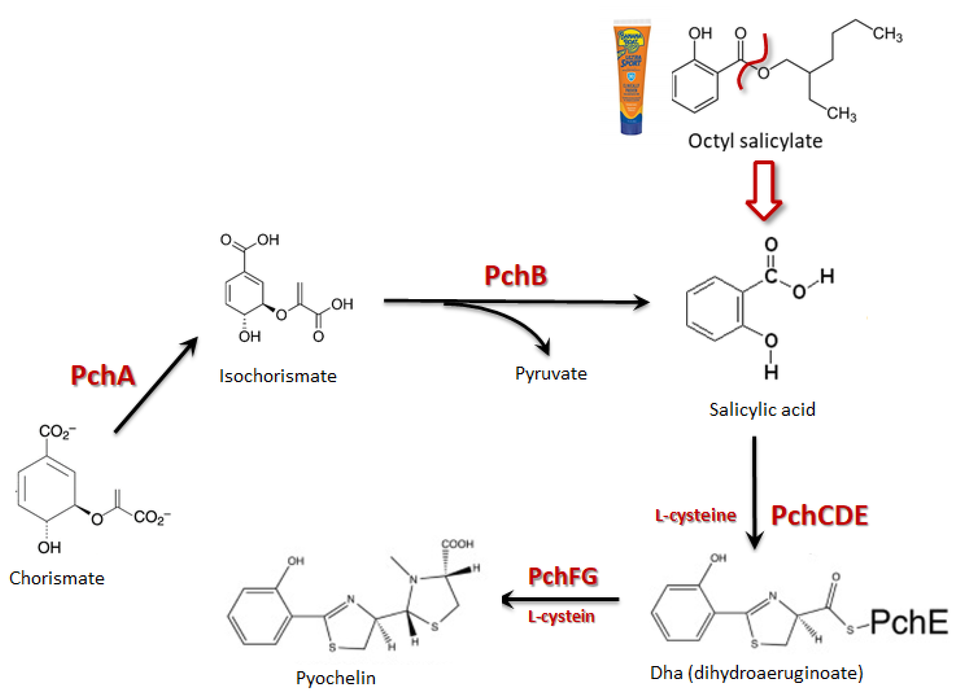
  5. Crosa, J.H.; Walsh, C.T. Genetics and Assembly Line Enzymology of Siderophore Biosynthesis in Bacteria. Microbiol. Mol. Biol. Rev. 2002, 66, 223–249. [Google Scholar] [CrossRef] [Green Version]
  6. Gasser, V.; Guillon, L.; Cunrath, O.; Schalk, I.J. Cellular organization of siderophore biosynthesis in Pseudomonas aeruginosa: Evidence for siderosomes. J. Inorg. Biochem. 2015. [Google Scholar] [CrossRef]
  7. Serino, L.; Reimmann, C.; Visca, P.; Beyeler, M.; Chiesa, V.D.; Haas, D. Biosynthesis of pyochelin and dihydroaeruginoic acid requires the iron- regulated pchDCBA operon in Pseudomonas aeruginosa. J. Bacteriol. 1997, 179, 248–257. [Google Scholar] [CrossRef] [Green Version]
  8. Chapman, J.S. Characterizing bacterial resistance to preservatives and disinfectants. Int. Biodeterior. Biodegrad. 1998, 41, 241–245. [Google Scholar] [CrossRef]
  9. Valkova, N.; Lépine, F.; Bollet, C.; Dupont, M.; Villemur, R. prbA, a Gene Coding for an Esterase Hydrolyzing Parabens in Enterobacter cloacae and Enterobacter gergoviae strains. J. Bacteriol. 2002, 184, 5011–5017. [Google Scholar] [CrossRef] [PubMed] [Green Version]
  10. Weiser, R.; Green, A.E.; Bull, M.J.; Cunningham-Oakes, E.; Jolley, K.A.; Maiden, M.C.J.; Hall, A.J.; Winstanley, C.; Weightman, A.J.; Donoghue, D.; et al. Not all Pseudomonas aeruginosa are equal: Strains from industrial sources possess uniquely large multireplicon genomes. Microb. Genom. 2019, 5. [Google Scholar] [CrossRef] [PubMed]
  11. Green, A.E.; Amézquita, A.; Le Marc, Y.; Bull, M.J.; Connor, T.R.; Mahenthiralingam, E. The consistent differential expression of genetic pathways following exposure of an industrial Pseudomonas aeruginosa strain to preservatives and a laundry detergent formulation. FEMS Microbiol. Lett. 2018, 365, 1–8. [Google Scholar] [CrossRef] [PubMed] [Green Version]
  12. Rushton, L.; Sass, A.; Baldwin, A.; Dowson, C.G.; Donoghue, D.; Mahenthiralingam, E. Key role for efflux in the preservative susceptibility and adaptive resistance of Burkholderia cepacia complex bacteria. Antimicrob. Agents Chemother. 2013, 57, 2972–2980. [Google Scholar] [CrossRef] [Green Version]
  13. Kutty, P.K.; Moody, B.; Gullion, J.S.; Zervos, M.; Ajluni, M.; Washburn, R.; Sanderson, R.; Kainer, M.A.; Powell, T.A.; Clarke, C.F.; et al. Multistate Outbreak of Burkholderia cenocepacia Colonization and Infection Associated with the Use of Intrinsically Contaminated Alcohol-Free Mouthwash. Chest 2007, 132, 1825–1831. [Google Scholar] [CrossRef] [PubMed]
  14. Molina-Cabrillana, J.; Bolaños-Rivero, M.; Alvarez-León, E.E.; Sánchez, A.M.M.; Sánchez-Palacios, M.; Alvarez, D.; Sáez-Nieto, J.A. Intrinsically Contaminated Alcohol-Free Mouthwash Implicated in a Nosocomial Outbreak of Burkholderia cepacia Colonization and Infection. Infect. Control Hosp. Epidemiol. 2006, 27, 1281–1282. [Google Scholar] [CrossRef] [PubMed] [Green Version]
  15. Stephenson, J.R.; Heard, S.R.; Richards, M.A.; Tabaqchali, S. Outbreak of Septicaemia Due to Contaminated Mouthwash. Br. Med. J. 1984, 289, 1584. [Google Scholar] [CrossRef] [PubMed] [Green Version]
  16. Matrician, L.; Ange, G.; Burns, S.; Fanning, W.L.; Kioski, C.; Cage, G.D.; Komatsu, K.K. Outbreak of Nosocomial Burkholderia cepacia Infection and Colonization Associated with Intrinsically Contaminated Mouthwash. Infect. Control Hosp. Epidemiol. 2000, 21, 739–741. [Google Scholar] [CrossRef] [PubMed]
  17. Neza, E.; Centini, M. Microbiologically Contaminated and Over-Preserved Cosmetic Products According Rapex 2008–2014. Cosmetics 2016, 3, 3. [Google Scholar] [CrossRef] [Green Version]
  18. Savli, H.; Karadenizli, A.; Kolayli, F.; Gundes, S.; Ozbek, U.; Vahaboglu, H. Expression stability of six housekeeping genes: A proposal for resistance gene quantification studies of Pseudomonas aeruginosa by real-time quantitative RT-PCR. J. Med. Microbiol. 2003, 52, 403–408. [Google Scholar] [CrossRef] [Green Version]
  19. Vial, L.; Marie-Christine, G.; Dekimpe, V.; Deziel, E. Burkholderia Diversity and Versatility: An Inventory of the Extracellular Products. J. Microbiol. Biotechnol. 2007, 17, 1407–1429. [Google Scholar] [CrossRef]
  20. Breidenstein, E.B.M.; de la Fuente-Núñez, C.; Hancock, R.E.W. Pseudomonas aeruginosa: All roads lead to resistance. Trends Microbiol. 2011, 19, 419–426. [Google Scholar] [CrossRef]
  21. Nair, B.M.; Cheung, K.-J.; Griffith, A.; Burns, J.L. Salicylate induces an antibiotic efflux pump in Burkholderia cepacia complex genomovar III (B. cenocepacia). J. Clin. Invest. 2004, 113, 464–473. [Google Scholar] [CrossRef] [PubMed] [Green Version]
  22. Jones, A.M.; Lindow, S.E.; Wildermuth, M.C. Salicylic acid, Yersiniabactin, and Pyoverdin Production by the Model Phytopathogen Pseudomonas syringae pv. Tomato DC3000: Synthesis, Regulation, and Impact on Tomato and Arabidopsis Host Plants. J. Bacteriol. 2007, 189, 6773–6786. [Google Scholar] [CrossRef] [Green Version]
  23. Prithiviraj, B.; Bais, H.P.; Weir, T.; Suresh, B.; Najarro, E.H.; Dayakar, B.V.; Schweizer, H.P.; Vivanco, J.M. Down Regulation of Virulence Factors of Pseudomonas aeruginosa by Salicylic Acid Attenuates Its Virulence on Arabidopsis thaliana and Caenorhabditis elegans. Infect. Immun. 2005, 73, 5319–5328. [Google Scholar] [CrossRef] [PubMed] [Green Version]
  24. Lattab, A.; Djibaoui, R.; Arabi, A.; Hichem, D. Effect of salicylic acid on biofilm formation and on some virulence factors in Pseudomonas aeruginosa. Int. J. Biosci. 2017, 10, 60–71. [Google Scholar] [CrossRef]
  25. Bandara, M.; Sankaridurg, P.; Zhu, H.; Hume, E.; Willcox, M. Effect of salicylic acid on the membrane proteome and virulence of Pseudomonas aeruginosa. Investig. Ophthalmol. Vis. Sci. 2016, 57, 1213–1220. [Google Scholar] [CrossRef] [Green Version]
  26. Nguyen, A.T.; Jones, J.W.; Ruge, M.A.; Kane, M.A.; Oglesby-Sherrouse, A.G. Iron depletion enhances production of antimicrobials by Pseudomonas aeruginosa. J. Bacteriol. 2015, 197, 2265–2275. [Google Scholar] [CrossRef] [Green Version]
  27. Leinweber, A.; Weigert, M.; Kümmerli, R. The bacterium Pseudomonas aeruginosa senses and gradually responds to interspecific competition for iron. Evolution (N. Y.) 2018, 72, 1515–1528. [Google Scholar] [CrossRef]
  28. Burns, J.L.; Clark, D.K. Salicylate-inducible antibiotic resistance in Pseudomonas cepacia associated with absence of a pore-forming outer membrane protein. Antimicrob. Agents Chemother. 1992, 36, 2280–2285. [Google Scholar] [CrossRef] [Green Version]
Figure 1. Chorismate cycle in Pseudomonas aeruginosa and Burkholderia cepacia incorporated with the hypothesized hydrolysis of octyl salicylate ultraviolet (UV)-filter from sunscreen formulations to salicylic acid, a precursor for pyochelin biosynthesis.
Cosmetics 07 00063 g001
Figure 2. Challenge tests of typical sunscreen formulations against five pharmacopeia strains. Two different sunscreen formulations with SPF 30 (A) and SPF 45 (B), were challenged separately with 1 × 10⁶ CFU/mL of E. coli, S. aureus, and P. aeruginosa, and 1 × 10⁵ CFU/mL of C. albicans and A. brasiliensis. One gram from each inoculated formulation was sampled at 2, 7, 14, 21, and 28 days and was seeded using serial dilutions in order to enumerate the viable microorganisms. Data are presented as CFU/mL.
Cosmetics 07 00063 g002
Figure 3. Challenge tests of basic cream formulation supplemented with either octyl salicylate (OS) or homosalate (HS), against five pharmacopeia strains as well as B. cepacia. Basic cream formulation (Table 1), (A) without UV-filter (B) with 5% OS and (C) with 5% HS, were challenged separately with 1 × 10⁶ CFU/mL of E. coli, S. aureus, P. aeruginosa, and B. cepacia, and 1 × 10⁵ CFU/mL of C. albicans and A. brasiliensis. One gram from each inoculated formulation was sampled at 2, 7, 14, 21, and 28 days and was seeded using serial dilutions in order to enumerate the viable microorganisms. Data are presented as CFU/mL.
Cosmetics 07 00063 g003
Figure 4. Pseudomonas aeruginosa pchD gene expression during treatment with subinhibitory concentrations of octyl salicylate (OS) and homosalate (HS). (A) Increasing concentrations of OS and HS were added to a P. aeruginosa culture and incubated ON. pchD gene expression was assessed against the rpoS housekeeping gene. The surfactant used to dissolve the compounds into the growth medium act as an additional control. Data are presented as 2−ΔΔCT normalized expression levels to the 0% negative control treatment, ± SD. (B) Viability of P. aeruginosa was documented for each treatment while data are presented as mean CFU/mL ± SD. Significant differences in comparison to the control treatment (0%), are indicated in the graph (p-value * < 0.05, ** < 0.01, *** < 0.001, **** < 0.0001).
Cosmetics 07 00063 g004
Figure 5. Burkholderia cepacia pchD gene expression during the treatment with subinhibitory concentrations of octyl salicylate (OS) and homosalate (HS). (A) Increasing concentrations of OS and HS were added to a B. cepacia culture and incubated ON. pchD gene expression was assessed against the recA housekeeping gene. The surfactant used to dissolve the compounds into the growth medium act as an additional control. Data are presented as 2−ΔΔCT normalized expression levels to the 0% negative control treatment, ± SD. (B) Viability of B. cepacia was documented for each treatment while data are presented as mean CFU/mL ± SD. Significant differences in comparison to the control treatment (0%), are indicated in the graph (p-value * < 0.05, ** < 0.01, *** < 0.001, **** < 0.0001).
Cosmetics 07 00063 g005
Table 1. Ingredients within each of the formulations used in this study, SPF 30 with octyl salicylate (OS) (+OS), SPF 45 with OS (+OS), and basic cream with or without OS or homosalate (HS) (+/− OS/HS).
SPF 30 (+OS) with Preservative AM-25
Phase A
Trade NameINCI Name%w/w
WaterAquaUp to 100%
Xanthan gumXanthan gum0.5
Phase B
Montanov 68Cetearyl Glucoside (and) Cetearyl Alcohol3
Emulgade® 1000 NICetearyl Alcohol (and) Ceteareth-203
IPMIsopropyl myristate5
Octyl SalicylateOctyl Salicylate5
Octyl MethoxycinnamateEthylhexyl Methoxycinnamate7.5
Benzophenone-3Benzophenone-34
AvobenzoneButyl Methoxydibenzoylmethane1.6
Antaron™ V-220FVP/Eicosene Copolymer3
Phase C
Vitamin ETocopheryl acetate0.2
Sharomix™ Amplify AM25
(Preservative)
Phenoxyethanol, Chlorphenesin, Caprylyl glycol, Didecyldimethylammonium chloride0.5
Final pH 5.0–5.5
SPF 45 (+OS) with Preservative AM-25
Trade NameINCI Name%w/w
Phase A
WaterAquaUp to 100%
Xanthan gumXanthan gum0.5
Phase B
Montanov 68Cetearyl Glucoside (and) Cetearyl Alcohol3
Emulgade® 1000 NICetearyl Alcohol (and) Ceteareth-203
TCCCaprylic/Capric Triglyceride5
Octyl SalicylateOctyl Salicylate5
HomosalateHomosalate10
Octyl MethoxycinnamateEthylhexyl Methoxycinnamate7.5
Benzophenone-3Benzophenone-36
AvobenzoneButyl Methoxydibenzoylmethane1.6
Titanium DioxideTitanium Dioxide3
Antaron™ V-220FVP/Eicosene Copolymer3
Phase C
Vitamin ETocopheryl acetate0.1
Sharomix™ Amplify AM25
(Preservative)
Phenoxyethanol, Chlorphenesin, Caprylyl glycol, Didecyldimethylammonium chloride0.5
Final pH 6.5–7.0
Basic Cream (+/− OS/HS) with Preservative AM-25
Trade NameINCI Name%w/w
Phase A
WaterAquaUp to 100%
GlycerinGlycerin4
Phase B
Montanov 68Cetearyl Glucoside (and) Cetearyl Alcohol5
Emulgade® 1000 NICetearyl Alcohol (and) Ceteareth-204.8
TCC Caprylic/Capric Triglyceride3
Octyl Salicylate/HomosalateOctyl Salicylate/Homosalate5
Phase C
Vitamin ETocopheryl acetate0.1
Sharomix™ Amplify AM25 (Preservative)Phenoxyethanol, Chlorphenesin, Caprylyl glycol, Didecyldimethylammonium chloride0.5
Final pH 5.5–6.0
Table 2. Ingredients and formulation design for P. aeruginosa pchD gene expression assay in the TSB media supplemented with increasing concentrations of OS and HS.
Material NameFunction%w/w
Sample #1 (OS/HS)
Phase A
TSB mediaSolvent92
Tween 20 (INCI: Polysorbate 20)Surfactant1.5
Phase B
Span 20 (INCI: Sorbitan Laurate)Surfactant1.5
Octyl salicylate/HomosalateUV-filter5
Final pH 7
Sample #2 (Surfactant)
Phase A
TSB mediaSolvent97
Tween 20 (INCI: Polysorbate 20)Surfactant1.5
Phase B
Span 20 (INCI: Sorbitan Laurate)Surfactant1.5
Final pH 7
Table 3. Primers’ list.
PrimerSequenceGeneBacteriaTypeAmplicon
rpoSF [18]CTCCCCGGGCAACTCCAAAAGrpoSP. aeruginosaReference198 bp
rpoSR [18]CGATCATCCGCTTCCGACCAG
pchDF_PsGAGAAGATCGCTGCGGAAGApchDP. aeruginosaTarget170 bp
pchDR_PsGCGTGCAGGTATTGCTTCAG
recAFATCGAGAAGCAGTTCGGCAArecAB. cepaciaReference186 bp
recARCAGCGTGAGCGTGGTTTTAC
pchD_BcFGAAGTGGAGAACCTGCTGCTpchDB. cepaciaTarget146 bp
pchD_BcRCTGGTGCTGAAACGCTACCT

