Design of Cellulose Interfaces through Self-Assembly of Adhesive Peptides with Intrinsic Stress Sensitive Properties †


Institute for Materials Research (IMO-IMOMEC), Applied and Analytical Chemistry, University of Hasselt, 3500 Hasselt, Belgium
†
Presented at the First International Conference on “Green” Polymer Materials 2020, 5–25 November 2020; Available online: https://sciforum.net/conference/CGPM2020.
Proceedings 2021, 69(1), 32; https://doi.org/10.3390/CGPM2020-07157
Published: 3 November 2020
(This article belongs to the Proceedings of The First International Conference on “Green” Polymer Materials 2020)
Abstract
:The design of interfaces in green polymer composites is a crucial factor in ensuring mechanical strength in composite materials. While cellulose fibers have high intrinsic mechanical strength, their reinforcing effect in polymer composite materials relies greatly on the creation of a tight interface with the surrounding polymer matrix. In parallel, the hydrophilicity of the cellulose has to be compatibilized with often more hydrophobic polymer matrixes. In this study, the cellulose interface has been modified by the self-assembly of polymer-peptide nanoparticles regulating the adhesive strength in the interface. The incorporation of catecholic groups allows physical adsorption at the cellulose surface in parallel with the mimicking of mussel-inspired adhesion in the presence of dopamine groups. In this study, the cellulose surface modification was performed with different concentrations of the adhesive nanoparticles, and interesting trends in adhesive forces at macroscale length were observed. The macroscale adhesion was characterized by single-fiber pull out tests, indicating an optimum concentration of nanoparticles at the surface to provide high adhesive interface strength. In addition, the nanoparticles show colorimetric and fluorescent response to mechanical shear stresses, providing an evaluation tool to explore the interface phenomena upon failure.
1. Introduction
A main hurdle in regulating the performance of green polymer composites and/or fibrous non-woven materials is the control of interactions at the level of the fiber/matrix or fiber/fiber interface. The chemical interactions between both the continuous matrix phase and the dispersed reinforcing phase are regulated in polymer composites through the interphase compatiblization after chemical modification of the fiber. In particular, the strong hydrophilic properties of natural cellulose fibers owing to the intrinsic abundance of hydroxyl groups at the fiber surface provide reactive sites for easy access of chemical modification. The modification of cellulose fibers is often done by traditional chemical grafting reactions, including a variety of chemicals. In view of not interfering with the bio-based content of “green materials” as filler or matrix, innovative ways for controlling the interface are needed. Examples of the control of adhesive properties are found in natural phenomena, such as mussels, where the attachment of a mussel byssal thread to different surfaces is controlled by the secretion of peptides, including high concentrations of dihydroxy-phenyl alanine (dopa) [1]. The mechanisms for strong adhesion of catechol groups have been widely studied in recent years [2], and the reactivity of cellulose surfaces towards the catechol groups has been demonstrated through grafting reactions [3]; after modification of cellulose surfaces with dopamine functional groups, it was used in various applications of adhesives, or biomedically compatible surfaces.
In order to gain further insight in the performance and failure prediction of interfaces, the localization of shear stresses should be narrowly controlled with a homogeneous distribution over the fiber surface. However, it is often difficult to have access to the internal shear stresses at the fiber/matrix or fiber/fiber interfaces, and therefore, intrinsic stress sensitivity can bring local access. Many chemical functionalities may provide stress-sensitive functional groups, in particular due to the reorganization of the electronic structure within conjugated chemical bonds. In green materials such as lipid-like linear chains with conjugated triple bonds (e.g., diacetylenic chains), the structure is known to be sensitive to color change and/or fluorescence [4], depending on the conformation of the triple bonds, which is influenced by the external chemical and/or biological environment, temperature, pH and mechanical stress. Therefore, the coupling of adhesive groups to diacetylenic chains may result in the formation of a polymer-peptide conjugate with both adhesive and stress sensitive properties.
In this paper, we build further on the previously synthesized polymer-peptide amphiphiles with adhesive head groups and their organization into nanoscale polymer vesicles [5]. In future applications they can be interestingly applied as a “green” interface mediator in cellulose-based composite materials. Therefore, the feasibility and mechanical performance of different interface designs is evaluated through the variation of the conformation of the single amphiphiles and/or composition and morphology of the polymer vesicles. It seems that the concentration of dopa-groups significantly affects the interface morphology and hence adhesive properties. Therefore, an extensive screening study has been done on systematically varying the concentration of adhesive groups at the cellulose fiber surface. In addition, the failed adhesive interfaces are characterized and seem to show fluorescence at the surface corresponding to localized shear stresses, indicative of premature failure.
2. Experimental Details
2.1. Materials
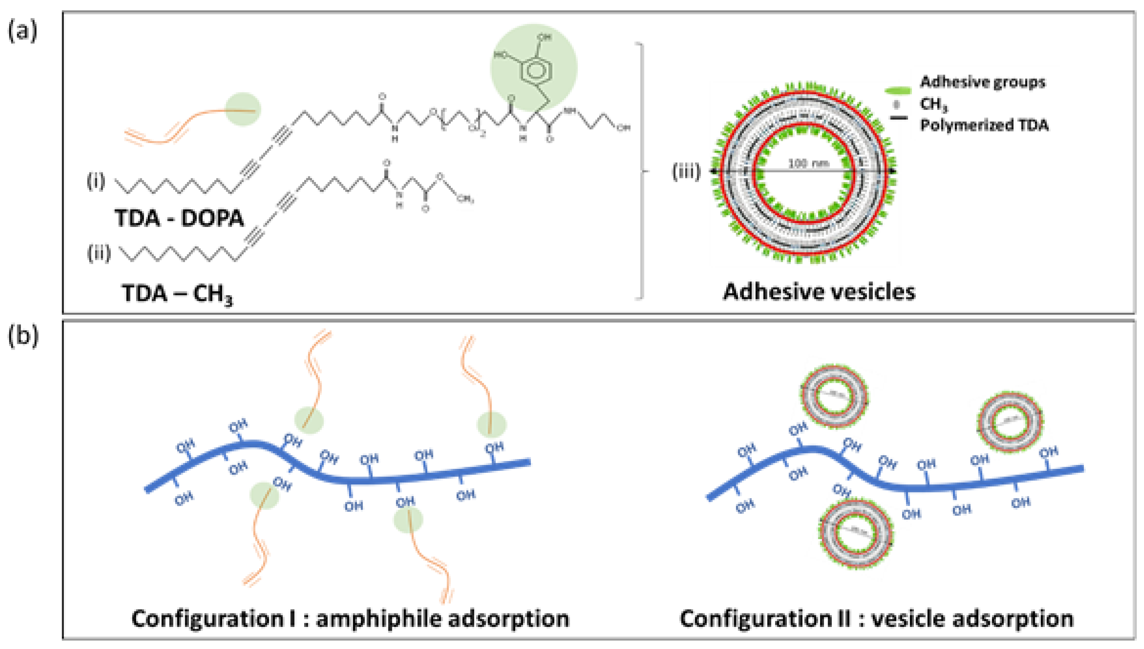
In first step, the polymer-peptides (TDA-DOPA, i.e., tricosadiynoic acid–dihydrox-phenyl-alanine) with adhesive functional groups were manually synthesized according to a previously established protocol [5]. The dopamine moieties were incorporated into a polymer-peptide amphiphile with a dopamine-like headgroup (hydrophilic part) and diacetylene fatty acid tail (hydrophobic part). The adhesive amphiphiles (Figure 1a, structure (i)) were obtained by manual solid-phase peptide synthesis with Fluorenylmethyloxycarbonyl (Fmoc) protecting groups starting from 10,12-tricosadiynoic acid (TDA) and Fmoc-protected (L-3,4-di-hydroxy-phenylalanine) (acetonid)-OH. An intermediate spacer of Fmoc-NH-(PEG)2-OH was inserted between the head group and peptide ligand head group.
In the second step, the polymer-peptides (TDA-CH3, i.e., tricosadiynoic acid–methyl) with neutral methylated head-groups were manually synthesized through the coupling of 10,12-tricosadiynoic acid with a glycine methyl ester (H-Gly-OMe3HCl), as before [5]. The neutral amphiphiles (Figure 1a, structure (ii)) were synthesized in a dimethyl formamide solution from dissolved 10,12-tricosadiynoic acid, modifying the carboxylic head group into a methyl ester group.
Both amphiphiles were combined into vesicular structures through a self-assembly mechanism (Figure 1a, structure (iii)) that included the different ratios of both amphiphiles. Therefore, various molar ratios of the TDA-DOPA and TDA-CH3 amphiphiles were combined by dissolving them in chloroform and removing the solvent by drying under vacuum conditions. The materials were consequently swollen by adding de-ionized water into the vials and heating near the melting temperature of the amphiphiles, about 70 °C. The heated mixtures were treated with a tip-sonicator, and after a cooling period of 6 h at 4 °C, the arranged amphiphiles were polymerized into vesicular structures under UV irradiation at a wavelength of 250 nm for 90 s. The polymerization reaction induced the 1,4-addition reaction between the unsaturated triple bonds in the TDA groups and caused the morphological stabilization of the vesicular structures with a diameter of around 100 nm. As such, the stable vesicles were made with following composition, containing a variation in the molar ratio of constituting amphiphiles TDA-DOPA/TDA-CH3 = 0/100, 10/90, 20/80, 35/65 and 100/0. In this series, the content of adhesive catechol groups steadily increased. The aqueous stock solutions with different concentrations of vesicles were prepared by consequent dilution with de-ionized water towards vesicle concentrations of c = 0.015, 0.03, 0.06, 0.125 and 0.25 mg/mL.
2.2. Cellulose Fiber Modification
The bleached softwood kraft pulp fibers were obtained from Sappi (Lanaken, Belgium) and modified into two configurations (Figure 1b), including (i) the adsorption of single adhesive amphiphiles, and (ii) the adsorption of vesicular structures with adhesive groups.
The cellulose fibers in configuration I were made by soaking the single cellulose fibers for a time of 1 h in an aqueous solution of individual TDA-DOPA adhesive amphiphiles (i.e., structure (i) in Figure 1a before the amphiphiles were organized into vesicular structures), resulting in the spontaneous adsorption of the amphiphiles at the cellulose surface. The individual cellulose fibers were soaked in the aqueous solutions with different concentrations of TDA-DOPA adhesive amphiphiles, similar to the mentioned vesicle concentrations, followed by drawing the modified fibers from the solution and drying them under ambient conditions for one week. The application of different concentrations of the amphiphile solutions consequently resulted in varying degrees of surface modification of the cellulose fibers through adsorption of the TDA-DOPA amphiphiles.
The cellulose fibers in configuration II were created by soaking the cellulose fibers for 1 h in the prepared aqueous solutions with different concentrations of vesicles (i.e., structure (iii) in Figure 1a after the TDA-DOPA and TDA-CH3 amphiphiles were combined into vesicles with different molar ratios for both amphiphiles. The aqueous solutions contained different concentrations of the vesicles, resulting in a different degree of vesicle deposits at the fiber surface. The modified fibers were dried under ambient conditions for one week.
2.3. Composite Formulation
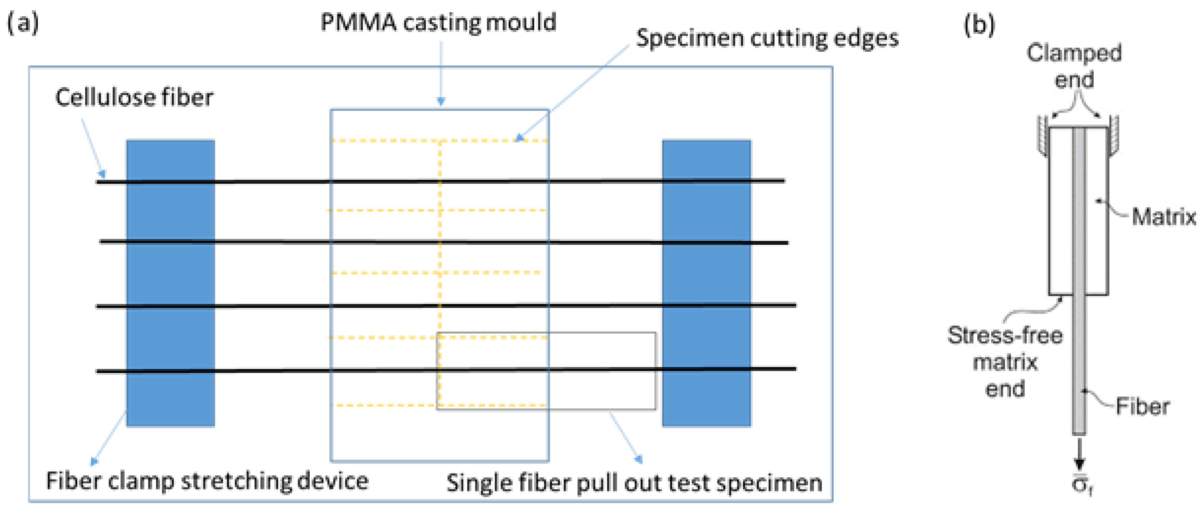
In order to evaluate the effect of surface modification of the cellulose fibers on the interfacial strength in a polymer composite, the fibers were embedded into a film with a PMMA polymer matrix by means of solution casting from toluene. The samples were prepared for subsequent single-fiber pull-out tests (SFPTs) by casting the films with embedded fibers in a designed casting mold (Figure 2a).
The selected single cellulose fibers were organized in between a stretching device with two supporting clamps to ensure they were straight and had parallel orientation. After casting the PMMA films into the mold, the films formed after evaporation of the solvent, and with a polymer concentration of 20% (w/v), composite films with a thickness of 1 mm were formed. The samples were subsequently cut along the indicated paths, using close control for the embedment depth of the fiber.
2.4. Analytical Testing Methods
The failure strength at the interface was determined during a single-fiber pull-out test (SFPT), using a universal tensile tester of Instron (Norwood, MA, USA), by attaching the embedded fiber in the upper clamp and the free end of the fiber in the lower clamp (Figure 2b). The clamps were moved with a velocity of 0.5 mm/min until fracture of the fiber at the interface from the matrix. The embedding length, L, was chosen after preliminary testing of single cellulose fibers, in order to not exceed the intrinsic tensile strength of the fiber, but rather to induce failure at the fiber/matrix interface. The maximum strength was determined from the maximum force at break as indicated from the force-displacement curves.
The wettability of the fibers was determined from contact angle measurements with sessile water drops on GBX equipment (Bourg-de-Peage, France). A droplet of 0.5 µL was deposited on the cellulose fibers, and the shape was fitted with an elliptical geometry to determine the average contact angle.
The optical and fluorescence microscopy on single fiber surfaces after the pull-out test was done on a BX51 microscope (Olympus, Düsseldorf, Germany), representing the fluorescent images as black-white pictures.
3. Results and Discussion
3.1. Cellulose Fiber Surface Properties
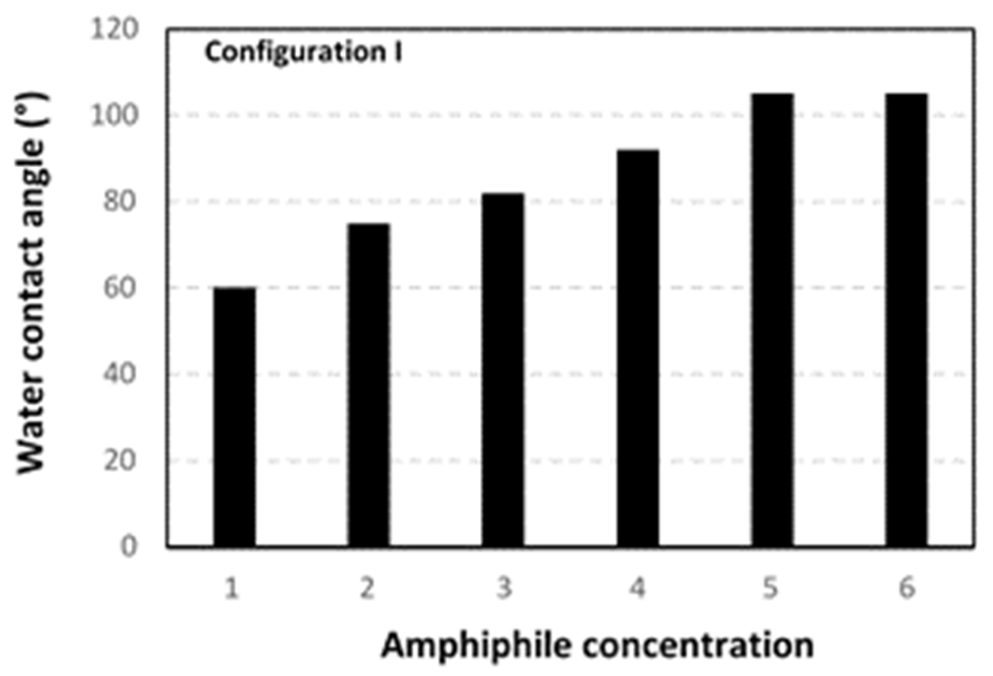
The surface properties of the cellulose fibers modified according to configuration I or configuration II were first analyzed by water contact angles, in order to illustrate the variations in hydrophilic properties of the native cellulose fibers. The results for water contact angles were repeated over five measurements per fiber sample, resulting in a statistical variation of ± 2° for the reported values. Therefore, significant trends could be noticed in water contact angles depending on the configuration or concentration of the surface modification (Figure 3 and Figure 4). Where the initial cellulose fibers were hydrophilic with a characteristic water contact angle of 60°, the surface modified fibers showed significantly higher water contact angles.
For configuration I (Figure 3), contact angles increased up to a maximum of 105° at a concentration of 0.125 mg/mL. The high water contact angles proved a successful surface modification with adsorption of the TDA-DOPA amphiphiles at the cellulose surface. Moreover, the transition from hydrophilic into hydrophobic surface properties of the cellulose fibers confirmed the assumption for self-assembly of the amphiphiles at the cellulose surface. The amphiphiles were characterized by the presence of a hydrophobic tail (TDA) and a hydrophilic head-group (DOPA), which allowed for the spontaneous self-assembly of the amphiphiles at the cellulose fiber surface. The hydrophilic TDA-part of the amphiphiles likely interacted with the hydroxyl groups at the cellulose surface and was consequently oriented towards the cellulose surface, while the hydrophobic DOPA-part of the amphiphiles was likely oppositely oriented towards the outer side of the modified fibers. The interactions between the DOPA catechol groups and the cellulose hydroxyl groups were preferentially established through hydrophilic surface interactions and attractive hydrogen bonding. The latter seemed to be relatively strong and stable due to the abundancy in hydroxyl groups both at the cellulose surface and the amphiphilic structures. The intrinsic hydrophobic (fatty-acid–like) properties of the TDA tails were successfully reflected in a hydrophobic surface modification of the cellulose fiber. At highest concentrations, however, there seemed to be an upper level for hydrophobic modification that could be related to the efficiency for surface modification by assembly of the amphiphiles at the cellulose surface. At the higher concentrations, surface confinement may have hindered the access of additional catechol groups to the cellulose surface, and/or counteracting hydrophobic interactions at the strongly modified cellulose fibers may have hindered the further efficiency of surface modification. However, the increase in contact angle from 60 to 105° by the presentation of fatty-acid–like chemical structures at the outer surface side represented a significant improvement in hydrophobicity.
For configuration II (Figure 4), the variation in contact angle strongly depended on the concentration of TDA-DOPA amphiphiles, with a maximum up to 118° for the vesicles with 20 mol-% TDA-DOPA and concentration c = 0.125 mg/mL. Depending on the vesicle composition, the absence of DOPA groups and/or low concentrations of DOPA groups obviously did not result in favorable adsorption of the vesicles to the cellulose structure due to lack of sufficient interaction between the catechol groups in the vesicles and the hydroxyl groups at the cellulose surface. The surface of the cellulose fibers was consequently hardly modified with vesicles containing 100 mol-% TDA-CH3 amphiphiles, while there a progressive increase was seen in contact angles and number of deposits for vesicles with 10 and 20 mol-% TDA-DOPA amphiphiles. For the vesicles with 35, 50 and 100 mol-% TDA-DOPA amphiphiles, there sufficient deposits were seen at the fiber surface; however, the hydrophilic properties of the DOPA-groups became more dominating above the hydrophobic properties of the CH3 groups in the number of amphiphiles. Considering the variation in vesicle concentration, a maximum for water contact angles was noticeable at c = 0.125 mg/mL, which was likely a result in combination with the variations in nanoscale surface roughness effects. The deposition of multilayer nanoparticle layers at high concentrations, rather than single nanoparticle deposits at lower concentrations, could result in a decrease in particle interaction with the cellulose surface and smoothening of the nanoscale roughness caused by single nanoparticle deposits. As a result, an optimum for highest hydrophobicity of modified cellulose fibers is observed under specific conditions of vesicle composition and concentration.
3.2. Single-Fiber Pull-Out Tests
The average results from SFPTs were reported as the maximum pull-out force, representing the loosening of the fiber from the matrix as a measure for the interfacial strength (Figure 5). The values were represented as pull-out forces and were comparable for the similar geometries of the used fibers with a statistical variation of ±0.10 N.
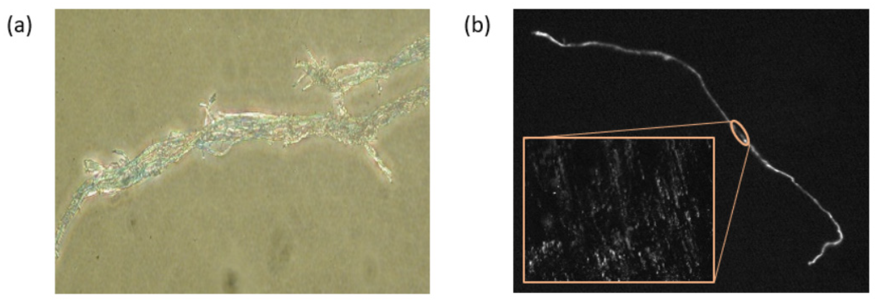
The improvement of interfacial strength occurred at an optimum composition of the vesicles with 20 mol-% TDA-DOPA amphiphiles at a corresponding concentration c = 0.125 mg/mL, comparable to the observations for surface hydrophobicity of the fibers. In addition to the improvement in the adhesive interaction between fibers and matrix observed in the presence of DOPA for the configuration II samples, the hydrophobic interactions between the fiber and the PMMA matrix may improve the interface strength for the configuration I samples. However, the latter may become better visualized in a better dispersion of the cellulose fibers within a hydrophobic polymer matrix. In this research, only a better wettability between the fibers and the PMMA matrix was measured. An evaluation of the surface morphology of cellulose fibers with 20 mol-% TDA-DOPA amphiphiles after shear failure at the interface showed local color transition from blue into red, associated with the fluorescence corresponding to the locations of maximum shear stress at the fiber surface (Figure 6).
4. Conclusions
Cellulose fibers can be modified through the adsorption of vesicles containing amphiphiles with an adhesive head-group. The catechol groups favorably interact with the cellulose surface and provide improved interface adhesion at given optimum concentrations. The intrinsic fluorescent response of the polymerized adhesive vesicles provides stress sensitivity response at the interface.
Funding
Funding for this research was provided by the Robert Bosch Foundation (Germany, Stuttgart) under the framework of the Junior professorship program on research into sustainable use of natural resources.
References
- Lee, H.; Scherer, N.F.; Messersmith, P.B. Single-molecule mechanics of mussel adhesion. Proc. Nat. Acad. Sci. USA 2006, 103, 12999–13003. [Google Scholar] [CrossRef] [PubMed]
- Saiz-Poseu, J. The chemistry behind catechol-based adhesion. Agew. Chem. Int. Ed. 2019, 58, 696–714. [Google Scholar] [CrossRef] [PubMed]
- Zhang, L.; Li, Z.; Pan, Y.T.; Yanez, A.P.; Hu, S.; Zhang, X.Q.; Wang, R.; Wang, D.Y. Polydopamine induced natural fiber surface functionalization: a way towards flame retardancy of flax/polylacticc acid biocomposites. Composites B 2018, 154, 56–63. [Google Scholar] [CrossRef]
- Warta, R.; Sixl, H. Optical absorption and fluorescence spectroscopy of stable diacetylene oligomer molecules. J. Chem. Phys. 1988, 88, 95. [Google Scholar] [CrossRef]
- Samyn, P.; Rühe, J.; Biesalski, M. Polymerizable biomimetic vesicles with controlled local presentation of adhesive functional dopa groups. Langmuir 2010, 26, 8573–8581. [Google Scholar] [CrossRef] [PubMed]
Figure 1.
Synthesis of adhesive and sensitive interfaces at cellulose fibers. (a) Chemical structure of adhesive vesicles with self-organization of synthesized amphiphiles. (b) Surface modification of cellulose fibers with single amphiphiles or vesicles.
Figure 1.
Synthesis of adhesive and sensitive interfaces at cellulose fibers. (a) Chemical structure of adhesive vesicles with self-organization of synthesized amphiphiles. (b) Surface modification of cellulose fibers with single amphiphiles or vesicles.

Figure 2.
Formulation method of polymer composites with embedded fibers for the creation of the test specimen. (a) Organization of cellulose fibers and casting mold for PMMA films with embedded fibers and making the test pieces. (b) Single-fiber pull-out tests (SFPTs) in universal tensile tester.
Figure 2.
Formulation method of polymer composites with embedded fibers for the creation of the test specimen. (a) Organization of cellulose fibers and casting mold for PMMA films with embedded fibers and making the test pieces. (b) Single-fiber pull-out tests (SFPTs) in universal tensile tester.

Figure 3.
Water contact angle measurements representing hydrophobicity of surface-modified cellulose fibers, according to configuration I, for different concentrations (mg/mL) c = 0.00 (1), 0.015 (2), 0.03 (3), 0.06 (4), 0.125 (5), and 0.25 (6).
Figure 3.
Water contact angle measurements representing hydrophobicity of surface-modified cellulose fibers, according to configuration I, for different concentrations (mg/mL) c = 0.00 (1), 0.015 (2), 0.03 (3), 0.06 (4), 0.125 (5), and 0.25 (6).

Figure 4.
Water contact angle measurements representing hydrophobicity of surface-modified cellulose fibers, according to configuration II, including different vesicle compositions as indicated in the figures, for different concentrations (mg/mL) c = 0.00 (1), 0.015 (2), 0.03 (3), 0.06 (4), 0.125 (5), and 0.25 (6).

Figure 5.
Forces from SFPTs for cellulose fibers, according to configuration II and including different vesicle compositions, or according to configuration I for different concentrations (mg/mL) c = 0.00 (1), 0.015 (2), 0.03 (3), 0.06 (4), 0.125 (5), and 0.25 (6).
Figure 5.
Forces from SFPTs for cellulose fibers, according to configuration II and including different vesicle compositions, or according to configuration I for different concentrations (mg/mL) c = 0.00 (1), 0.015 (2), 0.03 (3), 0.06 (4), 0.125 (5), and 0.25 (6).

Figure 6.
Local coloration and fluorescence at the fiber surface after SFPT evaluation: (a) optical coloration of fiber with red and blue zones, (b) fluorescent response corresponding to local zones of intense shear stress over full fiber and detail of fiber surface (inset).
Figure 6.
Local coloration and fluorescence at the fiber surface after SFPT evaluation: (a) optical coloration of fiber with red and blue zones, (b) fluorescent response corresponding to local zones of intense shear stress over full fiber and detail of fiber surface (inset).

Publisher’s Note: MDPI stays neutral with regard to jurisdictional claims in published maps and institutional affiliations. |
© 2020 by the author. Licensee MDPI, Basel, Switzerland. This article is an open access article distributed under the terms and conditions of the Creative Commons Attribution (CC BY) license (https://creativecommons.org/licenses/by/4.0/).
Share and Cite
MDPI and ACS Style
Samyn, P. Design of Cellulose Interfaces through Self-Assembly of Adhesive Peptides with Intrinsic Stress Sensitive Properties. Proceedings 2021, 69, 32. https://doi.org/10.3390/CGPM2020-07157
AMA Style
Samyn P. Design of Cellulose Interfaces through Self-Assembly of Adhesive Peptides with Intrinsic Stress Sensitive Properties. Proceedings. 2021; 69(1):32. https://doi.org/10.3390/CGPM2020-07157
Chicago/Turabian StyleSamyn, Pieter. 2021. "Design of Cellulose Interfaces through Self-Assembly of Adhesive Peptides with Intrinsic Stress Sensitive Properties" Proceedings 69, no. 1: 32. https://doi.org/10.3390/CGPM2020-07157