Article

Terpene Glycosides from the Roots of Sanguisorba officinalis L. and Their Hemostatic Activities

School of Pharmaceutical Sciences, Jilin University, Changchun 130021, China
*
Author to whom correspondence should be addressed.
Molecules 2012, 17(7), 7629-7636; https://doi.org/10.3390/molecules17077629
Submission received: 15 May 2012 / Revised: 13 June 2012 / Accepted: 15 June 2012 / Published: 25 June 2012

Abstract

:
Guided by a hemostasis bioassay, seven terpene glycosides were isolated from the roots of Sanguisorba officinalis L. by silica gel column chromatography and preparative HPLC. On the grounds of chemical and spectroscopic methods, their structures were identified as citronellol-1-O-α-L-arabinofuranosyl-(1→6)-β-D-glucopyranoside (1), geraniol-1-O-α-L-arabinofuranosyl-(1→6)-β-D-glucopyranoside (2), geraniol-1-O-α-L-arabinopyranosyl-(1→6)-β-D-glucopyranoside (3), 3β-[(α-L-arabinopyranosyl)oxy]-19α-hydroxyolean-12-en-28-oic acid 28-β-D-glucopyranoside (4), 3β-[(α-L-arabinopyranosyl)-oxy]-19α-hydroxyurs-12-en-28-oic acid 28-β-D-glucopyranoside (ziyu-glycoside I, 5), 3β,19α-hydroxyolean-12-en-28-oic acid 28-β-D-glucopyranoside (6) and 3β,19α-di-hydroxyurs-12-en-28-oic acid 28-β-D-glucopyranoside (7). Compound 1 is a new mono-terpene glycoside and compounds 2, 3 and 5 were isolated from the Sanguisorba genus for the first time. Compounds 17 were assayed for their hemostatic activities with a Goat Anti-Human α2-plasmin inhibitor ELISA kit, and ziyu-glycoside I (5) showed the strongest hemostatic activity among the seven terpene glycosides. This is the first report that ziyu-glycoside I has strong hemostatic activity.

1. Introduction

Sanguisorba officinalis L. (Rosaceae) is a perennial plant widely distributed in China, and its roots have been used as a traditional Chinese medicine for the treatment of hemostasis and inflammation [1]. A variety of chemical constituents, including tannins, triterpenoids, flavonoids, anthraquinones, steroids were isolated from S. officinalis L., and pharmacological studies on its hemostatic and anti-inflammatory properties have been reported [2,3], but the molecular level mechanisms of these activities have not been reported until now. In order to study the mechanism of hemostasis, we have carried out the bioassay-guided isolation and identification of hemostatic constituents of the roots of S. officinalis L. In the present study, we report the isolation and identification of a new monoterpene glycoside 1, together with two known monoterpene glycosides 2, 3 and four known triterpenoids 47, and their hemostatic activities.

2. Results and Discussion
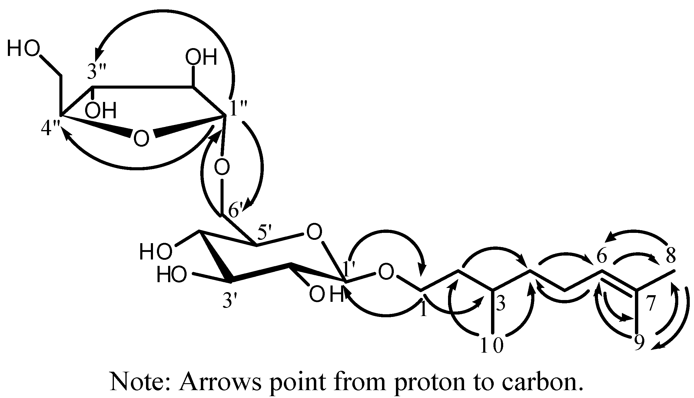
Compound 1, a colorless amorphous powder, produced a positive reaction to Molish reagent, and had the molecular formula C21H38O10 as determined by HRESIMS ([M+Na]+ m/z 473.2369). Its IR spectrum indicated the presence of hydroxyl (3450 cm−1) groups. Acid hydrolysis of compound 1 gave D-glucose and L-arabinose. The 1H-, 13C- and DEPT-NMR spectrum (DMSO-d6) of compound 1 showed signals of a monoterpene moiety consisting of two singlets and one double methyl groups [δH 1.64 (3H, s, H-8), 1.56 (3H, s, H-9), and 0.85 (3H, d, J = 6.4 Hz H-10); δC 25.5 (C-8), 17.5 (C-9), and 19.3 (C-10)], four methylenes [δH 3.41 and 3.76 (each 1H, m, H-1), 1.32 and 1.56 (each 1H, m, H-2), 1.12 and 1.29 (each 1H, m, H-4), and 1.93 (2H, m, H-5); δC 66.9 (C-1), 36.3 (C-2), 36.8 (C-4), and 24.9 (C-5)], one olefinic methine [δH 1.52 (1H, m, H-3); δC 28.9 (C-3)], and one carbon-carbon double bond [δH 5.09 (1H, t-like, J = 7.2 Hz, H-6); δC 124.7 (C-6) and 130.4 (C-4)]. Based on the 2D NMR data of H-H COSY, HMQC and HMBC experiments, the monoterpene moiety was identified as citronellol [4]. The coupling constant of the anomeric proton of glucose at δ 4.11 (d, 1H, J = 8.0 Hz) indicated that glucose moiety was in a β-configuration. The HMBC correlation signal of the anomeric proton of glucose, 1'-H (δH 4.11) to C-1 (δC 66.9) showed that glucose was linked to C-1 of the alycone. The downfield shift by about 5 ppm of the signal of C-6' of glucose (δC 67.2) showed that arabinose was linked to C-6' of the glucose, which was further confirmed by the HMBC correlations of 6'-H (δH 3.39 and 3.85) to C-1" (δC 108.5) of arabinose and 1"-H (δH 4.79) to C-6' (δC 67.2). The 13C-NMR signals of compound 1 assignable to the arabinose moiety [δC 82.0 (C-2"), 83.8 (C-2")] and the coupling constant of the anomeric proton of arabinose at δH 4.79 (d, 1H, J = 1.6 Hz) indicated that arabinose moiety was an α- L -arabinofuranose moiety [5], which was further confirmed by the HMBC correlation of 1"-H (δH 4.79) to C-4" (δC 83.8). The complete assignment of the signals of compound 1 was based on DEPT 13C-NMR and 2D-NMR H-H COSY, HMQC and HMBC data. For all the 1H-, 13C-, and HMBC NMR data of compound 1 see Table 1, and for the structure of compound 1, see Figure 1. Therefore, the structure of compound 1 was elucidated as citronellol-1-O-α-L-arabinofuranosyl-(1→6)-α-D-glucopyranoside.
Using similar methods as described above, compounds 27 were identified as geraniol-1-O-α-L-arabinofuranosyl-(1→6)-β-D-glucopyranoside (2) [5], geraniol-1-O-α-L-arabinopyranosyl-(1→6)-β-D-glucopyranoside (3) [5], 3β-[(α-L-arabinopyranosyl)oxy]-19α-hydroxyolean-12-en-28-oic acid 28-β-D-glucopyranoside (4) [6], 3β-[(α-L-arabinopyranosyl)oxy]-19α-hydroxyurs-12-en-28-oic acid 28-β-D-glucopyranoside (ziyu-glycoside І, 5) [7], 3β,19α-hydroxyolean-12-en-28-oic acid 28-β-D-gluco-pyranoside (6) [8], 3β,19α-dihydroxyurs-12-en-28-oic acid 28-β-D-glucopyranoside (7) [8], respectively.
Table 1. 1H-NMR (400 MHz), 13C-NMR (100 MHz), HMQC and HMBC data of compound 1 (DMSO-d6, δppm).
No.δCδHHMBC(H→C)No.δCδHHMBC(H→C)
aglycone glc
166.93.41, 3.76 (m, each 1H)28.9, 102.81'102.84.11 (d, 1H, J = 8.0 Hz)66.9, 75.4, 76.7
236.31.32, 1.56 (m, each 1H)19.3, 36.8,2'73.42.93 (t-like, 1H, J = 8.0 Hz)
328.91.52 (m, 1H)66.93'76.73.13 (t, 1H, J = 8.8 Hz)
436.81.12, 1.29 (m, each 1H)19.3, 36.3, 124.74'70.42.98 (t-like, 1H, J = 8.4 Hz)73.4
524.91.93 (m, 2H)28.9, 130.45'75.43.28 (t-like, 1H, J = 8.4 Hz)102.8
6124.75.09 (t-like, 1H, J = 7.2 Hz)17.5, 25.5, 36.86'67.23.85 (d-like, 1H, J = 10.8 Hz), 3.39 (dd, 1H, J = 10.8, 8.4 Hz)108.5
7130.4 ara(f)
825.51.64 (s, 3H)17.5, 124.71"108.54.79 (d, 1H, J = 1.6 Hz)67.2, 77.2, 83.8
917.51.56 (s, 3H)25.5, 124.72"82.03.79 (m, 1H)
1019.30.85 (d, 3H, J = 6.4 Hz)36.3, 36.83"77.23.62 (m, 1H)
4"83.83.72 (m, 1H)
5"61.43.55 (dd, 1H, J = 11.6, 2.0 Hz), 3.40 (m, 1H)
All assignments based on extensive 1D and 2D NMR experiments (HMQC, HMBC, 1H-1H COSY).
Figure 1. The key HMBC correlations of compound 1.
Molecules 17 07629 g001
Compounds 17 were next assayed for hemostatic activity with a Goat Anti-Human α2-plasmin inhibitor ELISA kit, and the results are shown in Table 2. The data proved that ziyu-glycoside I showed strongest hemostasis activity within 7 kinds of terpene glycosides. It is the first report that ziyu-glycoside І (5) has strong hemostatic activity.
Table 2. The hemostasis assay data of the separated fractions and the isolated compounds.
FractionsH2O30% EtOH70% EtOH95% EtOHIIIIIIIVControl blankStandard(10 g/L)
OD value (n = 3)0.056 ± 0.0110.053 ± 0.0020.131 ± 0.0140.051 ± 0.0060.051 ± 0.0050.051 ± 0.0060.490 ± 0.0170.061 ± 0.0040.028 ± 0.0040.828 ± 0.031
Percent inhibition 3.53.112.82.92.92.957.84.1
Compounds1234567 Control blankStandard(10 g/L)
OD value (n = 3)0.138 ± 0.0160.111 ± 0.0090.122 ± 0.0030.260 ± 0.0130.741 ± 0.0120.227 ± 0.0100.214 ± 0.015 0.060 ± 0.0040.828 ± 0.031
Percent inhibition10.26.68.126.088.721.720.1
Note: Percent inhibition = [(ODsample − ODblank) ÷ (ODstandard − ODblank)] × 100; the OD value is directly proportional the concentration of α2-plasmin inhibitor present in the samples.

3. Experimental

3.1. General

IR spectra were recorded on a FT-IR 5DX Nicolet/Nicolet Magna IR-560 spectrometer (Thermo Scientific, Osaka, Japan). 1H- and 13C-NMR spectra were recorded on a Bruker AV-400 spectrometer (Zürich, Switzerland). HR-ESI-MS were recorded on a Bruker microOTOF-Q II mass spectrometer. Prep. HPLC was performed on a Shimadzu LC-10A equipped with a SPD-10A detector and Gemini 5μm C18 110A column (250 mm × 10.00 mm, 5 μm, flow rate: 3.0 mL/min). The bioactivities were measured on a DG5033A Enzyme immunoassay spectrophotometer (Nanjing, China), using Goat Anti-Human α2-plasmin inhibitor ELISA kit [96/48-wells microtiter plates, plastic cover, standard (40 g/L), blank control, standard diluent, biotinylated anti-α2-plasmin inhibitor, streptavidin-HRP, washing buffer, substrate A, substrate B, stopping solution, sample diluent)] (R&D Systems, Minneapolis, MN, USA). The roots of S. officinalis L. were collected in Tong-Hua County in Jilin Province, China. They were identified by Prof. Jing-min Zhang of School of Pharmaceutical Sciences, Jilin University.

3.2. Extraction and Isolation

The air-dried the roots of S. officinalis (4.0 kg) were extracted with hot 70% EtOH (ca. 20 L, 24 h, 45 °C). The EtOH extract was concentrated under reduced pressure, and the viscous concentrate (420 g) was passed through a D101 polyporous resin column eluting successively with H2O, 30% EtOH, 70% EtOH, and 95% EtOH, and by vacuum distillation recovery, four fractions (I–IV) were obtained. The bioassay experiments suggested that the 70% EtOH portion had hemostatic activity. Therefore the 70% EtOH fraction was subjected to silica gel column chromatography eluted with a stepwise gradient mixture of CHCl3–MeOH (9:1; 6:1; 3:1), and finally with MeOH alone, and four fractionons (I–IV) were obtained. Fraction III having hemostasis activity was further subjected to a silica gel column eluted with CHCl3–MeOH–EtOAc–H2O (6.5:5:4:1.7), and three fractions (A, B, C) were obtained. Fraction B was applied to a ODS-A (50 μm, 12 nm, YMC, Kyoto, Japan) column eluted with a stepwise gradient mixture of MeOH-H2O (2:3; 3:2; 4:1), and thus compound 5 (230 mg) and Fraction D were obtained. Fractions A, C, and D were separated by preparative HPLC using MeOH–H2O (60:40, 70:30, 65:35, respectively), and compound 1 (22 mg), 2 (25 mg), and 3 (27 mg) were isolated from fraction A, compound 6 (30 mg) and 7 (33 mg) from fraction C, and compound 4 (30 mg) from Fraction B.
Compound 1: Colorless amorphous powder, [α]24D: −21.0° (c 0.6, MeOH). HRESIMS, m/z: 473.2369 ([M+Na]+; calcd for C21H38O10Na, 473.2363). IR (KBr) νmax: 3450 cm−1. 1H and 13C-NMR: See Table 1.
Compound 2: Colorless amorphous powder. HRESIMS, m/z: 471.2209 ([M+Na]+; calcd for C21H36O10Na, 471.2206). IR (KBr) νmax: 3440 cm1. 1H-NMR (DMSO-d6) δ: 1.57 (s, 3H, H-9), 1.61 (s, 3H, H-10), 1.64 (s, 3H, H-8), 1.99 (2H, t, J = 6.8 Hz, H-4), 2.05 (2H, m, H-5), 4.08 (1H, dd, J = 11.9, 7.6 Hz, H-1a), 4.19(1H, dd, J = 11.9, 6.1 Hz, H-1b), 5.07 (1H, t-like, J = 6.8 Hz, H-6), 5.26 (1H, m, H-2); 4.11 (1H, d, J = 7.8 Hz, glc-H1), 2.95 (1H, t-like, J = 8.0 Hz, glc-H2), 3.12 (1H, t-like, J = 8.8 Hz, glc-H3), 2.98 (1H, t-like, J = 8.8 Hz, glc-H4), 3.25 (1H, t-like, J = 8.4 Hz, glc-H5), 3.86 (1H, d-like, J = 9.9 Hz, glc-H6a), 3.39 (1H, m, glc-H6b); 4.79 (1H, d, J = 1.6 Hz, ara-H1), 3.79 (1H, m, ara-H2), 3.62 (1H, m, ara-H3), 3.72 (1H, m, ara-H4), 3.52 (1H, d-like, J = 13.4Hz, ara-H5a),3.40 (m, 1H, ara-H5b). 13C-NMR (DMSO-d6) δ: 16.1(C-10), 17.6 (C-9), 25.6 (C-8), 25.9 (C-5), 39.4(C-4), 64.2 (C-1), 120.6 (C-2), 123.9 (C-6), 130.4 (C-7), 139.4 (C-3); 101.3 (glc-C1), 73.3 (glc-C2), 76.7 (glc-C3), 70.5 (glc-C4), 75.5 (glc-C5), 67.2 (glc-C6); 108.5 (ara(f)-C1), 82.1 (ara(f)-C2), 77.3 (ara(f)-C3), 83.8 (ara(f)-C4), 61.4 (ara(f)-C4).
Compound 3: Colorless amorphous powder. HRESIMS, m/z: 471.2208 ([M+Na]+; calcd for C21H36O10Na, 471.2206). IR (KBr) νmax: 3445 cm−1. 1H-NMR (DMSO-d6) δ: 1.57 (s, 3H, H-9), 1.61 (s, 3H, H-10), 1.64 (s, 3H, H-8), 1.99 (2H, t, J = 6.8 Hz, H-4), 2.05 (2H, t, J = 6.8 Hz, H-5), 4.06 (1H, dd, J = 12.7, 7.3 Hz, H-1a), 4.22(1H, dd, J = 12.7, 6.0 Hz, H-1b), 5.08(1H, t-like, J = 6.5 Hz, H-6), 5.28 (1H, J = 6.5 Hz, H-2); 4.12 (1H, d, J = 7.8 Hz, glc-H1), 4.20 (1H, d, J = 6.0 Hz, ara-H1). 13C-NMR (DMSO-d6) δ: 16.1 (C-10), 17.6 (C-9), 25.5 (C-8), 25.8 (C-5), 39.3 (C-4), 64.4 (C-1), 120.7 (C-2), 123.9 (C-6), 130.9 (C-7), 139.1 (C-3); 101.5 (glc-C1), 73.3 (glc-C2), 76.6 (glc-C3), 70.5 (glc-C4), 75.6 (glc-C5), 67.2 (glc-C6); 103.4 (ara(p)-C1), 70.1 (ara(p)-C2), 72.5 (ara(p)-C3), 68.5(ara(p)-C4), 64.7 (ara(p)-C4).
Compound 4: Colorless amorphous powder. HRESIMS, m/z: 789.4404 ([M+Na]+; calcd for C41H66O13Na, 789.4401). IR (KBr) νmax: 3440, 1720 cm−1. 1H-NMR (DMSO-d6) δ: 5.24 (1H, d, J = 7.8 Hz, glc-H1), 5.23(1H, br.s, H-12), 4.45 (1H, d, J = 7.0Hz, ara-H1), 3.11 (1H, br.s, H-19), 3.01 (1H, dd, J = 11.5, 3.9 Hz, H-3), 2.91(1H, br.s, H-18), 1.23 (3H, s, Me-27), 0.97 (3H, s, Me-23), 0.87 (3H × 2, s, Me-26 and Me-29), 0.85 (3H, s, Me-30), 0.76 (3H, s, Me-24), 0.65(3H, s, Me-25). 13C-NMR (DMSO-d6) δ: 38.0(C-1), 25.6 (C-2), 87.7 (C-3), 38.8 (C-4), 55.1 (C-5), 17.9 (C-6), 32.3 (C-7), 40.2 (C-8), 47.3 (C-9), 36.4 (C-10); 23.2 (C-11), 122.3 (C-12), 143.2 (C-13), 41.1 (C-14), 28.3 (C-15), 27.0 (C-16); 45.2 (C-17), 43.2 (C-18), 80.0 (C-19), 34.8 (C-20), 27.8 (C-21), 31.8 (C-22), 27.6 (C-23), 16.4 (C-24), 15.1 (C-25), 16.6 (C-26); 24.5 (C-27), 175.8 (C-28), 28.0 (C-29), 24.0 (C-30); 94.1 (glc-C1), 72.4 (glc-C2), 76.7 (glc-C3), 69.5 (glc-C4), 77.7 (glc-C5), 60.6 (glc-C6); 105.8 (ara(p)-C1), 71.0 (ara(p)-C2), 72.7 (ara(p)-C3), 67.6 (ara-C4), 65.1 (ara-C4).
Compound 5: Colorless amorphous powder. HRESIMS, m/z: 789.4403 ([M+Na]+; calcd for C41H66O13Na, 789.4401). IR (KBr) νmax: 3475, 1740 cm−1. 1H-NMR (DMSO-d6) δ: 5.16 (1H, d, J = 7.8 Hz, glc-H1), 5.17(1H, br.s, H-12), 4.12 (1H, d, J = 6.0Hz, ara-H1), 3.11 (1H, br.s, H-19), 3.01 (1H, dd, J = 11.1, 3.9 Hz, H-3), 2.36(1H, br.s, H-18), 1.23 (3H, s, Me-27), 1.09 (3H, s, Me-29), 0.97 (3H, s, Me-23), 0.88 (3H, s, Me-26), 0.84(3H, d, J = 6.6 Hz, Me-30), 0.76 (3H, s, Me-25), 0.67 (3H, s, Me-24). 13C-NMR (DMSO-d6) δ: 38.2(C-1), 25.7 (C-2), 87.8 (C-3), 38.7 (C-4), 55.0 (C-5), 17.9 (C-6), 32.5 (C-7), 39.4 (C-8), 46.7 (C-9), 36.2 (C-10); 23.2 (C-11), 127.0 (C-12), 138.2 (C-13), 41.2 (C-14), 28.1 (C-15), 25.1 (C-16); 47.3 (C-17), 53.2 (C-18), 71.6 (C-19), 41.0 (C-20), 25.8 (C-21), 36.6 (C-22), 27.6 (C-23), 16.4 (C-24), 15.2 (C-25), 16.4 (C-26); 23.8 (C-27), 175.5 (C-28), 26.4 (C-29), 16.2 (C-30); 94.1 (glc-C1), 72.2 (glc-C2), 76.7 (glc-C3), 69.5 (glc-C4), 77.6 (glc-C5), 60.6 (glc-C6); 105.8 (ara(p)-C1), 71.0 (ara(p)-C2), 72.7 (ara(p)-C3), 67.6 (ara-C4), 65.1 (ara-C4).
Compound 6: Colorless amorphous powder. HRESIMS, m/z: 657.39783 ([M+Na]+; calcd for C36H58O9Na, 657.39785). IR (KBr) νmax: 3440, 1720 cm−1. 1H-NMR (DMSO-d6) δ: 5.23 (1H, d, J = 7.6 Hz, glc-H1), 5.23(1H, br.s, H-12), 3.11 (1H, br.s, H-19), 3.00 (1H, dd, J = 11.2, 3.9 Hz, H-3), 2.91(1H, br.s, H-18), 1.23 (3H, s, Me-27), 0.89 (3H, s, Me-23), 0.87 (3H, s, Me-29), 0.84 (3H × 2, s, Me-25 and Me-30), 0.68 (3H, s, Me-24), 0.64(3H, s, Me-25). 13C-NMR (DMSO-d6) δ: 38.0 (C-1), 26.9 (C-2), 77.7 (C-3), 38.4 (C-4), 54.9 (C-5), 18.1 (C-6), 32.3 (C-7), 40.2 (C-8), 47.3 (C-9), 36.7 (C-10); 23.2 (C-11), 122.3 (C-12), 143.2 (C-13), 41.1 (C-14), 28.3 (C-15), 27.0 (C-16); 45.2 (C-17), 43.1 (C-18), 80.0 (C-19), 34.8 (C-20), 27.8 (C-21), 31.8 (C-22), 28.3 (C-23), 15.9 (C-24), 15.0 (C-25), 16.6 (C-26); 24.1 (C-27), 175.8 (C-28), 28.0 (C-29), 24.5 (C-30); 94.1 (glc-C1), 72.4 (glc-C2), 76.7 (glc-C3), 69.5 (glc-C4), 76.9 (glc-C5), 60.6 (glc-C6).
Compound 7: Colorless amorphous powder. HRESIMS, m/z: 657.39784 ([M+Na]+; calcd for C36H58O9Na, 657.39785). IR (KBr) νmax: 3475, 1740 cm−1. 1H-NMR (DMSO-d6) δ: 5.16 (1H, d, J = 8.0 Hz, glc-H1), 5.17(1H, br.s, H-12), 4.12 (1H, d, J = 6.0Hz, ara-H1), 3.11 (1H, br.s, H-19), 3.00 (1H, dd, J = 11.0, 3.9 Hz, H-3), 2.37(1H, br.s, H-18), 1.27 (3H, s, Me-27), 1.08 (3H, s, Me-29), 0.89 (3H, s, Me-23), 0.85 (3H, s, Me-26), 0.84 (3H, d, J = 6.6 Hz, Me-30), (3H, s,), 0.67 (3H × 2, s, Me-25 and Me-24). 13C-NMR (DMSO-d6) δ: 38.2 (C-1), 27.0 (C-2), 77.6 (C-3), 38.4 (C-4), 54.8 (C-5), 18.1 (C-6), 32.6 (C-7), 39.2 (C-8), 46.7 (C-9), 36.5 (C-10); 23.2 (C-11), 127.0 (C-12), 138.1 (C-13), 41.2 (C-14), 28.1 (C-15), 25.1 (C-16); 47.3 (C-17), 53.2 (C-18), 71.6 (C-19), 41.1 (C-20), 25.4 (C-21), 36.6 (C-22), 28.2 (C-23), 16.4 (C-24), 15.1 (C-25), 16.2 (C-26); 23.8 (C-27), 175.5 (C-28), 26.4 (C-29), 16.0 (C-30); 94.0 (glc-C1), 72.2 (glc-C2), 76.7 (glc-C3), 69.5 (glc-C4), 76.9 (glc-C5), 60.6 (glc-C6).

3.3. Acid Hydrolysis of 1–7

Solutions of 17 (each 1.0 mg) in 0.5 M H2SO4 (2.0 mL) were heated under reflux for 3 h. After cooling, each reaction mixture was diluted with H2O, neutralized with BaCO3, then filtered. The solution was partitioned with EtOAc to give two layers. The aqueous layer was evaporated and then subjected to TLC analysis with authentic sugar samples using n-BuOH–MeOH–CHCl3–HOAc (12.5:4.5:9:1.5:1, detection with aniline-phthalic acid). Compounds 15 afforded D-glucose (Rf = 0.30) and L-arabinose (Rf = 0.36), and 67 gave D-glucose (Rf = 0.30).

3.4. Bioactivity Assay

The hemostasis assay was carried out by using a Goat Anti-Human α2-plasmin inhibitor kit which is a solid phase sandwich enzyme-linked quantitative immunoabsorbent assay (ELISA) with a purified antibody specific for α2-plasmin inhibitors. The above separated fractions and compounds were dissolved in dimethylsulfoxide (DMSO) (0.188 mg/μL for the fractions; 0.094 mg/μL for compounds), and were diluted with sample diluent (1:1). After an aliquot (50 μL) of the above samples or standards was added to each microplate well, a portion of biotinylated anti-α2-plasmin inhibitor (50 μL) was immediately added to each well, and the microplate was incubated for 1 h at 37 °C. After a wash with washing buffer, streptavidin-HRP was added, and the microplate was incubated for 30 min at 37 °C. Again, after a wash with washing buffer, substrate A and substrate B (50 μL each) were added to each well, and the microplate was incubated for 10 min at 37 °C. The enzyme-substrate reaction was stopped by quickly pipetting stopping solution (50 μL) into each well. The optical density absorbance (OD value) of each well was measured at 450 nm wavelength on a DG5033A enzyme immunoassay spectrophotometer. All the separated fractions and isolated compounds were tested for their hemostasis activities, and the results are summarized in Table 2.
The correlation of calibration curve test data is shown in Table 3. By using OD value as Y-axis(Y) and standards cocentration as X-axis (X), the linear regression equation, y = 0.0687x + 0.0317, r = 0.9997, was obtained. The results showed that the linear relation between OD value and concentration of standards were good and linearity domain of the measure is 0~40g/L. Therefore the OD value is directly proportional the concentration of α2-plasmin inhibitor present in the samples.
Table 3. The correlation of calibration curve test data.
Standards Concentration (g/L)4020105.02.51.25Control blank
OD2.94041.51710.8280.49050.29550.08950.0214
Note: Y = 0.0687x + 0.0317, r = 0.9997, linearity domain: 0~40 g/L; the OD value is directly proportional the concentration of α2-plasmin inhibitor present in the samples.

4. Conclusions

Compound 1 is a new monoterpene glycoside and compounds 2, 3 and 5 were isolated from the Sanguisorba genus for the first time. Compounds 17 were assayed for their hemostatic activities with a Goat Anti-Human α2-plasmin inhibitor ELISA kit, and the hemostatic constituent of S. officinalis wasidentified as ziyu-glycoside I (5). This is the first report that ziyu-glycoside I has strong hemostatic activity.

Acknowledgments

This work was supported by National Science and Technology Major Project of China (2009ZX09502-011). The authors gratefully thank Jing-Min Zhang (School of Pharmaceutical Sciences, Jilin University) for the identification of plants.

References and Notes

  1. The Editorial Board of Zhong Hua Ben Cao of State Administration of Traditional Chinese Medicine of the People’s Republic of China. In ZhongHua Ben Cao 4, 1st ed; Scientific and Technical Publishers: Shanghai, China, 1999; p. 281.
  2. Yu, B.B.; Zhong, F.X.; Dong, X. Progress on chemical ingredient of Sanguisorba officinalis L. Chin. J. Inf. TCM 2009, 16 Suppl., 103–105. [Google Scholar]
  3. Xia, H.M.; Sun, L.L.; Sun, J.Y.; Zhong, Y. Progress on chemical ingredient and pharmacological activity of Sanguisorba officinalis L. Food Drug 2009, 11, 67–69. [Google Scholar]
  4. Nakamura, S.; Li, X.Z.; Matsuda, H.; Yoshikawa, M. Bioactive constituents from Chinese natural medicines. XXVIII. Chemical structures of acyclic alcohol glycosides from the roots of Rhodiola crenulata. Chem. Pharm. Bull. 2008, 56, 536–540. [Google Scholar] [CrossRef]
  5. Ji, C.J.; Tan, N.H.; Fu, J.; Zhang, Y.M.; He, M. Monoterpene disaccharide glycosides from Rodgersia pinnata. Acta Bot. Yunnanica (Yunnan Zhiwu Yanjiu) 2004, 26, 465–470. [Google Scholar]
  6. Mimaki, Y.; Fukushima, M.; Yokosuka, A.; Sashida, Y.; Furuya, S.; Sakagami, H. Triterpene glycosides from the roots of Sanguisorba officinalis. Phytochemistry 2001, 57, 773–779. [Google Scholar] [CrossRef]
  7. Luo, Y.; Wang, H.; Yuan, Z. Triterpenoidsaponins of Sanguisorba officinalis and their anti-inflammatory activity. Chin. J. Med. Chem. 2008, 18, 138–141. [Google Scholar]
  8. Wu, Z.J.; Ouyang, M.A.; Wang, C.Z.; Zhang, Z.K.; Shen, J.G. Anti-tobacco mosaic virus (TMV) triterpenoid saponins from the leaves of Ilex oblonga. J. Agric. Food Chem. 2007, 55, 1712–1717. [Google Scholar]
  • Sample Availability: Samples of the compounds 5 are available from the authors.

