
Lights and Dots toward Therapy—Carbon-Based Quantum Dots as New Agents for Photodynamic Therapy
 ,
,  , , ,
, , ,  and
and
Abstract
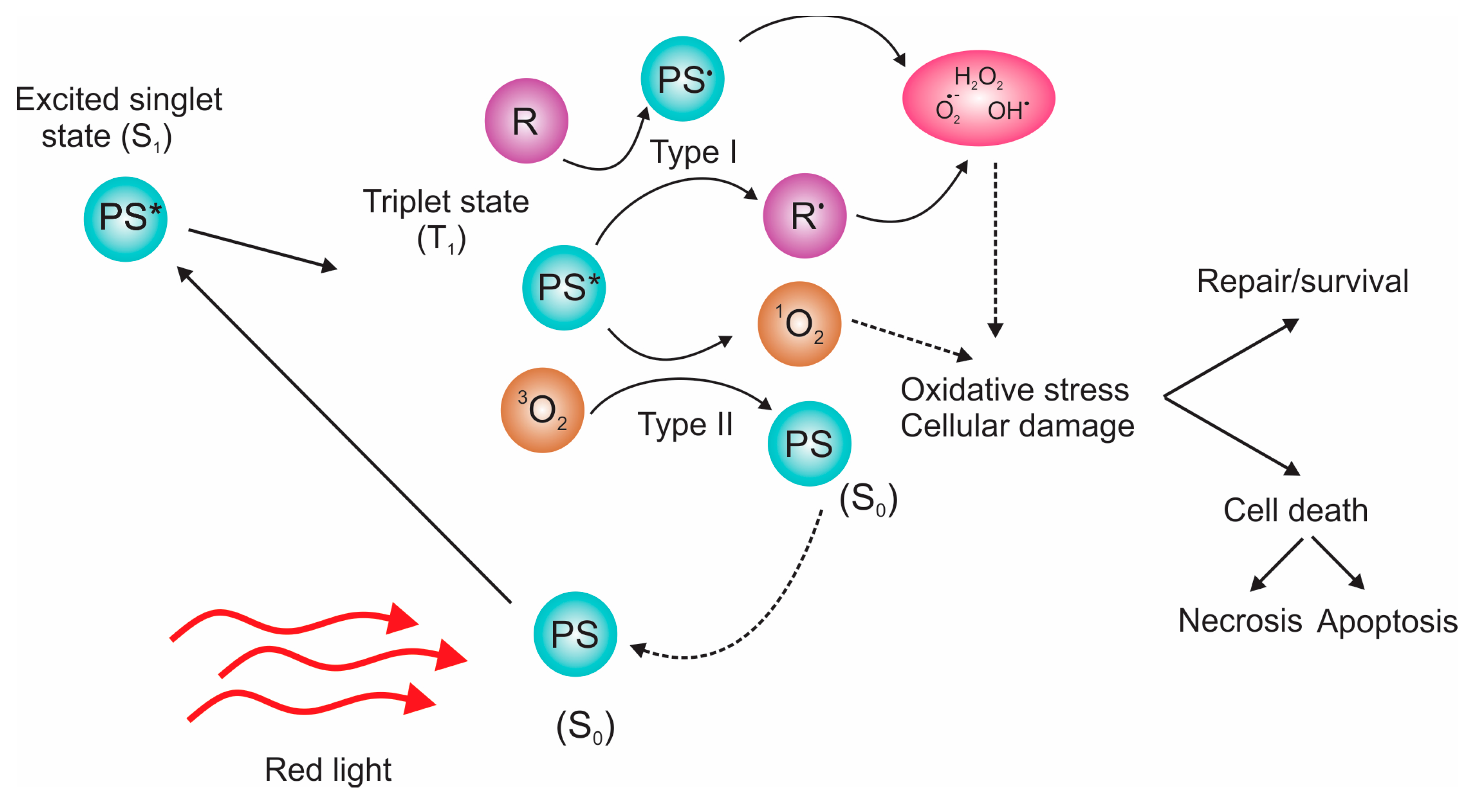
:1. Introduction to Photodynamic Therapy
- The PSs tend to build up in abnormal cells, and the application is focused strictly on them; that is why the damage to healthy cells is limited;
- No longterm side effects, and possibility for repeated treatments at the same position;
- Less invasive than surgery, and shorter recovery time;
- Immune activation ability;
- Usually costs less than other cancer treatments;
- Does not cause scarring, and is appropriate for treating skin or eye cancers.
2. Structure and Properties of Carbon-Based Dots
2.1. Graphene Quantum Dots, Structure, and Properties
- The size of the core (graphene);
- Edge configuration (zigzag or armchair);
- The physicochemical nature of functional groups.
- Carboxyl and amide groups mainly cause green emissions;
- Hydroxyl groups contribute to blue emissions [57];
- Red emissions depend on the sp2-conjugated size, and surface states control emission [58];
- Amine groups are electron-donating, increasing the electron density, and lowering the band gap [59];
- OH groups lead to various levels of disruption of the conjugated π-system, changing the dots’ structural flexibility, and making them more rigid [60].
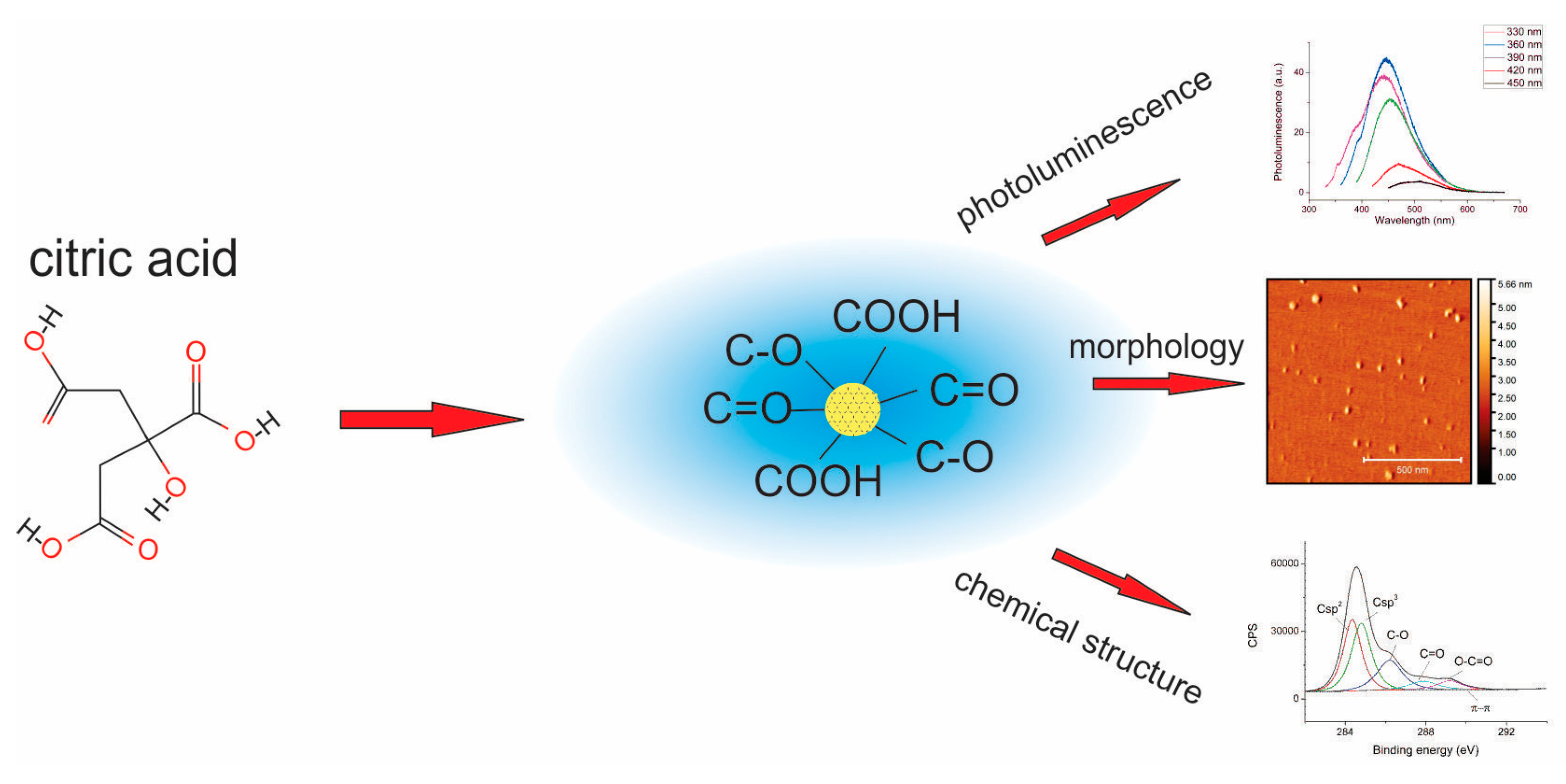
2.2. Carbon Quantum Dots
2.3. Doped Carbon Quantum Dots
3. ROS Production from Carbon-Based Dots
- Electrochemical cutting of graphite electrodes leads to the formation of GQDs functionalized with –OH groups (formed on the cathode), and GQDs functionalized with –CH groups (formed on the anode) [26]. The first type is water soluble, while the second one is soluble in acetone and toluene. Even though sp2 aromatic content is relatively large, –OH and –COOH groups quench singlet oxygen in aqueous solutions very efficiently. Acetone-soluble, and especially toluene-soluble GQDs, produce a significantly higher amount of singlet oxygen [unpublished data].
- CQDs are soluble in aqueous solvents, and have many functional groups that quench singlet oxygen. Some types of CQDs are excellent antioxidants [185,186,187,188,189,190,191]. As source material, fruits and vegetables with known antioxidant properties are commonly used. In Table 3, the DPPH scavenging activity for a variety of CQDs is presented:

| Title | Source Material | Scavenging Activity (%) |
|---|---|---|
| CQD [185] | Tannic acid | 84.5 |
| CQD [186] | Ananas | 23.3 |
| GQD [188] | Pyrene | 80 |
| Cl-CQD [189] | Citric acid, urea, NaCl | 88 |
| CQD [190] | Tomato | 63.8 |
| CQD [191] | Pomelo | 56 |
| T-CQD [192] | Thumbai | 89 |
| CQD [193] | Taurine | 82.5 |
| S-CQD [194] | Turmeric and ammonium persulfate | 79.5 |
| CD [195] | Carica papaya leaves | 86 |
| CD [196] | Beta vulgaris | 94.5 |
| CQD [197] | Citrus clementina peel | 81.4 |
| TCD [198] | Green tea | 75 |
| rcCQD [199] | Red cabbage | 61 |
| N-CD [200] | Black soya | 93.8 |
| N,S-CD [201] | Pomelo and sulfamic acid | 82 |
- The CNDs possess rather similar ROS generation and quenching properties to CQDs.
- The polymer content of CPDs provides excellent solubility in solvents, as well as a low quenching ability of singlet oxygen [112,202]. Quenching is especially low if copolymers are used for synthesis, with hydrophobic parts rich in methyl groups [109]. The architecture of the carbon core and the size of the π-conjugated domain are crucial for singlet oxygen generation.
4. Anticancer PDT with Carbon-Based Dots
4.1. Photodynamic Therapy with GQDs
Photodynamic Therapy with GQDs-Based Composites
4.2. Photodynamic Therapy with CQDs
5. Antibacterial PDT with GQDs and CQDs as Agent
| Material | Diameter | Toxicity | Mechanism | Observations |
|---|---|---|---|---|
| + charged, −charged, and 0 CDs [253] | 2.69–3.04 | + CDs: 100%, − CDs: ~80% 0 CDs: ~15% viability loss of E. coli incubated with 300 µg mL−1 for 6 h. | ROS production disrupting cytoplasmic membrane by + charged CDs | + CDs had the highest antibacterial activity, while 0 charged had the lowest |
| CQD-EDA [263] | 5 nm | E. coli: ~95% viability loss in the light conditions after 6 h | ROS production under visible light | The first report on the visible/natural light-activated antibacterial activity of CDs |
| GQDs [26] | 20–67 nm | E. coli: 80% MRSA: 90% viability loss; | ROS production under blue light (470 nm) | Fast antibacterial action, only 15 min of exposure |
| N-GQD [240] | 8 nm, height ~ 1.03 nm | Killing 100% of E. coli in only 3 min of exposure | ROS production under 670 nm laser irradiation, the synergistic effect of ROS and RNS (reactive nitrogen species) | Higher nitrogen content in GQDs leads to more efficient PDT |
| GQD, CQDCA, and CQDNH [28] | GQD: 14 nm; CQDCA: 22.5 nm; CQDNH: 12.5 nm | E. coli, E. aerogenes, P. aeruginosa, K. pneumoniae, B. subtilis MIC: 3.905–250 µg mL−1 | ROS production under blue light (470 nm) | N-CQDs showed the best antibacterial properties |
| CDs three groups according to sizes [261] | Small (s-CGCD): ~2 nm Middle (m-CGCD): ~3.9 nm Large (l-CGCD): ~5.3 nm | For E. coli: the concentration of s-CGCD ˃ 100 µg mL−1, for m-CGC and l-CGCD ˃150 µg mL−1 For S. aureus: 50, 75, and 100 µg mL−1 for s-CGCD, m-CGCD, and l-CGCD | No ROS production. The mechanism includes destroying the cytoplasmic membrane of bacteria by causing the leaking of cellular components | The antibacterial effect was increased with the decrease in particle size |
| Curcumin carbon dots from curcumin, neutral red, and citrate (Cur-NRCQDs) [264] | ~3.83 nm | Cur-NRCQDs inactivated 100% S. aureus and E. coli at concentrations of 10 and 15 μM | ROS production under the xenon lamp 555–850 nm | Cur-NRCQDs efficient against biofilms |
| Graphitic carbon nitride quantum dots: g-CNQDs [265] | 2–7 nm | Inhibition of ~99% of E. coli and ~90% of S. aureus at a concentration of 100 µg mL−1 | ROS production under visible light | Antibacterial activity of g-CNQDs was equivalent to silver nanoparticles |
| CDs from vitamin C [262] | ~5 nm | Killing 100% of a broad spectrum of bacteria at a concentration of 100 µg mL−1 at 150 µg mL−1, inhibiting the growth of fungus | CDs can enter the bacteria by diffusion, destroy the cell wall, bind to the DNA and RNA of bacteria, and finally kill them | These CDs could be degraded into CO2, CO, and H2O under visible light in the air after 20 days |
6. Cytotoxicity of GQDs and CQDs
6.1. Toxicity of GQDs
| Sample | Structure | Size | Toxicity |
|---|---|---|---|
| GQDs [269] | Oxygen-containing, no specific data | 20 nm | MGC-803 and MCF-7, 80% cell viability at 400 μg mL−1 |
| GQDs [270] | Carboxylated GQDs | 5 nm | KB, MDA-MB231, A549, and MDCK 80% cell viability at 500 μg mL−1 |
| GQDs [268] | GQDs-PEG, 36% O | 5 nm | 20 mg kg−1 every second day for 14 days |
| GQDs [272] | O-GQDs, C–O, C=O | 1.5–4 nm | Slight toxicity on macrophage at 400 μg mL−1 |
| GQDs [273] | PEG-GQDs | 6.6 nm to 88 nm PEG | Not toxic on HeLa cells at 8 μg mL−1 |
| N-GQDs [40] | NH2 groups, pyrrolic, pyridinic | 3.5 nm | No toxic effect on HeLa cells at 100 μg mL−1 (72 h) |
| GQDs [267] | NH2, COOH, and CO–N (CH3)2 | Low cytotoxicity to A549 at up to 200 μg mL−1 | |
| N-GQDs [274] | NH2 groups, pyridinic | 2.3–6.4 nm | No toxic effects on HeLa up to 200 μg mL−1, low effect on zebrafish embryos and larvae |
| Chiral [275] | L- or D-cysteine moieties attached to GQDs | 3–7 nm | HepG2 cells to l/d-GQDs general biocompatibility and d-GQDs accumulate in cellular membrane |
| N-GQDs [276] | N-doped | 5.1 nm | No hemolysis and release of ATP in RBCs, up to 200 μg mL−1 |
| N-doped GQDs [277] | N-doped, NH2 groups | 2.1 nm | No toxic effect SW480 cells at 0−1000 μg mL−1 |
| P,N-doped [278] | 41.79% C1s, 43.65% O1s, 5.85% N1s, 8.71% P2p | 4.2 | 90% T24 cell viability at 20 to 100 μg mL−1 |
| GQDs [279] | C 51 %, O 42%, N 8% | 20 | No toxic effect at 1000 μg mL−1 |
| N-doped [280] | Amino, and pyrrolic groups | 2.3–5.0 | 100% HeLa cells viability at 200 μg mL−1 after 48 h |
| GQDs [271] | C, O | 4–6 | 80% cell viability at 1 mg mL−1 |
| FA-GQDs [281] | Folic acid encapsulated N-GQDs | 33.59 | 80% HeLa cells viability at 2.0 mg mL−1 |
| GQDs [282] | / | / | In vivo, mice, 300 mg kg−1 |
6.2. Toxicity of CQDs
6.3. In Vivo Toxicity and Genotoxicity of GQDs and CQDs
7. Current Challenges and Future Prospects
- The development of anticancer and antibacterial medication;
- Establishing new procedures for treating these conditions;
- Creating the foundation for new products, such as antibacterial plastics, for medical and domestic usage.
Author Contributions
Funding
Institutional Review Board Statement
Informed Consent Statement
Data Availability Statement
Conflicts of Interest
References
- Ferlay, J.; Colombet, M.; Soerjomataram, I.; Parkin, D.M.; Piñeros, M.; Znaor, A.; Bray, F. Cancer statistics for the year 2020: An overview. Int. J. Cancer 2021, 149, 778–789. [Google Scholar] [CrossRef] [PubMed]
- Macdonald, I.J.; Dougherty, T.J. Basic principles of photodynamic therapy. Phthalocyanines 2001, 5, 105–129. [Google Scholar] [CrossRef]
- Figge, F.H.J.; Weiland, G.S.; Manganiello, L.O.J. Cancer Detection and Therapy. Affinity of Neoplastic, Embryonic, and Traumatized Tissues for Porphyrins and Metalloporphyrins. Exp. Biol. Med. 1948, 68, 640–641. [Google Scholar] [CrossRef] [PubMed]
- Rasmussen-Taxdal, D.S.; Ward, G.E.; Figge, F.H. Fluorescence of human lymphatic and cancer tissues following high doses of intravenous hematoporphyrin. Surg. Forum 1955, 5, 619–624. [Google Scholar] [CrossRef]
- Jesionek, A.; Tappeiner, H. Zur behandlung der haufcarcinome mit fluorescierenden stoffen. Deut. Arch. Klin. Med. 1904, 82, 223–227. [Google Scholar]
- Lipson, R.L.; Baldes, E.J.; Olsen, A.M. The use of a derivative of hematoporhyrin in tumor detection. J. Natl. Cancer Inst. 1961, 26, 1–11. [Google Scholar] [PubMed]
- Dougherty, T.J. Activated dyes as antitumor agents. J. Natl. Cancer Inst. 1974, 52, 1333–1336. [Google Scholar] [CrossRef]
- Dougherty, T.J. A brief history of clinical photodynamic therapy development at Roswell Park Cancer Institute. J. Clin. Laser Med. Surg. 1996, 14, 219–221. [Google Scholar] [CrossRef]
- Correia, J.H.; Rodrigues, J.A.; Pimenta, S.; Dong, T.; Yang, Z. Photodynamic Therapy Review: Principles, Photosensitizers, Applications, and Future Directions. Pharmaceutics 2021, 13, 1332. [Google Scholar] [CrossRef]
- Freitas, L.; Hamblin, M. Antimicrobial photoinactivation with functionalized fullerenes. In Nanobiomaterials in Antimicrobial Therapy; William Andrew Publishing: New York, NY, USA, 2016; pp. 1–27. [Google Scholar]
- Fekrazad, R.; Nejat, A.; Kalhori, K.A.M. Chapter 10—Antimicrobial Photodynamic Therapy With Nanoparticles Versus Conventional Photosensitizer in Oral Diseases. In Nanostructures for Antimicrobial Therapy; Ficai, A., Grumezescu, A.M., Eds.; Elsevier: Amsterdam, The Netherlands, 2017; pp. 237–259. [Google Scholar]
- He, H.; Zheng, X.; Liu, S.; Zheng, M.; Xie, Z.; Wang, Y.; Yu, M.; Shuai, X. Diketopyrrolopyrrole-based carbon dots for photodynamic therapy. Nanoscale 2018, 10, 10991–10998. [Google Scholar] [CrossRef]
- Xie, J.; Wang, Y.; Choi, W.; Jangili, P.; Ge, Y.; Xu, Y.; Kang, J.; Liu, L.; Zhang, B.; Xie, Z.; et al. Overcoming barriers in photodynamic therapy harnessing nano-formulation strategies. Chem. Soc. Rev. 2021, 50, 9152–9201. [Google Scholar] [CrossRef] [PubMed]
- Photodynamic Therapy to Treat Cancer. Available online: https://www.cancer.gov/about-cancer/treatment/types/photodynamic-therapy (accessed on 15 October 2022).
- Jacques, S.L. Optical properties of biological tissues: A review. Phys. Med. Biol. 2013, 58, R37–R61. [Google Scholar] [CrossRef]
- Agostinis, P.; Berg, K.; Cengel, K.A.; Foster, T.H.; Girotti, A.W.; Gollnick, S.O.; Hahn, S.M.; Hamblin, M.R.; Juzeniene, A.; Kessel, D.; et al. Photodynamic therapy of cancer: An update. CA Cancer J. Clin. 2011, 61, 250–281. [Google Scholar] [CrossRef] [PubMed]
- Chen, W.; Zhang, J. Using Nanoparticles to Enable Simultaneous Radiation and Photodynamic Therapies for Cancer Treatment. J. Nanosci. Nanotechnol. 2006, 6, 1159–1166. [Google Scholar] [CrossRef]
- Wang, P.; Wang, X.; Ma, L.; Sahi, S.; Li, L.; Wang, X.; Wang, Q.; Chen, Y.; Chen, W.; Liu, Q. Nanosonosensitization by Using Copper–Cysteamine Nanoparticles Augmented Sonodynamic Cancer Treatment. Part. Part. Syst. Charact. 2018, 35, 1700378. [Google Scholar] [CrossRef]
- Pandey, N.K.; Xiong, W.; Wang, L.; Chen, W.; Bui, B.; Yang, J.; Amador, E.; Chen, M.; Xing, C.; Athavale, A.A.; et al. Aggregation-induced emission luminogens for highly effective microwave dynamic therapy. Bioact. Mater. 2022, 7, 112–125. [Google Scholar] [CrossRef] [PubMed]
- Dong, X.; Liang, W.; Meziani, M.J.; Sun, Y.-P.; Yang, L. Carbon Dots as Potent Antimicrobial Agents. Theranostics 2020, 10, 671–686. [Google Scholar] [CrossRef]
- Cieplik, F.; Deng, D.; Crielaard, W.; Buchalla, W.; Hellwig, E.; Al-Ahmad, A.; Maisch, T. Antimicrobial photodynamic therapy—What we know and what we don’t. Crit. Rev. Microbiol. 2018, 44, 571–589. [Google Scholar] [CrossRef] [Green Version]
- Rajesh, S.; Koshi, E.; Philip, K.; Mohan, A. Antimicrobial photodynamic therapy: An overview. J. Indian Soc. Periodontol. 2011, 15, 323–327. [Google Scholar]
- Mahmoudi, H.; Bahador, A.; Pourhajibagher, M.; Alikhani, M.Y. Antimicrobial Photodynamic Therapy: An Effective Alternative Approach to Control Bacterial Infections. J. Lasers Med. Sci. 2018, 9, 154–160. [Google Scholar] [CrossRef] [Green Version]
- Zhou, C.; Peng, C.; Shi, C.; Jiang, M.; Chau, J.H.C.; Liu, Z.; Bai, H.; Kwok, R.T.K.; Lam, J.W.Y.; Shi, Y.; et al. Mitochondria-Specific Aggregation-Induced Emission Luminogens for Selective Photodynamic Killing of Fungi and Efficacious Treatment of Keratitis. ACS Nano 2021, 15, 12129–12139. [Google Scholar] [CrossRef] [PubMed]
- Tram, G.; Korolik, V.; Day, C.J. MBDS Solvent: An Improved Method for Assessment of Biofilms. J. Adv. Microbiol. 2013, 03, 200–204. [Google Scholar] [CrossRef] [Green Version]
- Ristic, B.Z.; Milenkovic, M.M.; Dakic, I.R.; Todorovic-Markovic, B.M.; Milosavljevic, M.S.; Budimir, M.D.; Paunovic, V.G.; Dramicanin, M.D.; Markovic, Z.M.; Trajkovic, V.S. Photodynamic antibacterial effect of graphene quantum dots. Biomaterials 2014, 35, 4428–4435. [Google Scholar] [CrossRef]
- Jovanovic, S.P.; Syrgiannis, Z.; Markovic, Z.M.; Bonasera, A.; Kepic, D.P.; Budimir, M.D.; Milivojevic, D.D.; Spasojevic, V.D.; Dramicanin, M.D.; Pavlovic, V.B.; et al. Modification of Structural and Luminescence Properties of Graphene Quantum Dots by Gamma Irradiation and Their Application in a Photodynamic Therapy. ACS Appl. Mater. Interfaces 2015, 7, 25865–25874. [Google Scholar] [CrossRef] [PubMed]
- Markovic, Z.M.; Jovanovic, S.P.; Maskovic, P.Z.; Danko, M.; Micusik, M.; Pavlovic, V.B.; Milivojevic, D.D.; Kleinova, A.; Spitalsky, Z.; Markovic, B.M.T. Photo-induced antibacterial activity of four graphene based nanomaterials on a wide range of bacteria. RSC Adv. 2018, 8, 31337–31347. [Google Scholar] [CrossRef] [Green Version]
- Marković, Z.M.; Jovanović, S.P.; Mašković, P.Z.; Mojsin, M.M.; Stevanović, M.J.; Danko, M.; Mičušík, M.; Jovanović, D.J.; Kleinová, A.; Špitalský, Z.; et al. Graphene oxide size and structure pro-oxidant and antioxidant activity and photoinduced cytotoxicity relation on three cancer cell lines. J. Photochem. Photobiol. B Biol. 2019, 200, 111647. [Google Scholar] [CrossRef] [PubMed]
- Stanković, N.K.; Todorović Marković, B.M.; Marković, Z.M. Self-assembly of carbon based nanoparticles films by Langmuir-Blodgett method—Review. J. Serb. Chem. Soc. 2020, 85, 1095–1127. [Google Scholar] [CrossRef] [Green Version]
- Xia, C.; Zhu, S.; Feng, T.; Yang, M.; Yang, B. Evolution and Synthesis of Carbon Dots: From Carbon Dots to Carbonized Polymer Dots. Adv. Sci. 2019, 6, 1901316. [Google Scholar] [CrossRef]
- Ponomarenko, L.A.; Schedin, F.; Katsnelson, M.I.; Yang, R.; Hill, E.W.; Novoselov, K.S.; Geim, A.K. Chaotic Dirac billiard in graphene quantum dots. Science 2008, 320, 356–358. [Google Scholar] [CrossRef] [Green Version]
- Novoselov, K.S.; Jiang, D.; Schedin, F.; Booth, T.J.; Khotkevich, V.V.; Morozov, S.V.; Geim, A.K. Two-dimensional atomic crystals. Proc. Natl. Acad. Sci. USA 2005, 102, 10451–10453. [Google Scholar] [CrossRef] [Green Version]
- Yan, X.; Cui, X.; Li, L.-S. Synthesis of Large, Stable Colloidal Graphene Quantum Dots with Tunable Size. J. Am. Chem. Soc. 2010, 132, 5944–5945. [Google Scholar] [CrossRef] [PubMed]
- Li, Y.; Hu, Y.; Zhao, Y.; Shi, G.; Deng, L.; Hou, Y.; Qu, L. An Electrochemical Avenue to Green-Luminescent Graphene Quantum Dots as Potential Electron-Acceptors for Photovoltaics. Adv. Mater. 2011, 23, 776–780. [Google Scholar] [CrossRef] [PubMed]
- Liu, R.L.; Wu, D.Q.; Feng, X.L.; Mullen, K. Bottom-Up Fabrication of Photoluminescent Graphene Quantum Dots with Uniform Morphology. J. Am. Chem. Soc. 2011, 133, 15221–15223. [Google Scholar] [CrossRef]
- Dong, Y.; Shao, J.; Chen, C.; Li, H.; Wang, R.; Chi, Y.; Lin, X.; Chen, G. Blue luminescent graphene quantum dots and graphene oxide prepared by tuning the carbonization degree of citric acid. Carbon 2012, 50, 4738–4743. [Google Scholar] [CrossRef]
- Lin, L.; Zhang, S. Creating high yield water soluble luminescent graphene quantum dots via exfoliating and disintegrating carbon nanotubes and graphite flakes. Chem. Commun. 2012, 48, 10177–10179. [Google Scholar] [CrossRef] [PubMed]
- Atienzar, P.; Primo, A.; Lavorato, C.; Molinari, R.; García, H. Preparation of graphene quantum dots from pyrolyzed alginate. Langmuir 2013, 29, 6141–6146. [Google Scholar] [CrossRef]
- Hu, C.; Liu, Y.; Yang, Y.; Cui, J.; Huang, Z.; Wang, Y.; Yang, L.; Wang, H.; Xiao, Y.; Rong, J. One-step preparation of nitrogen-doped graphene quantum dots from oxidized debris of graphene oxide. J. Mater. Chem. B 2013, 1, 39–42. [Google Scholar] [CrossRef]
- Luo, Z.M.; Yang, D.L.; Qi, G.Q.; Shang, J.Z.; Yang, H.P.; Wang, Y.L.; Yuwen, L.H.; Yu, T.; Huang, W.; Wang, L.H. Microwave-assisted solvothermal preparation of nitrogen and sulfur co-doped reduced graphene oxide and graphene quantum dots hybrids for highly efficient oxygen reduction. J. Mater. Chem. A 2014, 2, 20605–20611. [Google Scholar] [CrossRef]
- Shin, Y.; Lee, J.; Yang, J.; Park, J.; Lee, K.; Kim, S.; Park, Y.; Lee, H. Mass Production of Graphene Quantum Dots by One-Pot Synthesis Directly from Graphite in High Yield. Small 2014, 10, 866–870. [Google Scholar] [CrossRef]
- Cai, Z.W.; Li, F.M.; Wu, P.; Ji, L.J.; Zhang, H.; Cai, C.X.; Gervasio, D.F. Synthesis of Nitrogen-Doped Graphene Quantum Dots at Low Temperature for Electrochemical Sensing Trinitrotoluene. Anal. Chem. 2015, 87, 11803–11811. [Google Scholar] [CrossRef]
- Li, L.; Li, L.; Wang, C.; Liu, K.; Zhu, R.; Qiang, H.; Lin, Y. Synthesis of nitrogen-doped and amino acid-functionalized graphene quantum dots from glycine, and their application to the fluorometric determination of ferric ion. Microchim. Acta 2015, 182, 763–770. [Google Scholar] [CrossRef]
- Dai, Y.; Pang, H.; Huang, J.; Yang, Y.; Huang, H.; Wang, K.; Ma, Z.; Liao, B. Tailoring of ammonia reduced graphene oxide into amine functionalized graphene quantum dots through a Hofmann rearrangement. RSC Adv. 2016, 6, 34514–34520. [Google Scholar] [CrossRef]
- Ahirwar, S.; Mallick, S.; Bahadur, D. Electrochemical Method To Prepare Graphene Quantum Dots and Graphene Oxide Quantum Dots. ACS Omega 2017, 2, 8343–8353. [Google Scholar] [CrossRef] [PubMed] [Green Version]
- Pan, D.Y.; Zhang, J.C.; Li, Z.; Wu, M.H. Hydrothermal Route for Cutting Graphene Sheets into Blue-Luminescent Graphene Quantum Dots. Adv. Mater. 2010, 22, 734–738. [Google Scholar] [CrossRef] [PubMed]
- Novoselov, K.S.; Geim, A.K.; Morozov, S.V.; Jiang, D.; Katsnelson, M.I.; Grigorieva, I.V.; Dubonos, S.V.; Firsov, A.A. Two-dimensional gas of massless Dirac fermions in graphene. Nature 2005, 438, 197–200. [Google Scholar] [CrossRef] [Green Version]
- Ritter, K.A.; Lyding, J.W. The influence of edge structure on the electronic properties of graphene quantum dots and nanoribbons. Nat. Mater. 2009, 8, 235–242. [Google Scholar] [CrossRef]
- Kim, S.; Hwang, S.W.; Kim, M.-K.; Shin, D.Y.; Shin, D.H.; Kim, C.O.; Yang, S.B.; Park, J.H.; Hwang, E.; Choi, S.-H.; et al. Anomalous Behaviors of Visible Luminescence from Graphene Quantum Dots: Interplay between Size and Shape. ACS Nano 2012, 6, 8203–8208. [Google Scholar] [CrossRef]
- Song, Y.; Wang, J.; Wan, H.; Zhang, Y.; Ningcong, Y.; Yang, B. Photoluminescence mechanism in graphene quantum dots: Quantum confinement effect and surface/edge state. Nano Today 2016, 13, 10–14. [Google Scholar]
- Zhu, S.; Wang, L.; Li, B.; Song, Y.; Zhao, X.; Zhang, G.; Zhang, S.; Lu, S.; Zhang, J.; Wang, H.; et al. Investigation of photoluminescence mechanism of graphene quantum dots and evaluation of their assembly into polymer dots. Carbon 2014, 77, 462–472. [Google Scholar] [CrossRef]
- He, S.J.; Turnbull, M.J.; Nie, Y.T.; Sun, X.H.; Ding, Z.F. Band structures of blue luminescent nitrogen-doped graphene quantum dots by synchrotron-based XPS. Surf. Sci. 2018, 676, 51–55. [Google Scholar] [CrossRef]
- Guin, J.P.; Guin, S.K.; Debnath, T.; Ghosh, H.N. Chemically clean single-step oxido-reductive synthesis of green luminescent graphene quantum dots as impending electrocatalyst. Carbon 2016, 109, 517–528. [Google Scholar] [CrossRef]
- Wang, Z.; Chen, D.; Gu, B.; Gao, B.; Liu, Z.; Yang, Y.; Guo, Q.; Zheng, X.; Wang, G. Yellow emissive nitrogen-doped graphene quantum dots as a label-free fluorescent probe for Fe3+ sensing and bioimaging. Diam. Relat. Mater. 2020, 104, 107749. [Google Scholar] [CrossRef]
- Lin, L.P.; Song, X.H.; Chen, Y.Y.; Rong, M.C.; Zhao, T.T.; Jiang, Y.Q.; Wang, Y.R.; Chen, X. One-pot synthesis of highly greenish-yellow fluorescent nitrogen-doped graphene quantum dots for pyrophosphate sensing via competitive coordination with Eu3+ ions. Nanoscale 2015, 7, 15427–15433. [Google Scholar] [CrossRef]
- Zhu, S.; Shao, J.; Song, Y.; Zhao, X.; Du, J.; Wang, L.; Wang, H.; Zhang, K.; Zhang, J.; Yang, B. Investigating the surface state of graphene quantum dots. Nanoscale 2015, 7, 7927–7933. [Google Scholar] [CrossRef] [PubMed]
- Yan, Y.; Chen, J.; Li, N.; Tian, J.; Li, K.; Jiang, J.; Liu, J.; Tian, Q.; Chen, P. Systematic Bandgap Engineering of Graphene Quantum Dots and Applications for Photocatalytic Water Splitting and CO2 Reduction. ACS Nano 2018, 12, 3523–3532. [Google Scholar] [CrossRef]
- Jin, S.H.; Kim, D.H.; Jun, G.H.; Hong, S.H.; Jeon, S. Tuning the Photoluminescence of Graphene Quantum Dots through the Charge Transfer Effect of Functional Groups. ACS Nano 2013, 7, 1239–1245. [Google Scholar] [CrossRef] [PubMed]
- Cui, P.; Xue, Y. Influence of edge and center oxidation configurations on non-radiative relaxation in graphene quantum dots. J. Mater. Sci. Mater. Electron. 2022, 33, 5024–5036. [Google Scholar] [CrossRef]
- Tang, S.; Chen, D.; Yang, Y.; Wang, C.; Li, X.; Wang, Y.; Gu, C.; Cao, Z. Mechanisms behind multicolor tunable Near-Infrared triple emission in graphene quantum dots and ratio fluorescent probe for water detection. J. Colloid Interface Sci. 2022, 617, 182–192. [Google Scholar] [CrossRef]
- Zhu, W.; Feng, X.; Zhao, M.; Wei, Z.; Liu, Z.; Wang, G.; Guo, Q.; Chen, D. Scalable and atom economic preparation of red-near-infrared emitted N-doped graphene quantum dots with a high quantum yield. Diam. Relat. Mater. 2021, 116, 108395. [Google Scholar] [CrossRef]
- Ge, J.; Lan, M.; Zhou, B.; Liu, W.; Guo, L.; Wang, H.; Jia, Q.; Niu, G.; Huang, X.; Zhou, H.; et al. A graphene quantum dot photodynamic therapy agent with high singlet oxygen generation. Nat. Commun. 2014, 5, 4596. [Google Scholar] [CrossRef] [Green Version]
- Tan, X.; Li, Y.; Li, X.; Zhou, S.; Fan, L.; Yang, S. Electrochemical synthesis of small-sized red fluorescent graphene quantum dots as a bioimaging platform. Chem. Commun. 2015, 51, 2544–2546. [Google Scholar] [CrossRef] [PubMed]
- Wu, X.; Tian, F.; Wang, W.; Chen, J.; Wu, M.; Zhao, J.X. Fabrication of highly fluorescent graphene quantum dots using L-glutamic acid for in vitro/in vivo imaging and sensing. J. Mater. Chem. C 2013, 1, 4676–4684. [Google Scholar] [CrossRef] [PubMed] [Green Version]
- Juang, R.S.; Hsieh, C.T.; Kao, C.P.; Gandomi, Y.A.; Fu, C.C.; Liu, S.H.; Gu, S. Highly fluorescent green and red emissions from boron-doped graphene quantum dots under blue light illumination. Carbon 2021, 176, 61–70. [Google Scholar] [CrossRef]
- Cao, H.; Qi, W.; Gao, X.; Wu, Q.; Tian, L.; Wu, W. Graphene Quantum Dots prepared by Electron Beam Irradiation for Safe Fluorescence Imaging of Tumor. Nanotheranostics 2022, 6, 205–214. [Google Scholar] [CrossRef] [PubMed]
- Yang, Y.; Tang, S.; Chen, D.; Wang, C.; Gu, B.; Li, X.; Xie, F.; Wang, G.; Guo, Q. Multifunctional red-emission graphene quantum dots with tunable light emissions for trace water sensing, WLEDs and information encryption. Colloids Surf. A Physicochem. Eng. Asp. 2021, 622, 126593. [Google Scholar] [CrossRef]
- Wang, C.; Chen, D.; Tang, S.; Yang, Y.; Li, X.; Xie, F.; Guo, Q. PVDF-triggered multicolor fluorine-doped graphene quantum dots for water detection and anti-counterfeiting. Microchim. Acta 2021, 189, 6. [Google Scholar] [CrossRef]
- Zhao, J.; Zheng, Y.; Pang, Y.; Chen, J.; Zhang, Z.; Xi, F.; Chen, P. Graphene quantum dots as full-color and stimulus responsive fluorescence ink for information encryption. J. Colloid Interf. Sci. 2020, 579, 307–314. [Google Scholar] [CrossRef] [PubMed]
- Kadian, S.; Manik, G. Sulfur doped graphene quantum dots as a potential sensitive fluorescent probe for the detection of quercetin. Food Chem. 2020, 317, 126457. [Google Scholar] [CrossRef]
- Ye, R.; Peng, Z.; Metzger, A.; Lin, J.; Mann, J.A.; Huang, K.; Xiang, C.; Fan, X.; Samuel, E.L.G.; Alemany, L.B.; et al. Bandgap engineering of coal-derived graphene quantum dots. ACS Appl. Mater. Interfaces 2015, 7, 7041–7048. [Google Scholar] [CrossRef]
- Gao, T.; Wang, X.; Yang, L.-Y.; He, H.; Ba, X.-X.; Zhao, J.; Jiang, F.-L.; Liu, Y. Red, Yellow, and Blue Luminescence by Graphene Quantum Dots: Syntheses, Mechanism, and Cellular Imaging. ACS Appl. Mater. Interfaces 2017, 9, 24846–24856. [Google Scholar] [CrossRef]
- Yang, G.; Wu, C.; Luo, X.; Liu, X.; Gao, Y.; Wu, P.; Cai, C.; Scott Saavedra, S. Exploring the Emissive States of Heteroatom-Doped Graphene Quantum Dots. J. Phys. Chem. C 2018, 122, 6483–6492. [Google Scholar] [CrossRef]
- Wang, R.; Fan, H.; Jiang, W.; Ni, G.; Qu, S. Amino-functionalized graphene quantum dots prepared using high-softening point asphalt and their application in Fe3+ detection. Appl. Surf. Sci. 2019, 467–468, 446–455. [Google Scholar] [CrossRef]
- Sivaselvam, S.; Viswanathan, C.; Ponpandian, N. One-step preparation of N-doped grapheme quantum dots with high quantum yield for bioimaging and highly sensitive electrochemical detection of isoniazid. Biomater. Adv. 2022, 135, 212731. [Google Scholar] [CrossRef] [PubMed]
- Wang, H.; Qi, C.; Yang, A.; Wang, X.; Xu, J. One-pot synthesis of bright blue luminescent N-doped GQDs: Optical properties and cell imaging. Nanomaterials 2021, 11, 2798. [Google Scholar] [CrossRef]
- Selvakumar, T.; Rajaram, M.; Natarajan, A.; Harikrishnan, L.; Alwar, K.; Rajaram, A. Highly Efficient Sulfur and Nitrogen Codoped Graphene Quantum Dots as a Metal-Free Green Photocatalyst for Photocatalysis and Fluorescent Ink Applications. ACS Omega 2022, 7, 12825–12834. [Google Scholar] [CrossRef]
- Cunci, L.; González-Colón, V.; Lee Vargas-Pérez, B.; Ortiz-Santiago, J.; Pagán, M.; Carrion, P.; Cruz, J.; Molina-Ontoria, A.; Martinez, N.; Silva, W.; et al. Multicolor Fluorescent Graphene Oxide Quantum Dots for Sensing Cancer Cell Biomarkers. ACS Appl. Nano Mater. 2021, 4, 211–219. [Google Scholar] [CrossRef]
- Ngoc Anh, N.T.; Chang, P.-Y.; Doong, R.-A. Sulfur-doped graphene quantum dot-based paper sensor for highly sensitive and selective detection of 4-nitrophenol in contaminated water and wastewater. RSC Adv. 2019, 9, 26588–26597. [Google Scholar] [CrossRef] [Green Version]
- Cui, P.; Xue, Y. Tuning nonradiative recombination loss by selective oxidation patterns of epoxy groups bound to different sites of graphene quantum dots. J. Chem. Eng. 2021, 431, 134052. [Google Scholar] [CrossRef]
- Shen, J.; Zhu, Y.; Yang, X.; Zong, J.; Zhang, J.; Li, C. One-Pot Hydrothermal Synthesis of Graphene Quantum Dots Surface-Passivated by Polyethylene Glycol and Their Photoelectric Conversion under Near-Infrared Light. New J. Chem. 2011, 36, 97–101. [Google Scholar] [CrossRef]
- Tetsuka, H.; Asahi, R.; Nagoya, A.; Okamoto, K.; Tajima, I.; Ohta, R.; Okamoto, A. Optically Tunable Amino-Functionalized Graphene Quantum Dots. Adv. Mater. 2012, 24, 5333–5338. [Google Scholar] [CrossRef]
- Kong, W.; Wang, Y.; Wang, L.; Li, Y.; Li, Y.; Xue, W. Investigation of photoluminescence behavior of reduced graphene quantum dots. Inorg. Chem. Commun. 2019, 99, 199–205. [Google Scholar] [CrossRef]
- Jovanović, S.; Marković, Z.; Budimir, M.; Spitalsky, Z.; Vidoeski, B.; Todorović Marković, B. Effects of low gamma irradiation dose on the photoluminescence properties of graphene quantum dots. Opt. Quantum. Electron. 2016, 48, 259. [Google Scholar] [CrossRef]
- Jovanović, S.; Dorontić, S.; Jovanović, D.; Ciasca, G.; Budimir, M.; Bonasera, A.; Scopelliti, M.; Marković, O.; Todorović Marković, B. Gamma irradiation of graphene quantum dots with ethylenediamine: Antioxidant for ion sensing. Ceram. Int. 2020, 46, 23611–23622. [Google Scholar] [CrossRef]
- Xie, M.; Su, Y.; Lu, X.; Zhang, Y.; Yang, Z.; Zhang, Y. Blue and green photoluminescence graphene quantum dots synthesized from carbon fibers. Mater. Lett. 2013, 93, 161–164. [Google Scholar] [CrossRef]
- Zhang, M.; Bai, L.; Shang, W.; Xie, W.; Ma, H.; Fu, Y.; Fang, D.; Sun, H.; Fan, L.; Han, M.; et al. Facile synthesis of water-soluble, highly fluorescent graphene quantum dots as a robust biological label for stem cells. J. Mater. Chem. 2012, 22, 7461–7467. [Google Scholar] [CrossRef]
- Tang, L.; Ji, R.; Cao, X.; Lin, J.; Jiang, H.; Li, X.; Teng, K.S.; Luk, C.M.; Zeng, S.; Hao, J.; et al. Deep Ultraviolet Photoluminescence of Water-Soluble Self-Passivated Graphene Quantum Dots. ACS Nano 2012, 6, 5102–5110. [Google Scholar] [CrossRef] [PubMed]
- Ding, Z.; Hao, Z.; Meng, B.; Xie, Z.; Liu, J.; Dai, L. Few-layered graphene quantum dots as efficient hole-extraction layer for high-performance polymer solar cells. Nano Energy 2015, 15, 186–192. [Google Scholar] [CrossRef]
- Wolk, A.; Rosenthal, M.; Neuhaus, S.; Huber, K.; Brassat, K.; Lindner, J.K.N.; Grothe, R.; Grundmeier, G.; Bremser, W.; Wilhelm, R. A Novel Lubricant Based on Covalent Functionalized Graphene Oxide Quantum Dots. Sci. Rep. 2018, 8, 5843. [Google Scholar] [CrossRef] [Green Version]
- Wang, Y.; Hu, A. Carbon quantum dots: Synthesis, properties and applications. J. Mater. Chem. C 2014, 2, 6921–6939. [Google Scholar] [CrossRef] [Green Version]
- Marković, Z.; Todorović-Marković, B. Treating of Aquatic Pollution by Carbon Quantum Dots. In Nanostructured Materials for Treating Aquatic Pollution; Gonçalves, G.A.B., Marques, P., Eds.; Springer: Berlin/Heidelberg, Germany, 2019; pp. 121–145. [Google Scholar]
- Lim, S.Y.; Shen, W.; Gao, Z. Carbon quantum dots and their applications. Chem. Soc. Rev. 2015, 44, 362–381. [Google Scholar] [CrossRef]
- Travlou, N.A.; Giannakoudakis, D.A.; Algarra, M.; Labella, A.M.; Rodríguez-Castellón, E.; Bandosz, T.J. S- and N-doped carbon quantum dots: Surface chemistry dependent antibacterial activity. Carbon 2018, 135, 104–111. [Google Scholar] [CrossRef]
- Dong, Y.; Wang, R.; Li, H.; Shao, J.; Chi, Y.; Lin, X.; Chen, G. Polyamine-functionalized carbon quantum dots for chemical sensing. Carbon 2012, 50, 2810–2815. [Google Scholar] [CrossRef]
- Zhu, S.; Meng, Q.; Wang, L.; Zhang, J.; Song, Y.; Jin, H.; Zhang, K.; Sun, H.; Wang, H.; Yang, B. Highly Photoluminescent Carbon Dots for Multicolor Patterning, Sensors, and Bioimaging. Angew. Chem. Int. Ed. 2013, 52, 3953–3957. [Google Scholar] [CrossRef] [PubMed]
- Yang, Z.-C.; Wang, M.; Yong, A.M.; Wong, S.Y.; Zhang, X.-H.; Tan, H.; Chang, A.Y.; Li, X.; Wang, J. Intrinsically fluorescent carbon dots with tunable emission derived from hydrothermal treatment of glucose in the presence of monopotassium phosphate. Chem. Commun. 2011, 47, 11615–11617. [Google Scholar] [CrossRef]
- Yang, Y.; Cui, J.; Zheng, M.; Hu, C.; Tan, S.; Xiao, Y.; Yang, Q.; Liu, Y. One-step synthesis of amino-functionalized fluorescent carbon nanoparticles by hydrothermal carbonization of chitosan. Chem. Commun. 2012, 48, 380–382. [Google Scholar] [CrossRef]
- De, B.; Karak, N. A green and facile approach for the synthesis of water soluble fluorescent carbon dots from banana juice. RSC Adv. 2013, 3, 8286–8290. [Google Scholar] [CrossRef]
- Alam, A.-M.; Park, B.-Y.; Ghouri, Z.K.; Park, M.; Kim, H.-Y. Synthesis of carbon quantum dots from cabbage with down- and up-conversion photoluminescence properties: Excellent imaging agent for biomedical applications. Green Chem. 2015, 17, 3791–3797. [Google Scholar] [CrossRef]
- Arumugam, N.; Kim, J. Synthesis of carbon quantum dots from Broccoli and their ability to detect silver ions. Mater. Lett. 2018, 219, 37–40. [Google Scholar] [CrossRef]
- Liu, Y.; Xiao, N.; Gong, N.; Wang, H.; Shi, X.; Gu, W.; Ye, L. One-step microwave-assisted polyol synthesis of green luminescent carbon dots as optical nanoprobes. Carbon 2014, 68, 258–264. [Google Scholar] [CrossRef]
- Chae, A.; Choi, Y.; Jo, S.; Nur’aeni; Paoprasert, P.; Park, S.Y.; In, I. Microwave-assisted synthesis of fluorescent carbon quantum dots from an A2/B3 monomer set. RSC Adv. 2017, 7, 12663–12669. [Google Scholar] [CrossRef] [Green Version]
- Wang, J.; Cheng, C.; Huang, Y.; Zheng, B.; Yuan, H.; Bo, L.; Zheng, M.-W.; Yang, S.-Y.; Guo, Y.; Xiao, D. A facile large-scale microwave synthesis of highly fluorescent carbon dots from benzenediol isomers. J. Mater. Chem. C 2014, 2, 5028–5035. [Google Scholar] [CrossRef]
- Bhunia, S.K.; Saha, A.; Maity, A.R.; Ray, S.C.; Jana, N.R. Carbon Nanoparticle-based Fluorescent Bioimaging Probes. Sci. Rep. 2013, 3, 1473. [Google Scholar] [CrossRef] [PubMed] [Green Version]
- Zhao, P.; Zhu, L. Dispersibility of carbon dots in aqueous and/or organic solvents. Chem. Commun. 2018, 54, 5401–5406. [Google Scholar] [CrossRef] [PubMed]
- Mao, Q.-X.; Wang, W.-J.; Hai, X.; Shu, Y.; Chen, X.-W.; Wang, J.-H. The regulation of hydrophilicity and hydrophobicity of carbon dots via a one-pot approach. J. Mater. Chem. B 2015, 3, 6013–6018. [Google Scholar] [CrossRef]
- Stanković, N.K.; Bodik, M.; Šiffalovič, P.; Kotlar, M.; Mičušik, M.; Špitalsky, Z.; Danko, M.; Milivojević, D.D.; Kleinova, A.; Kubat, P.; et al. Antibacterial and Antibiofouling Properties of Light Triggered Fluorescent Hydrophobic Carbon Quantum Dots Langmuir–Blodgett Thin Films. ACS Sustain. Chem. Eng. 2018, 6, 4154–4163. [Google Scholar] [CrossRef]
- Pan, D.; Zhang, J.; Li, Z.; Zhang, Z.; Guo, L.; Wu, M. Blue fluorescent carbon thin films fabricated from dodecylamine-capped carbon nanoparticles. J. Mater. Chem. 2011, 21, 3565–3567. [Google Scholar] [CrossRef]
- Mitra, S.; Chandra, S.; Kundu, T.; Banerjee, R.; Pramanik, P.; Goswami, A. Rapid microwave synthesis of fluorescent hydrophobic carbon dots. RSC Adv. 2012, 2, 12129–12131. [Google Scholar] [CrossRef]
- Kováčová, M.; Marković, Z.M.; Humpolíček, P.; Mičušík, M.; Švajdlenková, H.; Kleinová, A.; Danko, M.; Kubát, P.; Vajďák, J.; Capáková, Z.; et al. Carbon Quantum Dots Modified Polyurethane Nanocomposite as Effective Photocatalytic and Antibacterial Agents. ACS Biomater. Sci. Eng. 2018, 4, 3983–3993. [Google Scholar] [CrossRef]
- Marković, Z.M.; Kováčová, M.; Humpolíček, P.; Budimir, M.D.; Vajďák, J.; Kubát, P.; Mičušík, M.; Švajdlenková, H.; Danko, M.; Capáková, Z.; et al. Antibacterial photodynamic activity of carbon quantum dots/polydimethylsiloxane nanocomposites against Staphylococcus aureus, Escherichia coli and Klebsiella pneumoniae. Photodiagnosis Photodyn. Ther. 2019, 26, 342–349. [Google Scholar] [CrossRef]
- Budimir, M.; Marković, Z.; Vajdak, J.; Jovanović, S.; Kubat, P.; Humpoliček, P.; Mičušik, M.; Danko, M.; Barras, A.; Milivojević, D.; et al. Enhanced visible light-triggered antibacterial activity of carbon quantum dots/polyurethane nanocomposites by gamma rays induced pre-treatment. Radiat. Phys. Chem. 2021, 185, 109499. [Google Scholar] [CrossRef]
- Kováčová, M.; Bodík, M.; Mičušík, M.; Humpolíček, P.; Šiffalovič, P.; Špitálsky, Z. Increasing the effectivity of the antimicrobial surface of carbon quantum dots-based nanocomposite by atmospheric pressure plasma. Clin. Plasma Med. 2020, 19–20, 100111. [Google Scholar] [CrossRef]
- Langer, M.; Paloncýová, M.; Medveď, M.; Pykal, M.; Nachtigallová, D.; Shi, B.; Aquino, A.J.A.; Lischka, H.; Otyepka, M. Progress and challenges in understanding of photoluminescence properties of carbon dots based on theoretical computations. Appl. Mater. Today 2021, 22, 100924. [Google Scholar] [CrossRef]
- Zhu, S.; Song, Y.; Zhao, X.; Shao, J.; Zhang, J.; Yang, B. The photoluminescence mechanism in carbon dots (graphene quantum dots, carbon nanodots, and polymer dots): Current state and future perspective. Nano Res. 2015, 8, 355–381. [Google Scholar] [CrossRef]
- Xu, Y.; Yu, H.; Chudal, L.; Pandey, N.K.; Amador, E.H.; Bui, B.; Wang, L.; Ma, X.; Deng, S.; Zhu, X.; et al. Striking luminescence phenomena of carbon dots and their applications as a double ratiometric fluorescence probes for H2S detection. Mater. Today Phys. 2021, 17, 100328. [Google Scholar] [CrossRef]
- Tang, X.-D.; Yu, H.-M.; Nguyen, W.; Amador, E.; Cui, S.-P.; Ma, K.; Chen, M.-L.; Wang, S.-Y.; Hu, Z.-Z.; Chen, W. New Observations on Concentration-Regulated Carbon Dots. Adv. Photonics Res. 2023, 4, 2200314. [Google Scholar] [CrossRef]
- Kundelev, E.V.; Tepliakov, N.V.; Leonov, M.Y.; Maslov, V.G.; Baranov, A.V.; Fedorov, A.V.; Rukhlenko, I.D.; Rogach, A.L. Amino Functionalization of Carbon Dots Leads to Red Emission Enhancement. J. Phys. Chem. Lett. 2019, 10, 5111–5116. [Google Scholar] [CrossRef] [PubMed]
- Janus, Ł.; Radwan-Pragłowska, J.; Piątkowski, M.; Bogdał, D. Facile Synthesis of Surface-Modified Carbon Quantum Dots (CQDs) for Biosensing and Bioimaging. Materials 2020, 13, 3313. [Google Scholar] [CrossRef] [PubMed]
- Wang, H.-y.; Zhou, L.; Yu, H.; Tang, X.; Xing, C.; Nie, G.; Akafzade, H.; Wang, S.; Chen, W. Exploration of Room-Temperature Phosphorescence and New Mechanism on Carbon Dots in a Polyacrylamide Platform and their Applications for Anti-Counterfeiting and Information Encryption. Adv. Opt. Mater. 2022, 10, 2200678. [Google Scholar] [CrossRef]
- Yoon, H.; Chang, Y.H.; Song, S.H.; Lee, E.-S.; Jin, S.H.; Park, C.; Lee, J.; Kim, B.H.; Kang, H.J.; Kim, Y.-H.; et al. Intrinsic Photoluminescence Emission from Subdomained Graphene Quantum Dots. Adv. Mater. 2016, 28, 5255–5261. [Google Scholar] [CrossRef]
- Kandasamy, G. Recent Advancements in Doped/Co-Doped Carbon Quantum Dots for Multi-Potential Applications. C 2019, 5, 24. [Google Scholar] [CrossRef] [Green Version]
- Ge, J.; Jia, Q.; Liu, W.; Guo, L.; Liu, Q.; Lan, M.; Zhang, H.; Meng, X.; Wang, P. Red-Emissive Carbon Dots for Fluorescent, Photoacoustic, and Thermal Theranostics in Living Mice. Adv. Mater. 2015, 27, 4169–4177. [Google Scholar] [CrossRef] [PubMed]
- Yan, F.; Jiang, Y.; Sun, X.; Bai, Z.; Zhang, Y.; Zhou, X. Surface modification and chemical functionalization of carbon dots: A review. Microchim. Acta 2018, 185, 424. [Google Scholar] [CrossRef] [PubMed]
- Dimos, K. Carbon Quantum Dots: Surface Passivation and Functionalization. Curr. Org. Chem. 2015, 19, 682–695. [Google Scholar] [CrossRef]
- Alaghmandfard, A.; Sedighi, O.; Tabatabaei Rezaei, N.; Abedini, A.A.; Malek Khachatourian, A.; Toprak, M.S.; Seifalian, A. Recent advances in the modification of carbon-based quantum dots for biomedical applications. Mater. Sci. Eng. C 2021, 120, 111756. [Google Scholar] [CrossRef] [PubMed]
- Park, Y.; Yoo, J.; Lim, B.; Kwon, W.; Rhee, S.W. Improving the functionality of carbon nanodots: Doping and surface functionalization. J. Mater. Chem. A 2016, 4, 11582–11603. [Google Scholar] [CrossRef]
- Lin, L.; Luo, Y.; Tsai, P.; Wang, J.; Chen, X. Metal ions doped carbon quantum dots: Synthesis, physicochemical properties, and their applications. TrAC Trends Anal. Chem. 2018, 103, 87–101. [Google Scholar] [CrossRef]
- Tejwan, N.; Saini, A.K.; Sharma, A.; Singh, T.A.; Kumar, N.; Das, J. Metal-doped and hybrid carbon dots: A comprehensive review on their synthesis and biomedical applications. J. Control. Release 2021, 330, 132–150. [Google Scholar] [CrossRef]
- Yuan, F.; Li, S.; Fan, Z.; Meng, X.; Fan, L.; Yang, S. Shining carbon dots: Synthesis and biomedical and optoelectronic applications. Nano Today 2016, 11, 565–586. [Google Scholar] [CrossRef]
- Lin, L.; Song, X.; Chen, Y.; Rong, M.; Wang, Y.; Zhao, L.; Zhao, T.; Chen, X. Europium-decorated graphene quantum dots as a fluorescent probe for label-free, rapid and sensitive detection of Cu2+ and l-cysteine. Anal. Chim. Acta 2015, 891, 261–268. [Google Scholar] [CrossRef]
- Li Liu, M.; Chen, B.B.; Yang, T.; Wang, J.; Dong Liu, X.; Zhi Huang, C. One-pot carbonization synthesis of europium-doped carbon quantum dots for highly selective detection of tetracycline. Methods Appl. Fluoresc. 2017, 5, 015003. [Google Scholar] [CrossRef]
- Chen, B.B.; Liu, Z.X.; Zou, H.Y.; Huang, C.Z. Highly selective detection of 2,4,6-trinitrophenol by using newly developed terbium-doped blue carbon dots. Analyst 2016, 141, 2676–2681. [Google Scholar] [CrossRef] [PubMed]
- Wu, W.; Zhan, L.; Fan, W.; Song, J.; Li, X.; Li, Z.; Wang, R.; Zhang, J.; Zheng, J.; Wu, M.; et al. Cu–N Dopants Boost Electron Transfer and Photooxidation Reactions of Carbon Dots. Angew. Chem. Int. Ed. Engl. 2015, 54, 6540–6544. [Google Scholar] [CrossRef] [PubMed]
- Zhang, Q.; Xu, W.; Han, C.; Wang, X.; Wang, Y.; Li, Z.; Wu, W.; Wu, M. Graphene structure boosts electron transfer of dual-metal doped carbon dots in photooxidation. Carbon 2018, 126, 128–134. [Google Scholar] [CrossRef]
- Zhu, C.; Yang, S.; Sun, J.; He, P.; Yuan, N.; Ding, J.; Mo, R.; Wang, G.; Ding, G.; Xie, X. Deep ultraviolet emission photoluminescence and high luminescece efficiency of ferric passivated graphene quantum dots: Strong negative inductive effect of Fe. Synth. Met. 2015, 209, 468–472. [Google Scholar] [CrossRef]
- Ma, Y.; Cen, Y.; Sohail, M.; Xu, G.; Wei, F.; Shi, M.; Xu, X.; Song, Y.; Ma, Y.; Hu, Q. A Ratiometric Fluorescence Universal Platform Based on N, Cu Codoped Carbon Dots to Detect Metabolites Participating in H2O2-Generation Reactions. ACS Appl. Mater. Interfaces 2017, 9, 33011–33019. [Google Scholar] [CrossRef] [PubMed]
- Liu, T.; Li, N.; Dong, J.X.; Luo, H.Q.; Li, N.B. Fluorescence detection of mercury ions and cysteine based on magnesium and nitrogen co-doped carbon quantum dots and IMPLICATION logic gate operation. Sens. Actuators B Chem. 2016, 231, 147–153. [Google Scholar] [CrossRef]
- Yuan, Y.H.; Li, R.S.; Wang, Q.; Wu, Z.L.; Wang, J.; Liu, H.; Huang, C.Z. Germanium-doped carbon dots as a new type of fluorescent probe for visualizing the dynamic invasions of mercury(ii) ions into cancer cells. Nanoscale 2015, 7, 16841–16847. [Google Scholar] [CrossRef]
- Gong, N.; Wang, H.; Li, S.; Deng, Y.; Chen, X.a.; Ye, L.; Gu, W. Microwave-Assisted Polyol Synthesis of Gadolinium-Doped Green Luminescent Carbon Dots as a Bimodal Nanoprobe. Langmuir 2014, 30, 10933–10939. [Google Scholar] [CrossRef]
- Bourlinos, A.B.; Rathi, A.K.; Gawande, M.B.; Hola, K.; Goswami, A.; Kalytchuk, S.; Karakassides, M.A.; Kouloumpis, A.; Gournis, D.; Deligiannakis, Y.; et al. Fe(III)-functionalized carbon dots—Highly efficient photoluminescence redox catalyst for hydrogenations of olefins and decomposition of hydrogen peroxide. Appl. Mater. Today 2017, 7, 179–184. [Google Scholar] [CrossRef]
- Cheng, J.; Wang, C.-F.; Zhang, Y.; Yang, S.; Chen, S. Zinc ion-doped carbon dots with strong yellow photoluminescence. RSC Adv. 2016, 6, 37189–37194. [Google Scholar] [CrossRef]
- Han, C.; Xu, H.; Wang, R.; Wang, K.; Dai, Y.; Liu, Q.; Guo, M.; Li, J.; Xu, K. Synthesis of a multifunctional manganese(ii)–carbon dots hybrid and its application as an efficient magnetic-fluorescent imaging probe for ovarian cancer cell imaging. J. Mater. Chem. B 2016, 4, 5798–5802. [Google Scholar] [CrossRef] [PubMed]
- Yu, C.; Xuan, T.; Chen, Y.; Zhao, Z.; Liu, X.; Lian, G.; Li, H. Gadolinium-doped carbon dots with high quantum yield as an effective fluorescence and magnetic resonance bimodal imaging probe. J. Alloys Compd. 2016, 688, 611–619. [Google Scholar] [CrossRef]
- Sajid, P.A.; Chetty, S.S.; Praneetha, S.; Murugan, A.V.; Kumar, Y.; Periyasamy, L. One-pot microwave-assisted in situ reduction of Ag+ and Au3+ ions by Citrus limon extract and their carbon-dots based nanohybrids: A potential nano-bioprobe for cancer cellular imaging. RSC Adv. 2016, 6, 103482–103490. [Google Scholar] [CrossRef]
- Wang, Y.; Meng, H.; Jia, M.; Zhang, Y.; Li, H.; Feng, L. Intraparticle FRET of Mn(ii)-doped carbon dots and its application in discrimination of volatile organic compounds. Nanoscale 2016, 8, 17190–17195. [Google Scholar] [CrossRef]
- Kou, X.; Jiang, S.; Park, S.-J.; Meng, L.-Y. A review: Recent advances in preparations and applications of heteroatom-doped carbon quantum dots. Dalton Trans. 2020, 49, 6915–6938. [Google Scholar] [CrossRef]
- Ren, X.; Liu, L.; Li, Y.; Dai, Q.; Zhang, M.; Jing, X. Facile preparation of gadolinium(iii) chelates functionalized carbon quantum dot-based contrast agent for magnetic resonance/fluorescence multimodal imaging. J. Mater. Chem. B 2014, 2, 5541–5549. [Google Scholar] [CrossRef]
- Xu, Y.; Jia, X.-H.; Yin, X.-B.; He, X.-W.; Zhang, Y.-K. Carbon Quantum Dot Stabilized Gadolinium Nanoprobe Prepared via a One-Pot Hydrothermal Approach for Magnetic Resonance and Fluorescence Dual-Modality Bioimaging. Anal. Chem. 2014, 86, 12122–12129. [Google Scholar] [CrossRef]
- Li, F.; Liu, C.; Yang, J.; Wang, Z.; Liu, W.; Tian, F. Mg/N double doping strategy to fabricate extremely high luminescent carbon dots for bioimaging. RSC Adv. 2014, 4, 3201–3205. [Google Scholar] [CrossRef]
- Qi, H.; Teng, M.; Liu, M.; Liu, S.; Li, J.; Yu, H.; Teng, C.; Huang, Z.; Liu, H.; Shao, Q.; et al. Biomass-derived nitrogen-doped carbon quantum dots: Highly selective fluorescent probe for detecting Fe3+ ions and tetracyclines. J. Colloid. Interface Sci. 2019, 539, 332–341. [Google Scholar] [CrossRef]
- Yang, X.; Guo, Y.; Liang, S.; Hou, S.; Chu, T.; Ma, J.; Chen, X.; Zhou, J.; Sun, R. Preparation of sulfur-doped carbon quantum dots from lignin as a sensor to detect Sudan I in an acidic environment. J. Mater. Chem. B 2020, 8, 10788–10796. [Google Scholar] [CrossRef]
- Kalaiyarasan, G.; Joseph, J.; Kumar, P. Phosphorus-Doped Carbon Quantum Dots as Fluorometric Probes for Iron Detection. ACS Omega 2020, 5, 22278–22288. [Google Scholar] [CrossRef] [PubMed]
- Ma, Y.; Chen, A.Y.; Huang, Y.Y.; He, X.; Xie, X.F.; He, B.; Yang, J.H.; Wang, X.Y. Off-on fluorescent switching of boron-doped carbon quantum dots for ultrasensitive sensing of catechol and glutathione. Carbon 2020, 162, 234–244. [Google Scholar] [CrossRef]
- Huang, G.; Lin, Y.; Zhang, L.; Yan, Z.; Wang, Y.; Liu, Y. Synthesis of Sulfur-Selenium Doped Carbon Quantum Dots for Biological Imaging and Scavenging Reactive Oxygen Species. Sci. Rep. 2019, 9, 19651. [Google Scholar] [CrossRef] [PubMed] [Green Version]
- Hu, Y.; Yang, J.; Tian, J.; Jia, L.; Yu, J.-S. Waste frying oil as a precursor for one-step synthesis of sulfur-doped carbon dots with pH-sensitive photoluminescence. Carbon 2014, 77, 775–782. [Google Scholar] [CrossRef]
- Atchudan, R.; Edison, T.N.J.I.; Perumal, S.; Clament Sagaya Selvam, N.; Lee, Y.R. Green synthesized multiple fluorescent nitrogen-doped carbon quantum dots as an efficient label-free optical nanoprobe for in vivo live-cell imaging. J. Photochem. Photobiol. A 2019, 372, 99–107. [Google Scholar] [CrossRef]
- Luo, X.; Zhang, W.; Han, Y.; Chen, X.; Zhu, L.; Tang, W.; Wang, J.; Yue, T.; Li, Z. N,S co-doped carbon dots based fluorescent “on-off-on” sensor for determination of ascorbic acid in common fruits. Food Chem. 2018, 258, 214–221. [Google Scholar] [CrossRef]
- Dsouza, S.D.; Buerkle, M.; Brunet, P.; Maddi, C.; Padmanaban, D.B.; Morelli, A.; Payam, A.F.; Maguire, P.; Mariotti, D.; Svrcek, V. The importance of surface states in N-doped carbon quantum dots. Carbon 2021, 183, 1–11. [Google Scholar] [CrossRef]
- Bian, S.; Shen, C.; Qian, Y.; Liu, J.; Xi, F.; Dong, X. Facile synthesis of sulfur-doped graphene quantum dots as fluorescent sensing probes for Ag+ ions detection. Sens. Actuators B Chem. 2017, 242, 231–237. [Google Scholar] [CrossRef]
- Zhou, J.; Shan, X.; Ma, J.; Gu, Y.; Qian, Z.; Chen, J.; Feng, H. Facile synthesis of P-doped carbon quantum dots with highly efficient photoluminescence. RSC Adv. 2014, 4, 5465–5468. [Google Scholar] [CrossRef]
- Van Tam, T.; Kang, S.G.; Babu, K.F.; Oh, E.-S.; Lee, S.G.; Choi, W.M. Synthesis of B-doped graphene quantum dots as a metal-free electrocatalyst for the oxygen reduction reaction. J. Mater. Chem. A 2017, 5, 10537–10543. [Google Scholar] [CrossRef]
- Wang, H.; Mu, Q.; Revia, R.; Wang, K.; Tian, B.; Lin, G.; Lee, W.; Hong, Y.-K.; Zhang, M. Iron oxide-carbon core-shell nanoparticles for dual-modal imaging-guided photothermal therapy. J. Control. Release 2018, 289, 70–78. [Google Scholar] [CrossRef] [PubMed]
- Sachdev, A.; Gopinath, P. Monitoring the Intracellular Distribution and ROS Scavenging Potential of Carbon Dot–Cerium Oxide Nanocomposites in Fibroblast Cells. ChemNanoMat 2016, 2, 226–235. [Google Scholar] [CrossRef]
- Yang, K.; Li, F.; Che, W.; Hu, X.; Liu, C.; Tian, F. Increment of the FRET efficiency between carbon dots and photosensitizers for enhanced photodynamic therapy. RSC Adv. 2016, 6, 101447–101451. [Google Scholar] [CrossRef]
- Zhang, M.; Wang, W.; Zhou, N.; Yuan, P.; Su, Y.; Shao, M.; Chi, C.; Pan, F. Near-infrared light triggered photo-therapy, in combination with chemotherapy using magnetofluorescent carbon quantum dots for effective cancer treating. Carbon 2017, 118, 752–764. [Google Scholar] [CrossRef]
- Zhang, M.; Wang, W.; Yuan, P.; Chi, C.; Zhang, J.; Zhou, N. Synthesis of lanthanum doped carbon dots for detection of mercury ion, multi-color imaging of cells and tissue, and bacteriostasis. J. Chem. Eng. 2017, 330, 1137–1147. [Google Scholar] [CrossRef]
- Raina, S.; Thakur, A.; Sharma, A.; Pooja, D.; Minhas, A.P. Bactericidal activity of Cannabis sativa phytochemicals from leaf extract and their derived Carbon Dots and Ag@Carbon Dots. Mater. Lett. 2020, 262, 127122. [Google Scholar] [CrossRef]
- Priyadarshini, E.; Rawat, K.; Prasad, T.; Bohidar, H.B. Antifungal efficacy of Au@ carbon dots nanoconjugates against opportunistic fungal pathogen, Candida albicans. Colloids Surf. B 2018, 163, 355–361. [Google Scholar] [CrossRef]
- Teymourinia, H.; Salavati-Niasari, M.; Amiri, O. Simple synthesis of Cu2O/GQDs nanocomposite with different morphologies fabricated by tuning the synthesis parameters as novel antibacterial material. Compos. B Eng. 2019, 172, 785–794. [Google Scholar] [CrossRef]
- Prekodravac, J.; Vasiljević, B.; Marković, Z.; Jovanović, D.; Kleut, D.; Špitalský, Z.; Mičušik, M.; Danko, M.; Bajuk–Bogdanović, D.; Todorović–Marković, B. Green and facile microwave assisted synthesis of (metal-free) N-doped carbon quantum dots for catalytic applications. Ceram. Int. 2019, 45, 17006–17013. [Google Scholar] [CrossRef]
- Jiang, Y.; Wang, Z.; Dai, Z. Preparation of Silicon-Carbon-Based Dots@Dopamine and Its Application in Intracellular Ag(+) Detection and Cell Imaging. ACS Appl. Mater. Interfaces 2016, 8, 3644–3650. [Google Scholar] [CrossRef]
- Maruthapandi, M.; Nagvenkar, A.P.; Perelshtein, I.; Gedanken, A. Carbon-Dot Initiated Synthesis of Polypyrrole and Polypyrrole@CuO Micro/Nanoparticles with Enhanced Antibacterial Activity. ACS Appl. Polym. Mater. 2019, 1, 1181–1186. [Google Scholar] [CrossRef]
- Habiba, K.; Bracho-Rincon, D.P.; Gonzalez-Feliciano, J.A.; Villalobos-Santos, J.C.; Makarov, V.I.; Ortiz, D.; Avalos, J.A.; Gonzalez, C.I.; Weiner, B.R.; Morell, G. Synergistic antibacterial activity of PEGylated silver–graphene quantum dots nanocomposites. Appl. Mater. Today 2015, 1, 80–87. [Google Scholar] [CrossRef] [Green Version]
- Pandey, S.; Thakur, M.; Mewada, A.; Anjarlekar, D.; Mishra, N.; Sharon, M. Carbon dots functionalized gold nanorod mediated delivery of doxorubicin: Tri-functional nano-worms for drug delivery, photothermal therapy and bioimaging. J. Mater. Chem. B 2013, 1, 4972–4982. [Google Scholar] [CrossRef] [PubMed]
- Wang, J.; Hu, S.; Nie, S.; Yu, Q.; Xie, M. Reviews on Mechanisms of In Vitro Antioxidant Activity of Polysaccharides. Oxid. Med. Cell Longev. 2016, 2016, 5692852. [Google Scholar] [CrossRef] [Green Version]
- Yokoi, T.; Goto, T.; Hara, M.; Sekino, T.; Seki, T.; Kamitakahara, M.; Ohtsuki, C.; Kitaoka, S.; Takahashi, S.; Kawashita, M. Incorporation of tetracarboxylate ions into octacalcium phosphate for the development of next-generation biofriendly materials. Commun. Chem. 2021, 4, 4. [Google Scholar] [CrossRef]
- Buglak, A.A.; Filatov, M.A.; Hussain, M.A.; Sugimoto, M. Singlet oxygen generation by porphyrins and metalloporphyrins revisited: A quantitative structure-property relationship (QSPR) study. J. Photochem. Photobiol. A 2020, 403, 112833. [Google Scholar] [CrossRef]
- Hamblin, M.R. Fullerenes as photosensitizers in photodynamic therapy: Pros and cons. Photochem. Photobiol. Sci. 2018, 17, 1515–1533. [Google Scholar] [CrossRef]
- Kou, J.; Dou, D.; Yang, L. Porphyrin photosensitizers in photodynamic therapy and its applications. Oncotarget 2017, 8, 81591–81603. [Google Scholar] [CrossRef] [Green Version]
- Chou, P.-T.; Wei, G.-T.; Lin, C.-H.; Wei, C.-Y.; Chang, C.-H. Direct Spectroscopic Evidence of Photosensitized O2 765 nm (1Σ+g → 3Σ-g) and O2 Dimol 634 and 703 nm ((1Δg)2 → (3Σ-g)2) Vibronic Emission in Solution. J. Am. Chem. Soc. 1996, 118, 3031–3032. [Google Scholar] [CrossRef]
- Markovic, Z.; Trajkovic, V. Biomedical potential of the reactive oxygen species generation and quenching by fullerenes (C60). Biomaterials 2008, 29, 3561–3573. [Google Scholar] [CrossRef]
- Son, M.H.; Park, S.W.; Jung, Y.K. Antioxidant and anti-aging carbon quantum dots using tannic acid. Nanotechnology 2021, 32, 415102. [Google Scholar] [CrossRef] [PubMed]
- Rajamanikandan, S.; Biruntha, M.; Ramalingam, G. Blue Emissive Carbon Quantum Dots (CQDs) from Bio-waste Peels and Its Antioxidant Activity. J. Clust. Sci. 2022, 33, 1045–1053. [Google Scholar] [CrossRef]
- Rosenkrans, Z.T.; Sun, T.; Jiang, D.; Chen, W.; Barnhart, T.E.; Zhang, Z.; Ferreira, C.A.; Wang, X.; Engle, J.W.; Huang, P.; et al. Selenium-Doped Carbon Quantum Dots Act as Broad-Spectrum Antioxidants for Acute Kidney Injury Management. Adv. Sci. 2020, 7, 2000420. [Google Scholar] [CrossRef] [PubMed]
- Ruiz, V.; Yate, L.; García, I.; Cabanero, G.; Grande, H.-J. Tuning the antioxidant activity of graphene quantum dots: Protective nanomaterials against dye decoloration. Carbon 2017, 116, 366–374. [Google Scholar] [CrossRef]
- Marković, Z.M.; Labudová, M.; Danko, M.; Matijašević, D.; Mičušík, M.; Nádaždy, V.; Kováčová, M.; Kleinová, A.; Špitalský, Z.; Pavlović, V.; et al. Highly Efficient Antioxidant F- and Cl-Doped Carbon Quantum Dots for Bioimaging. ACS Sustain. Chem. Eng. 2020, 8, 16327–16338. [Google Scholar] [CrossRef]
- Rodríguez-Varillas, S.; Fontanil, T.; Obaya, Á.J.; Fernández-González, A.; Murru, C.; Badía-Laíño, R. Biocompatibility and Antioxidant Capabilities of Carbon Dots Obtained from Tomato (Solanum lycopersicum). Appl. Sci. 2022, 12, 773. [Google Scholar] [CrossRef]
- Shen, J.; Shang, S.; Chen, X.; Wang, D.; Cai, Y. Highly fluorescent N, S-co-doped carbon dots and their potential applications as antioxidants and sensitive probes for Cr (VI) detection. Sens. Actuators B Chem. 2017, 248, 92–100. [Google Scholar] [CrossRef]
- Varsha Raveendran, P.T.; Renuka, N.K. Hydrothermal synthesis of biomass-derived carbon nanodots: Characterization and applications. Mater. Chem. Phys. 2022, 288, 126236. [Google Scholar] [CrossRef]
- Shinoda, K.; Suganami, A.; Moriya, Y.; Yamashita, M.; Tanaka, T.; Suzuki, A.S.; Suito, H.; Akutsu, Y.; Saito, K.; Shinozaki, Y.; et al. Indocyanine green conjugated phototheranostic nanoparticle for photodiagnosis and photodynamic therapy. Photodiagnosis Photodyn. Ther. 2022, 39, 103041. [Google Scholar] [CrossRef]
- Ezati, P.; Roy, S.; Rhim, J.-W. Pectin/gelatin-based bioactive composite films reinforced with sulfur functionalized carbon dots. Colloids Surf. A Physicochem. Eng. Asp. 2022, 636, 128123. [Google Scholar] [CrossRef]
- Gudimella, K.k.; Gedda, G.; Kumar, P.S.; Babu, B.K.; Yamajala, B.; Rao, B.V.; Singh, P.P.; Kumar, D.; Sharma, A. Novel synthesis of fluorescent carbon dots from bio-based Carica Papaya Leaves: Optical and structural properties with antioxidant and anti-inflammatory activities. Environ. Res. 2022, 204, 111854. [Google Scholar] [CrossRef]
- Smrithi, S.P.; Kottam, N.; Muktha, H.; Mahule, A.M.; Chamarti, K.; Vismaya, V.; Sharath, R. Carbon dots derived from Beta vulgaris: Evaluation of its potential as antioxidant and anticancer agent. Nanotechnology 2021, 33, 045403. [Google Scholar] [CrossRef]
- Šafranko, S.; Stanković, A.; Hajra, S.; Kim, H.-J.; Strelec, I.; Dutour-Sikirić, M.; Weber, I.; Bosnar, M.H.; Grbčić, P.; Pavelić, S.K.; et al. Preparation of Multifunctional N-Doped Carbon Quantum Dots from Citrus clementina Peel: Investigating Targeted Pharmacological Activities and the Potential Application for Fe3+ Sensing. Pharmaceuticals 2021, 14, 857. [Google Scholar] [CrossRef]
- Murru, C.; Badía-Laíño, R.; Díaz-García, M.E. Synthesis and Characterization of Green Carbon Dots for Scavenging Radical Oxygen Species in Aqueous and Oil Samples. Antioxidants 2020, 9, 1147. [Google Scholar] [CrossRef]
- Sharma, N.; Das, G.S.; Yun, K. Green synthesis of multipurpose carbon quantum dots from red cabbage and estimation of their antioxidant potential and bio-labeling activity. Appl. Microbiol. Biotechnol. 2020, 104, 7187–7200. [Google Scholar] [CrossRef]
- Jia, J.; Lin, B.; Gao, Y.; Jiao, Y.; Li, L.; Dong, C.; Shuang, S. Highly luminescent N-doped carbon dots from black soya beans for free radical scavenging, Fe(3+) sensing and cellular imaging. Spectrochim. Acta A Mol. Biomol. Spectrosc. 2019, 211, 363–372. [Google Scholar] [CrossRef]
- Shen, J.; Zhang, T.; Cai, Y.; Chen, X.; Shang, S.; Li, J. Highly fluorescent N,S-co-doped carbon dots: Synthesis and multiple applications. New J. Chem. 2017, 41, 11125–11137. [Google Scholar] [CrossRef]
- Lanzilotto, A.; Kyropoulou, M.; Constable, E.C.; Housecroft, C.E.; Meier, W.P.; Palivan, C.G. Porphyrin-polymer nanocompartments: Singlet oxygen generation and antimicrobial activity. J. Biol. Inorg. Chem. 2018, 23, 109–122. [Google Scholar] [CrossRef] [Green Version]
- Xia, C.; Zhu, S.; Zhang, S.-T.; Zeng, Q.; Tao, S.; Tian, X.; Li, Y.; Yang, B. Carbonized Polymer Dots with Tunable Room-Temperature Phosphorescence Lifetime and Wavelength. ACS Appl. Mater. Interfaces 2020, 12, 38593–38601. [Google Scholar] [CrossRef]
- Ru, Y.; Ai, L.; Jia, T.; Liu, X.; Lu, S.; Tang, Z.; Yang, B. Recent advances in chiral carbonized polymer dots: From synthesis and properties to applications. Nano Today 2020, 34, 100953. [Google Scholar] [CrossRef]
- Li, M.; Yu, C.; Hu, C.; Yang, W.; Zhao, C.; Wang, S.; Zhang, M.; Zhao, J.; Wang, X.; Qiu, J. Solvothermal conversion of coal into nitrogen-doped carbon dots with singlet oxygen generation and high quantum yield. J. Chem. Eng. 2017, 320, 570–575. [Google Scholar] [CrossRef]
- Courté, M.; Ng, Y.X.; Tang, S.; Fichou, D. Design of π-extended dipyranylidenes as redox-active materials. Dyes Pigm. 2021, 194, 109584. [Google Scholar] [CrossRef]
- Yue, J.; Li, L.; Jiang, C.; Mei, Q.; Dong, W.-F.; Yan, R. Riboflavin-based carbon dots with high singlet oxygen generation for photodynamic therapy. J. Mater. Chem. B 2021, 9, 7972–7978. [Google Scholar] [CrossRef] [PubMed]
- Ge, J.; Jia, Q.; Liu, W.; Lan, M.; Zhou, B.; Guo, L.; Zhou, H.; Zhang, H.; Wang, Y.; Gu, Y.; et al. Carbon Dots with Intrinsic Theranostic Properties for Bioimaging, Red-Light-Triggered Photodynamic/Photothermal Simultaneous Therapy In Vitro and In Vivo. Adv. Healthc. Mater. 2016, 5, 665–675. [Google Scholar] [CrossRef] [PubMed]
- Wang, S.; Ma, M.; Liang, Q.; Wu, X.; Abbas, K.; Zhu, J.; Xu, Q.; Tedesco, A.C.; Bi, H. Single-Atom Manganese Anchored on Carbon Dots for Promoting Mitochondrial Targeting and Photodynamic Effect in Cancer Treatment. ACS Appl. Nano Mater. 2022, 5, 6679–6690. [Google Scholar] [CrossRef]
- Zhao, S.; Yang, K.; Jiang, L.; Xiao, J.; Wang, B.; Zeng, L.; Song, X.; Lan, M. Polythiophene-Based Carbon Dots for Imaging-Guided Photodynamic Therapy. ACS Appl. Nano Mater. 2021, 4, 10528–10533. [Google Scholar] [CrossRef]
- Luo, Q.; Ding, H.; Hu, X.; Xu, J.; Sadat, A.; Xu, M.; Primo, F.L.; Tedesco, A.C.; Zhang, H.; Bi, H. Sn4+ complexation with sulfonated-carbon dots in pursuit of enhanced fluorescence and singlet oxygen quantum yield. Dalton. Trans. 2020, 49, 6950–6956. [Google Scholar] [CrossRef]
- Zhao, S.; Wu, S.; Jia, Q.; Huang, L.; Lan, M.; Wang, P.; Zhang, W. Lysosome-targetable carbon dots for highly efficient photothermal/photodynamic synergistic cancer therapy and photoacoustic/two-photon excited fluorescence imaging. Chem. Eng. J. 2020, 388, 124212. [Google Scholar] [CrossRef]
- Wang, J.; Xu, M.; Wang, D.; Li, Z.; Primo, F.L.; Tedesco, A.C.; Bi, H. Copper-Doped Carbon Dots for Optical Bioimaging and Photodynamic Therapy. Inorg. Chem. 2019, 58, 13394–13402. [Google Scholar] [CrossRef]
- Wen, Y.; Jia, Q.; Nan, F.; Zheng, X.; Liu, W.; Wu, J.; Ren, H.; Ge, J.; Wang, P. Pheophytin Derived Near-Infrared-Light Responsive Carbon Dot Assembly as a New Phototheranotic Agent for Bioimaging and Photodynamic Therapy. Chem. Asian J. 2019, 14, 2162–2168. [Google Scholar] [CrossRef]
- Jia, Q.; Ge, J.; Liu, W.; Zheng, X.; Chen, S.; Wen, Y.; Zhang, H.; Wang, P. A Magnetofluorescent Carbon Dot Assembly as an Acidic H2O2-Driven Oxygenerator to Regulate Tumor Hypoxia for Simultaneous Bimodal Imaging and Enhanced Photodynamic Therapy. Adv. Mater. 2018, 30, e1706090. [Google Scholar] [CrossRef]
- Markovic, Z.M.; Ristic, B.Z.; Arsikin, K.M.; Klisic, D.G.; Harhaji-Trajkovic, L.M.; Todorovic-Markovic, B.M.; Kepic, D.P.; Kravic-Stevovic, T.K.; Jovanovic, S.P.; Milenkovic, M.M.; et al. Graphene quantum dots as autophagy-inducing photodynamic agents. Biomaterials 2012, 33, 7084–7092. [Google Scholar] [CrossRef]
- Thakur, M.; Kumawat, M.K.; Srivastava, R. Multifunctional graphene quantum dots for combined photothermal and photodynamic therapy coupled with cancer cell tracking applications. RSC Adv. 2017, 7, 5251–5261. [Google Scholar] [CrossRef] [Green Version]
- Tabish, T.A.; Scotton, C.J.; Ferguson, D.C.J.; Lin, L.; Veen, A.v.d.; Lowry, S.; Ali, M.; Jabeen, F.; Ali, M.; Winyard, P.G.; et al. Biocompatibility and toxicity of graphene quantum dots for potential application in photodynamic therapy. Nanomedicine 2018, 13, 1923–1937. [Google Scholar] [CrossRef] [Green Version]
- Zhang, Y.; Yang, C.; Yang, D.; Shao, Z.; Hu, Y.; Chen, J.; Yuwen, L.; Weng, L.; Luo, Z.; Wang, L. Reduction of graphene oxide quantum dots to enhance the yield of reactive oxygen species for photodynamic therapy. Phys. Chem. Chem. Phys. 2018, 20, 17262–17267. [Google Scholar] [CrossRef]
- Li, Z.; Wang, D.; Xu, M.; Wang, J.; Hu, X.; Anwar, S.; Tedesco, A.C.; Morais, P.C.; Bi, H. Fluorine-containing graphene quantum dots with a high singlet oxygen generation applied for photodynamic therapy. J. Mater. Chem. B 2020, 8, 2598–2606. [Google Scholar] [CrossRef]
- Chen, J.; Wu, W.; Zhang, F.; Zhang, J.; Liu, H.; Zheng, J.; Guo, S.; Zhang, J. Graphene quantum dots in photodynamic therapy. Nanoscale Adv. 2020, 2, 4961–4967. [Google Scholar] [CrossRef]
- Li, Y.; Zhou, R.; Xiao, D.; Shi, S.; Peng, S.; Wu, S.; Wu, P.; Lin, Y. Polypeptide uploaded efficient nanophotosensitizers to overcome photodynamic resistance for enhanced anticancer therapy. Chem. Eng. J. 2021, 403, 126344. [Google Scholar] [CrossRef]
- Roeinfard, M.; Zahedifar, M.; Darroudi, M.; Khorsand Zak, A.; Sadeghi, E. Synthesis of Graphene Quantum Dots Decorated With Se, Eu and Ag As Photosensitizer and Study of Their Potential to Use in Photodynamic Therapy. J. Fluoresc. 2021, 31, 551–557. [Google Scholar] [CrossRef]
- Zhou, L.; Ge, X.; Zhou, J.; Wei, S.; Shen, J. Multicolor imaging and the anticancer effect of a bifunctional silica nanosystem based on the complex of graphene quantum dots and hypocrellin A. Chem. Commun. 2015, 51, 421–424. [Google Scholar] [CrossRef]
- Nafiujjaman, M.; Nurunnabi, M.; Kang, S.H.; Reeck, G.R.; Khan, H.A.; Lee, Y.K. Ternary graphene quantum dot-polydopamine-Mn3O4 nanoparticles for optical imaging guided photodynamic therapy and T1-weighted magnetic resonance imaging. J. Mater. Chem. B 2015, 3, 5815–5823. [Google Scholar] [CrossRef]
- Habiba, K.; Encarnacion-Rosado, J.; Garcia-Pabon, K.; Villalobos-Santos, J.C.; Makarov, V.I.; Avalos, J.A.; Weiner, B.R.; Morell, G. Improving cytotoxicity against cancer cells by chemo-photodynamic combined modalities using silver-graphene quantum dots nanocomposites. Int. J. Nanomed. 2015, 11, 107–119. [Google Scholar] [CrossRef] [Green Version]
- Li, Y.; Wu, Z.; Du, D.; Dong, H.; Shi, D.; Li, Y. A graphene quantum dot (GQD) nanosystem with redox-triggered cleavable PEG shell facilitating selective activation of the photosensitiser for photodynamic therapy. RSC Adv. 2016, 6, 6516–6522. [Google Scholar] [CrossRef]
- Du, D.; Wang, K.; Wen, Y.; Li, Y.; Li, Y.Y. Photodynamic Graphene Quantum Dot: Reduction Condition Regulated Photoactivity and Size Dependent Efficacy. ACS Appl. Mater. Interfaces 2016, 8, 3287–3294. [Google Scholar] [CrossRef]
- Nafiujjaman, M.; Revuri, V.; Park, H.K.; Kwon, I.K.; Cho, K.J.; Lee, Y.K. Enhanced photodynamic properties of graphene quantum dot conjugated Ce6 nanoparticles for targeted cancer therapy and imaging. Chem. Lett. 2016, 45, 997–999. [Google Scholar] [CrossRef] [Green Version]
- Li, C.-L.; Ou, C.-M.; Huang, C.-C.; Wu, W.-C.; Chen, Y.-P.; Lin, T.-E.; Ho, L.-C.; Wang, C.-W.; Shih, C.-C.; Zhou, H.-C.; et al. Carbon dots prepared from ginger exhibiting efficient inhibition of human hepatocellular carcinoma cells. J. Mater. Chem. B 2014, 2, 4564–4571. [Google Scholar] [CrossRef]
- Wu, Y.-F.; Wu, H.-C.; Kuan, C.-H.; Lin, C.-J.; Wang, L.-W.; Chang, C.-W.; Wang, T.-W. Multi-functionalized carbon dots as theranostic nanoagent for gene delivery in lung cancer therapy. Sci. Rep. 2016, 6, 21170. [Google Scholar] [CrossRef] [Green Version]
- Vasimalai, N.; Vilas-Boas, V.; Gallo, J.; Cerqueira, M.F.; Menéndez-Miranda, M.; Costa-Fernández, J.M.; Diéguez, L.; Espiña, B.; Fernández-Argüelles, M.T. Green synthesis of fluorescent carbon dots from spices for in vitro imaging and tumour cell growth inhibition. Beilstein J. Nanotechnol. 2018, 9, 530–544. [Google Scholar] [CrossRef]
- Zhao, J.; Li, F.; Zhang, S.; An, Y.; Sun, S. Preparation of N-doped yellow carbon dots and N, P co-doped red carbon dots for bioimaging and photodynamic therapy of tumors. New J. Chem. 2019, 43, 6332–6342. [Google Scholar] [CrossRef]
- Li, Y.; Zheng, X.; Zhang, X.; Liu, S.; Pei, Q.; Zheng, M.; Xie, Z. Porphyrin-Based Carbon Dots for Photodynamic Therapy of Hepatoma. Adv. Healthc. Mater. 2017, 6, 1600924. [Google Scholar] [CrossRef]
- Beack, S.; Kong, W.H.; Jung, H.S.; Do, I.H.; Han, S.; Kim, H.; Kim, K.S.; Yun, S.H.; Hahn, S.K. Photodynamic therapy of melanoma skin cancer using carbon dot—Chlorin e6—Hyaluronate conjugate. Acta Biomater. 2015, 26, 295–305. [Google Scholar] [CrossRef]
- Jin, H.; Feura, E.S.; Schoenfisch, M.H. Theranostic Activity of Nitric Oxide-Releasing Carbon Quantum Dots. Bioconjug. Chem. 2021, 32, 367–375. [Google Scholar] [CrossRef]
- Raju, L.; Jacob, M.S.; Rajkumar, E. Don’t dust off the dust!—A facile synthesis of graphene quantum dots derived from indoor dust towards their cytotoxicity and antibacterial activity. New J. Chem. 2022, 46, 14859–14866. [Google Scholar] [CrossRef]
- Sheik Mydeen, S.; Raj Kumar, R.; Sivakumar, R.; Sambathkumar, S.; Kottaisamy, M.; Vasantha, V.S. Graphene quantum dots/ZnO nanocomposite: Synthesis, characterization, mechanistic investigations of photocatalytic and antibacterial activities. Chem. Phys. Lett. 2020, 761, 138009. [Google Scholar] [CrossRef]
- Zhang, L.; Liu, L.; Wang, J.; Niu, M.; Zhang, C.; Yu, S.; Yang, Y. Functionalized silver nanoparticles with graphene quantum dots shell layer for effective antibacterial action. J. Nanoparticle Res. 2020, 22, 124. [Google Scholar] [CrossRef]
- Kuo, W.S.; Chen, H.H.; Chen, S.Y.; Chang, C.Y.; Chen, P.C.; Hou, Y.I.; Shao, Y.T.; Kao, H.F.; Lilian Hsu, C.L.; Chen, Y.C.; et al. Graphene quantum dots with nitrogen-doped content dependence for highly efficient dual-modality photodynamic antimicrobial therapy and bioimaging. Biomaterials 2017, 120, 185–194. [Google Scholar] [CrossRef]
- Kuo, W.S.; Shao, Y.T.; Huang, K.S.; Chou, T.M.; Yang, C.H. Antimicrobial Amino-Functionalized Nitrogen-Doped Graphene Quantum Dots for Eliminating Multidrug-Resistant Species in Dual-Modality Photodynamic Therapy and Bioimaging under Two-Photon Excitation. ACS Appl. Mater. Interfaces 2018, 10, 14438–14446. [Google Scholar] [CrossRef]
- Kuo, W.S.; Yeh, T.S.; Chang, C.Y.; Liu, J.C.; Chen, C.H.; So, E.C.; Wu, P.C. Amino-Functionalized Nitrogen-Doped Graphene Quantum Dots for Efficient Enhancement of Two-Photon-Excitation Photodynamic Therapy: Functionalized Nitrogen as a Bactericidal and Contrast Agent. Int. J. Nanomed. 2020, 15, 6961–6973. [Google Scholar] [CrossRef]
- Openda, Y.I.; Sen, P.; Managa, M.; Nyokong, T. Acetophenone substituted phthalocyanines and their graphene quantum dots conjugates as photosensitizers for photodynamic antimicrobial chemotherapy against Staphylococcus aureus. Photodiagnosis Photodyn. Ther. 2020, 29, 101607. [Google Scholar] [CrossRef] [PubMed]
- Chen, S.; Quan, Y.; Yu, Y.L.; Wang, J.H. Graphene Quantum Dot/Silver Nanoparticle Hybrids with Oxidase Activities for Antibacterial Application. ACS Biomater. Sci. Eng. 2017, 3, 313–321. [Google Scholar] [CrossRef]
- Yunjian, Y.; Mei, L.; Shi, Y.; Zhang, X.; Cheng, K.; Cao, F.-Y.; Liuxue, Z.; Xu, J.; Li, X.M.; Xu, Z. Ag-Conjugated graphene quantum dots with blue light-enhanced singlet oxygen generation for ternary-mode highly-efficient antimicrobial therapy. J. Mater. Chem. B 2020, 8, 1371–1382. [Google Scholar]
- Pourhajibagher, M.; Parker, S.; Chiniforush, N.; Bahador, A. Photoexcitation triggering via semiconductor Graphene Quantum Dots by photochemical doping with Curcumin versus perio-pathogens mixed biofilms. Photodiagnosis Photodyn. Ther. 2019, 28, 125–131. [Google Scholar] [CrossRef] [PubMed]
- Mushtaq, S.; Yasin, T.; Saleem, M.; Dai, T.; Yameen, M.A. Potentiation of Antimicrobial Photodynamic Therapy by Curcumin-loaded Graphene Quantum Dots. Photochem. Photobiol. 2022, 98, 202–210. [Google Scholar] [CrossRef] [PubMed]
- Wang, N.; Xu, H.; Sun, S.; Guo, P.; Wang, Y.; Qian, C.; Zhong, Y.; Yang, D. Wound therapy via a photo-responsively antibacterial nano-graphene quantum dots conjugate. J. Photochem. Photobiol. B 2020, 210, 111978. [Google Scholar] [CrossRef] [PubMed]
- Mei, L.; Gao, X.; Shi, Y.; Cheng, C.; Shi, Z.; Jiao, M.; Cao, F.; Xu, Z.; Li, X.; Zhang, J. Augmented Graphene Quantum Dot-Light Irradiation Therapy for Bacteria-Infected Wounds. ACS Appl. Mater. Interfaces 2020, 12, 40153–40162. [Google Scholar] [CrossRef] [PubMed]
- Varghese, M.; Balachandran, M. Antibacterial efficiency of carbon dots against Gram-positive and Gram-negative bacteria: A review. J. Environ. Chem. Eng. 2021, 9, 106821. [Google Scholar] [CrossRef]
- Domingues, M.M.; Silva, P.M.; Franquelim, H.G.; Carvalho, F.A.; Castanho, M.A.R.B.; Santos, N.C. Antimicrobial protein rBPI21-induced surface changes on Gram-negative and Gram-positive bacteria. Nanomed. NBM 2014, 10, 543–551. [Google Scholar] [CrossRef]
- Li, Y.-J.; Harroun, S.G.; Su, Y.-C.; Huang, C.-F.; Unnikrishnan, B.; Lin, H.-J.; Lin, C.-H.; Huang, C.-C. Synthesis of Self-Assembled Spermidine-Carbon Quantum Dots Effective against Multidrug-Resistant Bacteria. Adv. Healthc. Mater. 2016, 5, 2545–2554. [Google Scholar] [CrossRef]
- Bing, W.; Sun, H.; Yan, Z.; Ren, J.; Qu, X. Programmed Bacteria Death Induced by Carbon Dots with Different Surface Charge. Small 2016, 12, 4713–4718. [Google Scholar] [CrossRef]
- Shahshahanipour, M.; Rezaei, B.; Ensafi, A.A.; Etemadifar, Z. An ancient plant for the synthesis of a novel carbon dot and its applications as an antibacterial agent and probe for sensing of an anti-cancer drug. Mater. Sci. Eng. C 2019, 98, 826–833. [Google Scholar] [CrossRef]
- Zhao, C.; Wang, X.; Wu, L.; Wu, W.; Zheng, Y.; Lin, L.; Weng, S.; Lin, X. Nitrogen-doped carbon quantum dots as an antimicrobial agent against Staphylococcus for the treatment of infected wounds. Colloids Surf. B 2019, 179, 17–27. [Google Scholar] [CrossRef] [PubMed]
- Park, S.Y.; Lee, C.Y.; An, H.-R.; Kim, H.; Lee, Y.-C.; Park, E.C.; Chun, H.-S.; Yang, H.Y.; Choi, S.-H.; Kim, H.S.; et al. Advanced carbon dots via plasma-induced surface functionalization for fluorescent and bio-medical applications. Nanoscale 2017, 9, 9210–9217. [Google Scholar] [CrossRef] [PubMed] [Green Version]
- Kang, J.-W.; Kang, D.-H. Effect of amino acid-derived nitrogen and/or sulfur doping on the visible-light-driven antimicrobial activity of carbon quantum dots: A comparative study. J. Chem. Eng. 2021, 420, 129990. [Google Scholar] [CrossRef]
- Chai, S.; Zhou, L.; Pei, S.; Zhu, Z.; Chen, B. P-Doped Carbon Quantum Dots with Antibacterial Activity. Micromachines 2021, 12, 1116. [Google Scholar] [CrossRef]
- Kováčová, M.; Kleinová, A.; Vajďák, J.; Humpolíček, P.; Kubát, P.; Bodík, M.; Marković, Z.; Špitálský, Z. Photodynamic-active smart biocompatible material for an antibacterial surface coating. J Photochem. Photobiol. B 2020, 211, 112012. [Google Scholar] [CrossRef]
- Zmejkoski, D.Z.; Marković, Z.M.; Mitić, D.D.; Zdravković, N.M.; Kozyrovska, N.O.; Bugárová, N.; Todorović Marković, B.M. Antibacterial composite hydrogels of graphene quantum dots and bacterial cellulose accelerate wound healing. J. Biomed. Mater. Res. B Appl. Biomater. 2022, 110, 1796–1805. [Google Scholar] [CrossRef]
- Sun, B.; Wu, F.; Zhang, Q.; Chu, X.; Wang, Z.; Huang, X.; Li, J.; Yao, C.; Zhou, N.; Shen, J. Insight into the effect of particle size distribution differences on the antibacterial activity of carbon dots. J. Colloid Interface Sci. 2021, 584, 505–519. [Google Scholar] [CrossRef]
- Li, H.; Huang, J.; Song, Y.; Zhang, M.; Wang, H.; Lu, F.; Huang, H.; Liu, Y.; Dai, X.; Gu, Z.; et al. Degradable Carbon Dots with Broad-Spectrum Antibacterial Activity. ACS Appl. Mater. Interfaces 2018, 10, 26936–26946. [Google Scholar] [CrossRef]
- Meziani, M.J.; Dong, X.; Zhu, L.; Jones, L.P.; LeCroy, G.E.; Yang, F.; Wang, S.; Wang, P.; Zhao, Y.; Yang, L.; et al. Visible-Light-Activated Bactericidal Functions of Carbon “Quantum” Dots. ACS Appl. Mater. Interfaces 2016, 8, 10761–10766. [Google Scholar] [CrossRef] [Green Version]
- Su, R.; Yan, H.; Jiang, X.; Zhang, Y.; Li, P.; Su, W. Orange-red to NIR emissive carbon dots for antimicrobial, bioimaging and bacteria diagnosis. J. Mater. Chem. B 2022, 10, 1250–1264. [Google Scholar] [CrossRef]
- Yadav, P.; Nishanthi, S.T.; Purohit, B.; Shanavas, A.; Kailasam, K. Metal-free visible light photocatalytic carbon nitride quantum dots as efficient antibacterial agents: An insight study. Carbon 2019, 152, 587–597. [Google Scholar] [CrossRef]
- Zuo, P.; Lu, X.; Sun, Z.; Guo, Y.; He, H. A review on syntheses, properties, characterization and bioanalytical applications of fluorescent carbon dots. Microchim. Acta 2016, 183, 519–542. [Google Scholar] [CrossRef]
- Yuan, X.; Liu, Z.; Guo, Z.; Ji, Y.; Jin, M.; Wang, X. Cellular distribution and cytotoxicity of graphene quantum dots with different functional groups. Nanoscale Res. Lett. 2014, 9, 108. [Google Scholar] [CrossRef] [PubMed] [Green Version]
- Chong, Y.; Ma, Y.; Shen, H.; Tu, X.; Zhou, X.; Xu, J.; Dai, J.; Fan, S.; Zhang, Z. The in vitro and in vivo toxicity of graphene quantum dots. Biomaterials 2014, 35, 5041–5048. [Google Scholar] [CrossRef]
- Wu, C.; Wang, C.; Han, T.; Zhou, X.; Guo, S.; Zhang, J. Insight into the cellular internalization and cytotoxicity of graphene quantum dots. Adv. Healthc. Mater. 2013, 2, 1613–1619. [Google Scholar] [CrossRef]
- Nurunnabi, M.; Khatun, Z.; Huh, K.M.; Park, S.Y.; Lee, D.Y.; Cho, K.J.; Lee, Y.-k. In Vivo Biodistribution and Toxicology of Carboxylated Graphene Quantum Dots. ACS Nano 2013, 7, 6858–6867. [Google Scholar] [CrossRef]
- Halder, A.; Godoy-Gallardo, M.; Ashley, J.; Feng, X.; Zhou, T.; Hosta-Rigau, L.; Sun, Y. One-pot green synthesis of biocompatible graphene quantum dots and their cell uptake studies. ACS Appl. Bio Mater. 2018, 1, 452–461. [Google Scholar] [CrossRef]
- Qin, Y.; Zhou, Z.W.; Pan, S.T.; He, Z.X.; Zhang, X.; Qiu, J.X.; Duan, W.; Yang, T.; Zhou, S.F. Graphene quantum dots induce apoptosis, autophagy, and inflammatory response via p38 mitogen-activated protein kinase and nuclear factor-κB mediated signaling pathways in activated THP-1 macrophages. Toxicology 2015, 327, 62–76. [Google Scholar] [CrossRef]
- Chandra, A.; Deshpande, S.; Shinde, D.B.; Pillai, V.K.; Singh, N. Mitigating the cytotoxicity of graphene quantum dots and enhancing their applications in bioimaging and drug delivery. ACS Macro Lett. 2014, 3, 1064–1068. [Google Scholar] [CrossRef]
- Jiang, D.; Chen, Y.; Li, N.; Li, W.; Wang, Z.; Zhu, J.; Zhang, H.; Liu, B.; Xu, S. Synthesis of luminescent graphene quantum dots with high quantum yield and their toxicity study. PLoS ONE 2015, 10, e0144906. [Google Scholar] [CrossRef]
- Suzuki, N.; Wang, Y.; Elvati, P.; Qu, Z.B.; Kim, K.; Jiang, S.; Baumeister, E.; Lee, J.; Yeom, B.; Bahng, J.H.; et al. Chiral Graphene Quantum Dots. ACS Nano 2016, 10, 1744–1755. [Google Scholar] [CrossRef] [PubMed] [Green Version]
- Wang, T.; Zhu, S.; Jiang, X. Toxicity mechanism of graphene oxide and nitrogen-doped graphene quantum dots in RBCs revealed by surface-enhanced infrared absorption spectroscopy. Toxicol. Res. 2015, 4, 885–894. [Google Scholar] [CrossRef]
- Zhang, C.; Cui, Y.; Song, L.; Liu, X.; Hu, Z. Microwave assisted one-pot synthesis of graphene quantum dots as highly sensitive fluorescent probes for detection of iron ions and pH value. Talanta 2016, 150, 54–60. [Google Scholar] [CrossRef] [PubMed]
- Liu, R.; Zhao, J.; Huang, Z.; Zhang, L.; Zou, M.; Shi, B.; Zhao, S. Nitrogen and phosphorus co-doped graphene quantum dots as a nano-sensor for highly sensitive and selective imaging detection of nitrite in live cell. Sens. Actuators B Chem. 2017, 240, 604–612. [Google Scholar] [CrossRef]
- Nafiujjaman, M.; Kim, J.; Park, H.-K.; Lee, Y.-K. Preparation of blue-color-emitting graphene quantum dots and their in vitro and in vivo toxicity evaluation. J. Ind. Eng. Chem. 2018, 57, 171–180. [Google Scholar] [CrossRef]
- Pang, Y.; Gao, H.; Lai, L.; Li, X. Facile synthesis of the nitrogen-doped graphene quantum dots at low temperature for cellular labeling. Mater. Res. Bull. 2018, 104, 83–86. [Google Scholar] [CrossRef]
- Hai, X.; Wang, Y.; Hao, X.; Chen, X.; Wang, J. Folic acid encapsulated graphene quantum dots for ratiometric pH sensing and specific multicolor imaging in living cells. Sens. Actuators B Chem. 2018, 268, 61–69. [Google Scholar] [CrossRef]
- Zhang, D.; Zhang, Z.; Wu, Y.; Fu, K.; Chen, Y.; Li, W.; Chu, M. Systematic evaluation of graphene quantum dot toxicity to male mouse sexual behaviors, reproductive and offspring health. Biomaterials 2019, 194, 215–232. [Google Scholar] [CrossRef]
- Bagheri, Z.; Ehtesabi, H.; Hallaji, Z.; Latifi, H.; Behroodi, E. Investigation the cytotoxicity and photo-induced toxicity of carbon dot on yeast cell. Ecotoxicol. Environ. Saf. 2018, 161, 245–250. [Google Scholar] [CrossRef]
- Mewada, A.; Pandey, S.; Shinde, S.; Mishra, N.; Oza, G.; Thakur, M.; Sharon, M.; Sharon, M. Green synthesis of biocompatible carbon dots using aqueous extract of Trapa bispinosa peel. Mater. Sci. Eng. C 2013, 33, 2914–2917. [Google Scholar] [CrossRef]
- Luo, J.; Kong, H.; Zhang, M.; Cheng, J.; Sun, Z.; Xiong, W.; Zhu, Y.; Zhao, Y.; Qu, H. Novel Carbon Dots-Derived from Radix Puerariae Carbonisata Significantly Improve the Solubility and Bioavailability of Baicalin. J. Biomed. Nanotechnol. 2019, 15, 151–161. [Google Scholar] [CrossRef] [PubMed]
- Fu, P.P.; Xia, Q.; Hwang, H.M.; Ray, P.C.; Yu, H. Mechanisms of nanotoxicity: Generation of reactive oxygen species. J. Food Drug. Anal. 2014, 22, 64–75. [Google Scholar] [CrossRef] [PubMed] [Green Version]
- Rakovich, A.; Rakovich, T. Semiconductor versus graphene quantum dots as fluorescent probes for cancer diagnosis and therapy applications. J. Mater. Chem. B 2018, 6, 2690–2712. [Google Scholar] [CrossRef] [Green Version]
- Li, H.; Kong, W.; Liu, J.; Liu, N.; Huang, H.; Liu, Y.; Kang, Z. Fluorescent N-doped carbon dots for both cellular imaging and highly-sensitive catechol detection. Carbon 2015, 91, 66–75. [Google Scholar] [CrossRef]
- Sadhanala, H.K.; Nanda, K.K. Boron-doped carbon nanoparticles: Size-independent color tunability from red to blue and bioimaging applications. Carbon 2016, 96, 166–173. [Google Scholar] [CrossRef]
- Han, Y.; Tang, D.; Yang, Y.; Li, C.; Kong, W.; Huang, H.; Liu, Y.; Kang, Z. Non-metal single/dual doped carbon quantum dots: A general flame synthetic method and electro-catalytic properties. Nanoscale 2015, 7, 5955–5962. [Google Scholar] [CrossRef] [PubMed]
- Li, L.; Yu, B.; You, T. Nitrogen and sulfur co-doped carbon dots for highly selective and sensitive detection of Hg (Ⅱ) ions. Biosens. Bioelectron. 2015, 74, 263–269. [Google Scholar] [CrossRef]
- Zhang, J.H.; Sun, T.; Niu, A.; Tang, Y.M.; Deng, S.; Luo, W.; Xu, Q.; Wei, D.; Pei, D.S. Perturbation effect of reduced graphene oxide quantum dots (rGOQDs) on aryl hydrocarbon receptor (AhR) pathway in zebrafish. Biomaterials 2017, 133, 49–59. [Google Scholar] [CrossRef]
- Wang, Z.G.; Zhou, R.; Jiang, D.; Song, J.E.; Xu, Q.; Si, J.; Chen, Y.P.; Zhou, X.; Gan, L.; Li, J.Z.; et al. Toxicity of Graphene Quantum Dots in Zebrafish Embryo. Biomed. Environ. Sci 2015, 28, 341–351. [Google Scholar]
- Deng, S.; Jia, P.-P.; Zhang, J.-H.; Junaid, M.; Niu, A.; Ma, Y.-B.; Fu, A.; Pei, D.-S. Transcriptomic response and perturbation of toxicity pathways in zebrafish larvae after exposure to graphene quantum dots (GQDs). J. Hazard. Mater. 2018, 357, 146–158. [Google Scholar] [CrossRef]
- Gao, J.; Zhu, M.; Huang, H.; Liu, Y.; Kang, Z. Advances, challenges and promises of carbon dots. Inorg. Chem. Front. 2017, 4, 1963–1986. [Google Scholar] [CrossRef]
- Zhang, S.; Pei, X.; Xue, Y.; Xiong, J.; Wang, J. Bio-safety assessment of carbon quantum dots, N-doped and folic acid modified carbon quantum dots: A systemic comparison. Chin. Chem. Lett. 2020, 31, 1654–1659. [Google Scholar] [CrossRef]
- Iyer, R.; Wolf, J.; Zhukova, D.; Padanilam, D.; Nguyen, K.T. Chapter 12—Nanomaterial Based Photo-Triggered Drug Delivery Strategies for Cancer Theranostics. In Handbook of Nanomaterials for Cancer Theranostics; Conde, J., Ed.; Elsevier: Amsterdam, The Netherlands, 2018; pp. 351–391. [Google Scholar]
- Allison, R.R.; Downie, G.H.; Cuenca, R.; Hu, X.-H.; Childs, C.J.H.; Sibata, C.H. Photosensitizers in clinical PDT. Photodiagnosis Photodyn. Ther. 2004, 1, 27–42. [Google Scholar] [CrossRef] [PubMed]
- Duchi, S.; Sotgiu, G.; Lucarelli, E.; Ballestri, M.; Dozza, B.; Santi, S.; Guerrini, A.; Dambruoso, P.; Giannini, S.; Donati, D.; et al. Mesenchymal stem cells as delivery vehicle of porphyrin loaded nanoparticles: Effective photoinduced in vitro killing of osteosarcoma. J. Control. Release 2013, 168, 225–237. [Google Scholar] [CrossRef] [PubMed]
- Mfouo-Tynga, I.S.; Dias, L.D.; Inada, N.M.; Kurachi, C. Features of third generation photosensitizers used in anticancer photodynamic therapy: Review. Photodiagnosis Photodyn. Ther. 2021, 34, 102091. [Google Scholar] [CrossRef]
- Sharker, S.M. Chapter: Nanoparticle for Photoresponsive Minimal-Invasive Cancer Therapy, In Cancer Nanotheranostics. Nanotechnology in the Life Sciences; Saravanan, M., Barabadi, H., Eds.; Springer: Berlin/Heidelberg, Germany, 2021; pp. 201–216. [Google Scholar]
- Kim, S.H.; Kang, E.B.; Jeong, C.J.; Sharker, S.M.; In, I.; Park, S.Y. Light Controllable Surface Coating for Effective Photothermal Killing of Bacteria. ACS Appl. Mater. Interfaces 2015, 7, 15600–15606. [Google Scholar] [CrossRef] [PubMed]


| Types | GQDs | CQDs | CNDs | CPDs |
|---|---|---|---|---|
| Structure | Graphene sheets; disc-like shape; chemical groups on the edge or within the interlayer defect | Spherical and possess obvious crystal lattices and chemical groups on the surface | High carbonization degree with some chemical groups on the surface | Polymer/carbon hybrid structure comprising abundant functional groups/polymer chains on the surface and a carbon core |
| Precursor | Graphite, carbon nanotube | Organic molecules | Organic molecules | Organic molecules, polymers |
| Crystallinity | yes | yes | no | no |
| Polymer content | no | no | no | yes |
| Synthesis | Top-down | Bottom-up | Bottom-up | Bottom-up |
| Properties | Quantum confinement effect and edge effect | Intrinsic state luminescence and quantum confinement effect | Photoluminescence mainly originates from the defect/surface state and subdomain state within the graphitic carbon core, without the quantum confinement effect of the particle size | High oxygen/nitrogen content, excellent solubility in polar and nonpolar solvents, outstanding photoluminescence, and singlet oxygen quantum yields |
| GQDs | Em (nm) | QY (%) | Diameter/Height | Structures (Identified Groups in at. % Where It Is Available) |
|---|---|---|---|---|
| N-GQDs [61] | Near-infrared | 35 | pyrrolic, pyridinic N, –NH2 | |
| N-GQDs [62] | 698 | 62 | 2.7/0.7 nm | 10.8% N, aromatic, –NH2 |
| S,N-GQDs [63] | 680 | 5.4 1 | 2–6 nm | 1.6% N, 5.8 S, 68.2 C, 24.4 O, C–O, C–S, C–N |
| N-GQDs [59] | 528 | - | 2.45 nm | –NH2 |
| S,N-GQDs [64] | 610 | - | 3 nm | |
| S,N-GQDs [65] | 800–850 | 54.5 | 4.7 nm | 60 C, 5.4 N, 34.6 O |
| B-GQDs [66] | 617 541 | 17.1 99.8 | 4 nm | |
| GQDs-PEG [67] | 610 | 6 | 2.75 nm | GQD–C(O)NH–PEG |
| N-GQDs [68] | 532 to 624 | 29–35 | 3.9 ± 0.6 nm | pyridinic, graphitic, pyrrolic N |
| F-GQDs [69] | 455 551 | 56.7 | 6.1 nm, 2 to 4 layers | C–F and C–F2 bonds |
| N-doped (red) S,N-doped green S,N-doped blue [70] | 600 540 430 | 24.2/red 19.7/green 20.2/blue | 4.1 nm/r 3.0 nm/g 3.1 nm/b 0.8–0.9 nm height | |
| S-doped [71] | 460 | |||
| GQDs [72] | 520–620 | 1.1 0.89 0.65 0.38 | 4.5 ± 1.2 16 ± 3.3 41 ± 6.4 70 ± 15 nm | C and O |
| GQDs GQD- PEI 1800 GQD-PEG600 [73] | 550 445 622 | - | 2.4 6.0 57.3 nm | Csp2, –COOH, –OH, –NH2, –CONH– |
| GQDs N-doped B-doped [74] | 455 465 535 | - | 4.3 nm | C=C bonds, C=O, C–O, C–N, pyridinic and pyrrolic N, BC3, BC2O, BCO3 |
| N-doped [75] | 520 | 13.8 | 2.3 nm | Csp2, COOH, COC, C–NH2, CO–NH– |
| N-doped [76] | 451 | 41.8 | 3 nm | C=C, C–N, C–O, C–NH2, O=N–C groups |
| N-doped [77] | 447 | 54 | 2.65 nm | 60.01% C, 36.42 O, 3.57 N |
| S,N-doped [78] | 448 | - | 3.13 nm | 81.1% C, 9.9 N, 4.3 S, 4.7 O, f O–H, N–H, C=O, C–O–C, N=C=S, C–S |
| GOQDs [79] | 400–600 | - | 10 nm | 5.6% C=C, 16.5 C=O, 6.1 C–OOH, 71.8 C–OH |
| S-doped [80] | 450–530 | 11 | 3 nm | C–C, C=C, C–S, C=O, –S2− oxidized S species (–SOn−) |
| Title | ΦΔ (%) | Source Material | Measurement Method (Probe) |
|---|---|---|---|
| N-doped carbon dots [205] | 19 | Coal | UV-Vis (DBPF) |
| N,S-codoped carbon dots [206] | 11 | Dansyl chloride | UV-Vis (DBPF) |
| CQD [207] | 71 | Riboflavin | Visible photoluminescence (SOSG) |
| GQD [63] | 130 | Polythiphene derivative | UV-Vis (Na-ADPA) |
| CQD [208] | 27 | Polythiphene benzoic acid | UV-Vis (Na-ADPA) |
| Mn/HA-CQD [209] | 40 | Manganese atoms and hyaluronic acid | UV-Vis (Na-ADPA) |
| N,S-codoped CQDs [210] | 8 | Polythiphene derivative | UV-Vis (Na-ADMA) |
| Sn@S-CQD [211] | 37 | Sodium p-styrene sulfonate and SnCl4 | UV-Vis (ABDA) |
| CQD [212] | 5.7 | Trinitropyrene | Luminescence at 1270 nm |
| Cu-CQD [213] | 36 | Poly(acrilic acid) and Cu(NO3)2 | UV-Vis (ABDA) |
| CQD [214] | 62 | pheophytin | UV-Vis (DBPF) |
| Mn-CD [215] | 40 | Manganese-phthalocyanine | UV-Vis (DBPF) |
| CQD [109] | 33 | PF 68 copolymer | Luminescence at 1270 nm |
Disclaimer/Publisher’s Note: The statements, opinions and data contained in all publications are solely those of the individual author(s) and contributor(s) and not of MDPI and/or the editor(s). MDPI and/or the editor(s) disclaim responsibility for any injury to people or property resulting from any ideas, methods, instructions or products referred to in the content. |
© 2023 by the authors. Licensee MDPI, Basel, Switzerland. This article is an open access article distributed under the terms and conditions of the Creative Commons Attribution (CC BY) license (https://creativecommons.org/licenses/by/4.0/).
Share and Cite
Jovanović, S.; Marković, Z.; Budimir, M.; Prekodravac, J.; Zmejkoski, D.; Kepić, D.; Bonasera, A.; Marković, B.T. Lights and Dots toward Therapy—Carbon-Based Quantum Dots as New Agents for Photodynamic Therapy. Pharmaceutics 2023, 15, 1170. https://doi.org/10.3390/pharmaceutics15041170
Jovanović S, Marković Z, Budimir M, Prekodravac J, Zmejkoski D, Kepić D, Bonasera A, Marković BT. Lights and Dots toward Therapy—Carbon-Based Quantum Dots as New Agents for Photodynamic Therapy. Pharmaceutics. 2023; 15(4):1170. https://doi.org/10.3390/pharmaceutics15041170
Chicago/Turabian StyleJovanović, Svetlana, Zoran Marković, Milica Budimir, Jovana Prekodravac, Danica Zmejkoski, Dejan Kepić, Aurelio Bonasera, and Biljana Todorović Marković. 2023. "Lights and Dots toward Therapy—Carbon-Based Quantum Dots as New Agents for Photodynamic Therapy" Pharmaceutics 15, no. 4: 1170. https://doi.org/10.3390/pharmaceutics15041170