Coralmycin Derivatives with Potent Anti-Gram Negative Activity Produced by the Myxobacteria Corallococcus coralloides M23

1
Superbacteria Research Center, Korea Research Institute of Bioscience and Biotechnology, Yusong, Daejeon 305-806, Korea
2
Department of Bio-Molecular Science, KRIBB School of Bioscience, Korea University of Science and Technology (UST), Yusong, Daejeon 305-806, Korea
3
College of Pharmacy, Chungnam National University, Daejeon 34134, Korea
*
Author to whom correspondence should be addressed.
†
They contributed equally to this work.
Molecules 2019, 24(7), 1390; https://doi.org/10.3390/molecules24071390
Submission received: 20 March 2019
/
Revised: 6 April 2019
/
Accepted: 8 April 2019
/
Published: 9 April 2019
(This article belongs to the Section Natural Products Chemistry)
Abstract
:Seven new coralmycin derivatives, coralmycins C (1), D (2), E (3), F (4), G (5), H (6), and I (7), along with three known compounds, cystobactamids 891-2 (8), 905-2 (9), and 507 (10), were isolated from a large-scale culture of the myxobacteria Corallococcus coralloides M23. The structures of these compounds, including their relative stereochemistries, were elucidated by interpretation of their spectroscopic and CD data. The structure-activity relationships of their antibacterial and DNA gyrase inhibitory activities indicated that the para-nitrobenzoic acid unit is critical for the inhibition of DNA gyrase and bacterial growth, while the nitro moiety of the para-nitrobenzoic acid unit and the isopropyl chain at C-4 could be important for permeability into certain Gram-negative bacteria, including Pseudomonas aeruginosa and Klebsiella pneumoniae, and the β-methoxyasparagine moiety could affect cellular uptake into all tested bacteria. These results could facilitate the chemical optimization of coralmycins for the treatment of multidrug-resistant Gram-negative bacteria.
1. Introduction
Concerns about multidrug-resistant (MDR) bacteria have increased worldwide. In particular, infections caused by MDR Gram-negative bacteria have become an urgent problem because no therapeutic agents active against carbapenem-resistant Gram-negative bacteria are currently available [1,2]. The “ESKAPE” pathogens (i.e., Enterococcus faecium, Staphylococcus aureus, Klebsiella pneumoniae, Acinetobacter baumannii, Pseudomonas aeruginosa, and Enterobacter spp.), MDR bacteria capable of “escaping” the biocidal action of current antibiotics, include four MDR Gram-negative bacteria (K. pneumoniae, A. baumannii, P. aeruginosa, and Enterobacter sp.) [3]. Thus, there is an urgent need for new drugs to treat infections caused by MDR Gram-negative pathogens.
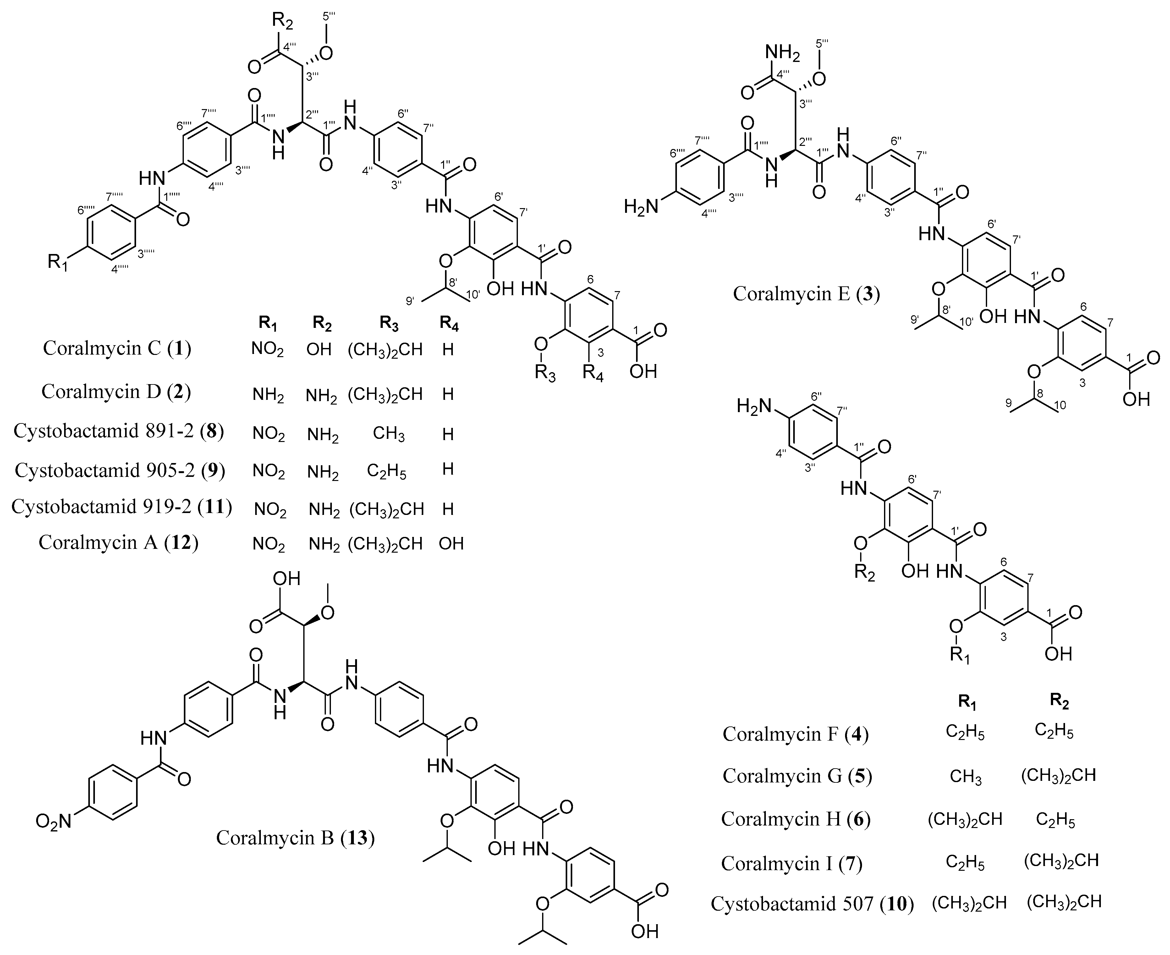
Recently, our group reported a new class of antibiotics, coralmycins A (12) and B (13), from cultures of the myxobacteria Corallococcus coralloides M23 [4]. Compound 12 shows potent antibacterial activity against clinically important Gram-negative pathogens as well as Gram-positive bacteria. Muller’s group reported cystobactamids 507 (10) and 919-2 (11) isolated from another myxobacteria, Cystobacter sp. [5]. Compound 12, a hydroxylated derivative of 11, has 10 times higher antibacterial activity than 11. Compound 10 shows weak antibacterial activity. Compound 11 and its structurally similar albicidin were reported to inhibit DNA gyrase [5,6]. Very recently, Muller’s group reported the discovery of new cystobactamids and 12 from a repeated-batch fermentation of another myxobacteria Myxococcus sp. for the sufficient production of cystobactamid derivatives occurred at very low product titers [7]. One of the new cystobactamids, cystobactamid 861-2, exhibit 4–10 times higher antibacterial activity against Gram-negative pathogens than 12.
Previously, compounds showing a UV absorption spectrum similar to those of the coralmycins were detected in minute amounts during isolation of 12 and 13. From a large-scale culture of Corallococcus coralloides M23 for the production of sufficient amounts of the coralmycin derivatives, we isolated seven new compounds (coralmycins C (1), D (2), E (3), F (4), G (5), H (6), and I (7)) and three known compounds (cystobactamids 891-2 (8), 905-2 (9), and 10) (Figure 1). Compound 1 was determined to be a new stereoisomer of 13 with the same configuration as 11, 2 is a new derivative of 11 with an amino moiety instead of the nitro moiety present in 11, and 3 is a new derivative of 11 that lacks the para-nitrobenzoic acid unit. Compounds 4–7 are new derivatives of 10 with variations on their two isopropyl chains. In this study, we report the fermentation, isolation, structural determination, and antibacterial and DNA gyrase inhibitory activities of 1–10.
2. Results and Discussion
2.1. Structural Elucidation
The chemical structures of 8 and 9 were independently elucidated using HRESIMS and 1D and 2D NMR analyses (Figures S1–S8 and Table 1 and Table 2). Their β-methoxyaspargine moieties were determined to be anti-relative configuration based on their large 3JHH coupling constant between H-2′′′ and H-3′′′. Additionally, the Cotton effects of 8 and 9 [[θ]25(nm)(MeOH): −703 (252), 508 (310)] and [[θ]25(nm)(MeOH): −746 (252), 488 (309), respectively], were almost the same as those of 11 [[θ]25(nm)(MeOH): −5344 (251), 3999 (305)] [4] in their circular dichroism (CD) spectra (Figure S9). Thus, 8 and 9 were identified as cystobactamids 891-2 and 905-2 [7], respectively. The complete assignments of the 13C-NMR data of 8 and 9 are reported for the first time in this study.
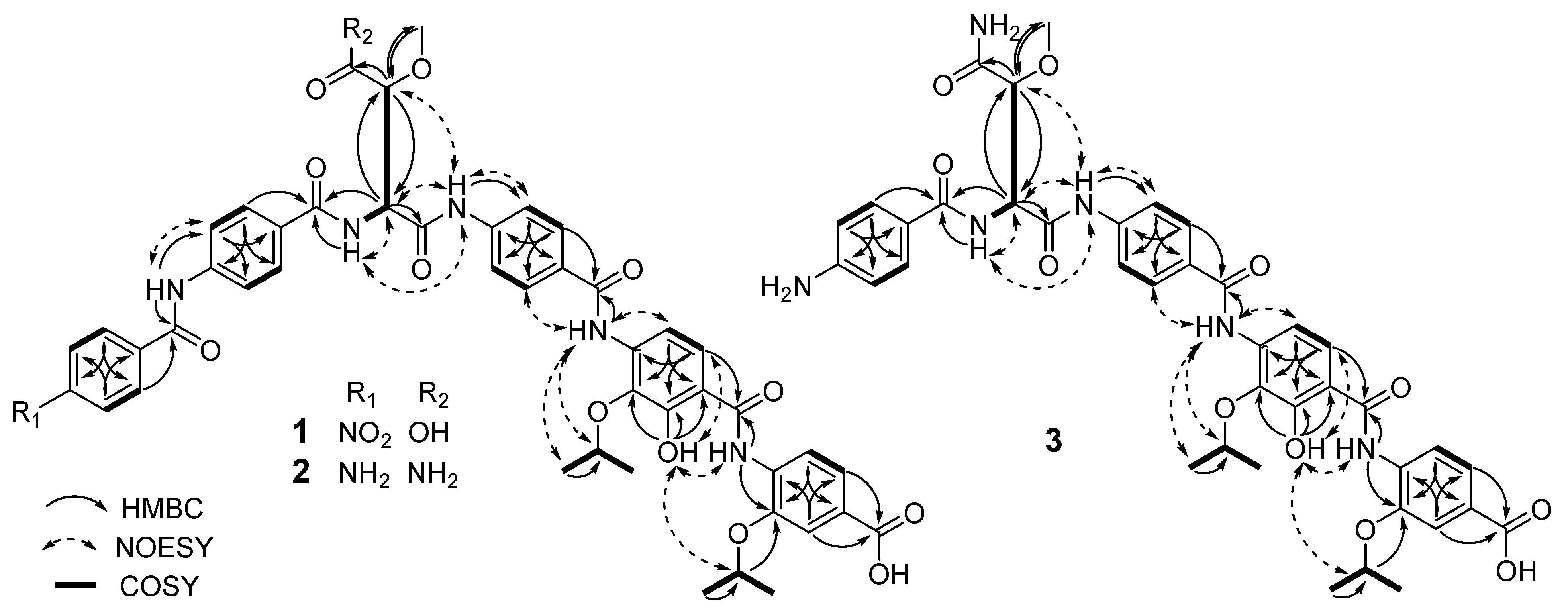
Compound 1 was not separated from 13 in SiO2 TLC but separated by ODS HPLC (Figure S10). Compound 1 showed the same molecular formula (C46H44N6O15) as that of 13 based on their HRESIMS data in combination with their 1H and 13C NMR data (Figures S11–S15 and Table 1 and Table 2). Various spectroscopic techniques, including 1D and 2D NMR experiments, were used for the structure elucidation of 1. The 1H-NMR and HMQC spectra of 1 were very similar to those of 13. The difference between the spectra of 1 and 13 is that the chemical shifts and coupling constants of H-2′′′ and H-3′′′ of the β-methoxyaspartic acid unit of 1 are more similar to those of 11 rather than 13 (Table 3), suggesting that 1 could be a stereoisomer of 13 at the β-methoxyaspartic acid moiety. The signals at δH 4.75 (1H, dt, J = 6.0, 12.1 Hz; δC 72.1), 4.31 (1H, dt, J = 6.1, 12.2 Hz; δC 76.1), 1.37 (6H, d, J = 6.0 Hz; δC 22.0), and 1.26 (6H, d, J = 6.1 Hz; δC 22.3) suggested the presence of two isopropyl groups. Additionally, the 1H-NMR, COSY, and HMQC spectra of 1 indicated the presence of two para-aminobenzoic acid units, a para-nitrobenzoic acid unit, a 4-amino-3-isopropoxybenzoic acid unit, a 4-amino-2-hydroxyl-3-isopropoxybenzoic acid unit, and a β-methoxyaspartic acid unit. The NH2 signal of the β-methoxyasparagine unit of 11 was not detected in the spectrum of 1 acquired in DMSO-d6 (Table 1), which, together with the molecular formula, suggested the presence of a β-methoxyaspartic acid fragment. The connectivities of these units were confirmed by the HMBC spectrum (Figure 2). Thus, the planar structure of 1 was elucidated to be the same as that of 13 [4].
The relative configuration at the β-methoxyaspartic acid unit of 1 was determined from 3JHH and 2JCH coupling constants and NOESY and NOE differential spectra (Table 2 and Figures S16–S18). The large coupling constant (8.1 Hz) between H-2′′′ and H-3′′′ indicated the anti-configuration. The 2JCH coupling constants were measured in DMSO-d6 by a HECADE experiment (Figure S16). The large coupling constant (5.11 Hz) between H-2′′′ and C-3′′′ indicated that H-2′′′ and OMe are in a gauche. Together with these coupling constants, the NOEs between H-3′′′ and NH-6′′′, from NH-8′′ to H-3′′′ and OMe, and from H-2′′′ to OMe confirmed that 1 had the same relative configuration as 11 (Figure S19). An unexpected NOE from H-2′′′ to H-3′′′ suggested the presence of the other rotamer as a minor conformer as detected previously in 13 [4]. The Cotton effect [[θ]25(nm)(MeOH): −415 (250), 345 (304)] in the CD spectra of 1 was almost the same as that [[θ]25(nm)(MeOH): −5344 (251), 3999 (305)] of 11 [4] (Figure S9). These data clearly indicated that 1 had the same configuration (S*R*) as 11. Thus, 1 was elucidated to be a stereoisomer of 13.
The HRESIMS data of 2 gave a protonated molecule at m/z 890.3415 [M + H]+ (calcd. 890.3362 for C46H48N7O12), which in combination with 1H and 13C NMR spectra suggested a molecular formula of C46H47N7O12 (Figures S20–S24 and Table 1 and Table 2). The 1H NMR spectrum of 2 was similar to that of 11. The difference was the appearance of the aromatic proton signals [δH 7.75 (2H, d, J = 8.8 Hz; δC 129.9) and 6.62 (2H, d, J = 8.6 Hz; δC 112.9)] for an 1,4-disubstituted phenyl moiety instead of signals (δH 8.39 and 8.21) for the para-nitrobenzoic acid unit in 11 (Table 1). In the HMBC spectrum (Figure 2), the proton at δH 6.62 showed long-range coupling to the carbons at δC 113.1 and 120.9, and the proton at δH 7.75 showed long-range coupling to the carbons at δC 165.9, 152.8, and 129.9. These 13C NMR data were consistent with those of para-aminobenzoic acid [8]. Together with the molecular formula, these spectroscopic data indicated the presence of a para-aminobenzoic acid unit instead of a para-nitrobenzoic acid unit. The connectivity of the remaining units was confirmed by the HMBC spectrum (Figure 2). Thus, the planar structure of 2 was identified as that of a new, C-5′′′′′ amino derivative of 11.
The relative configuration of the β-methoxyasparagine moiety of 2 was determined from 3JHH and 2JCH coupling constants and NOESY and NOE differential spectra (Table 1 and Figures S25–S27). The coupling constant between H-2′′′ and H-3′′′ was 8.0 Hz, indicating the anti configuration. Similar to 1, the large 2JCH coupling constant (5.49 Hz) between H-2′′′ and C-3′′′ indicated a gauche conformation of H-2′′′ and OMe. The NOEs between H-3′′′ and NH-6′′′, from NH-8′′ to H-3′′′ and OMe, and from H-2′′′ to OMe supported the same relative stereochemistry as 11. The Cotton effect [[θ]25(nm)(MeOH): −1278 (256), 1301 (313)] of 2 was almost the same as that [[θ]25(nm)(MeOH): −5344 (251), 3999 (305)] of 11 in CD spectra (Figure S9). Thus, 2 was determined to be a new, C-5′′′′′ amino derivative of 11.
The molecular formula of 3 was determined to be C39H24N6O11 based on its HRESIMS data in combination with its 1H and 13C NMR data (Figures S28–S30 and Table 1 and Table 2). The 1H NMR spectrum of 3 was similar to that of 2. The difference between the spectra of 3 and 2 was that the overlapping aromatic proton signals [δH 7.84 (4H, brs)] and the amino signal [δH 10.02 (1H, s)] of the para-aminobenzoic acid unit in the spectrum of 2 were not observed in the spectrum of 3. Additionally, detailed analysis of its 1H NMR, 13C NMR, COSY, and HMQC spectra suggested the presence of two para-aminobenzoic acid fragments, a 4-amino-3-isopropoxybenzoic acid fragment, a 4-amino-2-hydroxyl 3-isopropoxybenzoic acid fragment, and a β-methoxyasparagine fragment. These spectroscopic data suggested that 3 had one fewer para-aminobenzoic acid unit than were present in 2. The aromatic proton at δH 7.58 (H-3′′′′ and H-7′′′′) of one para-aminobenzoic acid unit [δH 7.58 (2H, overlapped; δC 129.0) and 6.57 (2H, d, J = 8.1 Hz; δC 112.7)] and the amine proton at δH 8.00 (6′′′-NH) have HMBC correlations with the carbonyl carbon at δC 166.0 (C-1′′′′), which in turn showed long-range coupling with the α-proton at 4.82 (H-2′′′) of the β-methoxyasparagine unit (Figure 2). These HMBC data indicated that the terminal para-aminobenzoic acid unit of 2 was absent in 3. The remaining structural fragments were confirmed by the HMBC spectrum (Figure 2). Thus, the planar structure of 3 was identified as that of a new derivative of 2 without the terminal para-aminobenzoic acid unit.
The relative configuration of the β-methoxyasparagine moiety of 3 was determined based on 3JHH and 2JCH coupling constants and NOESY and NOE differential spectra (Table 1 and Figures S34–S36). H-2′′′ and H-3′′′ were anti to each other based on their large 3JHH coupling constant (8.0 Hz). On the basis of the large 2JCH coupling constant (6.09 Hz) between H-2′′′ and C-3′′, H-2′′′ and OMe are in a gauche. The NOEs between H-3′′′ and NH-6′′′, from NH-8′′ to H-3′′′ and OMe, and from H-2′′′ to OMe supported the same relative stereochemistry as 2. The Cotton effects [[θ]25(nm)(MeOH): −707 (260), 976 (301)] of 3 were almost the same as those [[θ]25(nm)(MeOH): −1278 (256), 1301 (313)] of 2 (Figure S9). Thus, 3 was determined to be a new derivative of 2 without the terminal para-aminobenzoic acid unit.
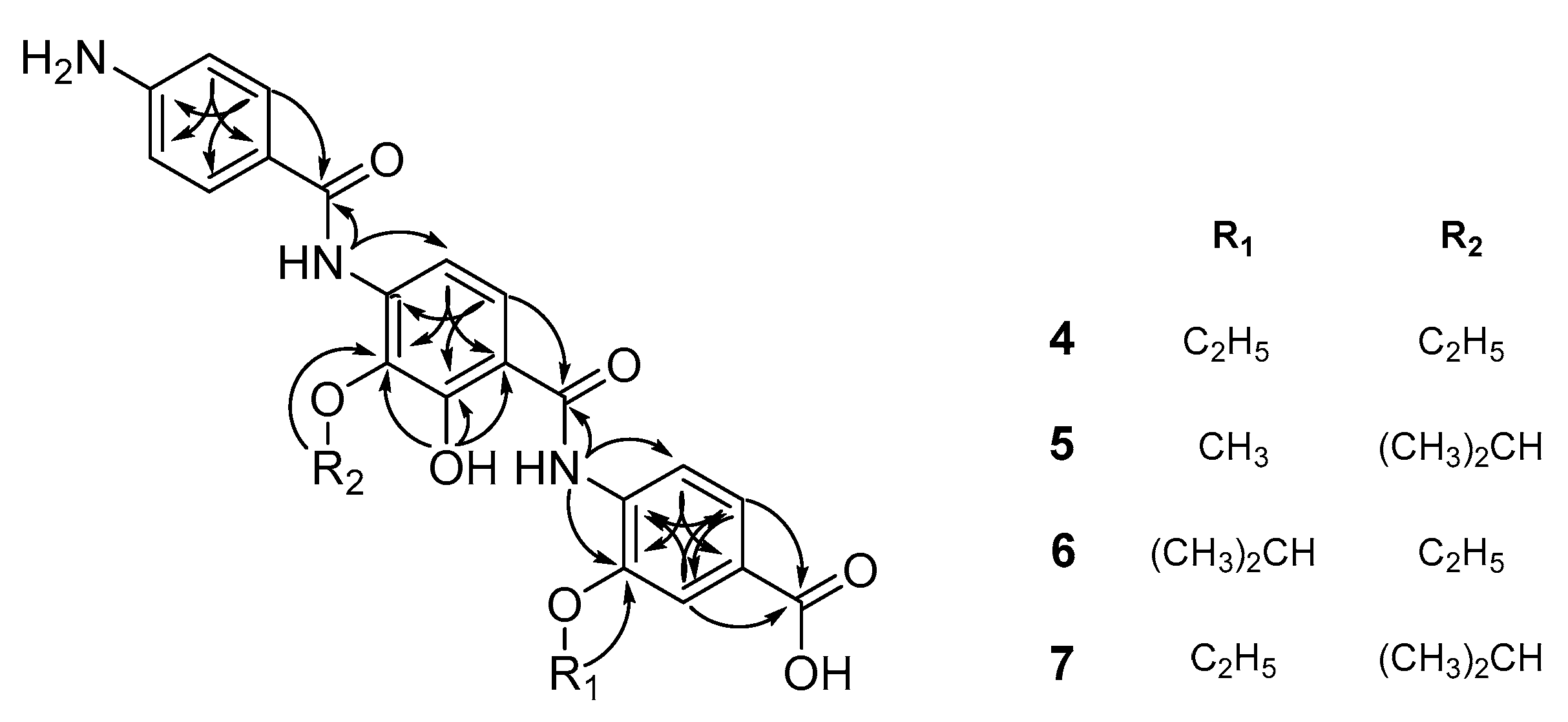
Compound 10 was identified as cystobactamid 507 [5] based on its ESIMS data in combination with its 1H and 13C NMR data (Figures S37–S40 and Table 4 and Table 5). The molecular formula of 4 was determined to be C25H25O7N3 based on its HRESIMS data in combination with its 1H and 13C NMR spectra (Figures S41–S44 and Table 4 and Table 5). The 1H NMR data acquired in CD3OD revealed the presence of nine aromatic protons, two oxygenated methylenes at δH 4.29 (2H, q, J = 7.0 Hz) and 4.16 (2H, q, J = 7.0 Hz), and two methyl groups at δH 1.58 (3H, t, J = 7.0 Hz) and 1.45 (3H, t, J = 7.0 Hz) (Table 4). The 1H-NMR and HMQC spectra of 4 indicated the presence of 1,4-disubstituted benzene, 1,2,4-trisubstituted benzene, 1,2,3,4-tetrasubstituted benzene, and two ethoxy groups. This result suggested the presence of two ethoxy groups in 4 instead of the two isopropoxyl groups seen in 10. This was confirmed by the HMBC correlations from the methylene protons at δH 4.16 of one ethoxy group to the carbon at δC 137.9 of the 1,2,4-trisubstituted benzene and from the methylene protons at δH 4.02 of the other ethoxy group to the carbon at δC 148.0 of the 1,2,4-trisubstituted benzene (Figure 3). Thus, 4 was determined to be a new, C-4 and C-4′ diethoxylated derivative at of 10.
The molecular formula of 5 was determined to be C25H25O7N3 based on its HRESIMS data in combination with its 1H and 13C NMR data (Figures S45–S48 and Table 4 and Table 5). The 1H NMR and HMQC data of 5 were similar to those of 10. The differences were that a signal for a methoxy group at δH 3.97 was observed instead of signals of an isopropoxy group. The location of the methoxy group was determined based on the HMBC correlation from the methoxy proton signal to the carbon at δC 149.4 of the 1,2,4-trisubstituted benzene (Figure 3). Thus, 5 was determined to be a new, C-4 methoxylated derivative of 10.
The molecular formula of 6 was determined to be C25H25O7N3 based on its HRESIMS data in combination with 1H and 13C NMR data (Figures S49–S52 and Table 4 and Table 5). The NMR spectra of 6 were similar to those of 10 except for the presence of ethoxy signals [δH 4.02 (1H, q, J = 7.0 Hz; δC 68.9) and 1.34 (3H, t, J = 7.0 Hz; δC 15.6)] instead signals for an isopropyl fragment. In the HMBC spectrum, the methylene protons at δH 4.02 of the ethoxy group showed long-range coupling to the carbon at δC 138.6, which in turn was correlated with the amine proton at δH 9.14 and the aromatic proton at δH 7.68 (Figure 3). These correlations indicated the linkage of the ethoxy group to C-3 of the 1,2,3,4-tetrasubstituted benzene. The HMBC spectrum confirmed the remaining structural features of 6. Thus, 6 was elucidated as a new, C-4′ ethoxylated derivative at of 10.
The molecular formula of 7 was determined to be C25H25O7N3 based on its HRESIMS data in combination with its 1H and 13C NMR data (Figures S53–S58 and Table 4 and Table 5). The NMR spectra of 7 were similar to those of 6. The major difference in the 1H NMR and HMQC data were that the methine proton of the isopropyl group of 6 was downfield-shifted from δH 4.77 to 4.36 in the spectra of 7. This result suggested that the isopropyl group might be connected to the 1,2,3,4-tetrasubstituted benzene as it is in 5. HMBC correlations were observed from the methine proton of the isopropyl group to the carbon at δC 138.1 (C-4′) of the 1,2,3,4-tetrasubstituted benzene and from the methylene proton of the ethoxy group to the carbon at δC 148.3 (C-4) of 1,2,4-trisubstituted benzene (Figure 3). The HMBC spectrum confirmed the remaining structural features of 7. Thus, 7 was determined to be as a new, C-4 ethoxylated derivative of 10.
2.2. Antibacterial and E. Coli DNA Gyrase-Inhibitory Activities of the New Compounds
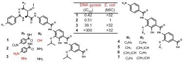
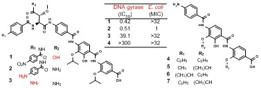
The antibacterial activities of 1–10 against clinically important Gram-positive and Gram-negative pathogens were evaluated compared with those of 11–13 and ciprofloxacin (Table 6 and Table S1). Additionally, their effects on the supercoiling activity of E. coli DNA gyrase were investigated using ciprofloxacin and nalidixic acid, as positive controls, (Figure S59 and Table 7) because the main target of 11 was reported to be DNA gyrase [5]. Compound 1, the β-methoxyaspartic acid derivative of 11, showed weaker antibacterial activity against Gram-positive bacteria (MICs of 8–4 μg/mL) compared with that of 11, and it exhibited no antibacterial activity against both Gram-negative bacteria at 32 μg/mL. However, 1 showed stronger E. coli DNA gyrase inhibition (IC50 of 0.42 μM) than was observed in 11 (0.95 μM). Considering that 13, the stereoisomer of 1, retained antibacterial activity against E. coli, these results suggested that the β-methoxyasparagine moiety and its stereochemistry in 11 influences cellular uptake in both Gram-positive and Gram-negative bacteria. The stronger antibacterial activity against E. coli of 12 could be due to its stronger E. coli DNA gyrase inhibition than 11. Compound 2, 8, and 9 showed antibacterial activities similar to that of 11 against both Gram-positive and Gram-negative bacteria except against P. aeruginosa and K. pneumonia. Compound 2, with an amino moiety instead of the nitro moiety on the para-nitrobenzoic acid unit (relative to 11), did not inhibit the growth of P. aeruginosa or K. pneumonia at 32 μg/mL, while 8 and 9, the derivatives in which the isopropyl chain at C-4 of 11 has been shortened, showed weak antibacterial activities against P. aeruginosa but did not inhibit the growth of K. pneumonia at 32 μg/mL. However, 2, 8, and 9 showed strong E. coli DNA gyrase inhibition with IC50 values of 0.05–0.51 μM, making them more potent than 11. Considering that 11 was reported to inhibit DNA gyrases of both E. coli and P. aeruginosa with similar potencies [9], these results suggested that the nitro group and the isopropyl chain could be important for permeability into P. aeruginosa and K. pneumonia. Another explanation of the discrepancy between E. coli DNA gyrase inhibition and antibacterial activity of these respective compounds might also be due to an improved efflux.
On the other hand, 3, a derivative of 11 with the para-nitrobenzoic acid unit removed, exhibited no antibacterial activities against any of the tested bacterial strains at 32 μg/mL, which was consistent with its dramatically lower ability to inhibit DNA gyrase (IC50 of 39.1 μM). These results suggested that the para-aminobenzoic acid unit is critical to the inhibition of DNA gyrase and bacterial growth. Compounds 4–7 and 10, derivatives missing the para-aminobenzoic acid unit and β-methoxyasparagine moiety, exhibited no antibacterial activities against any of the tested bacterial strains at 32 μg/mL and showed no DNA gyrase inhibition even at 300 μM. Although 10 was reported to show weak antibacterial and DNA gyrase-inhibitory activities against Gram-negative and Gram-positive bacteria (MICs of 4–65 μg/mL and IC50 of 20.2 μM, respectively) [5], 10 and its derivatives 4–7 did not display those activities in this study.
3. Materials and Methods
3.1. General Experimental Procedures
Optical rotations were determined on a JASCO P-1020 polarimeter (JASCO, Tokyo, Japan). UV spectra were measured on a Shimadzu UV-1601 UV−visible spectrophotometer (Shimadzu, Kyoto, Japan). IR spectra were obtained using a Bruker EQUINOX 55 spectrometer (Bruker Co., Ettlingen, Germany). NMR spectra were recorded on Bruker Biospin Avance 400, 500, 700, 800, or 900 MHz spectrometers (Korea Basic Science Institute, Ochang, Korea). HRESIMS data were recorded on a JEOL JMSHX110/110A mass spectrometer (JEOL, Tokyo, Japan).
3.2. Fermentation and Isolation
The 3000-L fermentation was carried out in a 5000-L fermenter using seed cultures from sequential 500-mL Erlenmeyer flask and 5-L, 50-L, and 500-L fermenters ((Supporting Information). The resin and cells were recovered and extracted twice with 100% acetone. The acetone was evaporated from the extract, and the remaining aqueous phase was extracted sequentially with chloroform and ethyl acetate. The ethyl acetate extract (8.1 g) was applied to Sephadex LH-20 column and eluted with MeOH to give four major fractions, Fr. I to IV, based on TLC analysis. Fr. II was purified by thin-layer chromatography (TLC) on silica gel 60 F254 plates (Merck No. 1.05715.0001, Darmstadt, Germany) with CHCl3:MeOH = 3:1 to give two major bands, Bands I and II, at Rf values of 0.3 and 0.8, respectively. Band I was further purified by TLC on silica gel 60 RP-18 F254 plates (Merck No. 1.15389.0001) with CH3CN:H2O (55:45) to yield 13 (2.1 mg) and 1 (2.2 mg) at Rf values of 0.60, and 0.64, respectively. In the same way, 12 (22.1 mg) was purified from band II at an Rf value of 0.5. Compounds 11 (657 mg) and 12 (2.8 mg) were obtained from Fr. III by silica gel TLC with CHCl3:MeOH = 6:1. Fr. IV was also purified by silica gel TLC with CHCl3−MeOH (3:1) and then subjected to preparative HPLC (YMC column 150 × 20 mm I.D., flow rate 3.0 mL/min) eluting with MeOH:H2O (50:50) to afford 2 (4.1 mg), 8 (3.2 mg), 9 (2.2 mg), and 11 (8.1 mg) at retention times of 19, 33, 43, and 52 min, respectively. The chloroform extract (500 g) was separated on a silica gel column eluted with a stepwise gradient of MeOH in CHCl3 containing 0.01% trifluoroacetic acid (TFA) to give 15 fractions, Fr. 1–16. Fr. 6 was purified by preparative HPLC (OP C18-101002510 column 250 × 10 mm I.D., flow rate 2.5 mL/min) eluting with CH3CN:H2O (50:50) containing 0.01% TFA to afford 4 (2.4 mg), 5 (3.5 mg), 6 (3.1 mg), 7 (6.2 mg), and 10 (12.3 mg) at retention times of 12.5, 14.0, 15.0, 18.5, and 22.0 min, respectively. Fr. 9 was purified on a Sephadex LH-20 column eluted with MeOH, and then purified by RP-18 TLC developed with MeOH:H2O (70:30) containing 0.01% TFA to yield 11 (7.1 mg), 10 (3.2 mg), and 3 (8.4 mg) at Rf values of 0.17, 0.30, and 0.38, respectively.
Compounds 1, 2, 3, 8, 9, 11, 12, and 13 appeared as single peaks at 220 nm using an analytical HPLC column (4.6 × 150 mm, S-4 μm, YMC C18) with CH3CN:H2O (50:50) containing 0.01% TFA at a flow rate of 0.8 mL/min with retention times of 18.2, 6.0, 5.1, 9.5, 12.8, 15.1, 19.8, and 18.3 min, respectively (Figure S60). Compounds 4, 5, 6, 7, and 10 were also determined to be single peaks under the HPLC conditions described above with retention times of 9.0, 9.9, 10.8, 13.6, and 16.7 min, respectively (Figure S61).
Coralmycin C (compound 1): yellow powder; = +24.8 (c 0.064, MeOH); UV (MeOH) λmax(log ε) 213 (4.80), 266 (sh) (4.13), 301 (4.26), 319 (4.23) nm; IR (KBr) νmax 3425, 2918, 2850, 1684, 1516, 1431, 1274, 1205, 1138, 1050, 1027, 1002 cm−1; CD (MeOH) [θ]25 −415 (250), 345 (304) nm; HRESIMS m/z 921.2941 (M + H)+ (cald for C46H45N6O15, 921.2937).
Coralmycin D (compound 2): yellow powder; = +16.0 (c 0.032, MeOH); UV (MeOH) λmax(log ε) 212 (4.73), 306 (4.24), 314 (4.24) nm; IR (KBr) 3424, 2917, 2850, 1682, 1602, 1512, 1428, 1272, 1203, 1138, 1026, 999 cm−1; CD (MeOH) [θ]25 −1278 (256), 1301 (313) nm; HRESIMS m/z 890.3415 (M + H)+ (cald for C46H48N7O12, 890.3362).
Coralmycin E (compound 3): yellow powder; = −20.0 (c 0.064, MeOH); UV (MeOH) λmax(log ε) 212 (4.56), 266 (sh) (3.99), 298 (4.17), 320 (4.12) nm; IR (KBr) 3426, 2958, 2918, 2850, 1683, 1603, 1538, 1432, 1277, 1204, 1139, 1052, 1027 cm−1; CD (MeOH) [θ]25 −707 (260), 976 (301) nm; HRESIMS m/z 771.2995 (M + H)+ (cald for C39H43N6O11, 771.2984).
Coralmycin F (compound 4): yellow powder; UV (MeOH) λmax(log ε) 212 (4.30), 263 (sh) (3.87), 323 (3.93) nm; IR (KBr) 2927, 1686, 1497, 1205 cm−1; HRESIMS m/z 480.1769 (M + H)+ (cald for C25H26N3O7, 480.1765).
Coralmycin G (compound 5): yellow powder; UV (MeOH) λmax(log ε) 210 (4.38), 263 (sh) (3.81), 318 (3.96) nm; IR (KBr) 3426, 2974, 2930, 1685, 1599, 1506, 1421, 1275, 1206, 1181, 1136 cm−1; HRESIMS m/z 480.1748 (M + H)+ (cald for C25H26N3O7, 480.1765).
Coralmycin H (compound 6): yellow powder; UV (MeOH) λmax(log ε) 213 (4.53), 263 (sh) (3.88), 325 (4.03) nm; IR (KBr) 3424, 2977, 2928, 1685, 1599, 1509, 1427, 1271, 1202, 1137, 1007 cm−1; HRESIMS m/z 494.1906 (M + H)+ (cald for C26H28N3O7, 494.1922).
Coralmycin I (compound 7): yellow powder; UV (MeOH) λmax(log ε) 210 (4.38), 263 (sh) (3.86), 319 (3.97) nm; IR (KBr) 3422, 2976, 2929, 1686, 1645, 1598, 1503, 1424, 1275, 1186, 1155, 1105, 1025, 1001 cm−1; HRESIMS m/z 494.1904 (M + H)+ (cald for C26H28N3O7, 494.1922).
3.3. Determination of Antibacterial Susceptibility
Whole-cell antimicrobial activities were determined using a broth microdilution method described previously [10]. Most of the test strains were grown to mid-log phase in Mueller–Hinton broth and diluted 1,000-fold in the same medium. Cells (105/mL) were dispensed at 0.2 mL/well in 96-well microtiter plates. Streptococcus pneumonia and Acinetobacter baumanii were grown in Todd–Hewitt medium and nutrient broth, respectively. The test compounds and ciprofloxacin (Sigma St. Louis, MO, USA) were soluble in DMSO, the final concentration of which did not exceed 0.05% in the cells. Cells were treated with 0.05% DMSO as a vehicle control. The MICs were determined in triplicate by serial two-fold dilutions of the test compounds. The MIC was defined as the concentration of a test compound that completely inhibited cell growth during a 24-h incubation period at 37 °C. Bacterial growth was determined by measuring the absorption at 650 nm using a microtiter enzyme-linked immunosorbent assay (ELISA) reader (Molecular Devices Corporation, Sunnyvale, CA, USA).
3.4. DNA Gyrase Assay
The inhibitory activities of coralmycins on the supercoiling activity of DNA gyrase were investigated using a ‘Purified E. coli DNA Gyrase Drug Screening Kit’ (TopoGEN #TG2001G, Buena Vista, CO, USA). The test compounds, ciprofloxacin (200–0.02 μM) and nalidixic acid (Sigma) (200–0.02 μM) were soluble in DMSO, the final concentration of which did not exceed 0.05% in the reactions (Figure S59). Relaxed plasmid (0.5 μg) was mixed with one unit of gyrase (~20.5 nM) and 1 μL of the test compound in the reaction buffer (final volume of 20 μL) and incubated at 37 °C for 30 min. The reaction was terminated by the addition of 2 μL of 10% (w/v) SDS and 2 μL of 10× DNA gel loading dye (bromophenol blue). Then, 20 μL of chloroform:isoamyl alcohol (24:1 mixture) was added, and the mixture was vortexed and then centrifuged. The samples in the blue phase were run on 1% (w/v) agarose gel and were visualized using ethidium bromide. All reactions were performed in triplicate. To calculate the IC50 values, the band densities were measured using ImageJ (National Institutes of Health, Bethesda, Maryland, MD, USA).
4. Conclusions
Seven new derivatives, 1–7, were isolated from a large-scale culture of the myxobacteria Corallococcus coralloides M23. Compound 2 showed potent antibacterial activity against both Gram-positive and Gram-negative bacteria except against P. aeruginosa and K. pneumonia with potent DNA gyrase inhibition. Interestingly, 1 exhibited antibacterial activity against Gram-positive bacteria but not against Gram-negative bacteria, although it potently inhibited DNA gyrase. Their structure-activity relationships between the antibacterial activity and DNA gyrase inhibitory activity indicated that the para-nitrobenzoic acid unit is critical for the inhibition of both DNA gyrase and bacterial growth, while the nitro moiety of the para-nitrobenzoic acid unit and the isopropyl chain at C-4 could be important for the permeability of the compound into certain Gram-negative bacteria, including P. aeruginosa and K. pneumonia, and the β-methoxyasparagine moiety could influence cellular uptake into all tested bacteria.
Supplementary Materials
The following are available online: 1D NMR, 2D NMR, and CD spectra (Figures S1–S58) of compounds 1–7 and related compounds.
Author Contributions
B.-M.K. and N.V.M. contributed equally; B.-M.K. performed the fermentation; B.-M.K. and N.V.M. performed the extraction and isolation; N.V.M. and W.-G.K. performed the spectral analysis and structure determination; B.-M.K. performed the antibacterial assay; H.-Y.C. performed the DNA gyrase assay and MRM analysis; W.-G.K. designed the experiments; N.V.M. and W.-G.K. wrote the paper.
Funding
This work was supported by a grant from the Korea Health Technology R&D Project through the Korea Health Industry Development Institute (KHIDI), which is funded by the Ministry of Health & Welfare, Republic of Korea (grant number HI17C1807) as well as a grant from the KRIBB Research Initiative Program, Republic of Korea.
Acknowledgments
The authors thank Keug Hyun Ahn (KRIBB) for his help in the large-scale fermentation.
Conflicts of Interest
The authors declare no conflict of interest.
References
- Boucher, H.W.; Talbot, G.H.; Benjamin, D.K., Jr.; Bradley, J.; Guidos, R.J.; Jones, R.N.; Murray, B.E.; Bonomo, R.A.; Gilbert, D. 10 × ’20 Progress–development of new drugs active against gram-negative bacilli: An update from the Infectious Diseases Society of America. Clin. Infect. Dis. 2013, 56, 1685–1694. [Google Scholar] [CrossRef] [PubMed]
- Giske, C.G.; Monnet, D.L.; Cars, O.; Carmeli, Y. Clinical and economic impact of common multidrug-resistant gram-negative bacilli. Antimicrob. Agents Chemother. 2008, 52, 813–821. [Google Scholar] [CrossRef] [PubMed]
- Boucher, H.W.; Talbot, G.H.; Bradley, J.S.; Edwards, J.E.; Gilbert, D.; Rice, L.B.; Scheld, M.; Spellberg, B.; Bartlett, J. Bad bugs, no drugs: No ESKAPE! An update from the Infectious Diseases Society of America. Clin. Infect. Dis. 2009, 48, 1–12. [Google Scholar] [CrossRef] [PubMed]
- Kim, Y.J.; Kim, H.J.; Kim, G.W.; Cho, K.; Takahashi, S.; Koshino, H.; Kim, W.G. Isolation of Coralmycins A and B, Potent Anti-Gram Negative Compounds from the Myxobacteria Corallococcus coralloides M23. J. Nat. Prod. 2016, 79, 2223–2228. [Google Scholar] [CrossRef] [PubMed]
- Baumann, S.; Herrmann, J.; Raju, R.; Steinmetz, H.; Mohr, K.I.; Huttel, S.; Harmrolfs, K.; Stadler, M.; Muller, R. Cystobactamids: Myxobacterial topoisomerase inhibitors exhibiting potent antibacterial activity. Angew. Chem. Int. Ed. Engl. 2014, 53, 14605–14609. [Google Scholar] [CrossRef] [PubMed]
- Cociancich, S.; Pesic, A.; Petras, D.; Uhlmann, S.; Kretz, J.; Schubert, V.; Vieweg, L.; Duplan, S.; Marguerettaz, M.; Noell, J.; et al. The gyrase inhibitor albicidin consists of p-aminobenzoic acids and cyanoalanine. Nat. Chem. Biol. 2015, 11, 195–197. [Google Scholar] [CrossRef] [PubMed]
- Huttel, S.; Testolin, G.; Herrmann, J.; Planke, T.; Gille, F.; Moreno, M.; Stadler, M.; Bronstrup, M.; Kirschning, A.; Muller, R. Discovery and Total Synthesis of Natural Cystobactamid Derivatives with Superior Activity against Gram-Negative Pathogens. Angew. Chem. Int. Ed. Engl. 2017, 56, 12760–12764. [Google Scholar] [CrossRef] [PubMed]
- Pouchert, C.J.; Behnke, J. The Aldrich Library of ¹³C and ¹H FT NMR Spectra; Aldrich Chemical Co.: Milwaukee, WI, USA, 1993; p. 1085. [Google Scholar]
- Cheng, B.; Muller, R.; Trauner, D. Total Syntheses of Cystobactamids and Structural Confirmation of Cystobactamid 919-2. Angew. Chem. Int. Ed. Engl. 2017, 56, 12755–12759. [Google Scholar] [CrossRef] [PubMed]
- Zheng, C.J.; Sohn, M.J.; Lee, S.; Kim, W.G. Meleagrin, a new FabI inhibitor from Penicillium chryosogenum with at least one additional mode of action. PLoS ONE 2013, 8, e78922. [Google Scholar] [CrossRef] [PubMed]
Sample Availability: Samples of the compound 11 are available from the authors. |
Figure 1.
The structures of coralmycins C (1), D (2), E (3), F (4), G (5), H (6), and I (7), and related compounds.
Figure 1.
The structures of coralmycins C (1), D (2), E (3), F (4), G (5), H (6), and I (7), and related compounds.

Figure 2.
Key HMBC, COSY, and NOE correlations of 1–3.

Figure 3.
Key HMBC and COSY correlations of 4–7.


Table 1.
1H NMR data (500 MHz, DMSO-d6) for 1–3.
| Position | 11 | 1 | 2 | 3 | 8 | 9 |
|---|---|---|---|---|---|---|
| 1 | - | - | - | - | - | - |
| 2 | - | - | - | - | - | - |
| 3 | 7.56, brs | 7.57, s | 7.59, s | 7.58 * | 7.57, s | 7.55, s |
| 4 | - | - | - | - | - | - |
| 5 | - | - | - | - | - | - |
| 6 | 8.50, d (8.3) | 8.51, d (8.3) | 8.52, d (8.4) | 8.51, d (8.2) | 8.39, d (8.3) | 8.46, d (8.3) |
| 7 | 7.58, d (8.3) | 7.59, d (8.7) | 7.60, d (8.3) | 7.58 * | 7.62, d (8.3) | 7.61, d (8.3) |
| 8 | 4.75, m | 4.75, m | 4.76, m | 4.75, m | 3.97, s | 4.2, q (7.0) |
| 9,10 | 1.37, d (6.0) | 1.37, d (6.0) | 1.38, d (6.0) | 1.37, d (5.8) | - | 1.46, t (7.1) |
| 11-NH | 10.97, brs | - | 11.00, s | 11.00, s | 10.89, s | 10.96, s |
| 1′ | - | - | - | - | - | - |
| 2′ | - | - | - | - | - | - |
| 3′ | - | - | - | - | - | - |
| 3′-OH | 11.21, brs | 11.30, s | 11.27, s | 11.27, s | 11.46, s | 11.31, s |
| 4′ | - | - | - | - | - | - |
| 5′ | - | - | - | - | - | - |
| 6′ | 7.50, d (8.5) | 7.52, d (8.7) | 7.54, d (8.8) | 7.52, d (8.7) | 7.55, m | 7.54, d (8.7) |
| 7′ | 7.80, d (8.5) | 7.81, d (8.7) | 7.84 * | 7.81 * | 7.82, m | 7.82, d (7.6) |
| 8′ | 4.32, m | 4.31, m | 4.33, m | 4.30, m | 4.38, m | 4.33, m |
| 9′,10′ | 1.26, d (6.1) | 1.26, d (6.1) | 1.28, d (6.1) | 1.26, d (6.0) | 1.27, d (6.4) | 1.26, d (6.1) |
| 11′-NH | 9.58, s | 9.60, s | 9.61, s | 9.60, s | 9.58, s | 9.59, s |
| 1′′ | - | - | - | - | - | - |
| 2′′ | - | - | - | - | - | - |
| 3′′,7′′ | 7.97, d (8.6) | 7.97, d (8.5) | 7.98, d (8.8) | 7.96, (8.3) | 7.98, d (8.7) | 7.97, d (8.8) |
| 4′′,6′′ | 7.83, d (8.6) | 7.84, d (8.5) | 7.84 * | 7.81 * | 7.83, m | 7.83, d (7.6) |
| 5′′ | - | - | - | - | - | - |
| 8′′-NH | 10.56, s | 10.52, s | 10.57, s | 10.50, s | 10.57, s | 10.57, s |
| 1′′′ | - | - | - | - | - | - |
| 2′′′ | 4.92, dd (8.0, 8.1) | 5.07, t (8.3) | 4.92, t (8.1) | 4.82, t (7.9) | 4.92, t (8.1) | 4.92, t (8.1) |
| 3′′′ | 4.09, d (8.0) | 4.16, d (8.1) | 4.11, d (8.0) | 4.06, d (8.0) | 4.09, d (8.1) | 4.09, d (8.1) |
| 4′′′-NH | 7.47, brs 7.54, brs | - | 7.48, brs | 7.45, brs | 7.48, brs | 7.48, brs |
| 5′′′ | 3.31, s | 3.35, s | 3.32, s | 3.28, s | 3.31, s | 3.31, s |
| 6′′′-NH | 8.46, d (8.1) | 8.72, d (8.5) | 8.41, d (8.2) | 8.00, d (8.0) | 8.47, d (8.1) | 8.47, d (8.1) |
| 1′′′′ | - | - | - | - | - | - |
| 2′′′′ | - | - | - | - | - | - |
| 3′′′′,7′′′′ | 7.90, d * | 7.94, d (8.6) | 7.84 * | 7.58 * | 7.91 * | 7.91 * |
| 4′′′′,6′′′′ | 7.90, d * | 7.90, d (8.6) | 7.84 * | 6.57, d (8.1) | 7.91* | 7.91* |
| 5′′′′ | - | - | - | - | - | - |
| 8′′′′-NH | 10.8, s | 10.84, s | 10.02, s | - | 10.81, s | 10.81, s |
| 1′′′′′ | - | - | - | - | - | - |
| 2′′′′′ | - | - | - | - | - | - |
| 3′′′′′,7′′′′′ | 8.21, d (8.6) | 8.21, d (8.6) | 7.75, d (8.8) | - | 8.21, d (8.8) | 8.21, d (8.8) |
| 4′′′′′,6′′′′′ | 8.38, d (8.6) | 8.38, d (8.6) | 6.62, d (8.6) | - | 8.39, d (8.8) | 8.39, d (8.8) |
| 5′′′′′ | - | - | - | - | - | - |
* Overlapped.
Table 2.
13C NMR data (500 MHz, DMSO-d6) for 1–3.
| Position | 11 | 1 | 2 | 3 | 8 | 9 |
|---|---|---|---|---|---|---|
| 1 | 166.9, C | 167.4, C | 167.3, C | 167.0, C | 166.8, C | 166.9, C |
| 2 | 125.7, C | 126.2, C | 126.2, C | 125.8, C | 126.1, C | 126.3, C |
| 3 | 113.9, CH | 114.3, CH | 114.4, CH | 113.9, CH | 111.7, CH | 111.9, CH |
| 4 | 146.3, C | 146.8, C | 146.8, C | 146.4, C | 148.4, C | 148.0, C |
| 5 | 133.3, C | 133.7, C | 133.4, C | 133.3, C | 131.4, C | 132.6, C |
| 6 | 119.6, CH | 120.0, CH | 120.1, CH | 119.6, CH | 120.4, CH | 119.6, CH |
| 7 | 122.6, CH | 123.1, CH | 123.0, CH | 122.7, CH | 122.4, CH | 122.6, CH |
| 8 | 71.7, CH | 72.1, CH | 72.2, CH | 71.8, CH | 56.5, CH3 | 64.1, CH2 |
| 9,10 | 21.6, CH3 | 22.0, CH3 | 22.0, CH3 | 21.7, CH3 | - | 14.3, CH3 |
| 1′ | 163.6, C | 164.0, C | 164.0, C | 163.6, C | 164.2, C | 136.6, C |
| 2′ | 116.4, C | 116.9, C | 117.0, C | 116.6, C | 115.5, C | 116.4, C |
| 3′ | 150.3, C | 150.9, C | 150.9, C | 150.4, C | 150.8, C | 150.3, C |
| 4′ | 138.4, C | 138.9, C | 139.0, C | 138.5, C | 138.5, C | 138.4, C |
| 5′ | 136.2, C | 136.7, C | 136.6, C | 136.3, C | 136.0, C | 136.2, C |
| 6′ | 115.3, CH | 115.8, CH | 115.6, CH | 115.3, CH | 115.1, CH | 114.9, CH |
| 7′ | 124.9, CH | 125.4, CH | 125.5, CH | 125.0, CH | 125.0, CH | 124.9, CH |
| 8′ | 75.6, CH | 76.1, CH | 76.1, CH | 75.7, CH | 75.1, CH | 75.6, CH |
| 9′,10′ | 22.0, CH3 | 22.3, CH3 | 22.3, CH3 | 22.0, CH3 | 21.8, CH3 | 22.0, CH3 |
| 1′′ | 164.3, C | 164.8, C | 164.8, C | 164.4, C | 164.1, C | 164.7, C |
| 2′′ | 128.6, C | 129, C | 129.3, C | 128.5, C | 128.3, C | 128.6, C |
| 3′′,7′′ | 128.4, CH | 128.9, CH | 128.9, CH | 128.5, CH | 128.3, CH | 128.4, CH |
| 4′′,6′′ | 118.8, CH | 119.4, CH | 119.3, CH | 118.8, CH | 118.6, CH | 118.8, CH |
| 5′′ | 142.1, C | 142.5, C | 142.6, C | 142.2, C | 142.0, C | 142.1, C |
| 1′′′ | 168.6, C | 168.6, C | 169.2, C | 169.2, C | 168.6, C | 168.6, C |
| 2′′′ | 55.7, CH | 56.0, CH | 56.1, CH | 55.7, CH | 55.6, CH | 55.5, CH |
| 3′′′ | 79.7, CH | 80.2, CH | 80.5, CH | 80.0, CH | 79.9, CH | 79.8, CH |
| 4′′′ | 170.8, C | 171.4, C | 171.3, C | 171.0, C | 171.2, C | 170.8, C |
| 5′′′ | 57.7, CH3 | 58.6, CH3 | 58.1, CH3 | 57.6, CH3 | 57.5, CH3 | 57.6, CH3 |
| 1′′′′ | 165.4, C | 166.2, C | 166.0, C | 166.0, C | 165.5, C | 165.4, C |
| 2′′′′ | 128.9, C | 129.4, C | 129.3, C | 120.3, C | 128.5, C | 128.9, C |
| 3′′′′,7′′′′ | 128.2, CH | 129.0, CH | 129.9, CH | 129.0, CH | 128.4, CH | 128.2, CH |
| 4′′′′,6′′′′ | 119.6, CH | 120.0, CH | 119.7, CH | 112.7, CH | 119.5, CH | 117.6, CH |
| 5′′′′ | 141.7, C | 142.1, C | 143.3, C | 151.8, C | 141.7, C | 141.7, C |
| 1′′′′′ | 164.2, C | 164.6, C | 165.9, C | - | 164.0, C | 164.2, C |
| 2′′′′′ | 140.4, C | 140.7, C | 120.9, C | - | 140.2, C | 140.4, C |
| 3′′′′′,7′′′′′ | 129.3, CH | 129.8, CH | 129.9, CH | - | 129.2, CH | 129.3, CH |
| 4′′′′′,6′′′′′ | 123.5, CH | 124.0, CH | 113.1, CH | - | 123.5, CH | 123.5, CH |
| 5′′′′′ | 149.2, C | 149.7, C | 152.8, C | 149.1, C | 149.2, C |
Table 3.
Comparison of the NMR data of the β-methoxyasparagine moiety of 1 with those of 11 and 13 (DMSO-d6).
Table 3.
Comparison of the NMR data of the β-methoxyasparagine moiety of 1 with those of 11 and 13 (DMSO-d6).
| Position | 11 | 13 | 1 | |||
|---|---|---|---|---|---|---|
| δC | δH | δC | δH | δC | δH | |
| 1′′′ | 168.6, C | - | 168.7, C | - | 168.6, C | - |
| 2′′′ | 55.7, CH | 4.92, t (8.0) | 54.9, CH | 4.87, brs | 56.0, CH | 5.07, t (8.3) |
| 3′′′ | 79.7, CH | 4.09, d (8.0) | 82.6, CH | 4.27, brs | 80.2, CH | 4.16, d (8.1) |
| 4′′′ | 170.8, C | - | 170.1, C | - | 171.4, C | - |
| 4′′′-NHa | - | 7.47, s | - | - | - | - |
| 4′′′-NHb | - | 7.54, s | - | - | - | - |
| 5′′′ | 57.7, CH3 | 3.31, s | 58.9, CH3 | 3.44, s | 58.6, CH3 | 3.35, s |
| 6′′′-NH | - | 8.46, d (8.1) | - | 8.37, brs | - | 8.72, d (8.5) |
Table 4.
1H NMR data for 4–7.
| Position | 10 a | 4 a | 5 b | 6 b | 7 b |
|---|---|---|---|---|---|
| 1 | - | - | - | - | |
| 2 | - | - | - | - | |
| 3 | 7.68, s | 7.68, d (1.6) | 7.59, d (1.7) | 7.58, d (1.5) | 7.55, d (1.7) |
| 4 | - | - | - | - | |
| 5 | - | - | - | - | |
| 6 | 8.50, d (8.8) | 8.53, d (8.4) | 8.36, d (8.3) | 8.54, d (8.3) | 8.44, d (8.4) |
| 7 | 7.69, d (7.4) | 7.71, dd (1.7, 8.4) | 7.63, dd (1.7, 8.3) | 7.60, dd (1.7, 8.4) | 7.61, dd (1.7, 8.4) |
| 8 | 4.80, m | 4.29, q (7.0) | 3.97, s | 4.77, m | 4.21, q (6.9) |
| 9,10 | 1.48, d (8.0) | 1.58, t (7.0) | - | 1.39, d (6.0) | 1.47, t (6.9) |
| 11-NH | - | 10.83, s | 10.96, s | 10.93, s | |
| 1′ | - | - | - | - | |
| 2′ | - | - | - | - | |
| 3′ | - | - | - | - | |
| 3′-OH | - | 11.51, s | 11.32, s | 11.34, s | |
| 4′ | - | - | - | - | |
| 5′ | - | - | - | - | |
| 6′ | 7.80, d (8.6) | 7.83, overlapped | 7.69, d (8.8) | 7.68, d (8.9) | 7.64, d (8.8) |
| 7′ | 7.77, d (9.0) | 7.83, overlapped | 7.81, d (8.8) | 7.80, d (8.9) | 7.81, d (8.8) |
| 8′ | 4.55, m | 4.16, q (7.0) | 4.42, m | 4.02, q (7.0) | 4.36, m |
| 9′,10′ | 1.37, d (6.14) | 1.45, t (7.0) | 1.28, d (6.1) | 1.34, t (7.0) | 1.28, d (6.2) |
| 11′-NH | - | 9.10, s | 9.14, s | 9.25, s | |
| 1′′ | - | - | - | - | |
| 2′′ | - | - | - | - | |
| 3′′,7′′ | 7.78, d (4.8) | 7.77, d (8.7) | 7.70, d (8.7) | 7.71, d (8.7) | 7.77, d (8.6) |
| 4′′,6′′ | 6.88, d (8.5) | 6.79, d (8.7) | 6.64, d (8.6) | 6.64, d (8.7) | 6.81, d (8.2) |
| 5′′ | - | - | - | - |
a measured in CD3OD at 500 MHz; b measured in DMSO-d6 at 500 MHz.
Table 5.
13C NMR data for 4–7.
| Position | 10 a | 4 a | 5 b | 6 b | 7 b |
|---|---|---|---|---|---|
| 1 | 169.5, C | 167.8, C | 167.4, C | 167.5, C | 167.4, C |
| 2 | 127.3, C | 125.9, C | 126.9, C | 126.0, C | 126.3, C |
| 3 | 116.3, CH | 111.8, CH | 111.7, CH | 114.3, CH | 112.6, CH |
| 4 | 148.4, C | 148.0, C | 149.4, C | 146.4, C | 148.3, C |
| 5 | 134.4, C | 132.1, C | 131.9, C | 133.8, C | 132.5, C |
| 6 | 121.2, CH | 119.5, CH | 120.8, CH | 120.0, CH | 120.4, CH |
| 7 | 124.4, CH | 122.7, CH | 123.0, CH | 122.9, CH | 122.8, CH |
| 8 | 73.3, CH | 64.5, CH2 | 56.5, CH3 | 72.1, CH | 64.8, CH2 |
| 9 | 22.3, CH3 | 13.5, CH3 | - | 22.0, CH3 | 14.9, CH3 |
| 10 | 22.3, CH3 | - | - | - | - |
| 1′ | 167.6, C | 165.2, C | 165.0, C | 164.1, C | 164.6, C |
| 2′ | 116.4, C | 114.9, C | 114.8, C | 116.0, C | 116.0, C |
| 3′ | 151.5, C | 150.7, C | 151.4, C | 150.3, C | 151.0, C |
| 4′ | 138.3, C | 137.9, C | 137.9, C | 138.6, C | 138.1, C |
| 5′ | 138.1, C | 136.5, C | 136.9, C | 136.9, C | 137.4, C |
| 6′ | 114.5, CH | 112.9, CH | 113.9, CH | 114.1, CH | 114.5, CH |
| 7′ | 125.5, CH | 124.7, CH | 125.1, CH | 125.9, CH | 125.2, CH |
| 8′ | 77.2, CH | 69.0, CH2 | 75.3, CH | 68.9, CH | 75.8, CH |
| 9′ | 22.7, CH3 | 14.3, CH3 | 22.3, CH3 | 15.6, CH3 | 22.6, CH3 |
| 10′ | 22.7, CH3 | - | 22.3, CH3 | 15.6, CH3 | 22.6, CH3 |
| 1′′ | 167.0, C | 166.4, C | 164.9, C | 165.3, C | 165.1, C |
| 2′′ | 124.1, C | 121.4, C | 120.5, C | 120.2, C | 122.9, C |
| 3′′,7′′ | 130.3, CH | 128.9, CH | 129.5, CH | 129.6, CH | 129.5, CH |
| 4′′,6′′ | 115.1, CH | 113.7, CH | 113.2, CH | 113.2, CH | 115.2, CH |
| 5′′ | 152.9, C | 152.2, C | 152.9, C | 152.9, C | 150.0, C |
a measured in CD3OD at 500 MHz; b measured in DMSO-d6 at 500 MHz.
Table 6.
Antibacterial activities of 1–3 and related compounds.
| Test Organisms | MIC (μg/mL) | ||||||||
|---|---|---|---|---|---|---|---|---|---|
| 11 | 12 | 13 | 1 | 2 | 3 | 8 | 9 | Cip * | |
| Staphylococcus aureus RN 4220 | 0.125 | 0.015 | 2 | 4 | 0.25 | >32 | 0.25 | 0.125 | 0.125 |
| MRSA CCARM 3167 | 0.25 | 0.015 | 1 | 4 | 0.5 | >32 | 0.5 | 0.25 | 4 |
| MRSA CCARM 3506 | 0.25 | 0.015 | 1 | 4 | 0.5 | >32 | 0.125 | 0.25 | 2 |
| QRSA CCARM 3505 | 1 | 0.125 | - | 4 | 1 | >32 | 2 | 2 | 128 |
| QRSA CCARM 3519 | 1 | 0.25 | - | 4 | 1 | >32 | 1 | 1 | 128 |
| Streptococcus pneumonia KCTC 5412 | 1 | 0.25 | >16 | 4 | 2 | >32 | 2 | 1 | 0.25 |
| Enterococcus faecalis KCTC 5191 | 0.25 | 0.03 | 4 | 8 | 1 | >32 | 1 | 0.5 | 0.5 |
| Acinetobacter baumannii KCTC 2508 | 2 | 0.125 | 4 | >32 | 4 | >32 | 1 | 0.5 | 0.25 |
| E. coli CCARM 1356 | 2 | 0.125 | 16 | >32 | 1 | >32 | 0.5 | 1 | 64 |
| E. coli KCTC 1682 | 1 | 0.125 | 4 | >32 | 1 | >32 | 0.25 | 0.5 | 0.06 |
| Pseudomonas aeruginosa KCTC 2004 | 4 | 4 | >16 | >32 | >32 | >32 | 4 | 8 | 0.03 |
| Klebsiella pneumoniae KCTC 22057 | 4 | 2 | >16 | >32 | >32 | >32 | >32 | >32 | 0.015 |
* Ciprofloxacin.
Table 7.
The inhibitory activity of 1–4 and related compounds on the supercoiling activity of E. coli DNA gyrase (IC50, μM) and their antibacterial activity on E. coli KCTC 1682 (MIC, μg/mL).
Table 7.
The inhibitory activity of 1–4 and related compounds on the supercoiling activity of E. coli DNA gyrase (IC50, μM) and their antibacterial activity on E. coli KCTC 1682 (MIC, μg/mL).
| 11 | 12 | 13 | 1 | 2 | 3 | 4 | 8 | 9 | 10 | Cip | |
|---|---|---|---|---|---|---|---|---|---|---|---|
| IC50 | 0.95 | 0.08 | 1.07 | 0.42 | 0.51 | 39.1 | >300 | 0.05 | 0.13 | >300 | 0.30 |
| MIC | 1 | 0.25 | 4 | >32 | 1 | >32 | >32 | 0.25 | 0.25 | >32 | 0.06 |
© 2019 by the authors. Licensee MDPI, Basel, Switzerland. This article is an open access article distributed under the terms and conditions of the Creative Commons Attribution (CC BY) license (http://creativecommons.org/licenses/by/4.0/).
Share and Cite
MDPI and ACS Style
Kim, B.-M.; Minh, N.V.; Choi, H.-Y.; Kim, W.-G. Coralmycin Derivatives with Potent Anti-Gram Negative Activity Produced by the Myxobacteria Corallococcus coralloides M23. Molecules 2019, 24, 1390. https://doi.org/10.3390/molecules24071390
AMA Style
Kim B-M, Minh NV, Choi H-Y, Kim W-G. Coralmycin Derivatives with Potent Anti-Gram Negative Activity Produced by the Myxobacteria Corallococcus coralloides M23. Molecules. 2019; 24(7):1390. https://doi.org/10.3390/molecules24071390
Chicago/Turabian StyleKim, Bo-Min, Nguyen Van Minh, Ha-Young Choi, and Won-Gon Kim. 2019. "Coralmycin Derivatives with Potent Anti-Gram Negative Activity Produced by the Myxobacteria Corallococcus coralloides M23" Molecules 24, no. 7: 1390. https://doi.org/10.3390/molecules24071390