
Synthesis of Silver Nanoparticles Using Buchu Plant Extracts and Their Analgesic Properties

 , , ,
, , ,
Abstract
:1. Introduction
2. Results and Discussion
2.1. Extraction and Phytochemical Screening
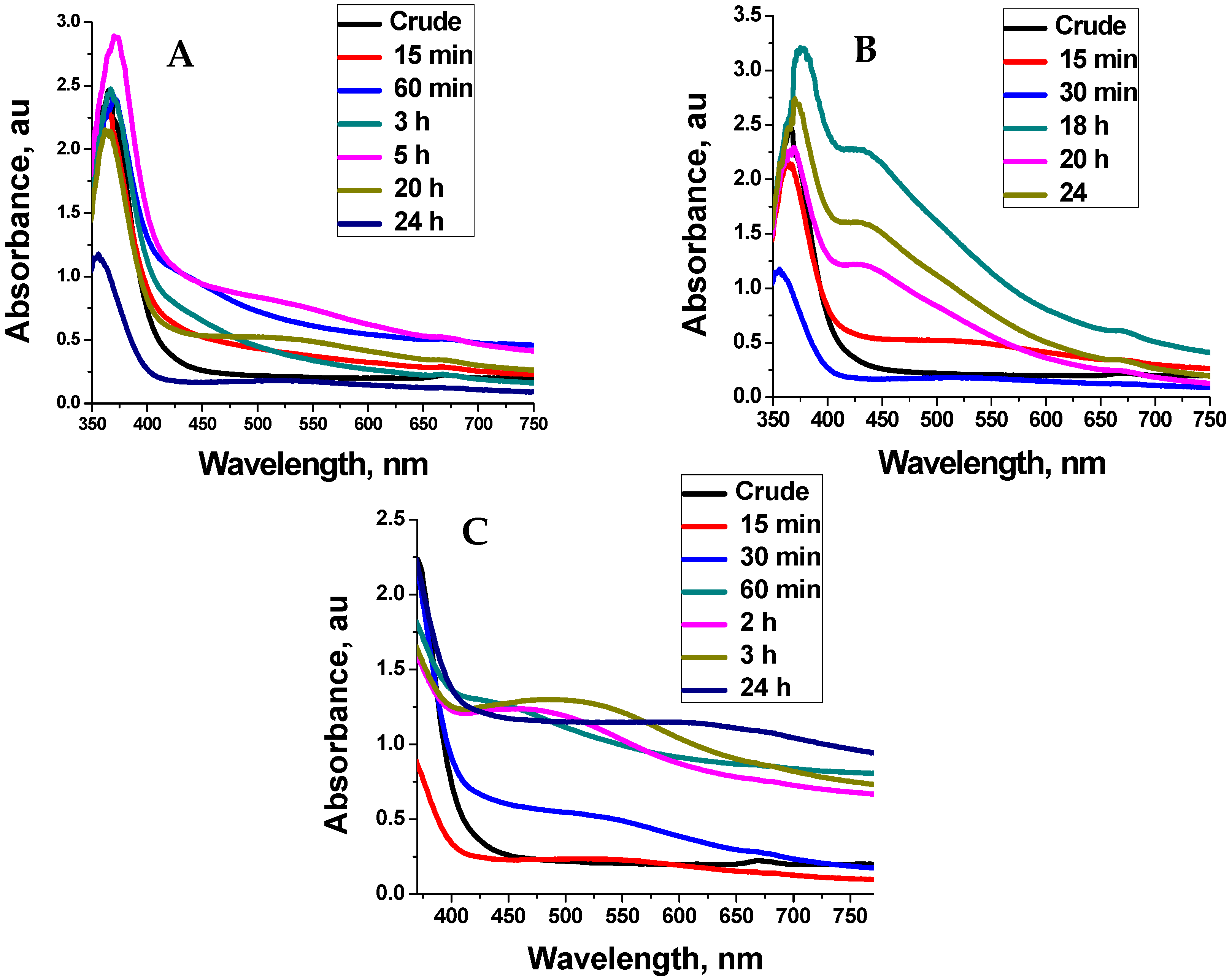
2.2. UV-Vis Analysis
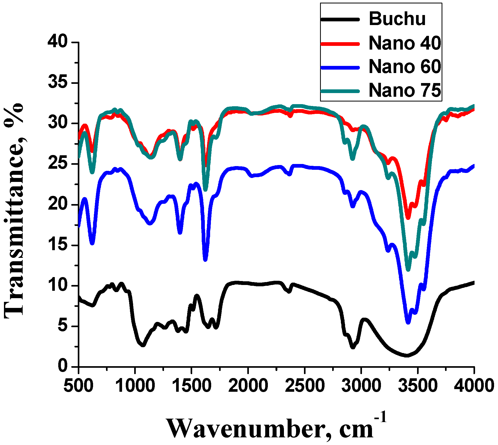
2.3. FTIR Analysis
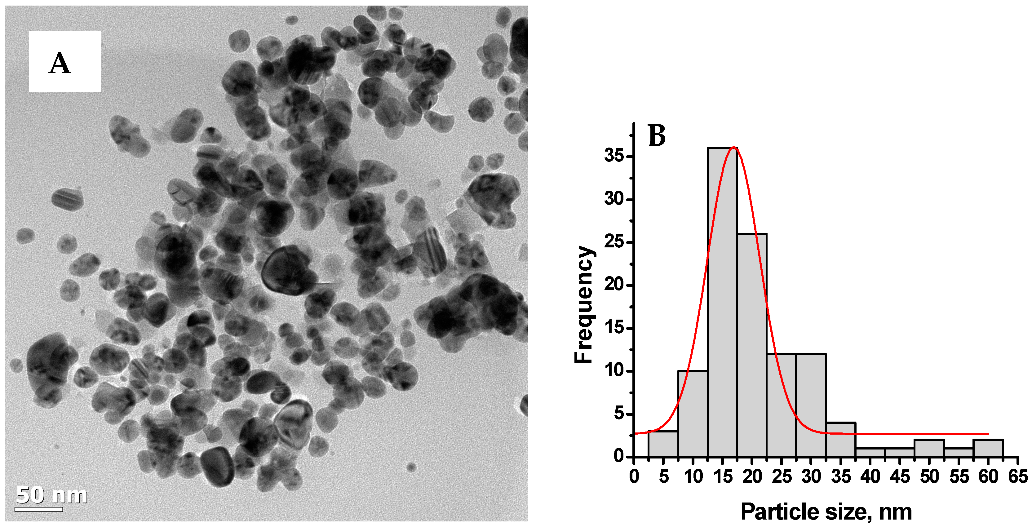
2.4. TEM Analysis
2.5. Analgesic Activity
3. Materials and Methods
3.1. Plant Material and Preparation of Extract
3.2. Synthesis of Silver Nanoparticles
3.3. Characterisation
3.4. Analgesic Activity
- Control group—0.9% w/v (normal saline)
- Standard group—100 mg/kg aspirin
- Group III—200 mg/kg ethanolic extract
- Group IV—200 mg/kg of nano particles produced at 40 °C
- Group V—200 mg/kg of nano particles produced at 60 °C
- Group VI—200 mg/kg of nano-particles at 75 °C (15 min reaction time)
- Group VII—200 mg/kg nanoparticles produced at 75 °C (24 h reaction time)
4. Conclusions
Acknowledgments
Author Contributions
Conflicts of Interest
References
- Coetzee, C.; Jesthas, J.; Rintein, E. Indigenous Plant Genetic Resources of South Africa. Perspectives on New Crops and New Uses; ASHS Press: Alexandria, VA, USA, 1999; pp. 160–163. [Google Scholar]
- Viljoen, A.M.; Moola, A.; van Vuuren, S.F.; Van Zyl, R.I.; Baser, K.H.C. The biological activity and essential oil composition of 17 Agathosma (Rutaceae) species. J. Essent. Oil Res. 2006, 18, 2–16. [Google Scholar]
- Posthumous, M.A.; Van Beek, T.A.; Collins, N.F.; Graven, E.F. Chemical composition of the essential oil of Agathosma betulina, A. crenulata and an A. betulina × crenulata hybrid (Buchu). J. Essent. Oil Res. 1996, 8, 223–228. [Google Scholar] [CrossRef]
- Firdhouse, J.M.; Lalitha, P.; Sripathi, K.S. Novel Synthesis of nanoparticles using leaf ethanol extract of Pisonia grandis. Pharm. Chem. 2012, 4, 2320–2326. [Google Scholar]
- Sharma, V.K.; Yngard, R.A.; Lin, Y. Silver nanoparticles: Green synthesis and their antimicrobial activities. Adv. ColloidInterface Sci. 2009, 145, 83–96. [Google Scholar] [CrossRef] [PubMed]
- El-Sheikh, M.A.; El-Rafie, S.M.; Abdel-Halim, E.S.; El-Rafie, M.H. Green Synthesis of Hydroxyethyl Cellulose-Stabilized Silver Nanoparticles. Polym. J. 2013, 2013, 1–11. [Google Scholar] [CrossRef]
- Harborne, A.J. Phytochemical Methods: A Guide to Modern Techniques of Plant Analysis; Chapman and Hall Ltd.: London, UK, 1973. [Google Scholar]
- Arunachalam, K.D.; Annamalai, S.K.; Hari, S. One step green synthesis of leaf extract-mediated biocompatible silver and gold nanoparticles from Memecylon umbellatum. Int. J. Nanomed. 2013, 8, 1307–1315. [Google Scholar] [CrossRef] [PubMed]
- Thilagam, M.; Tamiselvi, A.; Chandrasekeran, B.; Rose, C. Photosynthesis of silver nanoparticles using medicinal and dye yielding plant of Bixa orellana L. leaf extract. JPSI 2013, 2, 9–13. [Google Scholar]
- Ghosh, S.; Patil, S.; Ahire, M.; Kitture, R.; Kale, S. Synthesis of silver nanoparticles using Dioscora bulbifera tuber extract and evaluation of its synergistic potential in combination with antimicrobial agents. Int. J. Nanomed. 2012, 7, 483–496. [Google Scholar]
- Jancy, M.E.; Inbathamizh, L. Green Synthesis and Characterization of Nano Silver Using Leaf Extract of Morinda pubescens. Asian J. Pharm. Clin. Res. 2012, 5, 1–5. [Google Scholar]
- Harriot, M.; Marion, E.; Martha, A.; Welford, S.; William, A. Inflammation induced by histamine, serotonin, bradykinin, and compound 48/480 in the rat. Antagonists and mechanisms of action. J. Pharmacol. Exp. Ther. 2004, 19, 300–302. [Google Scholar]
- Morteau, O. Prostaglandins and Inflammation: The cyclooxygenase controversy. Arch. Immunol. Ther. Exp. (Warsz) 2000, 48, 473–480. [Google Scholar] [PubMed]
- Sofowora, A. Screening plants for bioactive agents. In Medicinal Plants and Traditional Medicinal in Africa, 2nd ed.; Spectrum books Ltd.: Ibadan, Nigeria, 1993. [Google Scholar]
- Prabhu, V.V.; Nalini, G.; Chidambaranathan, N.; Kisan, N.S. Evaluation of Anti-inflammation and analgesic activity of Tridax procumbens Linn against formalin, acetic acid and CFA induced pain models. Int. J. Pharm. Pharm. Sci. 2011, 3, 126–130. [Google Scholar]
- Ahmad, T.; Wani, I.A.; Manzoor, N.; Ahmed, J.; Asiri, A.M. Biosynthesis, structural characterization and antimicrobial activity of gold and silver nanoparticles. Colloids Surf. B 2013, 107, 227–234. [Google Scholar] [CrossRef] [PubMed]
- Sample Availability: Samples of the compounds are available from the authors.


| Metabolite | Ethanolic Extract |
|---|---|
| Glycosides | + |
| Flavonoids | + |
| Alkaloids | + |
| Terpenes | + |
| Steroids | + |
| Tannins | + |
| Saponins | + |
| Proteins | + |
© 2016 by the authors. Licensee MDPI, Basel, Switzerland. This article is an open access article distributed under the terms and conditions of the Creative Commons Attribution (CC-BY) license ( http://creativecommons.org/licenses/by/4.0/).
Share and Cite
Chiguvare, H.; Oyedeji, O.O.; Matewu, R.; Aremu, O.; Oyemitan, I.A.; Oyedeji, A.O.; Nkeh-Chungag, B.N.; Songca, S.P.; Mohan, S.; Oluwafemi, O.S. Synthesis of Silver Nanoparticles Using Buchu Plant Extracts and Their Analgesic Properties. Molecules 2016, 21, 774. https://doi.org/10.3390/molecules21060774
Chiguvare H, Oyedeji OO, Matewu R, Aremu O, Oyemitan IA, Oyedeji AO, Nkeh-Chungag BN, Songca SP, Mohan S, Oluwafemi OS. Synthesis of Silver Nanoparticles Using Buchu Plant Extracts and Their Analgesic Properties. Molecules. 2016; 21(6):774. https://doi.org/10.3390/molecules21060774
Chicago/Turabian StyleChiguvare, Herbert, Opeoluwa O. Oyedeji, Reuben Matewu, Olukayode Aremu, Idris A. Oyemitan, Adebola O. Oyedeji, Benedicta N. Nkeh-Chungag, Sandile P. Songca, Sneha Mohan, and Oluwatobi S. Oluwafemi. 2016. "Synthesis of Silver Nanoparticles Using Buchu Plant Extracts and Their Analgesic Properties" Molecules 21, no. 6: 774. https://doi.org/10.3390/molecules21060774