Article

The Nasopharyngeal, Ruminal, and Vaginal Microbiota and the Core Taxa Shared across These Microbiomes in Virgin Yearling Heifers Exposed to Divergent In Utero Nutrition during Their First Trimester of Gestation and in Pregnant Beef Heifers in Response to Mineral Supplementation

1
Department of Microbiological Sciences, North Dakota State University, Fargo, ND 58108, USA
2
Lacombe Research and Development Centre, Agriculture and Agri-Food Canada, 6000 C & E Trail, Lacombe, AB T4L 1W1, Canada
3
Department of Animal Sciences, North Dakota State University, Fargo, ND 58102, USA
4
Lethbridge Research and Development Centre, Agriculture and Agri-Food Canada, Lethbridge, AB T1J 4B1, Canada
5
Central Grasslands Research Extension Center, North Dakota State University, Streeter, ND 58483, USA
*
Author to whom correspondence should be addressed.
Microorganisms 2021, 9(10), 2011; https://doi.org/10.3390/microorganisms9102011
Submission received: 19 August 2021 / Revised: 11 September 2021 / Accepted: 20 September 2021 / Published: 23 September 2021
(This article belongs to the Special Issue Rumen Microbial Communities)

Abstract

:
In the present study, we evaluated whether the nasopharyngeal, ruminal, and vaginal microbiota would diverge (1) in virgin yearling beef heifers (9 months old) due to the maternal restricted gain during the first trimester of gestation; and (2) in pregnant beef heifers in response to the vitamin and mineral (VTM) supplementation during the first 6 months of pregnancy. As a secondary objective, using the microbiota data obtained from these two cohorts of beef heifers managed at the same location and sampled at the same time, we performed a holistic assessment of the microbial ecology residing within the respiratory, gastrointestinal, and reproductive tract of cattle. Our 16S rRNA gene sequencing results revealed that both α and β-diversity of the nasopharyngeal, ruminal and vaginal microbiota did not differ between virgin heifers raised from dams exposed to either a low gain (targeted average daily gain of 0.28 kg/d, n = 22) or a moderate gain treatment (0.79 kg/d, n = 23) during the first 84 days of gestation. Only in the vaginal microbiota were there relatively abundant genera that were affected by maternal rate of gain during early gestation. Whilst there was no significant difference in community structure and diversity in any of the three microbiota between pregnant heifers received no VTM (n = 15) and VTM supplemented (n = 17) diets, the VTM supplementation resulted in subtle compositional alterations in the nasopharyngeal and ruminal microbiota. Although the nasopharyngeal, ruminal, and vaginal microbiota were clearly distinct, a total of 41 OTUs, including methanogenic archaea, were identified as core taxa shared across the respiratory, gastrointestinal, and reproductive tracts of both virgin and pregnant heifers.

1. Introduction

Host genetic selection has been a primary target for improving animal health and productivity over the last several decades and has resulted in tremendous progress in both dairy and beef cattle production systems. Recently, the microbiota colonizing different mucosal surfaces of cattle have become a new target for manipulation and engineering with great potential to improve animal health and production [1,2]. Diverse and dynamic microbial communities present in the respiratory, gastrointestinal and reproductive tracts of cattle are vital to health and performance [2,3,4]. Among these microbial communities, the ruminal microbiota in cattle, which is the most densely populated and involved in both nutrient metabolism and immune system development, has become the primary target for manipulation and engineering [5].
Recent evidences suggest that the ruminal microbiome and host genetics can be targeted independently to improve feed efficiency and mitigate enteric methane emissions from cattle [6,7]. One of the challenges associated with manipulation of the ruminal microbiome in mature animals is its resiliency that allows the microbiome to revert to the original state following the cessation of an intervention [8]. To overcome this challenge, early life microbial programming in young ruminants was recommended and has shown some efficacy [9,10,11]. For example, Palma-Hidalgo et al. [12] reported that the direct inoculation of fresh ruminal fluid from adult goats to kids in early life accelerated ruminal microbial community development and improved the weaning process. Early life microbial programming is based on the current dogma that microbial colonization of the rumen starts at birth, and the developing ruminal microbiota within the first 3 weeks of life is less resilient to manipulation [9]. A recent study by Guzman and colleagues [13], however, provided sequencing and culture-based evidence indicating that the intestine of calf fetus is not sterile and colonization by so-called “pioneer” microbes may occur during gestation. This is further supported by our preliminary data which suggested that colonization of the fetal intestine by archaea and bacteria may take place within the first 12 weeks of gestation in cattle [14]. These observations highlight the potential and extended role of the maternal microbiome in calf ruminal microbiome development.
Although the role of maternal nutrition in programming of the offspring metabolic, immune and nervous system development has been relatively well documented in humans and food-producing animals including cattle [15,16,17,18], the potential involvement of the maternal microbiome in the developmental origins of health and disease has recently begun to be better appreciated [19,20,21]. It was hypothesized that undesired alterations in the maternal microbiota could indirectly influence fetal development, and that these effects may subsequently be transmitted to progeny, resulting in the development of an altered microbiota in offspring [20]. Undesirable outcomes in offspring resulting from changes in the maternal microbiota include increased susceptibility to the development of metabolic disorders and respiratory infections [20,22]. Recent evidence from studies in mice demonstrated that the maternal microbiota during pregnancy modulates the programming of fetal metabolic and nervous system development [23,24]. Considering the increased evidence showing the importance of the maternal microbiota in developmental programming in rodent models, and the greater evidence regarding the involvement of the microbiome in defining cattle health and productivity, exploring the role of the maternal microbiota in fetal programming and offspring microbiome development may provide important information for improving cattle health and feed efficiency.
In the present study, we used 16S rRNA gene sequencing to characterize the nasopharyngeal, ruminal and vaginal microbiota of virgin yearling heifers from dams given different nutritional diets during their first trimester of gestation, and in pregnant beef heifers in response to direct feeding of a mineral and vitamin (VTM) supplement during the first 6 months of gestation. Of note, a well-defined positive impact of maternal VTM supplementation exists on offspring health and performance in beef cattle, and the role of VTM on fetal programming assessed during the first trimester of pregnancy have been documented [25,26,27,28,29]. Questions remain, however, pertaining to whether these maternal VTM supplementation-associated positive outcomes are dependent on VTM-induced alterations of ruminal microbiota. To provide a more holistic view of the microbiota residing within the respiratory, gastrointestinal, and reproductive tract of cattle, the similarity of the microbiota within these sites was evaluated, and taxa shared amongst the three microbial habitats were identified. Given the relevance of these microbial communities to respiratory and reproductive health and rumen fermentation/nutrient metabolism, and most importantly, as potential maternal inoculant sources for seeding the fetal and offspring microbiota, a holistic evaluation of bovine microbiota is therefore necessary rather than focusing on only one microbial community.

2. Materials and Methods

All experimental procedures involving cattle were approved by the North Dakota State University Institutional Animal Care and Use Committee (#A20085 and #A20086, for virgin yearling heifers and for pregnant heifers, respectively).

2.1. Animal Husbandry and Experimental Design

2.1.1. Virgin Yearling Heifers

Deep nasopharyngeal swabs, ruminal fluid, and vaginal swabs were collected from 45 virgin heifers (9-month-old, Angus-cross, BW = 688 ± 57 kg) whose dams were assigned to either a low gain treatment (LG, targeted average daily gain of 0.28 kg/d, n = 22) or a moderate gain treatment (MG, 0.79 kg/d, n = 23) during the first 84 days of gestation. To achieve the LG, dams were maintained on a basal diet consisting of prairie grass hay, corn silage, and dried distillers grains plus solubles. To achieve the MG (0.79 kg/d), dams were fed the basal diet with the addition of a protein/energy supplement fed at the rate of 0.58% BW as-fed daily. Up to d 84 of gestation, dams were housed and individually fed (Insentec; Hokofarm B.V. Repelweg 10, 8316 PV Marknesse, The Netherlands) at the Beef Cattle Research and Extension Center (BCRC) in Fargo, ND. After day 84 of gestation, dams were transported to the Central Grasslands Research Extension Center (CGREC) in Streeter, ND, where they were managed as a single group on common diets until parturition and subsequent weaning of the F1 offspring. Upon weaning, the heifers (approx. 6-months old) were transported to the BCRC where they were housed in two pens and individually fed (Insentec; Hokofarm B.V. Repelweg 10, 8316 PV Marknesse, the Netherlands) a common diet (Table 1).

2.1.2. Pregnant Heifers

The nasopharyngeal, ruminal, and vaginal microbiota of the replacement pregnant heifers (1 year 9 months old, BW = 1001 ± 128 kg) during the sixth month of gestation were also evaluated. At breeding, heifers were assigned to one of two treatments: (1) vitamin and mineral supplementation (VTM; n = 17) or (2) no vitamin and mineral supplementation (Control; n = 15). Heifers were housed at the BCRC and individually fed (Insentec; Hokofarm B.V. Repelweg 10, 8316 PV Marknesse, the Netherlands) a total mixed ration containing triticale hay, corn silage, modified distillers grains plus solubles, ground corn, and, if indicated by treatment, a VTM premix (Table 1). The VTM premix was fed at 0.45 kg/heifer/day to provide macro and trace minerals and vitamins A, D, and E to meet 110% of the daily requirements [30]. The specific ingredients within the VTM supplement are as previously described [29].

2.2. Nasopharyngeal Swab, Ruminal Fluid, and Vaginal Swab Sampling

Nasopharyngeal swabs, ruminal fluid and vaginal swabs were collected simultaneously from each of the virgin yearling (9 months old) and pregnant heifers (at 6 months pregnancy) by same personnel on the same day. All sample collection was completed within 4 h (8 a.m.–12 p.m.).

2.2.1. Nasopharyngeal Sampling

Deep nasopharyngeal swabs were collected as previously described [31,32]. Briefly, prior to sampling, the right nostril of the heifer was wiped clean with 70% ethanol and an extended guarded swab (27 cm) with a rayon bud (MW 128, Medical Wire & Equipment, Corsham, England) was used for sampling. Swab tips were then cut and placed in a sterile 2 mL centrifuge tube on ice until processing. Upon arrival in the lab, the swab was transferred into 1 mL sterile brain heart infusion (BHI) broth containing 20% glycerol stock.

2.2.2. Rumen Fluid Sampling

Rumen fluid sample collection was performed using the method currently used in our laboratory which was modified from Paz et al. [33]. Briefly, a rigid metal speculum was placed into the mouth of the heifer and then a flexible plastic tube with multiple holes at the tip was passed through the speculum and into the esophagus. The speculum was used to ensure that the plastic tube was not damaged by the heifers and that the tube entered the esophagus. Once the tube entered the rumen, and was below the ruminal mat layer, suction pressure was applied to the tube to collect ruminal fluid. Up to 120 mL of ruminal fluid was collected on each sampling day. Separate tubing and containers were used for each heifer to avoid cross-contamination. After thorough mixing, an aliquot of 40 mL of rumen fluid was placed into a 50 mL falcon tube and immediately frozen with dry ice.

2.2.3. Vaginal Sampling

For vaginal sampling, the vulva was thoroughly cleaned with 70% ethanol and a paper towel. Then, the labia majora of the vulva was held open allowing the passage of a swab (15 cm, sterile cotton tipped applicators with aerated tip protector; Puritan). When the swab tip reached the midpoint of the vaginal cavity, it was placed against the vaginal wall, swirled four times, and then withdrawn carefully to minimize contamination. The vaginal swabs were immediately placed in sterile Whirl Pak bags and transported on ice to the lab. All nasopharyngeal and vaginal swabs as well as rumen fluid were stored at −80 °C until DNA extraction.

2.3. Metagenomic DNA Extraction

Metagenomic DNA was extracted from the nasopharyngeal and vaginal swabs using a Qiagen DNeasy Tissue kit (Qiagen Inc., Germantown, MD, USA) according to the kit manual with minor modifications. Briefly, the cotton tip of the nasopharyngeal swab was removed and placed back into the BHI-glycerol mixture, and then centrifuged at 20,000× g for 5 min at 4 °C to pellet the cotton and microbes. The pellet was then re-suspended in 180 μL of enzymatic buffer. The enzymatic buffer (20 mM Tris.CI (pH 8.0), 2 mM sodium EDTA, and 1.2% Triton X-100) contained 300 U/mL mutanolysin and 20 mg/mL lysozyme. The mixture was then vortexed and incubated for 1 h at 37 °C with agitation at 800 rpm. After incubation, 25 μL proteinase K and 200 μL Buffer AL (without ethanol) were added and vortexed, and then incubated at 56 °C for 30 min with agitation at 800 rpm. Approximately 400 mg of 0.1 mm zircon/silica beads were added to the tube and subjected to mechanical cell lysis using a FastPrep-24 Classic bead beater (MP Biomedicals, Irvine, CA, USA) at 4.0 m/s for 24 s. The mixture was then centrifuged (13,000× g for 5 min), and the supernatant (approx. 300–400 µL) was removed and placed in a new tube and mixed with an equal volume of 100% ethanol. From this step onward, the procedures were performed as described in the DNeasy Tissue Kit instruction manual.
The procedures for metagenomic DNA extraction from the vaginal swabs were identical to those used on the nasopharyngeal swabs with the following exceptions. First, the cotton swab was removed from applicator and placed in a sterile 2 mL centrifuge tube. Then, 360 µL of enzymatic buffer was added to the tube to ensure complete emersion of the swab in the enzymatic buffer. Metagenomic DNA from the rumen fluid samples was extracted using the Qiagen DNeasy PowerLyzer PowerSoil kit (Qiagen Inc., Germantown, MD, USA) according to the instructions of manufacturer. The frozen rumen fluid samples were thawed, and vortexed thoroughly before transfer to a sterile 2 mL microfuge tube. The sample was then centrifuged at 20,000× g for 10 min at 4 °C to pellet the microbes in the sample. From this point onwards, the protocol for the PowerLyzer PowerSoil kit was followed as per the instructions of the manufacturer. Negative extraction controls were included for all extraction kits.

2.4. 16S rRNA Gene Sequencing and Analysis

The V3-V4 hypervariable regions of the 16S rRNA gene were amplified using the 341-F (5′-CCTAYGGGRBGCASCAG-3′) and 806-R (5′-GGACTACNNGGGTATCTAAT-3′) primers. All PCR steps were carried out using the Phusion High-Fidelity PCR Master Mix (New England Biolabs, Ipswich, MA, USA). The PCR products were electrophoresed on a 2% agarose gel and stained with SYBR Safe DNA gel stain. The DNA fragment was excised from the gel and purified using the QIAquick Gel Extraction Kit (Qiagen Inc.). Sequencing libraries were generated with NEBNext Ultra DNA Library Prep Kit (New England BioLabs, Ipswich, MA, USA) for Illumina, following the recommendations of the manufacturer. The library quality was assessed with a Qubit 2.0 Fluorometer (Thermo Scientific, Waltham, MA, USA) and Agilent Bioanalyzer 2100 system. Libraries were then sequenced on a NovaSeq 6000 instrument with a SP flow cell (2 × 250 bp) (Illumina Inc., San Diego, CA, USA).
The 16S rRNA gene sequences were processed using DADA2 v. 1.18 [34] in R. 4.0.3. Briefly, the forward reads were truncated at 225 bp and the reverse reads at 220 bp. The reads were merged, chimeric sequences removed, and taxonomy assigned to each merged sequence, referred to here as operational taxonomic units (OTUs) at 100% similarity, using the naïve Bayesian RDP classifier [35] and the SILVA SSU database release 138 [36]. OTUs that were predominant in the negative extraction control samples and likely to be contaminants were removed prior to analyses as were those OTUs classified as chloroplasts, mitochondria, or eukaryota. The number of OTUs per sample (richness), the Shannon and inverse Simpson’s diversity indices, and Bray–Curtis dissimilarities were calculated in R using Phyloseq 1.34.0 [37] and vegan 2.5–7 [38]. To account for uneven sequence depths, samples were randomly subsampled to 7100, 73,500, and 10,300 for the nasopharyngeal, ruminal, and vaginal samples respectively, prior to the calculation of Bray–Curtis dissimilarities and diversity measures for the virgin heifers. For the pregnant heifers, these values were 6200, 74,500, and 8200.

2.5. Statistical Analysis

Permutational multivariate analysis of variance (PERMANOVA; adonis2 function; 10,000 permutations) of the Bray–Curtis dissimilarities was performed using vegan to determine the effect of maternal nutrition on the nasopharyngeal, ruminal and vaginal microbial community structure in virgin heifers whose dams were managed to targeted LG or MG during the first 84 days of gestation. The effect of VTM supplementation on the microbial community structure of these three microbiotas in pregnant heifers was also assessed. Differentially abundant genera between treatment groups for both the virgin and pregnant heifers were identified using MaAsLin2 v. 1.5.1 in R [39]. Only those genera with a relative abundance greater than 0.1% within each sample type were included. Diversity metrics were compared by treatment for both virgin and pregnant heifers using an unpaired t-test. The number of OTUs (richness), diversity indices, relative abundance of the most relatively abundant genera between the LG and MG groups of virgin yearling heifers, or between the VTM and Control groups of pregnant heifers, and the relative abundance of Methanobrevibacter spp. between the virgin and pregnant heifers were compared using the generalized liner mixed model estimation procedure (PROC GLIMMIX) in SAS (ver. 9.4, SAS Institute Inc. Cary, NC, USA). The means were compared using the LSMEANS statement and significance was declared at p < 0.05.
Spearman’s rank-based correlations between Methanobrevibacter and the other 24 most relatively abundant genera in the ruminal and vaginal microbiota were calculated using the CORR procedure in SAS with the SPEARMAN option. From these 24 genera, the genera that have significant effect on Methanobrevibacter abundance were predicted using a stepwise-selected GLIMMIX model with beta-binomial distribution as described previously [32]. The model used was
logit ( Y ˜ ) = ln ( π / ( 1 π ) ) = b 0 + b 1 ( X 1 ) + + b n ( X n ) ,
where π represents the relative abundance of the Methanobrevibacter genus (0–1) and Xn represents the relative abundance (0–100%) of a bacterial genus n. The stepwise selection method involved backward elimination and forward selection to eliminate any variables in the model that have no significant effect (p > 0.05) on the predicted outcome.

3. Results

3.1. Sequencing Results

An average of 66,045 ± 31,588 (SD) 16S rRNA gene sequences per sample (min. = 2374; max. = 139,012) were obtained from 219 nasopharyngeal, ruminal fluid, and vaginal samples. From these sequences, a total of 81,391 archaeal and bacterial OTUs were identified and classified into 58 unique phyla (8 archaeal and 50 bacterial phyla), and 1511 unique genera.

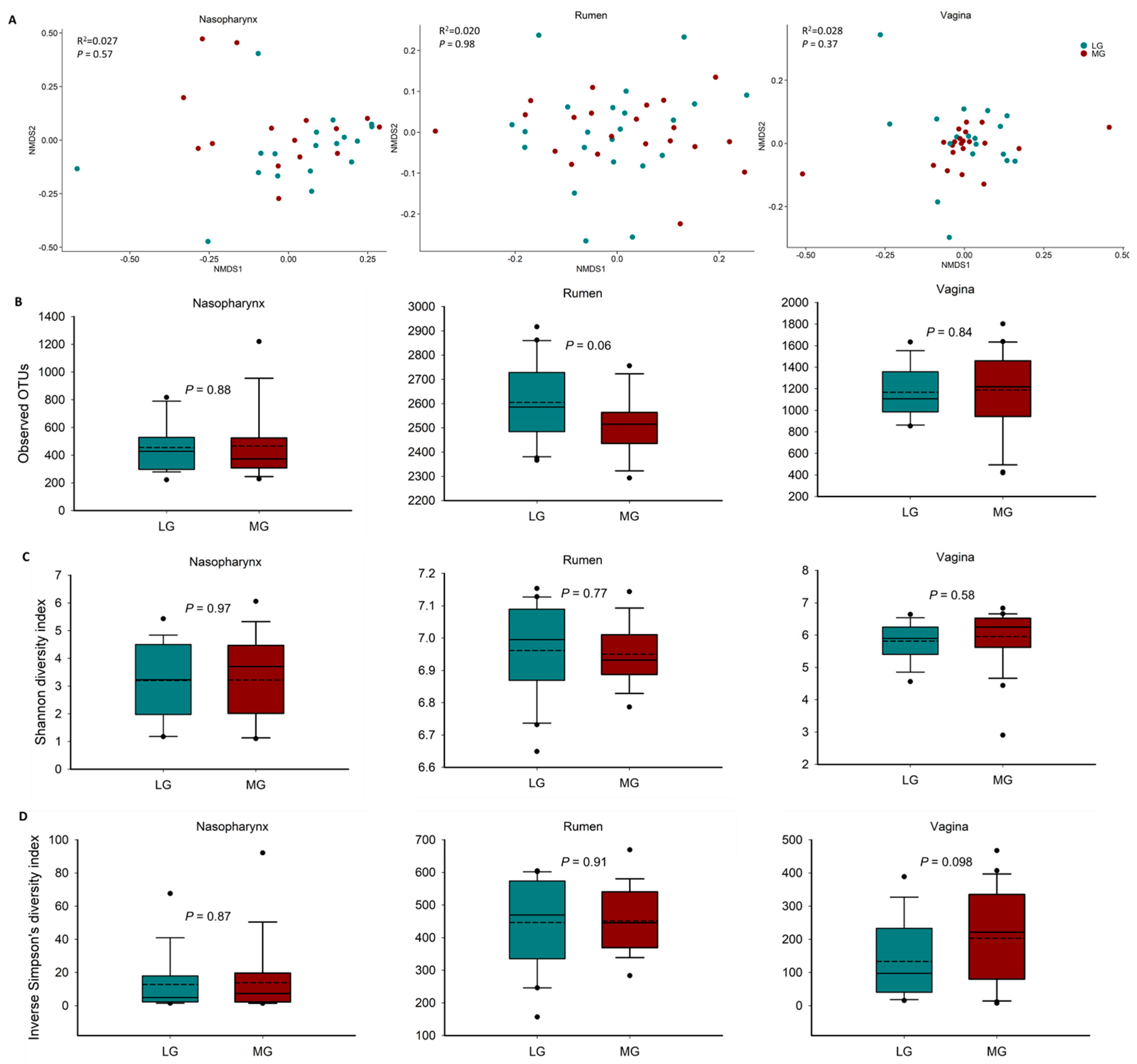
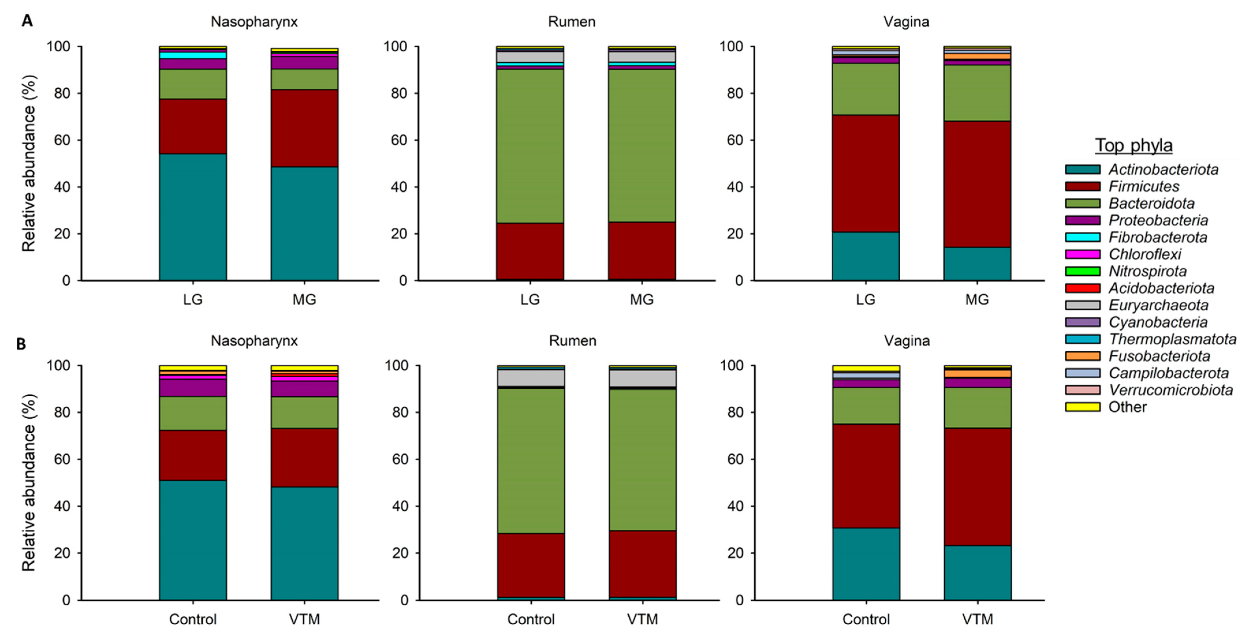
3.2. Effect of Maternal Restricted Gain during the First Trimester of Gestation on Offspring Microbiota Development

To determine the effect of maternal nutrition during the first trimester of gestation on microbial populations of their offspring, we characterized and compared the nasopharyngeal, ruminal and vaginal microbiota of virgin yearling heifers from two groups of dams that were subjected to diets resulting in either a LG or MG phenotype during the first 84 days of gestation.The microbial community structure of the nasopharynx (PERMANOVA: R2 = 0.027, p = 0.57), rumen (PERMANOVA: R2 = 0.02, p = 0.98) and vagina (PERMANOVA: R2 = 0.028, p = 0.37) in the virgin heifers did not differ between the LG and MG groups (Figure 1A). Microbial richness and diversity as measured by the number of OTUs, and the Shannon and inverse Simpson’s diversity indices of these microbiotas also did not significantly differ by maternal nutrition group (Figure 1B–D; p > 0.05). There was, however, a strong tendency (p = 0.06) in LG offspring to harbor a richer ruminal microbial community compared to MG offspring (2605 vs. 2515 OTUs).
The nasopharyngeal microbiota across all animals was dominated by Actinobacteriota (51%), Firmicutes (28.2%), Bacteroidota (10.8%) and Proteobacteria (4.9%). The relative abundance of the eight most relatively abundant phyla did not differ between LG and MG virgin heifers (p > 0.05) (Figure 2A). Bacteroidota was the most relatively abundant phylum in the ruminal microbiota (65.5%) followed by Firmicutes (24.2%). As with the nasopharyngeal microbiota, none of the eight most relatively abundant phyla in the rumen microbiota differed between the two treatment groups. The most relatively abundant phylum present in the vaginal tract was Firmicutes (52%) followed by Bacteroidota (23.0%) and Actinobacteriota (17.4%). Similar to the rumen and nasopharyngeal microbiota, no difference between treatments was detected in the relative abundance of eight most relatively abundant phyla in the vaginal microbiota (p > 0.05).
The 25 most relatively abundant genera in the nasopharyngeal, ruminal and vaginal microbiota are listed in Table 2. Overall, the predominant nasopharyngeal genera did not differ (to a statistically significant degree) in relative abundance between the LG and MG groups (p > 0.05). In the rumen, the relative abundance of only one genus ((Eubacterium) ruminantium group) was significantly different between the two groups, being greater in the MG group than in the LG group (p = 0.029). Within the vaginal microbial community, Alistipes, Ruminococcus and Oscillospiraceae NK4A214 group were significantly less relatively abundant in the LG group compared to the MG group (p < 0.05). The relative abundance of Romboutsia (p = 0.061) and Paeniclostridium (p = 0.092) tended to be lower while Arcanobacterium (p = 0.090) tended to be higher in MG group compared to LG group.

3.3. Effect of Vitamin and Mineral Supplementation during the First Six Months of Gestation on the Maternal Microbiota

To investigate whether VTM supplementation during the first 6 months of gestation affects the maternal microbiota, we compared the nasopharyngeal, ruminal and vaginal microbiota between the VTM and Control groups of replacement pregnant heifers. The community structure of the nasopharyngeal (PERMANOVA: R2 = 0.038, p > 0.05); ruminal (PERMANOVA: R2 = 0.032, p > 0.05); and vaginal (PERMANOVA: R2 = 0.049, p > 0.05) microbiota was not affected by VTM supplementation (Figure 2A). Microbial richness and diversity also did not differ by VTM supplementation in any of the three microbial communities (p > 0.05) (Figure 2B–D). The relative abundance of the eight most relatively abundant phyla in the nasopharynx, rumen and vagina did not differ between the control and VTM groups (Figure 3B). Nevertheless, the relative abundance of several genera present in the nasopharynx (5 genera) and rumen (3 genera) were affected by VTM supplementation (p < 0.05, Table 3).
Mycoplasma, the third most relative abundant genus in the nasopharyngeal microbiota, was enriched in pregnant heifers receiving the VTM supplement (8.95% vs. 2.74%, p = 0.039). VTM supplementation also resulted in a reduced relative abundance of Oscillospiraceae UCG-005, Christensenellaceae R-7 group, Ruminococcus and Ornithinimicrobium genera (p < 0.05). Among the 26 most predominant ruminal genera, statistically significant differences between the means of the relative abundance were observed for only three genera (Oscillospiraceae NK4A214 group, Butyrivibrio and Ruminococcaceae CAG-352), and all of which were enriched in the VTM group (p < 0.05). The relative abundance of the 27 relatively most abundant genera in the vaginal microbiota did not differ between the VTM and control groups (p > 0.05) (Table 3).

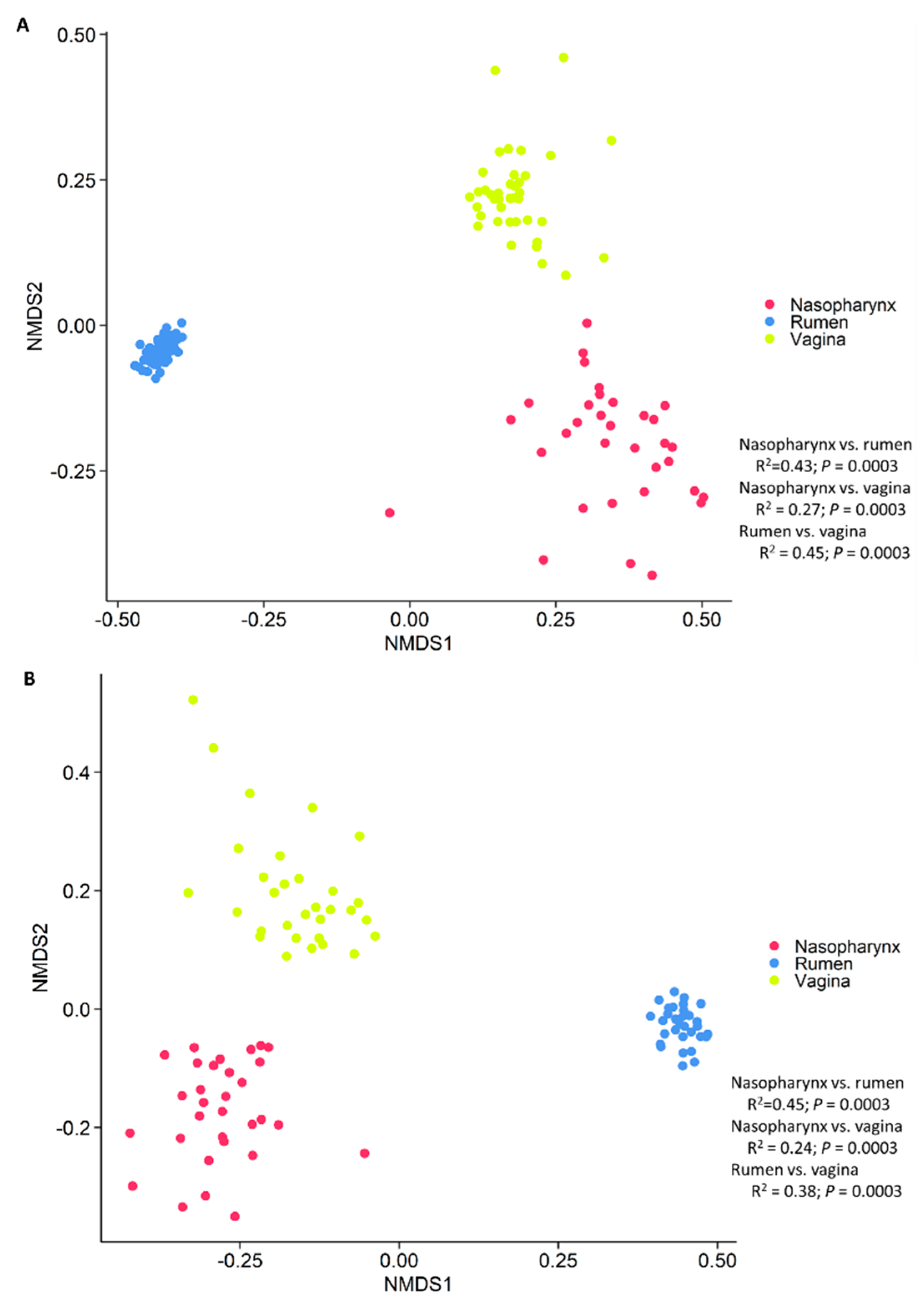
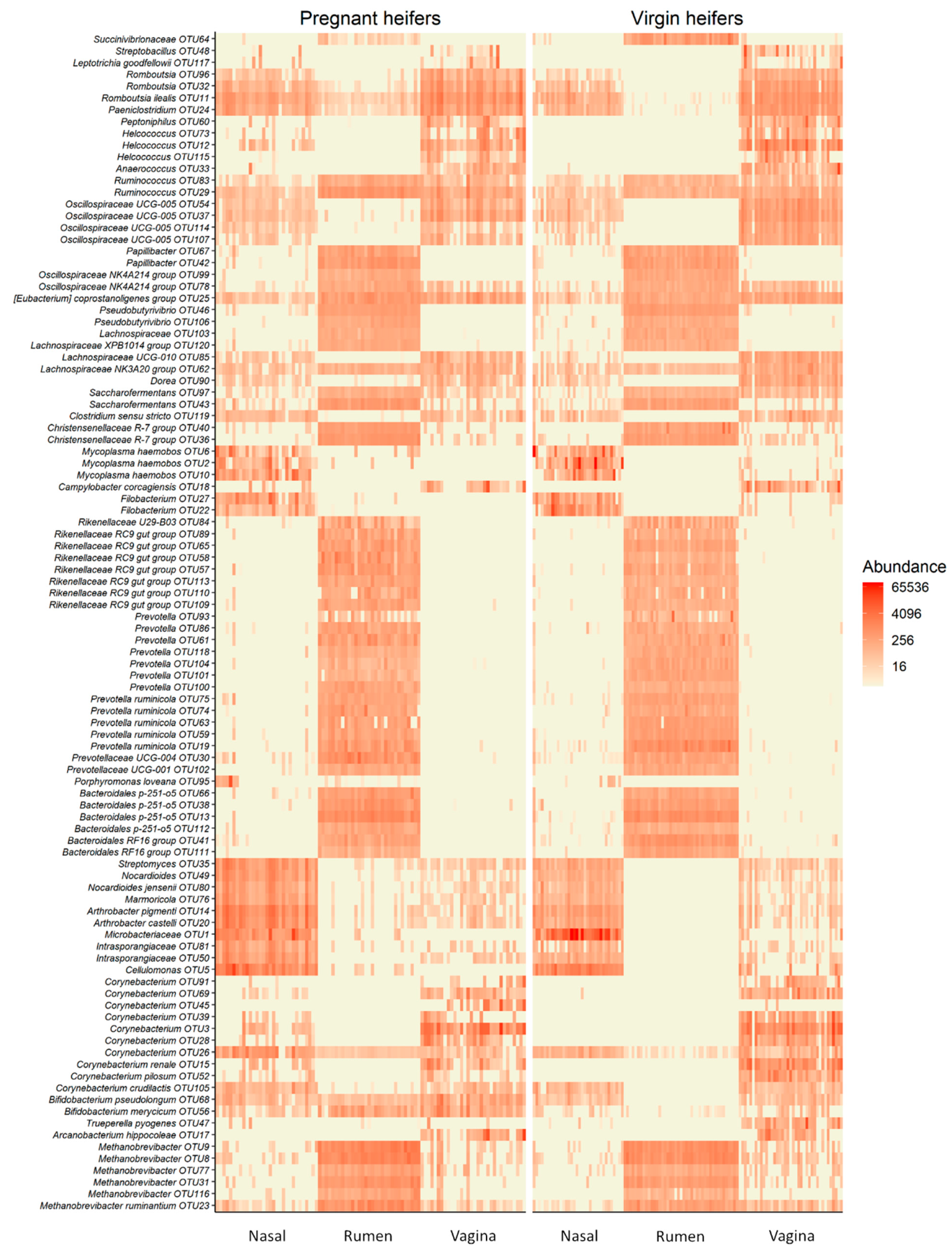
3.4. Holistic View of Nasopharyngeal, Ruminal and Vaginal Microbiota and Identification of Core Taxa Shared across These Microbiomes

To provide a holistic view of the microbiota residing within the respiratory, gastrointestinal, and reproductive tracts of each animal, we attempted to identify similarities among these microbial communities in virgin and pregnant heifers. To do this, the sequence data from all animal groups and treatments were combined and all samples were randomly subsampled to 6200 sequences. As expected, each anatomical site had a distinct microbiota (Figure 4). In terms of microbial richness, the rumen was characterized by e richest microbiota followed by the vagina and nasopharynx of virgin (Figure 1B) and pregnant heifers (Figure 2B). Overall, the ruminal and vaginal microbiota were also more diverse than the nasopharyngeal microbiota in both groups of heifers (Figure 1C,D and Figure 2C,D).
Many taxa appeared to be highly specific to one of the three microbial habitats as shown in the heatmap (Figure 5). For example, OTUs classified as Prevotella, Papillibacter, Oscillospiraceae NK4A214 group and Pseudobutyrivibrio were more exclusively present in the rumen. While the most of the OTUs within the archaeal Methanobrevibacter genus were present in three sampled habitats, the rumen was most predominantly colonized by members of this genera. Some taxa, including Mycoplasma, Filobacterium, Streptomyces, Nocardioides, Marmoricola, Arthrobacter and Cellulomonas spp., were associated with the nasopharynx. Certain Corynebacterium OTUs appeared to be specific to the vaginal microbiota.
Although the nasopharyngeal, ruminal, and vaginal microbiota were clearly distinct, a small number of taxa were present in all three microbial communities. We identified 43 OTUs that were shared by more than 60% of all samples from the virgin yearling heifers (Table S1). From these OTUs, two were classified as Methanobrevibacter (OTU8 and OTU23). The remaining shared OTUs were bacterial in origin, with 17% and 80% of them belonging to the Actinobacteriota and Firmicutes phyla, respectively. Of note, three bacterial OTUs (OTU25 (Eubacterium coprostanoligenes group; OTU561 (Colidextribacter) and OTU29 (Ruminococcus)) were shared by more than 90% of the samples. In pregnant heifers, 47 OTUs were present in more than 60% of all samples (Table S2), and most of them were identical to those taxa shared among the virgin yearling heifer samples. One taxon identified as Romboutsia ilealis (OTU11) was found in 100% of the samples, and OTU24 (Paeniclostridium), OTU25, OTU29 and OTU68 (Bifidobacterium pseudolongum) were identified in 95% of the samples.
As listed in Table 4, 41 OTUs were identified in 60% of virgin yearling and pregnant heifer samples. Nine of these OTUs were also present in more than 80% of the samples. These included OTU23 (Methanobrevibacter ruminantium), OTU26 (Corynebacterium), three OTUs (OTU25, OTU62 and OTU1688) within the Lachnospiraceae NK3A20 group, two Ruminococcus OTUs (OTU29 and OTU83), OTU24 and OTU11. OTU68 was also found in 75% of the samples. Regardless of animal group and diet, these OTUs were shared by a high proportion of the nasopharyngeal, ruminal fluid, and vaginal samples, suggesting that these taxa may be so-called “core taxa” among these anatomical locations.

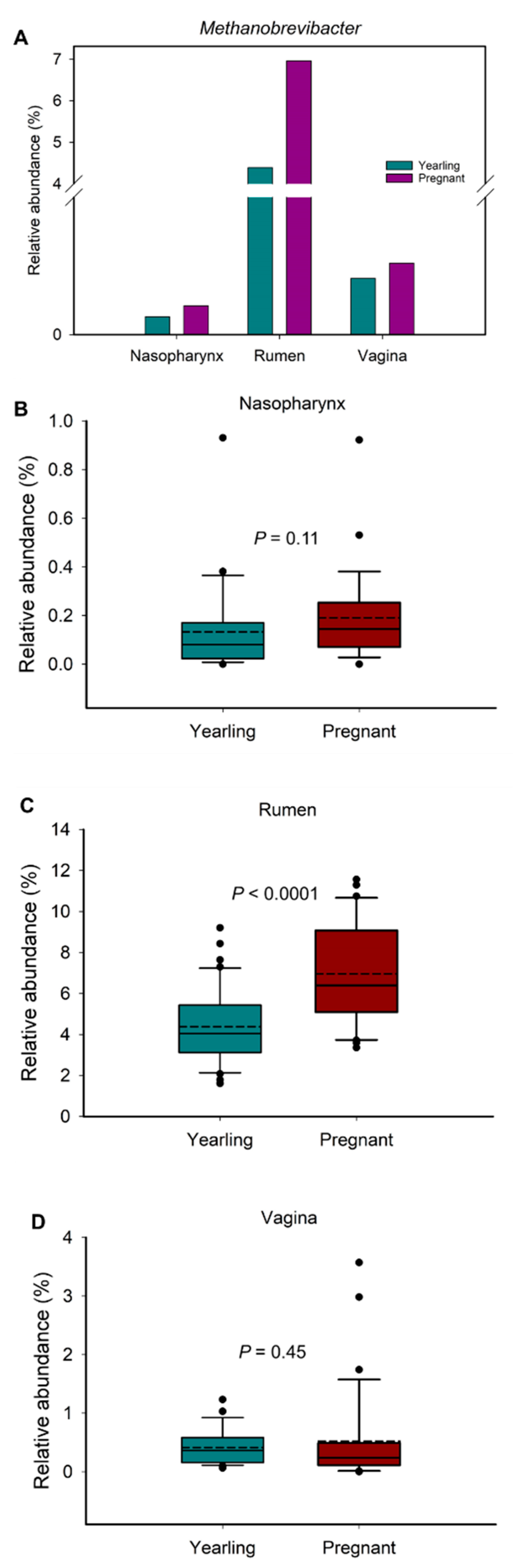
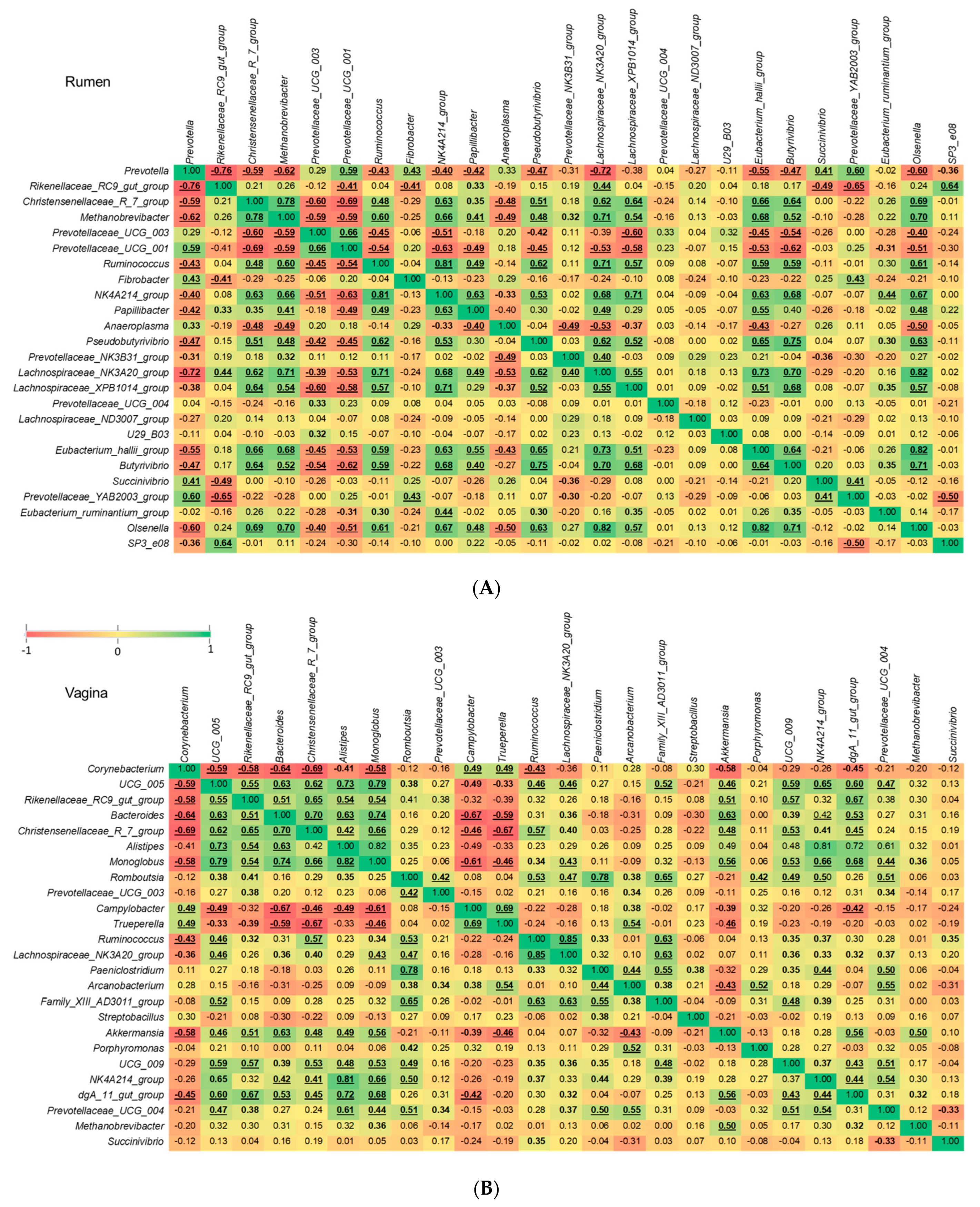
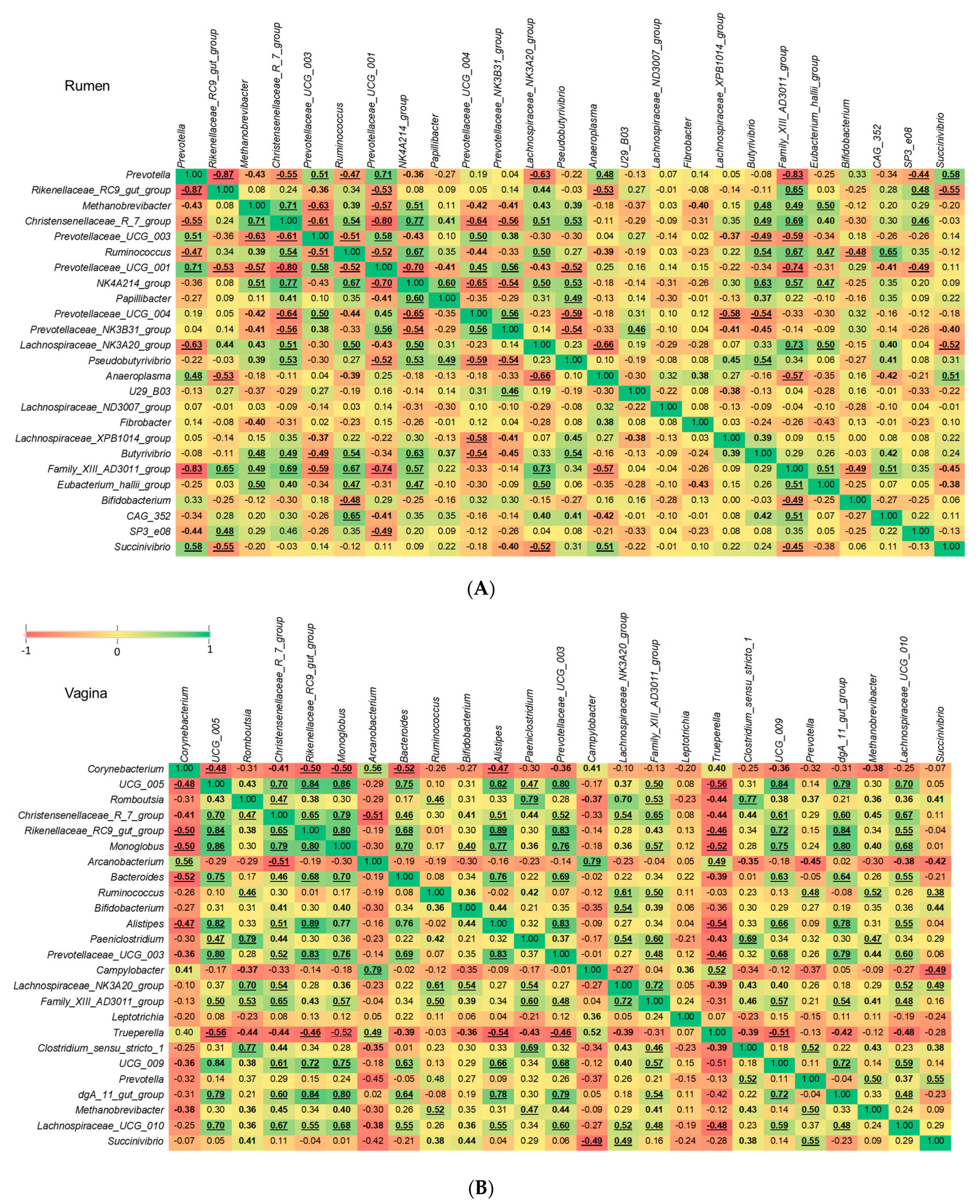
3.5. Comparison of Methanogenic Archaeal Relative Abundance between Virgin Yearling and Pregnant Heifers, and Identification of Bacterial Genera Associated with Methanobrevibacter

Methanogenic archaea, and in particular members of the Methanobrevibacter genus, have been reported to colonize the intestine of 83-day-old [14], and 5- to 7-month-old calf fetuses [13], as well as newborn calves [40,41]. In addition, we identified here two Methanobrevibacter OTUs (OTU8 and OTU23) that were shared by a relatively high portion (≥65%) of all samples collected from both virgin yearling and pregnant heifers (Table S1). Therefore, we assessed whether the relative abundance of Methanobrevibacter changed in response to pregnancy. For this, we compared the overall relative abundance of Methanobrevibacter spp. within each sample type between virgin yearling (non-pregnant) and pregnant heifers. Overall, the mean relative abundance of Methanobrevibacter in the nasopharynx, rumen fluid and vagina were 0.17%, 5.67%, and 0.47%, respectively (Figure 6A). The relative abundance of Methanobrevibacter in the rumen was greater in pregnant heifers compared to yearling heifers (p < 0.0001), but similar in the other two microbial habitats (p > 0.05) (Figure 6B,D).
There is increased research interest in the mitigation of enteric methane emissions from ruminant livestock via the manipulation of the rumen microbiota, primarily targeting commensal bacterial species involved in the supply or consumption of methanogenic substrates. Therefore, we assessed correlations between Methanobrevibacter and the other 24 most relatively abundant genera present in vaginal and ruminal microbiota of virgin yearling and pregnant heifers. Of note, the relative abundance of Methanobrevibacter in the nasopharynx was relatively low compared to the vagina and rumen and, therefore, only genera in the vaginal and ruminal microbiota were included for this correlation analysis. The Spearman correlation analysis revealed that 15 out of these 24 genera in the rumen of virgin yearling heifers exhibited significant (p < 0.05) correlations with Methanobrevibacter. Among which, the following 10 genera were positively correlated: Christensenellaceae R-7 group, Ruminococcus, Oscillospiraceae NK4A214 group, Papillibacter, Pseudobutyrivibrio, Prevotellaceae NK3B31 group, Lachnospiraceae NK3A20 group, Lachnospiraceae XPB1014 group, Eubacterium hallii group, Butyrivibrio and Olsenella. Whereas genera within the Prevotellaceae family (Prevotella, Prevotellaceae UCG-003 and Prevotellaceae UCG-001) and Anaeroplasma were strongly and inversely associated with Methanobrevibacter (Figure 7A). Varying degrees of positive or negative associations among the Methanobrevibacter-associated 15 genera and between other genera were also identified (Figure 7A). Within the vaginal microbial community of virgin yearling heifers, there were only three genera (Monoglobus, Akkermansia and Rikenellaceae dgA-11 gut group) that displayed significant correlations with Methanobrevibacter (p < 0.05) and these were positive correlations (Figure 7B).
In pregnant heifers, there were similar correlation patterns between Methanobrevibacter and other ruminal genera in the yearling heifers, with 14 genera significantly (p < 0.05) and positively (n = 8) or negatively (n = 6) correlated with this genus (Figure 8A). In contrast to the vaginal tract of virgin yearling heifers, there were 10 genera in the vaginal microbiota of pregnant heifers that were significantly associated with Methanobrevibacter, nine of them positively correlated. Interestingly, although Prevotella and Prevotellaceae UCG-003 were inversely correlated with Methanobrevibacter in the rumen of both virgin yearling and pregnant heifers, they were strongly and positively correlated with this genus in the vagina microbiota. Only the inverse correlations between Methanobrevibacter and Corynebacterium were significant (p < 0.05) (Figure 8B).
Next, we applied a stepwise-selected generalized linear mixed model to further identify genera that have a significant effect on the relative abundance of Methanobrevibacter spp. in the rumen and vagina. In the virgin yearling heifers, Prevotella and the Christensenellaceae R-7 group were predicted to have a significant effect (p < 0.0001) on Methanobrevibacter [1/(Methanobrevibacter2) = 0.02956 + (0.002514 × Prevotella) + (−0.00875 × Christensenellaceae R-7 group)]. As for pregnant heifers, Methanobrevibacter abundance was predicted to be negatively affected by Prevotellaceae UCG 003 (p = 0.037), and positively by Christensenellaceae R-7 group (p = 0.0326) [1/(Methanobrevibacter2) = 0.03793 + (−0.00602 × Christensenellaceae R-7 group) + (0.007088 × Prevotellaceae UCG-003)]. Within the vaginal microbial community of pregnant heifers: Ruminococcus (p = 0.0098), Prevotellaceae UCG-003 (p = 0.0005) and Prevotella (p <.0001) were predicted to have a significant negative effect on Methanobrevibacter [1/(Methanobrevibacter2) = −0.3780 + (0.1501 × Ruminococcus) + (0.2875 × Prevotella UCG-003) + (0.2898 × Prevotella)]. Among the 24 most relatively abundant genera in the vaginal microbiota of yearling heifers, only Oscillospiraceae UCG-005 (p = 0.0138) was predicted to have a negative effect on Methanobrevibacter [1/(Methanobrevibacter2) = 0.05975 + (0.04037 × Oscillospiraceae UCG-005).

4. Discussion

New evidence from our laboratory (Amat et al., unpublished data) and Guzman and colleagues [13] indicate that microbial colonization of the calf gastrointestinal tract may take place before birth. These observations suggest that the maternal microbiome may have a role in shaping the development of the offspring microbiome in cattle. In addition, it is believed that undesirable alterations of the maternal microbiota may indirectly influence fetal development, and that these effects may be transmitted to progeny, resulting in a dysbiotic microbiota [20] and increased offspring susceptibility to the development of metabolic disorders and respiratory infections [20,22]. Recent evidence from mouse studies has demonstrated that the maternal microbiota during pregnancy modulates the programming of fetal metabolic and nervous system development [23,24].
Although the role of maternal nutrition in developmental programming in cattle has been relatively well appreciated [18,29,42,43], the potential involvement of the maternal microbiota in fetal programming and offspring microbiome development remains largely unexplored. Considering the current evidence, it is important to explore whether bovine maternal nutrition/microbiome during pregnancy influences feto-maternal crosstalk, subsequently influencing offspring microbiome development. Maternal VTM supplementation before calving has been well documented to be associated with improved fetal programming and offspring health and productivity in cattle [25,26,27,28,29]. However, whether VTM supplementation-associated positive outcomes observed pre- and post-calving are dependent on alterations in the ruminal microbiota remains unexplored. In the present study, we evaluated whether differences in maternal weight gain during the first trimester of gestation affected the postnatal nasopharyngeal, ruminal, and vagina microbial communities of virgin heifers at 9 months of age. We also characterized and compared these three microbiota in pregnant heifers to evaluate the impact of VTM supplementation during the first six month of gestation on the maternal microbiome. Finally, we identified core taxa that are shared within the respiratory, gastrointestinal, and reproductive tract microbiota of cattle.

4.1. Effect of Maternal Restricted Gain during the First Trimester of Gestation on Offspring Microbiota Development

The virgin yearling heifers born from the dams that were subjected to LG (0.29 kg/d) during the first 84 days of gestation harbored a similar nasopharyngeal, ruminal and vaginal microbiota to those born from MG (0.79 kg/d) dams. The LG dams had a reduced average daily gain (p < 0.01) were 40 kg lighter than MG dams at calving (p < 0.01), and their calves had a lower birth weight than those from MG dams (28.6 vs. 30.8 kg, p = 0.03) [44]. As previously reported, fetuses harvested from a subset of the LG and MG dams at 84 days of gestation exhibited distinct fetal metabolic programming, including altered amino acid profiles in the fetal fluids [29]. In addition, we identified the presence of an archaeal and bacterial microbiota in intestinal and fluid samples from these 84-day-old calf fetuses (Amat et al., unpublished data). Therefore, we hypothesized that the divergent microbiome may be detected in virgin heifers that were exposed to divergent in utero nutrition (i.e., LG or MG) during their first trimester of gestation. However, no significant effect of maternal nutrition was found on the microbial community structure in the offspring nasopharynx, rumen, or vagina. There may be many reasons for this finding, including the timing of sample collection. For example, samples were collected when the offspring heifers were at 9 months of age, which was about 15 months after fetal exposure to the restrictive maternal diets. This may simply be too late in their development to detect microbial community alterations in the offspring as a result of maternal nutrition. In addition, the use of a single sampling time may have missed other time points where the LG and MG virgin heifers may have differed. Therefore, future studies investigating the impact of maternal nutrient and microbiome on offspring microbiome development should include longitudinal sampling.

4.2. Effect of Vitamin and Mineral Supplementation during the First 6 Months of Gestation on Maternal Microbiota

VTM supplementation during the first 6 months of gestation did not induce significant alterations in community structure and diversity of the nasopharyngeal, rumen or vaginal microbiota. While there is limited information on the effect of mineral and vitamin supplementation on the gut microbiota of ruminant animals, the impact of dietary mineral and vitamin intake on potentially beneficial or pathogenic gut microbes in humans and rodent animals have been relatively well documented [45]. For example, calcium and phosphorus supplementation increased the relative abundance of Clostridium, Ruminococcus and Lactobacillus spp. while reducing Bifidobacterium spp. in healthy men or mice [46,47,48]. The impact of dietary supplementation with selenium, magnesium, iron or zinc on certain gut commensal or pathogenic microbes was also reported in children and mice [45]. Of note, a significant effect of mineral supplementation on the gut microbiota was observed but only at the microbial taxa level and not on the microbial community structure and diversity [45].
The results from a limited number of studies performed on cattle also indicate that mineral supplementation may influence ruminal microbiota composition. Clay mineral supplementation increased the relative abundance of Butyrivibrio while reducing the relative abundance of Lactobacillus, Fusobacterium, and Treponema genera in the rumen of non-lactating Holstein cows [49]; however, it did not alter the rumen microbial community structure or diversity. Similarly, we observed that VTM supplementation increased the relative abundance of Butyrivibrio in the rumen (p < 0.05). Butyrivibrio spp. are considered commensal members of the rumen microbiota, producing butyrate through degradation of otherwise indigestible plant polysaccharides [50]. The Oscillospiraceae NK4A214 group and Succinivibrionaceae CAG-352 were the only other ruminal genera that responded to VTM supplementation in the present study. These are uncultured taxa and their role in the rumen is largely unknown. In contrast to our findings and those of Neubauer et al. [49], Liu and others [51] observed noticeable alterations in microbial richness and diversity of ruminal microbiota in both lactating Holstein cows (3–4 years old) and yearling heifers (10 months old) in response to feeding mineral salt bricks containing Mg, Co, Cu, Fe, Mn, Se, Zn, I and Na for one month. In the present study, we only evaluated ruminal microbiota and did not evaluate the fecal microbiota. Given that the fecal microbiota is one of the potential microbial sources for seeding the neonatal calf microbiota [52], and that the microbial community structure and composition differs between the ruminal and fecal microbiota in beef cattle [53], it would be interesting to investigate whether the fecal microbiota would be influenced by the maternal VTM supplementation in pregnant beef cattle.
Compared to the rumen microbiota, the effect of mineral supplementation on the bovine respiratory and reproductive microbiota has been less characterized. Feeding beef calves with selenium-biofortified alfalfa hay has been reported to alter the nasopharyngeal microbiota [54,55]. In the present study, although no significant changes were detected in the microbial community structure and diversity in the nasopharynx following VTM supplementation, changes were detected in relative abundance of five relatively abundant genera (Mycoplasma, Oscillospiraceae UCG-005, Christensenellaceae R-7 group, Ruminococcus and Ornithinimicrobium). Among these genera, Mycoplasma, which includes a bovine respiratory disease (BRD)-associated pathogen, Mycoplasma bovis, was enriched in pregnant heifers that receiving VTM supplementation. BRD is not a significant health concern among adult and pregnant cattle but it is the number one health problem affecting newly weaning calves arriving in the feedlot [56]. The positive association between VTM supplementation and nasopharyngeal Mycoplasma observed here poses the question of whether maternal VTM supplementation influences colonization of the offspring respiratory tract by Mycoplasma spp. No information has been reported regarding the impact of mineral supplementation on reproductive microbiota in cattle.
Vitamins A, D3 and E were included in the VTM supplement given to the pregnant heifers. Thus, it is impossible to discern whether the subtle changes observed at the taxa level in both the nasopharyngeal and ruminal microbiota are due to the minerals and vitamins supplemented. Evidence from human, rodent and pig studies suggest that the gut microbiota responds to vitamin supplementation [45]. Gastrointestinal-associated Bifidobacterium (vitamin A, C) Akkermansia (vitamin A), and Lactobacillus spp. (vitamin C) were more relatively abundant while E. coli (vitamin C) and Clostridium (vitamin D) spp. decreased in relative abundance after vitamin supplementation [45,57,58,59]. Our results indicate that vitamin supplementation has less impact on the ruminal microbiota. Overall, VTM supplementation for first 6 months of gestation did not affect the maternal microbiota. There could be due to several factors. Considering that mineral salt intake was reported to alter the ruminal microbiota in 3–4-year-old lactating cows [51], the resilience and robust of the mature ruminal microbiota can likely be ruled out as a reason for the absence of any VTM effect on the ruminal microbiota in pregnant heifers (1 year 9 months old).
Pregnancy status rather than age, however, could be associated with the non-responsiveness of the ruminal microbiota to VTM supplementation. In rodent studies, the maternal gut microbiota undergoes profound changes over the course of pregnancy [60,61,62,63]. As pregnancy progresses from the 1st to 3rd trimester, the maternal gut microbiota becomes less diverse [61] but with a higher microbial density, which may result in a microbiota that is more robust and resilient to perturbations. Hence, future studies are warranted to investigate the impact of VTM supplementation and other dietary interventions on the maternal microbiota of cattle using a non-pregnant control cohort.

4.3. Holistic View of Microbial Communities across Respiratory, Gastrointestinal and Reproductive Tract and the Core Taxa Shared across These Habitats

As expected, the overall microbial structure, diversity and composition were noticeably different among the nasopharyngeal, ruminal and vaginal microbiota in both virgin yearling and pregnant heifers. The ruminal microbiota was dominated by the anaerobic phylum Bacteroidota (66%), while the nasopharyngeal and vaginal microbiota the majority of 16S rRNA gene sequences were classified as Actinobacteriota (51%) and Firmicutes (52%), respectively. Various factors including niche-specific physiological factors (temperature, pH, oxygen and nutrient availability), dietary, and environmental factors are involved in shaping the microbiota of the bovine respiratory tract [4,64], rumen [5,65] and reproductive tract [3]. Subtle physiological and anatomical differences in the mucosal surfaces of the bovine respiratory tract have been shown to significantly influence the microbial distribution along the respiratory tract [66].
In the present study, although the nasopharynx, rumen and vagina have drastically different physiological and anatomical properties, we identified 41 OTUs that were shared by a high portion (60%) of all samples from both virgin yearling and pregnant heifers. This indicates that these core taxa can colonize and inhabit the respiratory, gastrointestinal and reproductive tracts regardless of the drastic differences in physiological conditions in these locations. The majority (80%) of these core taxa are members of the Firmicutes, which is one of the most ubiquitous and relatively abundant bacterial phyla in the respiratory, gastrointestinal and reproductive tract- (vagina, uterus) [3], mammary gland- [67], ocular- [68] and hoof- [69] -associated microbiota in cattle, demonstrating the adaptability of members of this phylum. Nine taxa within the Actinobacteria phylum including Bifidobacterium pseudolongum and several Corynebacterium spp. were among these core taxa. B. pseudolongum is widely found in the mammalian gut [70] and has long been noted for its probiotic properties in human, cattle and pigs [71,72]. Given the known beneficial effects of this species on the host, and as a core taxon present in the respiratory, gastrointestinal and reproductive tracts of cattle, B. pseudolongum may potentially enhance cattle health and productivity, as may some of the other core taxa identified in this study. Species and strain level resolution of these core taxa using shotgun metagenomic sequencing and characterization of their functional features using culturing should be the focus of future studies.
Two Methanobrevibacter OTUs were also identified among the core taxa. Although members of this methanogenic genus are well known for their involvement in ruminal methane production [73,74,75], and are frequently observed in the vaginal microbiota [76] in cattle, it is interesting to note that this genus is also found in the bovine respiratory tract. The presence of these Methanobrevibacter OTUs within the respiratory, gastrointestinal, and reproductive tracts has important implications for the identification of maternal seeding of the calf microbiota with pioneer methanogens before and during birth. Methanobrevibacter spp. are predominant in 5- to 7-month-old calf fetuses [13] as well as newborn calves [40,41]. Our results indicate that the respiratory microbiota may also seed the calf gastrointestinal tract with Methanobrevibacter spp. perinatally. This highlights the necessity of holistic assessment of respiratory, gastrointestinal, and reproductive tract microbiota to trace the origin of pioneer calf microbiota. To our best of knowledge, this is the first study to evaluate nasopharyngeal, ruminal and vaginal microbiota in an individual ruminant animal, and to identify the core taxa shared amongst these microbial ecologies.

4.4. Ruminal Methanobrevibacter Enriched in Pregnant Heifers and Associations of Methanobrevibacter with Predominant Ruminal and Vaginal Bacterial Genera

Given that lowering methane emissions in cattle benefits both environment and cattle production [77], and increasing evidence suggesting that the ruminal microbiome and host genetics can be targeted independently to improve feed efficiency and mitigate enteric methane emissions from cattle [6,7]; Li et al., 2019a), we therefore focused on this methanogenic archaeal genus, Methanobrevibacter. We identified that pregnant heifers harbored a greater relative abundance of ruminal Methanobrevibacter compared to non-pregnant virgin yearling heifers. Confounding factors associated with dietary (11% more hay fed to virgin heifers than pregnant heifers) and age differences makes it difficult to associate pregnancy with the colonization of the rumen with methanogenic archaea. However, the impact of pregnancy and mitigation of maternal ruminal methanogens on offspring enteric methane emissions warrants further investigation.
Our correlation analysis revealed that in comparison to vaginal Methanobrevibacter, the relative abundance of ruminal Methanobrevibacter is highly influenced by many other commensal genera in the rumen. For example, in the rumen microbiota many genera within the Prevotellaceae family were inversely associated with the relative abundance of Methanobrevibacter. Interestingly, the opposite was found in the vaginal microbiota, suggesting that the nature of the interaction between Methanobrevibacter and Prevotella and Prevotellaceae UCG-003 may be niche specific and that Prevotella spp. in the rumen may become pro-methanogenic if they present in reproductive microbial ecosystem.
The stepwise-selected GLM model identified Prevotella and Prevotellaceae UCG-003 as having a significant and negative effect on the relative abundance of Methanobrevibacter in rumen of both virgin and pregnant heifers and vaginal tract of pregnant heifers. This is in agreement with previous studies reporting that microbial communities with highly abundant lactate-consuming bacteria (Prevotella bryantii) and high H2-consuming (e.g., certain Prevotella spp.) has been associated with lower ruminal methane production [74,78,79,80]. Thus, members of the Prevotella and Prevotellaceae UCG-003 in the bovine rumen and vagina may have anti-methanogenic potential to mitigate methane emissions in cattle. The Christensenellaceae R-7 group was identified as the genus that can have significant positive effect on Methanobrevibacter in the present study. This may suggest that some species within this genus may be involved in producing methanogenic substrates such as H2 and acetate. Future in vitro studies are needed to confirm the anti-methanogenic properties of Prevotella and Prevotellaceae UCG-003 and pro-methanogenic activity of Christensenellaceae R-7 group spp. originating from the rumen of cattle.

5. Conclusions

In conclusion, no noticeable difference was observed in α and β-diversity in any of the nasopharyngeal, ruminal and vaginal microbiota between virgin heifers raised from dams exposed to divergent rates of gain during the first trimester of pregnancy, or between pregnant heifers consuming control and VTM diets. Only in the vaginal microbiota were there relatively abundant genera that were affected by maternal rate of gain during early gestation. Maternal VTM supplementation resulted in subtle compositional alterations in the nasopharyngeal and ruminal microbiota. A total of 41 archaeal and bacterial OTUs were shared by over 60% of all samples from both virgin and pregnant heifers. Two taxa within the Methanobrevibacter genus were among these core taxa and this genus was more relatively abundant in pregnant compared to virgin heifers. Compared to the vaginal Methanobrevibacter, Methanobrevibacter in the rumen was predicted to be highly interactive with other commensal members.
Among the 25 most relatively abundant genera, Prevotella and Prevotella UCG-003 (negative) and Christensenellaceae R-7 group (positive) were predicted to have a significant effect on the relative abundance of ruminal Methanobrevibacter spp. Overall, the results of this study suggest that there is little impact of maternal gestational nutrition during the first trimester on the calf microbiota assessed at 9 months of age, and that VTM supplementation during pregnancy may not alter the maternal microbiota. This study provides evidence that there are several microbial taxa, including methanogenic archaea, that are shared across the respiratory, gastrointestinal, and reproductive tracts. Therefore, this suggests that there is a need for a holistic evaluation of the bovine microbiota when considering potential maternal sources for seeding calves with pioneer microbiota, and when targeting the maternal microbiome to enhance offspring health and development.

Supplementary Materials

The following are available online at https://www.mdpi.com/article/10.3390/microorganisms9102011/s1, Table S1: OTUs identified in the nasopharyngeal, ruminal and vaginal microbiota of at least 60% of samples from virgin yearling heifers. Table S2: OTUs identified in the nasopharyngeal, ruminal, and vaginal microbiota of at least 60% of samples from pregnant heifers.

Author Contributions

Conceiving the idea, designing the study, and providing supervision—S.A. and C.R.D.; Cattle management—A.C.B.M., F.B., C.R.D. and K.K.S.; Animal care and sample collections—S.A., C.R.D., K.S., A.C.B.M., T.W., J.D.K. and F.B.; Sample processing—K.S. and S.A.; Bioinformatics analysis—D.B.H., T.L. and S.A.; Data processing and statistical analysis—T.D.S., D.B.H. and S.A.; Manuscript writing—S.A.; Manuscript review and editing—S.A., D.B.H., A.C.B.M. and C.R.D. All authors have read and agreed to the published version of the manuscript.

Funding

The work presented in this study was financially supported by the North Dakota Ag Experiment Station as part of a start-up package for S.A. Animal management portion of this project was supported by the North Dakota Corn Council, the Central Grasslands Research and Extension Center, and the ND Ag Experiment Station.

Institutional Review Board Statement

All experimental procedures involving cattle were approved by the North Dakota State University Institutional Animal Care and Use Committee (#A20085 and #A20086, for virgin yearling heifers and for pregnant heifers, respectively). Approval dates for #A20085 and #A20086 are 5 May 2020 and 25 February 2020, respectively.

Informed Consent Statement

Not applicable.

Data Availability Statement

The datasets generated for this study can be found in the Sequences that were submitted to the NCBI sequence read archive under BioProject accession PRJNA721423.

Acknowledgments

The authors acknowledge the support from the staff at NDSU Beef Cattle Research Complex.

Conflicts of Interest

The authors declare that they have no competing interest.

References

  1. Huws, S.A.; Creevey, C.J.; Oyama, L.B.; Mizrahi, I.; Denman, S.E.; Popova, M.; Muñoz-Tamayo, R.; Forano, E.; Waters, S.M.; Hess, M.; et al. Addressing Global Ruminant Agricultural Challenges Through Understanding the Rumen Microbiome: Past, Present, and Future. Front. Microbiol. 2018, 9, 2161. [Google Scholar] [CrossRef]
  2. Matthews, C.; Crispie, F.; Lewis, E.; Reid, M.; O’Toole, P.W.; Cotter, P.D. The rumen microbiome: A crucial consideration when optimising milk and meat production and nitrogen utilisation efficiency. Gut Microbes 2018, 10, 115–132. [Google Scholar] [CrossRef]
  3. Galvão, K.N.; Bicalho, R.C.; Jeon, S.J. Symposium review: The uterine microbiome associated with the development of uterine disease in dairy cows. J. Dairy Sci. 2019, 102, 11786–11797. [Google Scholar] [CrossRef]
  4. Timsit, E.; McMullen, C.; Amat, S.; Alexander, T.W. Respiratory Bacterial Microbiota in Cattle. Veter- Clin. N. Am. Food Anim. Pr. 2020, 36, 297–320. [Google Scholar] [CrossRef] [PubMed]
  5. O’Hara, E.; Neves, A.L.; Song, Y.; Guan, L.L. The Role of the Gut Microbiome in Cattle Production and Health: Driver or Passenger? Annu. Rev. Anim. Biosci. 2020, 8, 199–220. [Google Scholar] [CrossRef] [PubMed] [Green Version]
  6. Difford, G.F.; Plichta, D.R.; Løvendahl, P.; Lassen, J.; Noel, S.J.; Højberg, O.; Wright, A.-D.G.; Zhu, Z.; Kristensen, L.; Nielsen, H.B.; et al. Host genetics and the rumen microbiome jointly associate with methane emissions in dairy cows. PLoS Genet. 2018, 14, e1007580. [Google Scholar] [CrossRef] [PubMed] [Green Version]
  7. Li, F.; Li, C.; Chen, Y.; Liu, J.; Zhang, C.; Irving, B.; Fitzsimmons, C.; Plastow, G.; Guan, L.L. Host genetics influence the rumen microbiota and heritable rumen microbial features associate with feed efficiency in cattle. Microbiome 2019, 7, 1–17. [Google Scholar] [CrossRef] [Green Version]
  8. Weimer, P.J. Redundancy, resilience, and host specificity of the ruminal microbiota: Implications for engineering improved ruminal fermentations. Front. Microbiol. 2015, 6, 296. [Google Scholar] [CrossRef] [Green Version]
  9. Yáñez-Ruiz, D.R.; Abecia, L.; Newbold, C.J. Manipulating rumen microbiome and fermentation through interventions during early life: A review. Front. Microbiol. 2015, 6, 1133. [Google Scholar] [CrossRef] [Green Version]
  10. Saro, C.; Hohenester, U.M.; Bernard, M.; Lagrée, M.; Martin, C.; Doreau, M.; Boudra, H.; Popova, M.; Morgavi, D.P. Effectiveness of Interventions to Modulate the Rumen Microbiota Composition and Function in Pre-ruminant and Ruminant Lambs. Front. Microbiol. 2018, 9, 1273. [Google Scholar] [CrossRef]
  11. Belanche, A.; Palma-Hidalgo, J.; Nejjam, I.; Jiménez, E.; Martín-García, A.; Yáñez-Ruiz, D. Inoculation with rumen fluid in early life as a strategy to optimize the weaning process in intensive dairy goat systems. J. Dairy Sci. 2020, 103, 5047–5060. [Google Scholar] [CrossRef] [PubMed]
  12. Palma-Hidalgo, J.M.; Jiménez, E.; Popova, M.; Morgavi, D.P.; Martín-García, A.I.; Yáñez-Ruiz, D.R.; Belanche, A. Inoculation with rumen fluid in early life accelerates the rumen microbial development and favours the weaning process in goats. Anim. Microbiome 2021, 3, 11. [Google Scholar] [CrossRef] [PubMed]
  13. Guzman, C.E.; Wood, J.L.; Egidi, E.; White-Monsant, A.C.; Semenec, L.; Grommen, S.V.H.; Hill-Yardin, E.L.; De Groef, B.; Franks, A.E. A pioneer calf foetus microbiome. Sci. Rep. 2020, 10, 17712. [Google Scholar] [CrossRef] [PubMed]
  14. Amat, S.; Holman, D.B.; Schmidt, K.; McCarthy, K.L.; Dorsam, S.T.; Ward, A.K.; Borowicz, P.P.; Reynolds, L.P.; Caton, J.S.; Sedivec, K.K.; et al. Characterization of the microbiota associated with 12-week-old bovine fetuses exposed to divergent in utero nutrition. bioRxiv 2021. [Google Scholar] [CrossRef]
  15. Palmer, A.C. Nutritionally Mediated Programming of the Developing Immune System. Adv. Nutr. 2011, 2, 377–395. [Google Scholar] [CrossRef] [Green Version]
  16. Parlee, S.D.; MacDougald, O.A. Maternal nutrition and risk of obesity in offspring: The Trojan horse of developmental plasticity. Biochim. Biophys. Acta (BBA)—Mol. Basis Dis. 2013, 1842, 495–506. [Google Scholar] [CrossRef] [PubMed] [Green Version]
  17. Khanal, P.; Nielsen, M.O. Impacts of prenatal nutrition on animal production and performance: A focus on growth and metabolic and endocrine function in sheep. J. Anim. Sci. Biotechnol. 2017, 8, 75. [Google Scholar] [CrossRef] [PubMed] [Green Version]
  18. Caton, J.S.; Crouse, M.S.; Reynolds, L.P.; Neville, T.L.; Dahlen, C.R.; Ward, A.K.; Swanson, K.C. Maternal nutrition and programming of offspring energy requirements. Transl. Anim. Sci. 2019, 3, 976–990. [Google Scholar] [CrossRef] [Green Version]
  19. Stiemsma, L.T.; Michels, K.B. The Role of the Microbiome in the Developmental Origins of Health and Disease. Pediatrics 2018, 141, e20172437. [Google Scholar] [CrossRef] [Green Version]
  20. Calatayud, M.; Koren, O.; Collado, M.C. Maternal Microbiome and Metabolic Health Program Microbiome Development and Health of the Offspring. Trends Endocrinol. Metab. 2019, 30, 735–744. [Google Scholar] [CrossRef]
  21. Codagnone, M.; Spichak, S.; O’Mahony, S.M.; O’Leary, O.; Clarke, G.; Stanton, C.; Dinan, T.G.; Cryan, J.F. Programming Bugs: Microbiota and the Developmental Origins of Brain Health and Disease. Biol. Psychiatry 2018, 85, 150–163. [Google Scholar] [CrossRef] [PubMed]
  22. Yao, Y.; Cai, X.; Chen, C.; Fang, H.; Zhao, Y.; Fei, W.; Chen, F.; Zheng, C. The Role of Microbiomes in Pregnant Women and Offspring: Research Progress of Recent Years. Front. Pharmacol. 2020, 11, 643. [Google Scholar] [CrossRef] [PubMed]
  23. Kimura, I.; Miyamoto, J.; Ohue-Kitano, R.; Watanabe, K.; Yamada, T.; Onuki, M.; Aoki, R.; Isobe, Y.; Kashihara, D.; Inoue, D.; et al. Maternal gut microbiota in pregnancy influences offspring metabolic phenotype in mice. Science 2020, 367, eaaw8429. [Google Scholar] [CrossRef] [PubMed]
  24. Vuong, H.E.; Pronovost, G.N.; Williams, D.W.; Coley, E.J.L.; Siegler, E.L.; Qiu, A.; Kazantsev, M.; Wilson, C.J.; Rendon, T.; Hsiao, E.Y. The maternal microbiome modulates fetal neurodevelopment in mice. Nature 2020, 586, 281–286. [Google Scholar] [CrossRef] [PubMed]
  25. Mee, J.F.; Rogers, A.P.; O’Farrell, K.J. Effect of feeding a mineral-vitamin supplement before calving on the calving performance of a trace element deficient dairy herd. Veter Rec. 1995, 137, 508–512. [Google Scholar] [CrossRef]
  26. Wilde, D. Influence of macro and micro minerals in the peri-parturient period on fertility in dairy cattle. Anim. Reprod. Sci. 2006, 96, 240–249. [Google Scholar] [CrossRef]
  27. Van Emon, M.; Sanford, C.; McCoski, S. Impacts of Bovine Trace Mineral Supplementation on Maternal and Offspring Production and Health. Animals 2020, 10, 2404. [Google Scholar] [CrossRef]
  28. Diniz, W.; Reynolds, L.; Borowicz, P.; Ward, A.; Sedivec, K.; McCarthy, K.; Kassetas, C.; Baumgaertner, F.; Kirsch, J.; Dorsam, S.; et al. Maternal Vitamin and Mineral Supplementation and Rate of Maternal Weight Gain Affects Placental Expression of Energy Metabolism and Transport-Related Genes. Genes 2021, 12, 385. [Google Scholar] [CrossRef]
  29. Menezes, A.C.B.; McCarthy, K.L.; Kassetas, C.J.; Baumgaertner, F.; Kirsch, J.D.; Dorsam, S.; Neville, T.L.; Ward, A.K.; Borowicz, P.P.; Reynolds, L.P.; et al. Vitamin and mineral supplementation and rate of gain during the first trimester of gestation affect concentrations of amino acids in maternal serum and allantoic fluid of beef heifers. J. Anim. Sci. 2021, 99, skab024. [Google Scholar] [CrossRef] [PubMed]
  30. NASEM. Nutrient Requirements of Beef Cattle, 8th ed.; The National Academies Press: Washington, DC, USA, 2016. [Google Scholar]
  31. Holman, D.B.; Timsit, E.; Amat, S.; Abbott, D.W.; Buret, A.G.; Alexander, T.W. The nasopharyngeal microbiota of beef cattle before and after transport to a feedlot. BMC Microbiol. 2017, 17, 70. [Google Scholar] [CrossRef] [Green Version]
  32. Amat, S.; Holman, D.; Timsit, E.; Schwinghamer, T.; Alexander, T.W. Evaluation of the Nasopharyngeal Microbiota in Beef Cattle Transported to a Feedlot, With a Focus on Lactic Acid-Producing Bacteria. Front. Microbiol. 2019, 10, 1988. [Google Scholar] [CrossRef]
  33. Paz, H.A.; Anderson, C.; Muller, M.J.; Kononoff, P.J.; Fernando, S.C. Rumen Bacterial Community Composition in Holstein and Jersey Cows Is Different under Same Dietary Condition and Is Not Affected by Sampling Method. Front. Microbiol. 2016, 7, 1206. [Google Scholar] [CrossRef] [PubMed]
  34. Callahan, B.J.; McMurdie, P.J.; Rosen, M.J.; Han, A.W.; Johnson, A.J.; Holmes, S.P. DADA2: High-resolution sample inference from Illumina amplicon data. Nat. Methods 2016, 13, 581–583. [Google Scholar] [CrossRef] [PubMed] [Green Version]
  35. Wang, Q.; Garrity, G.M.; Tiedje, J.M.; Cole, J.R. Naive Bayesian Classifier for Rapid Assignment of rRNA Sequences into the New Bacterial Taxonomy. Appl. Environ. Microbiol. 2007, 73, 5261–5267. [Google Scholar] [CrossRef] [PubMed] [Green Version]
  36. Quast, C.; Pruesse, E.; Yilmaz, P.; Gerken, J.; Schweer, T.; Yarza, P.; Peplies, J.; Glöckner, F.O. The SILVA ribosomal RNA gene database project: Improved data processing and web-based tools. Nucleic Acids Res. 2012, 41, D590–D596. [Google Scholar] [CrossRef]
  37. McMurdie, P.; Holmes, S. phyloseq: An R Package for Reproducible Interactive Analysis and Graphics of Microbiome Census Data. PLoS ONE 2013, 8, e61217. [Google Scholar] [CrossRef] [PubMed] [Green Version]
  38. Oksanen, J.; Blanchet, F.G.; Kindt, R.; Legendre, P.; Minchin, P.R.; O’hara, R. Package ‘Vegan’. Community Ecology Package, Version 2. 2013. Available online: https://cran.r-project.org/web/packages/vegan/index.html (accessed on 15 September 2021).
  39. Mallick, H.; Rahnavard, A.; McIver, L.J.; Ma, S.; Zhang, Y.; Nguyen, L.H.; Tickle, T.L.; Weingart, G.; Ren, B.; Schwager, E.H. Multivariable association discovery in population-scale meta-omics studies. bioRxiv 2021. [Google Scholar] [CrossRef]
  40. Guzman, E.C.; Bereza-Malcolm, L.T.; De Groef, B.; Franks, A. Presence of Selected Methanogens, Fibrolytic Bacteria, and Proteobacteria in the Gastrointestinal Tract of Neonatal Dairy Calves from Birth to 72 Hours. PLoS ONE 2015, 10, e0133048. [Google Scholar] [CrossRef] [Green Version]
  41. Alipour, M.J.; Jalanka, J.; Pessa-Morikawa, T.; Kokkonen, T.; Satokari, R.; Hynönen, U.; Iivanainen, A.; Niku, M. The composition of the perinatal intestinal microbiota in cattle. Sci. Rep. 2018, 8, 10437. [Google Scholar] [CrossRef]
  42. McLean, K.J.; Crouse, M.; Crosswhite, M.R.; Pereira, N.N.; Dahlen, C.R.; Borowicz, P.P.; Reynolds, L.P.; Ward, A.K.; Neville, B.W.; Caton, J.S. Impacts of maternal nutrition on uterine and placental vascularity and mRNA expression of angiogenic factors during the establishment of pregnancy in beef heifers1. Transl. Anim. Sci. 2017, 1, 160–167. [Google Scholar] [CrossRef]
  43. Crouse, M.S.; Greseth, N.P.; McLean, K.J.; Crosswhite, M.R.; Pereira, N.N.; Ward, A.K.; Reynolds, L.P.; Dahlen, C.R.; Neville, B.W.; Borowicz, P.P.; et al. Maternal nutrition and stage of early pregnancy in beef heifers: Impacts on hexose and AA concentrations in maternal and fetal fluids1. J. Anim. Sci. 2019, 97, 1296–1316. [Google Scholar] [CrossRef] [PubMed]
  44. Baumgaertner, F.; Underdahl, S.R.; McCarthy, K.L.; Menezes, A.C.B.; Diniz, W.J.D.S.; Ward, A.K.; Sedivec, K.K.; Dorsam, S.T.; Kirsch, J.D.; Caton, J.; et al. 187 Effects of energy supplementation during early gestation in beef heifers on body weight, concentrations of IGF-1, and calf characteristics. J. Anim. Sci. 2020, 98, 163–164. [Google Scholar] [CrossRef]
  45. Yang, Q.; Liang, Q.; Balakrishnan, B.; Belobrajdic, D.P.; Feng, Q.-J.; Zhang, W. Role of Dietary Nutrients in the Modulation of Gut Microbiota: A Narrative Review. Nutrients 2020, 12, 381. [Google Scholar] [CrossRef] [PubMed] [Green Version]
  46. Aslam, M.N.; Bassis, C.; Zhang, L.; Zaidi, S.; Varani, J.; Bergin, I.L. Calcium Reduces Liver Injury in Mice on a High-Fat Diet: Alterations in Microbial and Bile Acid Profiles. PLoS ONE 2016, 11, e0166178. [Google Scholar] [CrossRef]
  47. Trautvetter, U.; Camarinha-Silva, A.; Jahreis, G.; Lorkowski, S.; Glei, M. High phosphorus intake and gut-related parameters―results of a randomized placebo-controlled human intervention study. Nutr. J. 2018, 17, 23. [Google Scholar] [CrossRef] [PubMed]
  48. Li, P.; Tang, T.; Chang, X.; Fan, X.; Chen, X.; Wang, R.; Fan, C.; Qi, K. Abnormality in Maternal Dietary Calcium Intake During Pregnancy and Lactation Promotes Body Weight Gain by Affecting the Gut Microbiota in Mouse Offspring. Mol. Nutr. Food Res. 2018, 63, e1800399. [Google Scholar] [CrossRef]
  49. Neubauer, V.; Humer, E.; Mann, E.; Kröger, I.; Reisinger, N.; Wagner, M.; Zebeli, Q.; Petri, R.M. Effects of clay mineral supplementation on particle-associated and epimural microbiota, and gene expression in the rumen of cows fed high-concentrate diet. Anaerobe 2019, 59, 38–48. [Google Scholar] [CrossRef]
  50. Kelly, W.J.; Leahy, S.; Altermann, E.; Yeoman, C.J.; Dunne, J.C.; Kong, Z.; Pacheco, D.M.; Li, D.; Noel, S.J.; Moon, C.; et al. The Glycobiome of the Rumen Bacterium Butyrivibrio proteoclasticus B316T Highlights Adaptation to a Polysaccharide-Rich Environment. PLoS ONE 2010, 5, e11942. [Google Scholar] [CrossRef]
  51. Liu, C.; Li, X.H.; Chen, Y.X.; Cheng, Z.H.; Duan, Q.H.; Meng, Q.H.; Tao, X.P.; Shang, B.; Dong, H.M. Age-Related Response of Rumen Microbiota to Mineral Salt and Effects of Their Interactions on Enteric Methane Emissions in Cattle. Microb. Ecol. 2016, 73, 590–601. [Google Scholar] [CrossRef] [PubMed]
  52. Barden, M.; Richards-Rios, P.; Ganda, E.; Lenzi, L.; Eccles, R.; Neary, J.; Oultram, J.; Oikonomou, G. Maternal influences on oral and faecal microbiota maturation in neonatal calves in beef and dairy production systems. Anim. Microbiome 2020, 2, 31. [Google Scholar] [CrossRef]
  53. Lourenco, J.M.; Kieran, T.; Seidel, D.S.; Glenn, T.C.; Da Silveira, M.F.; Callaway, T.R.R.L.S., Jr. Comparison of the ruminal and fecal microbiotas in beef calves supplemented or not with concentrate. PLoS ONE 2020, 15, e0231533. [Google Scholar] [CrossRef] [PubMed]
  54. Hall, J.A.; Isaiah, A.; Estill, C.T.; Pirelli, G.J.; Suchodolski, J.S. Weaned beef calves fed selenium-biofortified alfalfa hay have an enriched nasal microbiota compared with healthy controls. PLoS ONE 2017, 12, e0179215. [Google Scholar] [CrossRef]
  55. Hall, J.A.; Isaiah, A.; Bobe, G.; Estill, C.T.; Bishop-Stewart, J.K.; Davis, T.Z.; Suchodolski, J.S.; Pirelli, G.J. Feeding selenium-biofortified alfalfa hay during the preconditioning period improves growth, carcass weight, and nasal microbial diversity of beef calves. PLoS ONE 2020, 15, e0242771. [Google Scholar] [CrossRef] [PubMed]
  56. Johnson, K.K.; Pendell, D.L. Market Impacts of Reducing the Prevalence of Bovine Respiratory Disease in United States Beef Cattle Feedlots. Front. Veter-Sci. 2017, 4, 189. [Google Scholar] [CrossRef] [PubMed] [Green Version]
  57. Xu, J.; Xu, C.; Chen, X.; Cai, X.; Yang, S.; Sheng, Y.; Wang, T. Regulation of an antioxidant blend on intestinal redox status and major microbiota in early weaned piglets. Nutrition 2014, 30, 584–589. [Google Scholar] [CrossRef]
  58. Talsness, C.E.; Penders, J.; Jansen, E.H.J.M.; Damoiseaux, J.; Thijs, C.; Mommers, M. Influence of vitamin D on key bacterial taxa in infant microbiota in the KOALA Birth Cohort Study. PLoS ONE 2017, 12, e0188011. [Google Scholar] [CrossRef] [PubMed]
  59. Huda, M.N.; Ahmad, S.M.; Kalanetra, K.M.; Taft, D.; Alam, J.; Khanam, A.; Raqib, R.; Underwood, A.M.; Mills, A.; Stephensen, D.C.B. Neonatal Vitamin A Supplementation and Vitamin A Status Are Associated with Gut Microbiome Composition in Bangladeshi Infants in Early Infancy and at 2 Years of Age. J. Nutr. 2019, 149, 1075–1088. [Google Scholar] [CrossRef] [PubMed] [Green Version]
  60. Collado, M.C.; Isolauri, E.; Laitinen, K.; Salminen, S. Distinct composition of gut microbiota during pregnancy in overweight and normal-weight women. Am. J. Clin. Nutr. 2008, 88, 894–899. [Google Scholar] [CrossRef]
  61. Koren, O.; Goodrich, J.K.; Cullender, T.C.; Spor, A.; Laitinen, K.; Bäckhed, H.K.; Gonzalez, A.; Werner, J.J.; Angenent, L.; Knight, R.; et al. Host Remodeling of the Gut Microbiome and Metabolic Changes during Pregnancy. Cell 2012, 150, 470–480. [Google Scholar] [CrossRef] [Green Version]
  62. Nuriel-Ohayon, M.; Neuman, H.; Koren, O. Microbial Changes during Pregnancy, Birth, and Infancy. Front. Microbiol. 2016, 7, 1031. [Google Scholar] [CrossRef] [Green Version]
  63. Ricks, N.M.; Panzer, A.; McCoy, A.N.; Azcarate-Peril, M.A.; Keku, T.O.; Boggess, K.A.; Smid, M.C. Maternal Gut Microbiome Biodiversity in Pregnancy. Am. J. Perinatol. 2017, 35, 24–30. [Google Scholar] [CrossRef]
  64. Zeineldin, M.; Lowe, J.; Aldridge, B. Contribution of the Mucosal Microbiota to Bovine Respiratory Health. Trends Microbiol. 2019, 27, 753–770. [Google Scholar] [CrossRef] [PubMed]
  65. Cholewińska, P.; Górniak, W.; Wojnarowski, K. Impact of selected environmental factors on microbiome of the digestive tract of ruminants. BMC Vet. Res. 2021, 17, 25. [Google Scholar] [CrossRef] [PubMed]
  66. McMullen, C.; Alexander, T.W.; Léguillette, R.; Workentine, M.; Timsit, E. Topography of the respiratory tract bacterial microbiota in cattle. Microbiome 2020, 8, 91. [Google Scholar] [CrossRef] [PubMed]
  67. Derakhshani, H.; Fehr, K.B.; Sepehri, S.; Francoz, D.; De Buck, J.; Barkema, H.; Plaizier, J.C.; Khafipour, E. Invited review: Microbiota of the bovine udder: Contributing factors and potential implications for udder health and mastitis susceptibility. J. Dairy Sci. 2018, 101, 10605–10625. [Google Scholar] [CrossRef] [Green Version]
  68. Bartenslager, A.C.; Althuge, N.D.; Loy, J.D.; Hille, M.M.; Spangler, M.L.; Fernando, S.C. Longitudinal assessment of the bovine ocular bacterial community dynamics in calves. Anim. Microbiome 2021, 3, 16. [Google Scholar] [CrossRef]
  69. Zinicola, M.; Lima, F.; Lima, S.; Machado, V.; Gomez, M.; Döpfer, D.; Guard, C.; Bicalho, R. Altered Microbiomes in Bovine Digital Dermatitis Lesions, and the Gut as a Pathogen Reservoir. PLoS ONE 2015, 10, e0120504. [Google Scholar] [CrossRef]
  70. Lugli, G.A.; Duranti, S.; Albert, K.; Mancabelli, L.; Napoli, S.; Viappiani, A.; Anzalone, R.; Longhi, G.; Milani, C.; Turroni, F.; et al. Unveiling Genomic Diversity among Members of the Species Bifidobacterium pseudolongum, a Widely Distributed Gut Commensal of the Animal Kingdom. Appl. Environ. Microbiol. 2019, 85, e03065-18. [Google Scholar] [CrossRef] [Green Version]
  71. Abe, F.; Ishibashi, N.; Shimamura, S. Effect of administration of bifidobacteria and lactic acid bacteria to newborn calves and piglets. J. Dairy Sci. 1995, 78, 2838–2846. [Google Scholar] [CrossRef]
  72. Kissels, W.; Wu, X.; Santos, R. Short communication: Interaction of the isomers carvacrol and thymol with the antibiotics doxycycline and tilmicosin: In vitro effects against pathogenic bacteria commonly found in the respiratory tract of calves. J. Dairy Sci. 2017, 100, 970–974. [Google Scholar] [CrossRef] [Green Version]
  73. Hook, S.E.; Wright, A.-D.G.; McBride, B.W. Methanogens: Methane Producers of the Rumen and Mitigation Strategies. Archaea 2010, 2010, 945785. [Google Scholar] [CrossRef] [PubMed] [Green Version]
  74. Danielsson, R.; Dicksved, J.; Sun, L.; Gonda, H.; Müller, B.; Schnürer, A.; Bertilsson, J. Methane Production in Dairy Cows Correlates with Rumen Methanogenic and Bacterial Community Structure. Front. Microbiol. 2017, 8, 226. [Google Scholar] [CrossRef] [PubMed]
  75. Greening, C.; Geier, R.; Wang, C.; Woods, L.C.; Morales, S.E.; McDonald, M.J.; Rushton-Green, R.; Morgan, X.C.; Koike, S.; Leahy, S.C.; et al. Diverse hydrogen production and consumption pathways influence methane production in ruminants. ISME J. 2019, 13, 2617–2632. [Google Scholar] [CrossRef] [PubMed]
  76. Laguardia-Nascimento, M.; Branco, K.M.G.R.; Gasparini, M.R.; Giannattasio-Ferraz, S.; Leite, L.R.; Araújo, F.M.G.; Salim, A.C.D.M.; Nicoli, J.R.; De Oliveira, G.C.; Barbosa-Stancioli, E. Vaginal Microbiome Characterization of Nellore Cattle Using Metagenomic Analysis. PLoS ONE 2015, 10, e0143294. [Google Scholar] [CrossRef]
  77. Beauchemin, K.A.; Ungerfeld, E.; Eckard, R.J.; Wang, M. Review: Fifty years of research on rumen methanogenesis: Lessons learned and future challenges for mitigation. Animal 2020, 14, s2–s16. [Google Scholar] [CrossRef] [PubMed] [Green Version]
  78. Denman, S.E.; Fernandez, G.M.; Eshinkai, T.; Emitsumori, M.; McSweeney, C. Metagenomic analysis of the rumen microbial community following inhibition of methane formation by a halogenated methane analog. Front. Microbiol. 2015, 6, 1087. [Google Scholar] [CrossRef]
  79. Tapio, I.; Snelling, T.J.; Strozzi, F.; Wallace, R.J. The ruminal microbiome associated with methane emissions from ruminant livestock. J. Anim. Sci. Biotechnol. 2017, 8, 7. [Google Scholar] [CrossRef]
  80. Granja-Salcedo, Y.T.; Fernandes, R.M.; de Araujo, R.C.; Kishi, L.T.; Berchielli, T.T.; de Resende, F.D.; Berndt, A.; Siqueira, G.R. Long-Term Encapsulated Nitrate Supplementation Modulates Rumen Microbial Diversity and Rumen Fermentation to Reduce Methane Emission in Grazing Steers. Front. Microbiol. 2019, 10, 614. [Google Scholar] [CrossRef]
Figure 1. Beta and alpha diversity of the nasopharyngeal, ruminal and vaginal microbiota of virgin yearling heifers from low gain (LG) or moderate gain (MG) dams as determined during the first trimester of gestation. (A) Non-metric multidimensional scaling (NMDS) plots of the Bray–Curtis dissimilarities, (B) number of operational taxonomic units (OTUs), and Shannon (C) and inverse Simpson’s diversity index (D) of each microbial community.
Microorganisms 09 02011 g001
Figure 2. Beta and alpha diversity of the nasopharyngeal, ruminal and vaginal microbiota of pregnant heifers that received a vitamin and mineral supplement (VTM) or a control diet (Control) during the first six months of gestation. (A) Non-metric multidimensional scaling (NMDS) plots of the Bray–Curtis dissimilarities, (B) number of operational taxonomic units (OTUs), and Shannon (C) and inverse Simpson’s diversity index (D) of each microbial community.
Microorganisms 09 02011 g002
Figure 3. Percent relative abundance of the eight most relatively abundant phyla in the nasopharyngeal, ruminal and vaginal microbiota of (A) virgin yearling heifers from low gain (LG) or moderate gain (MG) dams and (B) pregnant heifers that received a vitamin and mineral supplement (VTM) or a control diet (Control) during the first 6 six months of gestation.
Microorganisms 09 02011 g003
Figure 4. Non-metric multidimensional scaling (NMDS) plots of the Bray–Curtis dissimilarities of the nasopharyngeal, ruminal and vaginal microbiota of (A) virgin yearling and (B) pregnant heifers.
Microorganisms 09 02011 g004
Figure 5. Heat map showing the 100 most abundant OTUs (log4) overall by sample type within each animal group (Pregnant and virgin heifers).
Microorganisms 09 02011 g005
Figure 6. Overall relative abundance of Methanobrevibacter in nasopharyngeal, ruminal and vaginal microbiota by sample types (A) and by animal groups (BD).
Microorganisms 09 02011 g006
Figure 7. Correlations between the 25 most relatively abundant genera in the ruminal (A) and vaginal (B) microbiota of virgin yearling heifers. Spearman’s rank correlation coefficients (r). Bold correlation coefficients with 0.01 ≤ p < 0.05, and underlined bold correlation coefficients with p < 0.01.
Microorganisms 09 02011 g007
Figure 8. Correlations between the 25 most relatively abundant genera in the ruminal (A) and vaginal (B) microbiota of pregnant heifers. Spearman’s rank correlation coefficients (r). Bold correlation coefficients with 0.01 ≤ p < 0.05, and underlined bold correlation coefficients with p < 0.01.
Microorganisms 09 02011 g008
Table 1. Nutrient composition of the diets fed to the dams of virgin yearling heifers during the first 84 days of gestation, and virgin yearling heifers and pregnant heifers at the time of sample collection.
Diet Composition, % DMDams of Virgin YearlingVirgin YearlingPregnant Heifers
LG 1MG 2ControlVTM 3
Corn silage3729203030
Prairie hay5341705959
Dried distillers grains plus solubles105566
Premix555
Energy and protein supplement 125
1 Basal total mixed rations (TMR) contained a commercially available mineral supplement (Purina® Wind & Rain® Storm® All-Season 7.5 Complete Mineral, Land O’Lakes Inc., Arden Hills, MN, USA) fed at a rate of 113.4 g per head per day, targeting gain of 0.28 kg/d. 2 The supplement fed was an energy/protein supplement formulated with a blend of ground corn, DDGS, wheat midds, fish oil and urea, targeting gain of 0.79 kg/d. 3 VTM: Vitamin mineral supplement was a pelleted product fed at 0.45 kg/head/day (consisting of 113 g of a vitamin and mineral supplement (Purina Wind & Rain Storm All-Season 7.5 Complete, Land O’Lakes Inc. (Tulare, CA, USA) Arden Hills, MN, USA and 337 g of a carrier) (Menezes et al., 2021).
Table 2. Percent relative abundance of the most relatively abundant genera in nasopharyngeal (n = 26), ruminal (n = 24) and vaginal (n = 27) microbiota of virgin yearling heifers that were born from the dams received a basal diet to achieve a moderate gain (MG) or to achieve a low gain (LG) during the first 84 days of gestation 1.
NasopharynxRumenVagina
GenusRankMGLGSEMp-ValueGenusRankMGLGSEMp-ValueGenusRankMGLGSEMp-Value
Mycoplasma223.812.39.160.218Prevotella131.430.21.850.505Corynebacterium18.7512.002.540.209
Cellulomonas35.416.401.920.608Rikenellaceae RC9 gut group28.899.140.840.769Oscillospiraceae UCG-00529.527.860.910.076
Filobacterium42.628.033.000.081Christensenellaceae R-7 group44.704.810.480.804Rikenellaceae RC9 gut group44.303.870.510.406
Arthrobacter52.362.050.510.546Methanobrevibacter54.304.470.550.756Bacteroides54.153.950.620.757
Corynebacterium61.601.810.450.636Prevotellaceae UCG-00373.823.770.190.803Christensenellaceae R-7 group83.253.220.370.952
Nocardioides71.771.650.600.842Prevotellaceae UCG-00182.772.850.190.660Alistipes93.552.550.400.017
Oscillospiraceae UCG-00591.351.360.580.975Ruminococcus92.552.360.180.305Monoglobus102.832.440.290.182
Streptomyces131.060.760.340.389Fibrobacter121.361.450.300.773Romboutsia112.441.890.280.061
Romboutsia150.850.800.220.809Oscillospiraceae NK4A214 group141.421.340.100.444Prevotellaceae UCG-003131.711.790.290.771
Christensenellaceae R-7 group160.830.710.310.702Papillibacter181.111.170.130.668Campylobacter161.241.691.030.668
Ornithinimicrobium180.550.720.310.600Anaeroplasma210.960.900.100.551Trueperella170.662.301.140.160
Rikenellaceae RC9 gut group190.490.560.180.697Pseudobutyrivibrio220.820.810.050.856Ruminococcus181.641.200.190.025
Bacteroides210.520.490.160.865Prevotellaceae NK3B31 group260.690.790.080.193Lachnospiraceae NK3A20 group211.070.900.170.307
Marmoricola220.580.410.130.187Lachnospiraceae NK3A20 group300.470.450.050.599Paeniclostridium221.130.790.190.092
Ruminococcus240.430.500.180.658Lachnospiraceae XPB1014 group310.440.440.050.957Arcanobacterium240.491.350.500.090
Lachnospiraceae NK3A20 group250.530.310.140.110Prevotellaceae UCG-004320.440.410.050.547Family XIII AD3011 group250.970.810.100.116
Prevotella260.580.240.260.196Lachnospiraceae ND3007 group330.390.440.060.429Streptobacillus261.580.071.000.139
Ornithinicoccus290.330.420.130.527Rikenellaceae U29-B03340.400.410.070.997Akkermansia270.850.790.200.754
Clostridium sensu stricto 1300.410.340.140.615[Eubacterium] hallii group360.400.380.030.573Porphyromonas280.341.231.010.382
Olsenella310.320.390.140.618Butyrivibrio380.380.330.050.295Butyricicoccaceae UCG-009300.740.660.090.395
Bifidobacterium320.330.370.170.819Succinivibrio390.320.360.040.402Oscillospiraceae NK4A214 group310.810.560.090.010
Paeniclostridium330.400.310.110.393Prevotellaceae YAB2003 group400.350.320.040.435Rikenellaceae dgA-11 gut group330.710.590.080.149
Prevotellaceae UCG-003350.430.220.130.111[Eubacterium] ruminantium group410.320.270.020.029Prevotellaceae UCG-004370.680.540.110.209
Monoglobus360.300.320.130.859Olsenella420.270.260.040.754Lachnospiraceae UCG-010380.620.590.080.683
Rhodococcus370.280.310.120.778 Clostridium sensu stricto 1390.540.680.210.514
Brachybacterium380.310.270.120.777 Olsenella400.300.920.550.263
Mannheimia * 0.010.00 Alloprevotella420.570.420.130.246
Pasteurella * 0.020.07 Fusobacterium * 0.010.02
Histophilus * 0.000.01 Trueperella * 0.000.00 Fusobacterium * 0.000.00
1 The genera whose relative abundance was ranked within the top 42 are listed in this table and any ones within top 42 rank that were unclassified at genus level were excluded. * These genera included in this table because of their relevance to bovine respiratory disease and liver abscesses in cattle.
Table 3. Percent relative abundance of the 42 most relatively abundant genera in nasopharyngeal (n = 29), ruminal (n = 26) and vaginal (n = 27) microbiota of received diets without (CON) and with vitamin and mineral supplementation (VTM) during the first 6 months of gestation 1.
NasopharynxRumenVagina
GenusRankCONVTMSEMp-ValueGenusRankCONVTMSEMp-ValueGenusRankCONVTMSEMp-Value
Cellulomonas19.286.032.870.265Prevotella120.6920.672.590.994Corynebacterium118.6812.255.220.227
Arthrobacter26.877.201.400.820Rikenellaceae RC9 gut group212.9012.931.480.985Oscillospiraceae UCG-00525.086.421.000.190
Mycoplasma32.748.952.880.039Methanobrevibacter36.956.970.840.985Romboutsia44.393.620.770.330
Corynebacterium55.234.261.010.342Christensenellaceae R-7 group55.625.980.380.352Christensenellaceae R-7 group53.163.860.600.244
Nocardioides64.183.870.670.646Prevotellaceae UCG-00373.223.000.230.348Rikenellaceae RC9 gut group73.092.970.750.875
Filobacterium83.291.701.320.238Ruminococcus92.382.580.180.283Monoglobus82.513.080.560.316
Streptomyces92.242.240.400.995Prevotellaceae UCG-001102.522.280.290.414Arcanobacterium94.091.012.380.203
Oscillospiraceae UCG-005102.091.160.380.023Oscillospiraceae NK4A214 group111.411.730.130.024Bacteroides102.102.750.460.166
Romboutsia111.561.300.190.194Papillibacter151.091.370.160.102Ruminococcus112.412.220.560.732
Bacteroides121.451.200.540.644Prevotellaceae UCG-004190.911.160.350.484Bifidobacterium132.271.930.540.538
Porphyromonas131.650.771.290.498Prevotellaceae NK3B31 group211.060.950.160.515Alistipes141.892.060.450.709
Marmoricola141.181.100.220.719Lachnospiraceae NK3A20 group230.860.840.100.841Paeniclostridium161.541.280.340.451
Salinimicrobium160.721.480.400.068Pseudobutyrivibrio250.750.750.060.980Prevotellaceae UCG-003171.521.200.420.460
Brachybacterium191.080.930.210.485Anaeroplasma260.680.590.080.303Campylobacter182.170.421.430.231
Ornithinicoccus210.920.880.170.835Rikenellaceae U29-B03290.410.450.110.761Lachnospiraceae NK3A20 group200.981.200.280.451
Rhodococcus220.840.780.160.683Lachnospiraceae ND3007 group300.460.400.090.520Family XIII AD3011 group210.951.090.190.477
Rikenellaceae RC9 gut group240.860.590.170.114Fibrobacter310.380.460.080.296Leptotrichia260.011.701.200.171
Christensenellaceae R-7 group250.840.560.120.033Lachnospiraceae XPB1014 group320.380.400.030.665Trueperella270.910.630.580.641
Bifidobacterium260.530.810.190.146Butyrivibrio330.360.420.030.046Clostridium sensu stricto280.860.510.220.117
Saccharopolyspora270.690.630.150.698Family XIII AD3011 group340.340.400.060.336Butyricicoccaceae UCG-009300.540.730.130.178
Alistipes280.710.610.160.510[Eubacterium] hallii group360.360.350.030.791Prevotella340.270.840.670.400
Paeniclostridium290.750.550.100.061Bifidobacterium370.350.340.100.918Rikenellaceae dgA-11 gut group350.550.540.160.950
Ruminococcus300.780.490.120.024Ruminococcaceae CAG-352380.280.390.050.021Methanobrevibacter370.590.450.290.618
Clostridium sensu stricto 1310.580.580.150.998Rikenellaceae SP3-e08400.290.340.080.537Lachnospiraceae UCG-010380.430.560.080.114
Lachnospiraceae NK3A20 group330.570.530.120.754Anaerovorax410.320.310.030.721Streptobacillus390.030.930.740.229
Ornithinimicrobium340.650.440.100.049Monoglobus420.330.270.040.149Oscillospiraceae NK4A214 group410.470.480.100.932
Altererythrobacter350.600.450.130.240 Akkermansia420.430.510.190.667
Microlunatus360.580.450.100.191
Monoglobus370.610.420.120.122
Mannheimia * 0.010.02
Pasteurella * 0.000.00 Fusobacterium * 0.170.07
Histophilus * 0.000.00 Trueperella * 0.000.00 Fusobacterium * 0.0040.005
1 The genera whose relative abundance was ranked within the top 42 are listed in this table and any ones within the top 42 rank that were unclassified at genus level were excluded. * These genera included in this table because of their relevance to bovine respiratory disease and liver abscesses in cattle.
Table 4. OTUs identified in the nasopharyngeal, ruminal and vaginal microbiota of 60% of samples from yearling and pregnant heifers.
OTUTaxa60%65%70%75%80%85%90%95%100%
OTU8[k__Archaea, p__Euryarchaeota, c__Methanobacteria, o__Methanobacteriales, f__Methanobacteriaceae, g__Methanobrevibacter, s__NA]
OTU23[k__Archaea, p__Euryarchaeota, c__Methanobacteria, o__Methanobacteriales, f__Methanobacteriaceae, g__Methanobrevibacter, s__ruminantium]
OTU56[k__Bacteria, p__Actinobacteriota, c__Actinobacteria, o__Bifidobacteriales, f__Bifidobacteriaceae, g__Bifidobacterium, s__merycicum]
OTU68[k__Bacteria, p__Actinobacteriota, c__Actinobacteria, o__Bifidobacteriales, f__Bifidobacteriaceae, g__Bifidobacterium, s__pseudolongum]
OTU105[k__Bacteria, p__Actinobacteriota, c__Actinobacteria, o__Corynebacteriales, f__Corynebacteriaceae, g__Corynebacterium, s__crudilactis]
OTU147[k__Bacteria, p__Actinobacteriota, c__Actinobacteria, o__Corynebacteriales, f__Corynebacteriaceae, g__Corynebacterium, s__marinum]
OTU26[k__Bacteria, p__Actinobacteriota, c__Actinobacteria, o__Corynebacteriales, f__Corynebacteriaceae, g__Corynebacterium, s__NA]
OTU272[k__Bacteria, p__Actinobacteriota, c__Actinobacteria, o__Corynebacteriales, f__Corynebacteriaceae, g__Corynebacterium, s__provencense]
OTU160[k__Bacteria, p__Actinobacteriota, c__Actinobacteria, o__Micrococcales, f__Intrasporangiaceae, g__Ornithinimicrobium, s__NA]
OTU35[k__Bacteria, p__Actinobacteriota, c__Actinobacteria, o__Streptomycetales, f__Streptomycetaceae, g__Streptomyces, s__NA]
OTU351[k__Bacteria, p__Actinobacteriota, c__Coriobacteriia, o__Coriobacteriales, f__Atopobiaceae, g__Atopobium, s__NA]
OTU537[k__Bacteria, p__Firmicutes, c__Clostridia, o__Christensenellales, f__Christensenellaceae, g__Christensenellaceae R-7 group, s__NA]
OTU370[k__Bacteria, p__Firmicutes, c__Clostridia, o__Clostridia UCG-014, f__NA, g__NA, s__NA]
OTU927[k__Bacteria, p__Firmicutes, c__Clostridia, o__Clostridia UCG-014, f__NA, g__NA, s__NA]
OTU43[k__Bacteria, p__Firmicutes, c__Clostridia, o__Hungateiclostridiaceae, f__Saccharofermentans, g__NA, s__NA]
OTU97[k__Bacteria, p__Firmicutes, c__Clostridia, o__Hungateiclostridiaceae, f__Saccharofermentans, g__NA, s__NA]
OTU133[k__Bacteria, p__Firmicutes, c__Clostridia, o__Lachnospirales, f__Lachnospiraceae, g__[Eubacterium] hallii group, s__NA]
OTU158[k__Bacteria, p__Firmicutes, c__Clostridia, o__Lachnospirales, f__Lachnospiraceae, g__[Eubacterium] hallii group, s__NA]
OTU489[k__Bacteria, p__Firmicutes, c__Clostridia, o__Lachnospirales, f__Lachnospiraceae, g__[Eubacterium] hallii group, s__NA]
OTU62[k__Bacteria, p__Firmicutes, c__Clostridia, o__Lachnospirales, f__Lachnospiraceae, g__Lachnospiraceae NK3A20 group, s__NA]
OTU373[k__Bacteria, p__Firmicutes, c__Clostridia, o__Lachnospirales, f__Lachnospiraceae, g__Lachnospiraceae NK3A20 group, s__NA]
OTU657[k__Bacteria, p__Firmicutes, c__Clostridia, o__Lachnospirales, f__Lachnospiraceae, g__Lachnospiraceae NK3A20 group, s__NA]
OTU1688[k__Bacteria, p__Firmicutes, c__Clostridia, o__Lachnospirales, f__Lachnospiraceae, g__Lachnospiraceae NK3A20 group, s__NA]
OTU25[k__Bacteria, p__Firmicutes, c__Clostridia, o__Oscillospirales, f__[Eubacterium] coprostanoligenes group, g__NA, s__NA]
OTU78[k__Bacteria, p__Firmicutes, c__Clostridia, o__Oscillospirales, f__Oscillospiraceae, g__NK4A214 group, s__NA]
OTU37[k__Bacteria, p__Firmicutes, c__Clostridia, o__Oscillospirales, f__Oscillospiraceae, g__UCG-005, s__NA]
OTU54[k__Bacteria, p__Firmicutes, c__Clostridia, o__Oscillospirales, f__Oscillospiraceae, g__UCG-005, s__NA]
OTU360[k__Bacteria, p__Firmicutes, c__Clostridia, o__Oscillospirales, f__Ruminococcaceae, g__NA, s__NA]
OTU29[k__Bacteria, p__Firmicutes, c__Clostridia, o__Oscillospirales, f__Ruminococcaceae, g__Ruminococcus, s__NA]
OTU83[k__Bacteria, p__Firmicutes, c__Clostridia, o__Oscillospirales, f__Ruminococcaceae, g__Ruminococcus, s__NA]
OTU188[k__Bacteria, p__Firmicutes, c__Clostridia, o__Oscillospirales, f__Ruminococcaceae, g__Ruminococcus, s__NA]
OTU201[k__Bacteria, p__Firmicutes, c__Clostridia, o__Oscillospirales, f__Ruminococcaceae, g__Ruminococcus, s__NA]
OTU307[k__Bacteria, p__Firmicutes, c__Clostridia, o__Oscillospirales, f__Ruminococcaceae, g__Ruminococcus, s__NA]
OTU244k__Bacteria, p__Firmicutes, c__Clostridia, o__Oscillospirales, f__Ruminococcaceae, g__UCG-001, s__NA]
OTU243[k__Bacteria, p__Firmicutes, c__Clostridia, o__Peptostreptococcales-Tissierellales, f__Anaerovoracaceae, g__Family XIII AD3011 group, s__NA]
OTU518[k__Bacteria, p__Firmicutes, c__Clostridia, o__Peptostreptococcales-Tissierellales, f__Anaerovoracaceae, g__Family XIII AD3011 group, s__NA]
OTU372[k__Bacteria, p__Firmicutes, c__Clostridia, o__Peptostreptococcales-Tissierellales, f__Anaerovoracaceae, g__Mogibacterium, s__NA]
OTU24[k__Bacteria, p__Firmicutes, c__Clostridia, o__Peptostreptococcales-Tissierellales, f__Peptostreptococcaceae, g__Paeniclostridium, s__NA]
OTU11[k__Bacteria, p__Firmicutes, c__Clostridia, o__Peptostreptococcales-Tissierellales, f__Peptostreptococcaceae, g__Romboutsia, s__ilealis]
OTU32[k__Bacteria, p__Firmicutes, c__Clostridia, o__Peptostreptococcales-Tissierellales, f__Peptostreptococcaceae, g__Romboutsia, s__NA]
OTU1655[k__Bacteria, p__Proteobacteria, c__Alphaproteobacteria, o__Acetobacterales, f__Acetobacteraceae, g__Acetobacter, s__pasteurianus]
Publisher’s Note: MDPI stays neutral with regard to jurisdictional claims in published maps and institutional affiliations.

