
Legionella pneumophila Infections during a 7-Year Retrospective Analysis (2016–2022): Epidemiological, Clinical Features and Outcomes in Patients with Legionnaires’ Disease
 ,
,  ,
,
Abstract
:1. Introduction
2. Materials and Methods
Statistical Analysis
3. Results
3.1. Patient Characteristics
3.2. Clinical Presentation
3.3. Laboratory and Radiological Findings
4. Discussion
5. Conclusions
Author Contributions
Funding
Institutional Review Board Statement
Data Availability Statement
Conflicts of Interest
References
- Fraser, D.W.; Tsai, T.R.; Orenstein, W.; Parkin, W.E.; Beecham, H.J.; Sharrar, R.G.; Harris, J.; Mallison, G.F.; Martin, S.M.; McDade, J.E.; et al. Legionnaires’ Disease: Description of an epidemic of pneumonia. N. Engl. J. Med. 1977, 297, 1189–1197. [Google Scholar] [CrossRef] [PubMed]
- McDade, J.E.; Shepard, C.C.; Fraser, D.W.; Tsai, T.R.; Redus, M.A.; Dowdle, W.R.; the Laboratory Investigation Team. Legionnaires’ Disease: Isolation of a bacterium and demonstration of its role in other respiratory disease. N. Engl. J. Med. 1977, 297, 1197–1203. [Google Scholar] [CrossRef] [PubMed]
- Winn, W.C., Jr. Legionnaires disease: Historical perspective. Clin. Microbiol. Rev. 1988, 1, 60–81. [Google Scholar] [CrossRef] [PubMed]
- Henry, R. Etymologia: Legionella pneumophila. Emerg. Infect. Dis. 2017, 23, 1851. [Google Scholar] [CrossRef]
- Welte, T.; Köhnlein, T. Global and Local Epidemiology of Community-Acquired Pneumonia: The Experience of the CAPNETZ Network. Semin. Respir. Crit. Care Med. 2009, 30, 127–135. [Google Scholar] [CrossRef] [PubMed]
- Available online: https://www.ecdc.europa.eu/sites/default/files/documents/leggionnaires-disease-annual-epidemiological-report-2020.pdf (accessed on 1 February 2023).
- Rota, M.C.; Caporali, M.G.; Bella, A.; Scaturro, M.; Giannitelli, S.; Ricci, M.L. I risultati del sistema di sorveglianza della legionellosi nel 2021. Boll. Epidemiol. Naz. 2022, 3, 30–37. [Google Scholar] [CrossRef]
- Garin, N.; Marti, C.; Lami, A.S.; Prendki, V. Atypical Pathogens in Adult Community-Acquired Pneumonia and Implications for Empiric Antibiotic Treatment: A Narrative Review. Microorganisms 2022, 10, 2326. [Google Scholar] [CrossRef]
- Available online: https://www.cdc.gov/legionella/about/causes-transmission.html (accessed on 4 January 2023).
- Phin, N.; Parry-Ford, F.; Harrison, T.; Stagg, H.R.; Zhang, N.; Kumar, K.; Lortholary, O.; Zumla, A.; Abubakar, I. Epidemiology and clinical management of Legionnaires’ disease. Lancet Infect. Dis. 2014, 14, 1011–1021. [Google Scholar] [CrossRef]
- Fields, B.S.; Benson, R.F.; Besser, R.E. Legionella and Legionnaires’ Disease: 25 Years of Investigation. Clin. Microbiol. Rev. 2002, 15, 506–526. [Google Scholar] [CrossRef] [Green Version]
- Correia, A.M.; Ferreira, J.S.; Borges, V.; Nunes, A.; Gomes, B.; Capucho, R.; Gonçalves, J.; Antunes, D.M.; Almeida, S.; Mendes, A.; et al. Probable Person-to-Person Transmission of Legionnaires’ Disease. N. Engl. J. Med. 2016, 374, 497–498. [Google Scholar] [CrossRef] [Green Version]
- Riccò, M.; Peruzzi, S.; Ranzieri, S.; Giuri, P.G. Epidemiology of Legionnaires’ Disease in Italy, 2004–2019: A Summary of Available Evidence. Microorganisms 2021, 9, 2180. [Google Scholar] [CrossRef] [PubMed]
- Cunha, B.A.; Burillo, A.; Bouza, E. Legionnaires’ disease. Lancet 2016, 387, 376–385. [Google Scholar] [CrossRef] [PubMed]
- Singer, M.; Deutschman, C.S.; Seymour, C.W.; Shankar-Hari, M.; Annane, D.; Bauer, M.; Bellomo, R.; Bernard, G.R.; Chiche, J.-D.; Coopersmith, C.M.; et al. The Third International Consensus Definitions for Sepsis and Septic Shock (Sepsis-3). JAMA 2016, 315, 801–810. [Google Scholar] [CrossRef] [PubMed]
- De Rosa, F.G.; Fanelli, V.; Corcione, S.; Urbino, R.; Bonetto, C.; Ricci, D.; Rinaldi, M.; Di Perri, G.; Bonora, S.; Ranieri, M.V. Extra Corporeal Membrane Oxygenation (ECMO) in three HIV-positive patients with acute respiratory distress syndrome. BMC Anesthesiol. 2014, 14, 37. [Google Scholar] [CrossRef] [Green Version]
- Franzin, L.; Dal Conte, I.; Cabodi, D.; Sinicco, A. Culture Proven Legionella Pneumophila Pneumonia in a HIV-infected Patient: Case Report and Review. J. Infect. 2002, 45, 199–201. [Google Scholar] [CrossRef]
- Available online: https://www.ecdc.europa.eu/en/publications-data/legionnaires-disease-annual-epidemiological-report-2020 (accessed on 16 December 2022).
- Chidiac, C.; Che, D.; Pires-Cronenberger, S.; Jarraud, S.; Campèse, C.; Bissery, A.; Weinbreck, P.; Brun-Buisson, C.; Sollet, J.-P.; Ecochard, R.; et al. Factors associated with hospital mortality in community-acquired legionellosis in France. Eur. Respir. J. 2012, 39, 963–970. [Google Scholar] [CrossRef]
- Fine, M.J.; Smith, M.A.; Carson, C.A.; Mutha, S.S.; Sankey, S.S.; Weissfeld, L.A.; Kapoor, W.N. Prognosis and Outcomes of Patients With Community-Acquired Pneumonia. A meta-analysis. JAMA 1996, 275, 134–141. [Google Scholar] [CrossRef]
- Fraser, D.W. The peculiarities of “Legionella”. Proc. Am. Philos. Soc. 1986, 130, 330–335. [Google Scholar]
- Swanson, M.S.; Isberg, R.R. Association of Legionella pneumophila with the macrophage endoplasmic reticulum. Infect. Immun. 1995, 63, 3609–3620. [Google Scholar] [CrossRef] [Green Version]
- Molmeret, M.; Bitar, D.M.; Han, L.; Abu Kwaik, Y. Disruption of the Phagosomal Membrane and Egress of Legionella pneumophila into the Cytoplasm during the Last Stages of Intracellular Infection of Macrophages and Acanthamoeba polyphaga. Infect. Immun. 2004, 72, 4040–4051. [Google Scholar] [CrossRef] [Green Version]
- Crooke, S.N.; Ovsyannikova, I.G.; Poland, G.A.; Kennedy, R.B. Immunosenescence: A systems-level overview of immune cell biology and strategies for improving vaccine responses. Exp. Gerontol. 2019, 124, 110632. [Google Scholar] [CrossRef] [PubMed]
- Straus, W.L.; Plouffe, J.F.; File, T.M., Jr.; Lipman, H.B.; Hackman, B.H.; Salstrom, S.-J.; Benson, R.F.; Breiman, R.F. Risk Factors for Domestic Acquisition of Legionnaires Disease. Arch. Intern. Med. 1996, 156, 1685–1692. [Google Scholar] [CrossRef] [PubMed]
- Almirall, J.; Blanquer, J.; Bello, S. Community-Acquired Pneumonia Among Smokers. Arch. Bronconeumol. 2014, 50, 250–254. [Google Scholar] [CrossRef] [PubMed]
- El-Ebiary, M.; Sarmiento, X.; Torres, A.; Nogué, S.; Mesalles, E.; Bodí, M.; Almirall, J. Prognostic Factors of Severe Legionella Pneumonia Requiring Admission to ICU. Am. J. Respir. Crit. Care Med. 1997, 156, 1467–1472. [Google Scholar] [CrossRef] [PubMed]
- Østergaard, L.; Huniche, B.; Andersen, P. Relative bradycardia in infectious diseases. J. Infect. 1996, 33, 185–191. [Google Scholar] [CrossRef]
- Fernandez, L.S.; Iturriaga, L.A.R.; Bonilla, A.G.; Moyano, M.G.; Llorente, E.G.; Iturrate, J.A.; Crespo, B.G.; De Urbina Antia, B.O.; Gajate, A.U.; Jorge, R.Z. Comunity acquired legionella pneumonia: Cardiovascular events and survival. Eur. Respir. J. 2020, 56 (Suppl. 64), 1773. [Google Scholar]
- Falcone, M.; Russo, A.; Tiseo, G.; Cesaretti, M.; Guarracino, F.; Menichetti, F. Predictors of intensive care unit admission in patients with Legionella pneumonia: Role of the time to appropriate antibiotic therapy. Infection 2021, 49, 321–325. [Google Scholar] [CrossRef]
- Nicolini, A.; Ferraioli, G.; Senarega, R. Severe Legionella pneumophila pneumonia and non-invasive ventilation: Presentation of two cases and brief review of the literature. Pneumonol. Alergol. Pol. 2013, 81, 399–403. [Google Scholar] [CrossRef]
- Tkatch, L.S.; Kusne, S.; Irish, W.D.; Krystofiak, S.; Wing, E. Epidemiology of legionella pneumonia and factors associated with legionella-related mortality at a tertiary care center. Clin. Infect. Dis. 1998, 27, 1479–1486. [Google Scholar] [CrossRef] [Green Version]
- Kroboth, F.; Yu, V.; Reddy, S.; Yu, A. Clinicoradiographic correlation with the extent of Legionnaire disease. Am. J. Roentgenol. 1983, 141, 263–268. [Google Scholar] [CrossRef]
- Soni, A.J.; Peter, A. Established association of legionella with rhabdomyolysis and renal failure: A review of the literature. Respir. Med. Case Rep. 2019, 28, 100962. [Google Scholar] [CrossRef]
- Halperin, J.J. Nervous System Abnormalities and Legionnaire’s Disease. Infect. Dis. Clin. N. Am. 2017, 31, 55–68. [Google Scholar] [CrossRef] [PubMed]
- Johnson, J.D.; Raff, M.J.; Van Arsdall, J.A. Neurologic Manifestations of Legionnaires’ Disease. Medicine 1984, 63, 303–310. [Google Scholar] [CrossRef] [PubMed]
- Kuikel, S.; Pathak, N.; Poudel, S.; Thapa, S.; Bhattarai, S.L.; Chaudhary, G.; Pandey, K.R. Neutrophil–lymphocyte ratio as a predictor of adverse outcome in patients with community-acquired pneumonia: A systematic review. Health Sci. Rep. 2022, 5, e630. [Google Scholar] [CrossRef]
- Kaya, Y. Relationship between neutrophil to lymphocyte ratio with presence and severity of pneumonia. J. Clin. Anal. Med. 2018, 9, 452–457. [Google Scholar]
- Miller, A.C. Early Clinical Differentiation Between Legionnaires’ Disease and Other Sporadic Pneumonias. Ann. Intern. Med. 1979, 90, 526. [Google Scholar] [CrossRef]
- De Jager, C.; Gemen, E.; De Jongh-Leuvenink, J.; Walsh, I.; Laheij, R.; Van Der Poll, T.; Wever, P. Dynamics of lymphocyte subpopulations during Legionnaires’ disease. Crit. Care 2012, 16 (Suppl. 3), P13. [Google Scholar] [CrossRef] [Green Version]
- Tateda, K.; Moore, T.A.; Deng, J.C.; Newstead, M.W.; Zeng, X.; Matsukawa, A.; Swanson, M.S.; Yamaguchi, K.; Standiford, T.J. Early Recruitment of Neutrophils Determines Subsequent T1/T2 Host Responses in a Murine Model of Legionella pneumophila Pneumonia. J. Immunol. 2001, 166, 3355–3361. [Google Scholar] [CrossRef] [Green Version]
- Haeuptle, J.; Zaborsky, R.; Fiumefreddo, R.; Trampuz, A.; Steffen, I.; Frei, R.; Christ-Crain, M.; Müller, B.; Schuetz, P. Prognostic value of procalcitonin in Legionella pneumonia. Eur. J. Clin. Microbiol. Infect. Dis. 2009, 28, 55–60. [Google Scholar] [CrossRef]
- Franzin, L.; Cabodi, D. Legionella Pneumonia and Serum Procalcitonin. Curr. Microbiol. 2005, 50, 43–46. [Google Scholar] [CrossRef]
- De Rosa, F.; Palazzo, A.; Rosso, T.; Shbaklo, N.; Mussa, M.; Boglione, L.; Borgogno, E.; Rossati, A.; Pinna, S.M.; Scabini, S.; et al. Risk Factors for Mortality in COVID-19 Hospitalized Patients in Piedmont, Italy: Results from the Multicenter, Regional, CORACLE Registry. J. Clin. Med. 2021, 10, 1951. [Google Scholar] [CrossRef] [PubMed]
- Couturier, M.R.; Graf, E.H.; Griffin, A.T. Urine Antigen Tests for the Diagnosis of Respiratory Infections. Clin. Lab. Med. 2014, 34, 219–236. [Google Scholar] [CrossRef] [PubMed]
- Kim, P.; Deshpande, A.; Rothberg, M.B. Urinary Antigen Testing for Respiratory Infections: Current Perspectives on Utility and Limitations. Infect. Drug Resist. 2022, 15, 2219–2228. [Google Scholar] [CrossRef] [PubMed]
- Ito, A.; Yamamoto, Y.; Ishii, Y.; Okazaki, A.; Ishiura, Y.; Kawagishi, Y.; Takiguchi, Y.; Kishi, K.; Taguchi, Y.; Shinzato, T.; et al. Evaluation of a novel urinary antigen test kit for diagnosing Legionella pneumonia. Int. J. Infect. Dis. 2021, 103, 42–47. [Google Scholar] [CrossRef] [PubMed]
- Jasper, A.S.; Musuuza, J.S.; Tischendorf, J.S.; Stevens, V.W.; Gamage, S.D.; Osman, F.; Safdar, N. Are Fluoroquinolones or Macrolides Better for Treating Legionella Pneumonia? A Systematic Review and Meta-analysis. Clin. Infect. Dis. 2020, 72, 1979–1989. [Google Scholar] [CrossRef]
- Heath, C.H.; Grove, D.I.; Looke, D.F.M. Delay in appropriate therapy ofLegionella pneumonia associated with increased mortality. Eur. J. Clin. Microbiol. Infect. Dis. 1996, 15, 286–290. [Google Scholar] [CrossRef] [PubMed]


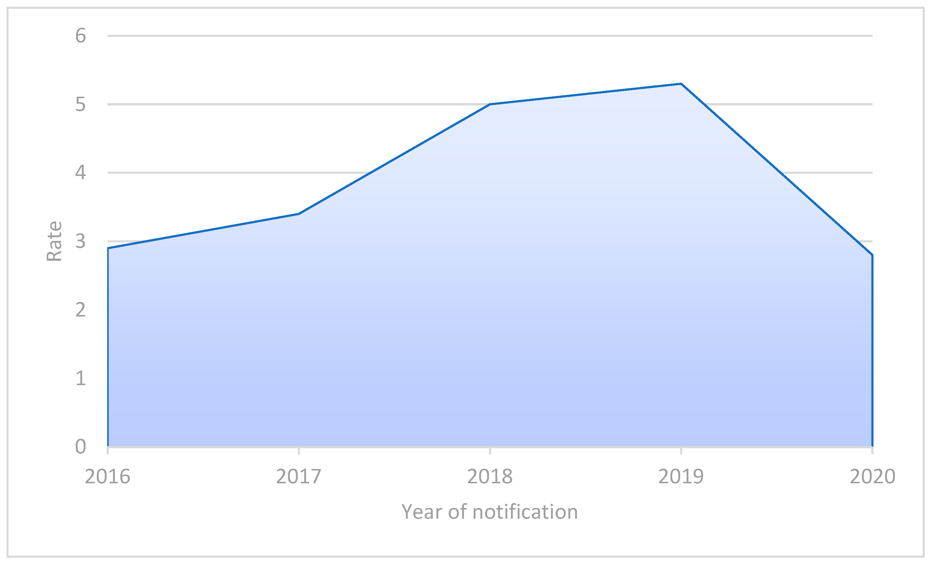
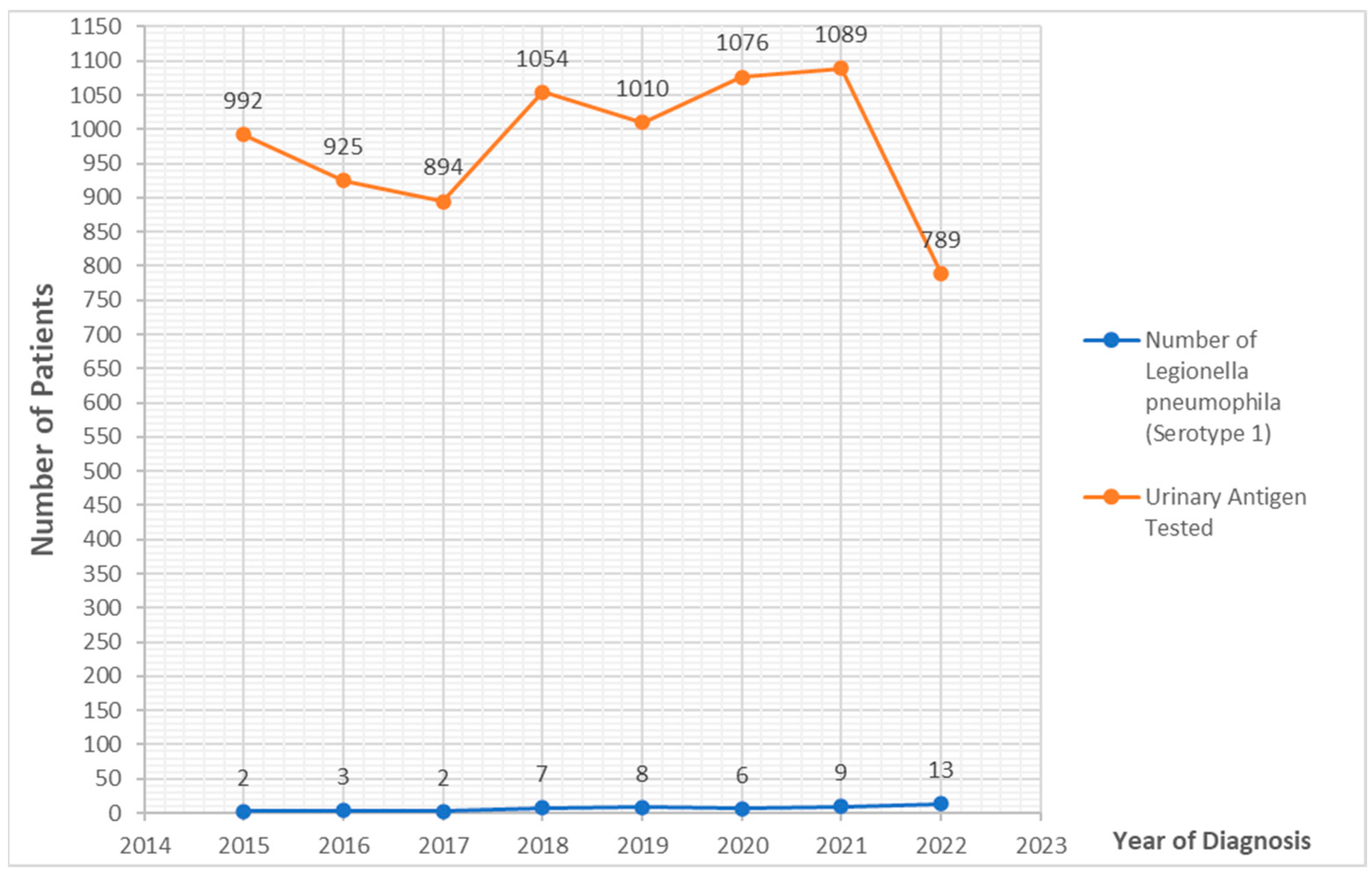
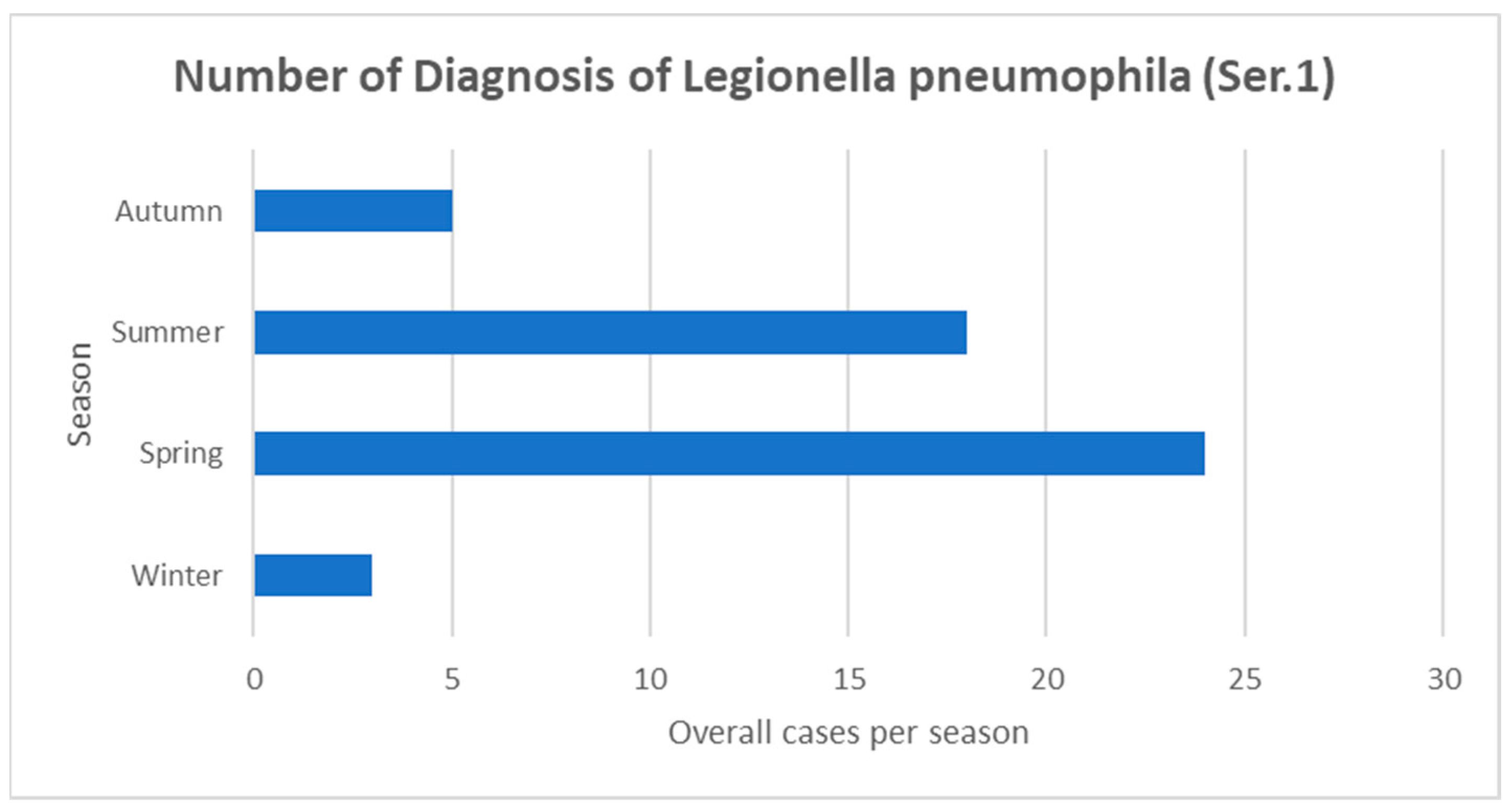
| Main Characteristics of Hospitalized Patients with Legionnaires’ Disease (N = 50) N (%) or Median (Range) | Survivors, N (%) or Median (Range) N = 44 (88) | Non-Survivors, N (%) or Median (Range) N = 6 (12) | p Value | |
|---|---|---|---|---|
| Age (median) | 69 (range 34–92) | 66 (34–92) | 85 (79–88) | 0.004 |
| Sex | 20% Female (10/50); 80% Male (40/50) | 16% Female (7/44); 84% Male (37/44) | 50% Female (3/6); 30% Male (3/6) | 0.086 |
| Ethnicity | 98% Caucasian (49/50); 2% Rom | 97% Caucasian (43/44); 2.3% Rom (1/44) | 100% Caucasian (6/6); 0% Rom | >0.99 |
| Smoking | 34% Active smokers (17/50) | 38.6% (17/44) | 0% (0/6) | 0.061 |
| Alcohol | 6% Daily Drinkers (3/50) | 6.8% (3/44) | 0% (0/6) | 0.509 |
| Comorbidities | ||||
| Cardiovascular diseases | 25% (25/50) | 43.2% (19/44) | 100% (6/6) | 0.009 |
| Kidney % | 8% (4/50) | 9.1% (4/44) | 0% (0/6) | 0.441 |
| Pulmonary diseases | 26% (13/50) | 25.6% (11/44) | 33.3% (2/6) | 0.687 |
| Neurological diseases | 12% (6/50) | 11.4% (5/44) | 16.7% (1/6) | 0.708 |
| Solid tumor | 10% (5/50) | 9.3% (4/44) | 16.7% (1/6) | 0.577 |
| Hematological disease | 8% (4/50) | 9.3% (4/44) | 0% (0/6) | 0.436 |
| Diabetes Mellitus | 6% (3/50) | 4.5% (2/44) | 16.7% (1/6) | 0.241 |
| SOT | 0% (0/50) | -- | -- | -- |
| Rheumatologic diseases % | 0% (0/50) | -- | -- | -- |
| HIV | 6% (3/50) | 6.8% (3/44) | 0% (0/6) | 0.509 |
| Hepatic diseases | 16% (8/50) | 12.2% (5/44) | 0% (0/6) | 0.366 |
| Obesity | 14% (7/50) | 23% (6/44) | 25% (1/6) | >0.99 |
| LD Acquisition Setting and Ward of Admission | ||||
| Community-acquired | 98% (49/50) | 97.7% (43/44) | 100% (6/6) | 0.709 |
| Hospital-acquired | 2% (1/50) | -- | -- | -- |
| Ward of admission | 94% MW; 4% ICU; 2% SW | -- | -- | -- |
| Diagnosis at Admission | ||||
| Urinary antigen for L. pneumophila serotype 1 | 100% (50/50) | -- | -- | -- |
| Diagnosis from admission (days) | Median 0 (range 0–12) | 0 (0–4) | 1 (1–9) | 0.035 |
| Specific therapy from admission | Median 0 (range 0–10) | 0 (0–2) | 0 (0–0) | 0.899 |
| Clinical Presentations and Complications at Admission | ||||
| Fever | 100% (50/50) | -- | -- | -- |
| Respiratory symptoms | 90% (45/50) | 90.9% (40/44) | 83.3% (5/6) | 0.562 |
| Gastro-intestinal symptoms | 12% (6/50) | 13.6% (6/44) | 0% (0/6) | 0.335 |
| Neurological symptoms | 10% (5/50) | 7% (3/44) | 33.3% (2/6) | 0.046 |
| Primary rhabdomiolysis | 16% (8/50) | -- | -- | -- |
| Acute Kidney Injury (without rhabdomiolysis) | 20% (10/50) | 18.2% (8/44) | 33.3% (2/6) | 0.384 |
| Pleural Effusion | 28% (14/50) | 27.3% (12/44) | 33.3% (2/6) | 0.756 |
| New onset arythmia | 10% (5/50) | 6.8% (3/44) | 33% (2/6) | 0.103 |
| Liver inflammation | 12% (6/50) | 13.6% (6/44) | 0 | >0.99 |
| Septic shock (according to SEPSIS-3) | 2% (1/50) | 0 | 16.7% (1/6)) | 0.006 |
| Respiratory failure | 74% (37/50) Low Flow Oxygen (34/50) NIV/C-PAP (3/50) | (30/44) (0/44) | (4/6) (3/6) | 0.941 <0.001 |
| Co-infections/Superinfections | ||||
| BSI | S. hominis (2); S. capitis (1) | |||
| CDI | Clostridioides difficile (1) | |||
| Viral pneumonia | SARS-CoV-2 (1) | |||
| Bacterial pneumonia | Pseudomonas aeruginosa (1) | |||
| Outcomes | ||||
| Survival 7 days after diagnosis | 92% (46/50) | |||
| Survival 28 days after diagnosis | 88% (44/50) | |||
| Main Characteristics of Hospitalized Patients with Legionnaires’ Disease (N = 50) N (%) or Median (Range) | Alive, N (%) or MEDIAN (Range) | Dead, N (%) or Median (Range) | p Value | |
|---|---|---|---|---|
| WBC | 13,715 (2300–31,210) | 12,510 (2300–31,210) | 14,130 (11,160–14,560) | 0.074 |
| PLTS | 191,000 (77,000–702,000) | 154,000 (77,000–466,000) | 191,000 (140,000–365,000) | 0.285 |
| Lymphocytes | 810 (170–28,210) | 830 (210–28,020) | 420 (220–810) | 0.005 |
| Monocytes | 400 (50–1770) | 340 (50–1640) | 530 (50–700) | 0.271 |
| Eosinophils | 40 (0–460) | 20 (0–260) | 110 (30–130) | 0.987 |
| Neutrophils | 11,090 (2030–28,850) | 9055 (2030–18,510) | 114 (8750–13,530) | 0.043 |
| Neutrophil-to-Lymphocyte Ratio | 11.9 (0.1–89.8) | 9.84 (0.10–30.81) | 20.83 (14.16–61.5) | <0.001 |
| GOT | 44 (15–288) | 60.5 (15–288) | 92 (37–117) | 0.771 |
| GPT | 44 (8–228) | 46.5 (14–228) | 39 (14–53) | 0.203 |
| Na | 129 (127–152) | 134.5 (126–152) | 137 (134–143) | 0.368 |
| K | 3.4 (3.0–4.0) | 3.9 (3–5) | 3.8 (3–4) | 0.261 |
| LDH | 528 (335–1098) | 547.5 (222–1098) | 771 (578–780) | 0.301 |
| Ferritin | 1634 (344–5810) | 1221 (344–5481) | - | - |
| Creatinine | 0.96 (0.42–4.95) | 0.89 (0.58–1.48) | 1.1 (0.87–1.5) | 0.493 |
| C-RP (mg/dL) | 277 (29–478) | 316 (103–478) | 386 (73–432) | 0.691 |
| PCT | 1.96 (0.12–37) | 2 (0.99–7.32) | 9.9 (6.6–17.5) | 0.005 |
| Microhematuria | Positive, 32% (16/50) Absent, 10% (5/50) Not Available, 58% (29/50) | Positive, 73.7% (14/50) Absent, 26.3% (5/50) | Positive, 100% (2/50) Absent, 0% (0/50) | 0.406 |
| Radiographic Involvement | ||||
| Isolated Median Lobe | 28% (14/50) | 27.3% (12/50) | 33.3% (2/50) | 0.756 |
| Isolated Apical Lobe | 16% (8/50) | 13.6% (6/50) | 33.3% (2/50) | 0.217 |
| Isolated RIL | 26% (13/50) | 22.7% (10/50) | 50% (3/50) | 0.153 |
| Isolated LIL | 12% (6/50) | 13.6% (6/50) | 0% (0/50) | 0.335 |
| Multifocal Involvement | 2% (1/50) | 2.3% (1/50) | 0% (0/50) | 0.709 |
| Choice of Treatment | ||||
| Levofloxacin (IV/OR) | 64% (32/50) | 80.6% (29/50) | 60% (3/50) | 0.567 |
| Levofloxacin (IV/OR) + Azithromicin (OR) | 10% (4/50) | 8.3% (3/50) | 205 (1/50) | |
| Levofloxacin (IV/OR) + Rifampicin (IV/OR) | 2% (1/50) | |||
| Azithromicin (OR) | 26% (13/50) | 83.3% (10/50) | 100% (3/50) | 0.749 |
| Duration of Treatment | ||||
| Days of treatment | 14 (1–21) | 14 (6–21) | 10 (1–14) | <0.001 |
Disclaimer/Publisher’s Note: The statements, opinions and data contained in all publications are solely those of the individual author(s) and contributor(s) and not of MDPI and/or the editor(s). MDPI and/or the editor(s) disclaim responsibility for any injury to people or property resulting from any ideas, methods, instructions or products referred to in the content. |
© 2023 by the authors. Licensee MDPI, Basel, Switzerland. This article is an open access article distributed under the terms and conditions of the Creative Commons Attribution (CC BY) license (https://creativecommons.org/licenses/by/4.0/).
Share and Cite
Lupia, T.; Corcione, S.; Shbaklo, N.; Rizzello, B.; De Benedetto, I.; Concialdi, E.; Navazio, A.S.; Penna, M.; Brusa, M.T.; De Rosa, F.G. Legionella pneumophila Infections during a 7-Year Retrospective Analysis (2016–2022): Epidemiological, Clinical Features and Outcomes in Patients with Legionnaires’ Disease. Microorganisms 2023, 11, 498. https://doi.org/10.3390/microorganisms11020498
Lupia T, Corcione S, Shbaklo N, Rizzello B, De Benedetto I, Concialdi E, Navazio AS, Penna M, Brusa MT, De Rosa FG. Legionella pneumophila Infections during a 7-Year Retrospective Analysis (2016–2022): Epidemiological, Clinical Features and Outcomes in Patients with Legionnaires’ Disease. Microorganisms. 2023; 11(2):498. https://doi.org/10.3390/microorganisms11020498
Chicago/Turabian StyleLupia, Tommaso, Silvia Corcione, Nour Shbaklo, Barbara Rizzello, Ilaria De Benedetto, Erika Concialdi, Anna Sara Navazio, Maurizio Penna, Maria Teresa Brusa, and Francesco Giuseppe De Rosa. 2023. "Legionella pneumophila Infections during a 7-Year Retrospective Analysis (2016–2022): Epidemiological, Clinical Features and Outcomes in Patients with Legionnaires’ Disease" Microorganisms 11, no. 2: 498. https://doi.org/10.3390/microorganisms11020498