
The Repetitive Detection of Toluene with Bioluminescence Bioreporter Pseudomonas putida TVA8 Encapsulated in Silica Hydrogel on an Optical Fiber


Abstract
:1. Introduction
2. Materials and Methods
2.1. Chemicals and Solutions
2.2. Microorganism and Cultivation
2.3. Entrapment of Cells into Silica Gel
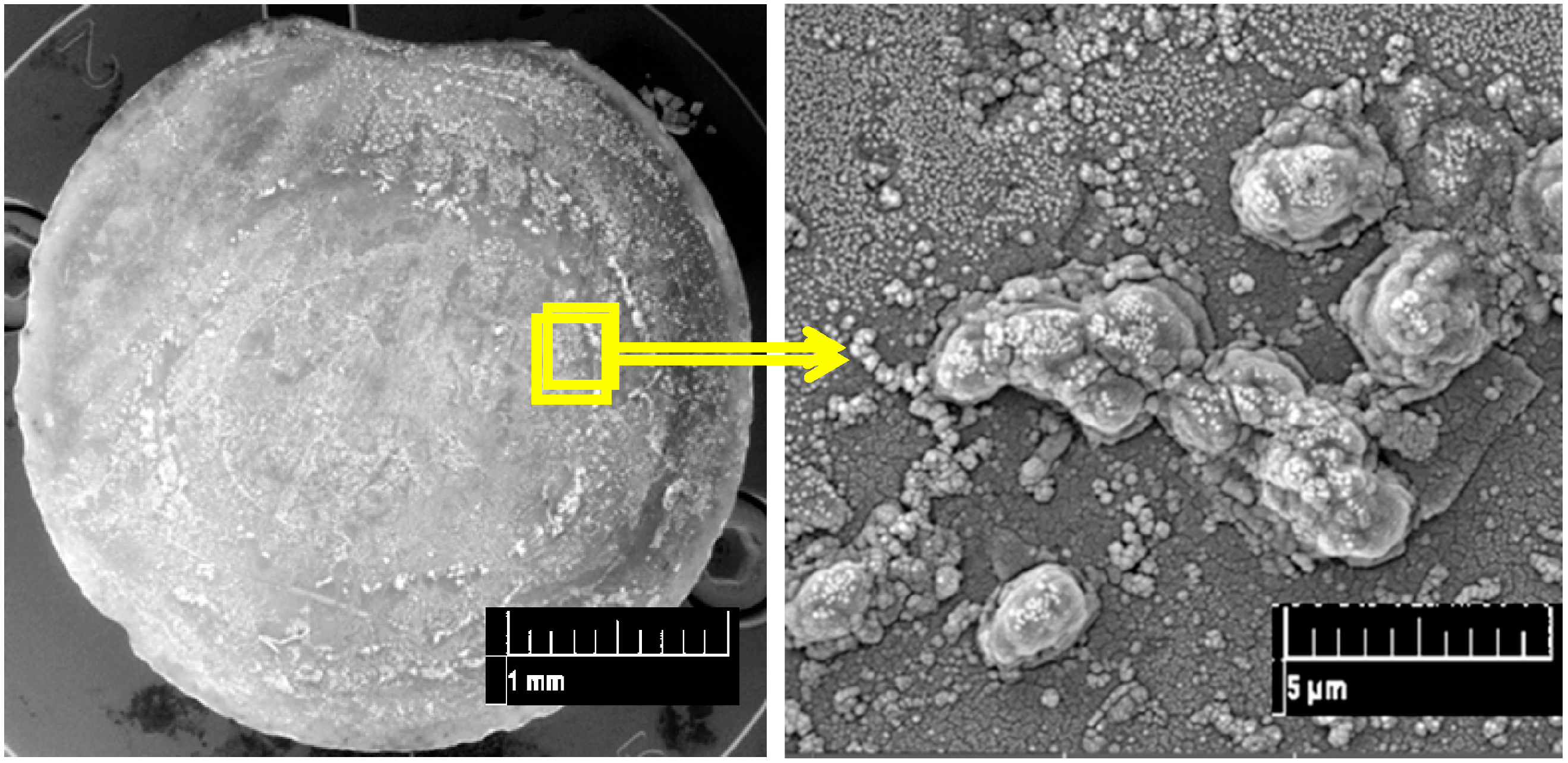
2.4. Scanning Electron Microscopy
2.5. Toluene Induction and Bioluminescence Measurements
3. Results and Discussion
4. Conclusions
Acknowledgments
Author Contributions
Conflicts of Interest
Abbreviations
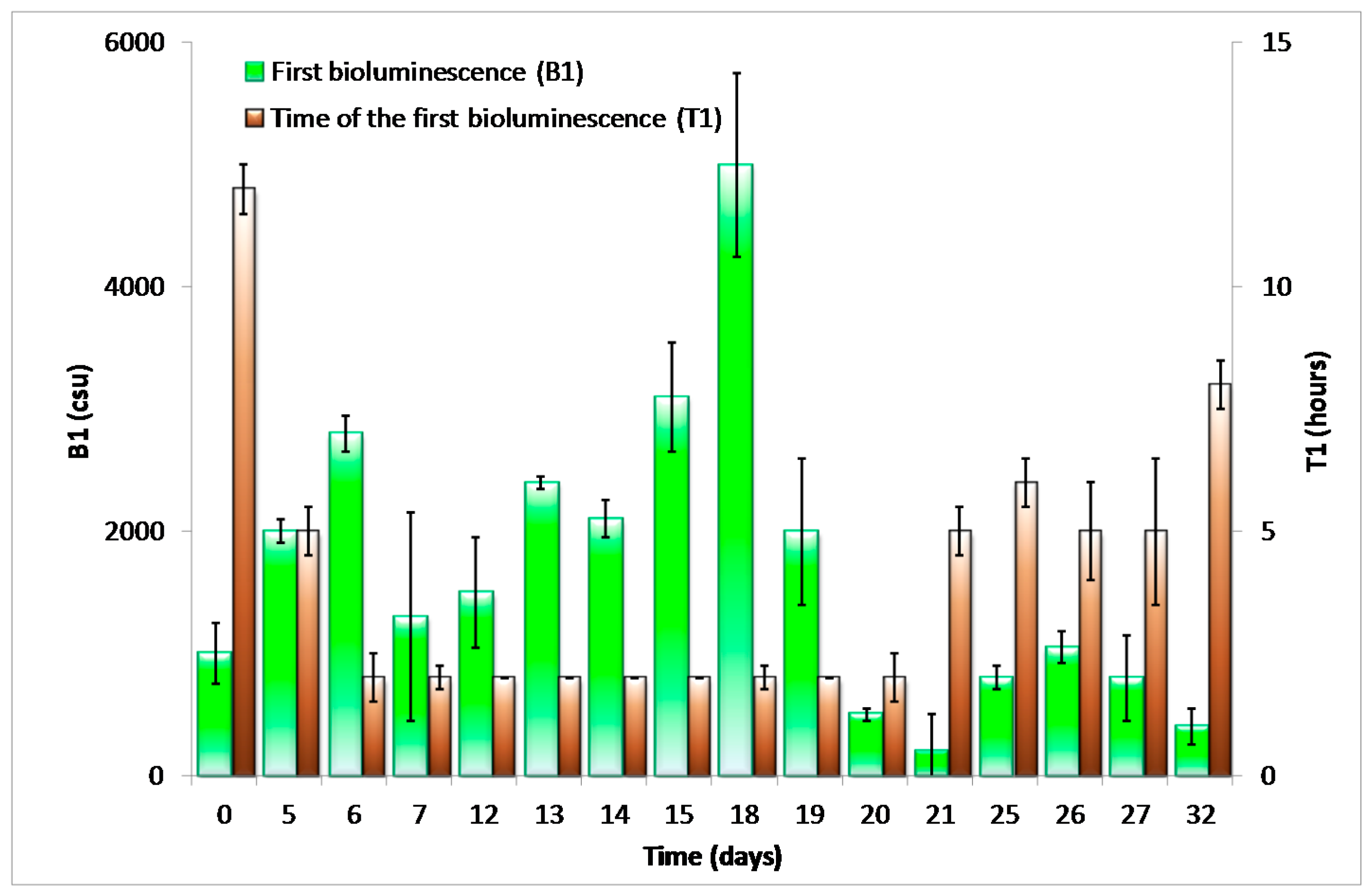
| B1 | |
| BTEX | benzene, toluene, ethylbenzene, and xylene |
| OD600 | optical density determined at λ = 600 nm |
| SEM | scanning electron microscopy |
| SMA connector | sub miniature version A optical fiber connector |
| T1 |
References
- Close, D.M.; Ripp, S.; Sayler, G.S. Reporter proteins in whole-cell optical bioreporter detection systems, biosensor integrations, and biosensing applications. Sensors 2009, 9, 9147–9174. [Google Scholar] [CrossRef] [PubMed]
- Van der Meer, J.R.; Belkin, S. Where microbiology meets microengineering: Design and applications of reporter bacteria. Nat. Rev. Microbiol. 2010, 8, 511–522. [Google Scholar] [CrossRef] [PubMed]
- Leveau, J.H.J.; Lindow, S.E. Bioreporters in microbial ecology. Curr. Opin. Microbiol. 2002, 5, 259–265. [Google Scholar] [CrossRef]
- Xu, T.T.; Close, D.M.; Sayler, G.S.; Ripp, S. Genetically modified whole-cell bioreporters for environmental assessment. Ecol. Indic. 2013, 28, 125–141. [Google Scholar] [CrossRef] [PubMed]
- King, J.M.H.; Digrazia, P.M.; Applegate, B.; Burlage, R.; Sanseverino, J.; Dunbar, P.; Larimer, F.; Sayler, G.S. Rapid, sensitive bioluminescent reporter technology for naphthalene exposure and biodegradation. Science 1990, 249, 778–781. [Google Scholar] [CrossRef] [PubMed]
- Trogl, J.; Chauhan, A.; Ripp, S.; Layton, A.C.; Kuncova, G.; Sayler, G.S. Pseudomonas fluorescens HK44: Lessons learned from a model whole-cell bioreporter with a broad application history. Sensors 2012, 12, 1544–1571. [Google Scholar] [CrossRef] [PubMed]
- Michelini, E.; Cevenini, L.; Calabretta, M.M.; Spinozzi, S.; Camborata, C.; Roda, A. Field-deployable whole-cell bioluminescent biosensors: So near and yet so far. Anal. Bioanal. Chem. 2013, 405, 6155–6163. [Google Scholar] [CrossRef] [PubMed]
- Wang, X.-D.; Wolfbeis, O.S. Fiber-optic chemical sensors and biosensors (2008–2012). Anal. Chem. 2013, 85, 487–508. [Google Scholar] [CrossRef] [PubMed]
- Zajic, J.; Bittner, M.; Branyik, T.; Solovyev, A.; Sabata, S.; Kuncova, G.; Pospisilova, M. Repetitive inductions of bioluminescence of Pseudomonas putida TVA8 immobilised by adsorption on optical fiber. Chem. Pap. 2016, 70, 877–887. [Google Scholar] [CrossRef]
- Vrbova, H.; Kuncova, G.; Pospisilova, M. Optical fiber element of sensor with bioluminescent cells. In Book of Abstracts of 10th European Conference on Optical Chemical Sensors and Biosensors, Europt(r)ode X; Institute of Photonics of CAV, v.v.i.: Prague, Czech Republic, 2010; p. 85. [Google Scholar]
- Kalabova, H.; Pospisilova, M.; Jirina, M.; Kuncova, G. Whole-cell biosensor for detection of environmental pollution enhancement of detected bioluminescence. Curr. Opin. Biotechnol. 2013, 24, S32. [Google Scholar] [CrossRef]
- Pospisilova, M.; Kuncova, G.; Troegl, J. Fiber-optic chemical sensors and fiber-optic bio-sensors. Sensors 2015, 15, 25208–25259. [Google Scholar] [CrossRef] [PubMed]
- Bjerketorp, J.; Hakansson, S.; Belkin, S.; Jansson, J.K. Advances in preservation methods: Keeping biosensor microorganisms alive and active. Curr. Opin. Biotechnol. 2006, 17, 43–49. [Google Scholar] [CrossRef] [PubMed]
- Depagne, C.; Roux, C.; Coradin, T. How to design cell-based biosensors using the sol-gel process. Anal. Bioanal. Chem. 2011, 400, 965–976. [Google Scholar] [CrossRef] [PubMed]
- Michelini, E.; Roda, A. Staying alive: New perspectives on cell immobilization for biosensing purposes. Anal. Bioanal. Chem. 2012, 402, 1785–1797. [Google Scholar] [CrossRef] [PubMed]
- Ben-Yoav, H.; Amzel, T.; Biran, A.; Sternheim, M.; Belkin, S.; Freeman, A.; Shacham-Diamand, Y. Bacterial biofilm-based water toxicity sensor. Sens. Actuators B Chem. 2011, 158, 366–371. [Google Scholar] [CrossRef]
- Jaroch, D.; McLamore, E.; Zhang, W.; Shi, J.; Garland, J.; Banks, M.K.; Porterfield, D.M.; Rickus, J.L. Cell-mediated deposition of porous silica on bacterial biofilms. Biotechnol. Bioeng. 2011, 108, 2249–2260. [Google Scholar] [CrossRef] [PubMed]
- Charrier, T.; Durand, M.-J.; Jouanneau, S.; Dion, M.; Pernetti, M.; Poncelet, D.; Thouand, G. A multi-channel bioluminescent bacterial biosensor for the on-line detection of metals and toxicity. Part I: Design and optimization of bioluminescent bacterial strains. Anal. Bioanal. Chem. 2011, 400, 1051–1060. [Google Scholar] [CrossRef] [PubMed]
- Trogl, J.; Ripp, S.; Kuncova, G.; Sayler, G.; Churava, A.; Parik, P.; Demnerova, K.; Halova, J.; Kubicova, L. Selectivity of whole cell optical biosensor with immobilized bioreporter Pseudomonas fluorescens HK44. Sens. Actuators B Chem. 2005, 107, 98–103. [Google Scholar] [CrossRef]
- Kuncova, G.; Trogl, J. Physiology of microorganisms immobilized into inorganic polymers. In Handbook of Inorganic Chemistry Research; Morrison, D.A., Ed.; Nova Science Publishers, Inc.: Hauppauge, NY, USA, 2010; pp. 53–101. [Google Scholar]
- Ripp, S.; Nivens, D.E.; Ahn, Y.; Werner, C.; Jarrell, J.; Easter, J.P.; Cox, C.D.; Burlage, R.S.; Sayler, G.S. Controlled field release of a bioluminescent genetically engineered microorganism for bioremediation process monitoring and control. Environ. Sci. Technol. 2000, 34, 846–853. [Google Scholar] [CrossRef]
- Polyak, B.; Bassis, E.; Novodvorets, A.; Belkin, S.; Marks, R.S. Bioluminescent whole cell optical fiber sensor to genotoxicants: System optimization. Sens. Actuators B Chem. 2001, 74, 18–26. [Google Scholar] [CrossRef]
- Ivask, A.; Green, T.; Polyak, B.; Mor, A.; Kahru, A.; Virta, M.; Marks, R. Fibre-optic bacterial biosensors and their application for the analysis of bioavailable Hg and As in soils and sediments from aznalcollar mining area in spain. Biosens. Bioelectron. 2007, 22, 1396–1402. [Google Scholar] [CrossRef] [PubMed]
- Premkumar, J.R.; Lev, O.; Marks, R.S.; Polyak, B.; Rosen, R.; Belkin, S. Antibody-based immobilization of bioluminescent bacterial sensor cells. Talanta 2001, 55, 1029–1038. [Google Scholar] [CrossRef]
- Polyak, B.; Geresh, S.; Marks, R.S. Synthesis and characterization of a biotin-alginate conjugate and its application in a biosensor construction. Biomacromolecules 2004, 5, 389–396. [Google Scholar] [CrossRef] [PubMed]
- Applegate, B.M.; Kehrmeyer, S.R.; Sayler, G.S. A chromosomally based tod-luxCDABE whole-cell reporter for benzene, toluene, ethybenzene, and xylene (BTEX) sensing. Appl. Environ. Microbiol. 1998, 64, 2730–2735. [Google Scholar] [PubMed]
- Kuncova, G.; Pazlarova, J.; Hlavata, A.; Ripp, S.; Sayler, G.S. Bioluminescent bioreporter Pseudomonas putida TVA8 as a detector of water pollution. Operational conditions and selectivity of free cells sensor. Ecol. Indic. 2011, 11, 882–887. [Google Scholar]
- Sambrook, J.; Fritsch, E.F.; Maniatis, T. Molecular Cloning; Cold Spring Harbor Laboratory Press: New York, NY, USA, 1989; Volume 2. [Google Scholar]
- Trogl, J.; Kuncova, G.; Kuran, P. Bioluminescence of Pseudomonas fluorescens HK44 in the course of encapsulation into silica gel. Effect of methanol. Folia Microbiol. 2010, 55, 569–575. [Google Scholar] [CrossRef] [PubMed]
- Kuncova, G.; Podrazky, O.; Ripp, S.; Trogl, J.; Sayler, G.; Demnerova, K.; Vankova, R. Monitoring of the viability of cells immobilized by sol-gel process. J. Sol-Gel Sci. Technol. 2004, 31, 335–342. [Google Scholar] [CrossRef]
- Troegl, J.; Jirkova, I.; Kuran, P.; Akhmetshina, E.; Brovdyova, T.J.; Sirotkin, A.; Kirilina, T. Phospholipid fatty acids as physiological indicators of paracoccus denitrificans encapsulated in silica sol-gel hydrogels. Sensors 2015, 15, 3426–3434. [Google Scholar] [CrossRef] [PubMed]
- Branyik, T.; Kuncova, G. Changes in phenol oxidation rate of a mixed microbial culture caused by sol-gel immobilization. Biotechnol. Lett. 2000, 22, 555–560. [Google Scholar] [CrossRef]
- Branyik, T.; Kuncova, G.; Paca, J. The use of silica gel prepared by sol-gel method and polyurethane foam as microbial carriers in the continuous degradation of phenol. Appl. Microbiol. Biotechnol. 2000, 54, 168–172. [Google Scholar] [CrossRef] [PubMed]
- Gavlasova, P.; Kuncova, G.; Kochankova, L.; Mackova, M. Whole cell biosensor for polychlorinated biphenyl analysis based on optical detection. Int. Biodeterior. Biodegrad. 2008, 62, 304–312. [Google Scholar] [CrossRef]
- Kuncova, G.; Pazlarova, J.; Adamova, N.; Hlavata, A.; Ripp, S.; Sayler, G.S. Bioluminiscent biosensor of toluene. In Proceedings of the XVIIth International Conference on Bioencapsulation, Groningen, The Netherlands, 24–26 September 2009; pp. 226–227.
- Verma, N.; Bansal, M.; Kumar, S. Whole cell based miniaturized fiber optic biosensor to monitor L-asparagine. Adv. Appl. Sci. Res. 2012, 3, 809–814. [Google Scholar]
- Lynch, A.M.; Sasaki, J.C.; Elespuru, R.; Jacobson-Kram, D.; Thybaud, V.; De Boeck, M.; Aardema, M.J.; Aubrecht, J.; Benz, R.D.; Dertinger, S.D.; et al. New and emerging technologies for genetic toxicity testing. Environ. Mol. Mutag. 2011, 52, 205–223. [Google Scholar] [CrossRef] [PubMed]
- Scholz, S.; Renner, P.; Belanger, S.E.; Busquet, F.; Davi, R.; Demeneix, B.A.; Denny, J.S.; Leonard, M.; McMaster, M.E.; Villeneuve, D.L.; et al. Alternatives to in vivo tests to detect endocrine disrupting chemicals (edcs) in fish and amphibians—Screening for estrogen, androgen and thyroid hormone disruption. Crit. Rev. Toxicol. 2013, 43, 45–72. [Google Scholar] [CrossRef] [PubMed]


© 2016 by the authors; licensee MDPI, Basel, Switzerland. This article is an open access article distributed under the terms and conditions of the Creative Commons Attribution (CC-BY) license (http://creativecommons.org/licenses/by/4.0/).
Share and Cite
Kuncová, G.; Ishizaki, T.; Solovyev, A.; Trögl, J.; Ripp, S. The Repetitive Detection of Toluene with Bioluminescence Bioreporter Pseudomonas putida TVA8 Encapsulated in Silica Hydrogel on an Optical Fiber. Materials 2016, 9, 467. https://doi.org/10.3390/ma9060467
Kuncová G, Ishizaki T, Solovyev A, Trögl J, Ripp S. The Repetitive Detection of Toluene with Bioluminescence Bioreporter Pseudomonas putida TVA8 Encapsulated in Silica Hydrogel on an Optical Fiber. Materials. 2016; 9(6):467. https://doi.org/10.3390/ma9060467
Chicago/Turabian StyleKuncová, Gabriela, Takayuki Ishizaki, Andrey Solovyev, Josef Trögl, and Steven Ripp. 2016. "The Repetitive Detection of Toluene with Bioluminescence Bioreporter Pseudomonas putida TVA8 Encapsulated in Silica Hydrogel on an Optical Fiber" Materials 9, no. 6: 467. https://doi.org/10.3390/ma9060467