Article

Two New Species of Backusella (Mucorales, Mucoromycota) from Soil in an Upland Forest in Northeastern Brazil with an Identification Key of Backusella from the Americas

by
Catarina Letícia Ferreira de Lima
1,
Joana D’arc Alves Leitão Lundgren
1,
Thuong Thuong Thi Nguyen
2,
Thalline Rafhaella Leite Cordeiro
1,
Diogo Xavier Lima
1,
Luciana Melo Sartori Gurgel
3,
Diogo Paes da Costa
4,
Hyang Burm Lee
2,* and
André Luiz Cabral Monteiro de Azevedo Santiago
1,*
1
Graduate Course in the Biology of Fungi, Department of Mycology, Universidade Federal de Pernambuco, Av. Prof. Moraes Rego, s/n, Recife 50740-465, PE, Brazil
2
Environmental Microbiology Laboratory C, Department of Agricultural Biological Chemistry, College of Agriculture and Life Sciences, Chonnam National University, Gwangju 61186, Korea
3
Instituto Agronômico de Pernambuco, Avenue General San Martin, 1371, Recife 50761-000, PE, Brazil
4
Microbiology and Enzymology Laboratory, Universidade Federal do Agreste de Pernambuco, Garanhuns 55292-270, PE, Brazil
*
Authors to whom correspondence should be addressed.
J. Fungi 2022, 8(10), 1038; https://doi.org/10.3390/jof8101038
Submission received: 11 August 2022 / Revised: 24 September 2022 / Accepted: 26 September 2022 / Published: 30 September 2022
(This article belongs to the Special Issue Diversity of Undiscovered Fungal Taxa and Novel Antifungal Agents)

Abstract

:
During a survey of Mucorales from a forest located in Pernambuco state, Brazil, two new Backusella species were discovered and described based on morphological and molecular data (internal transcribed spacer and large subunit ribosomal DNA sequences). Both species were characterized as unbranched sporangiophores and sporangia with columellae of varied shapes forming. Multispored sporangiola were frequent, whereas unispored sporangiola were rare. URM 8395 forms sporangiophores that may support hyaline, slightly curved or circinate pedicels with multispored sporangiola at their apical portion, and abundant giant cells and chlamydospores. Columellae of sporangia are hyaline, conical (majority), or ellipsoidal with a truncate base, globose to subglobose or subglobose to conical, and, rarely, with slight medial constriction. URM 8427 does not form sporangiola from pedicels, giant cells are not observed, and columellae of sporangia are globose to subglobose, cylindrical with a truncate base, some with a slight constriction, applanate, obovoid, ellipsoidal, or, rarely, conical. Some columellae may have one side more swollen than the other and some are arranged obliquely on the sporangiophores. Sterile sporangia may or may not be formed on short sporophores. The detailed description and illustration of both novel species as well as an identification key for Backusella from the Americas are provided.

1. Introduction

Backusella Hesseltine & J.J. Ellis belongs to the Backusellaceae family (established by K. Voigt and PM Kirk) and comprises fungi characterized by the production of sporangiophores that are transiently curved when young and erect when mature. These fungi possess non-apophysate and multisporate, deliquescent-walled sporangia [1], and most species form uni- and/or multispored sporangiola with a persistent wall [2]. Backusella species are saprobic in nature and are found in soil, litter, plant debris, herbivore dung, decaying wood, Fragaria, diseased roots, and Medicago sativa [3,4].
Backusella has been described as the member of the Mucoraceae family [5], with B. circina J.J. Ellis & Hesselt. as the type species of this genus. Subsequently, species of this genus were transferred to Thamnidiaceae Fitzp. [6], owing to their morphological similarity with Thamnidium Link [3]. However, Benny and Benjamin [7] monographed Backusella and grouped three species (B. circina J.J. Ellis & Hesselt., B. ctenidia (Durrell & M. Fleming) Pidopl. & Milko ex Benny & R.K. Benj., and B. lamprospora (Lendn.) Benny & R.K. Benj.) that produced sporangia and sporangiola into this genus [7].
In a study combining morphological and molecular data (internal transcribed spacer (ITS) and large subunit (LSU) of nuclear ribosomal DNA (rDNA) sequences), Walther et al. [2] transferred seven Mucor species that presented curved sporangiophores when young and erect at maturity to Backusella. In this study, B. ctenidia was transferred to Mucor (as M. ctenidius (Durrell & M. Fleming) Walther & de Hoog). Currently, this genus comprises 26 species, 10 of which were isolated from Australia [8], four from South Korea [3,4], and five from Brazil [9,10,11,12].
During a study on the diversity of Mucorales in an upland forest located in the municipalities of Arcoverde and Águas Belas, Pernambuco, Brazil, two species of Backusella that varied morphologically and genetically (via ITS and LSU rDNA analysis) from other species were isolated. The aim of this study was to describe and illustrate these new species. We also provide a key to the Backusella species from the Americas.

2. Materials and Methods

2.1. Sampling Site

Soil samples were collected from an upland forest in the city of Arcoverde (at 1000 m altitude), district of Mimoso (08°39′688″S 036°90′013″ W), and at Serra do Comunaty (09°05′441″ S 037°05′438″ W), in the municipality of Águas Belas (at 900 m altitude), both of which are located in the state of Pernambuco, Brazil. Mimoso has a tropical rainy climate with dry summers, an average temperature ranging between 12 °C and 25 °C, and an average annual rainfall of 653 mm [13,14]. The vegetation consists of deciduous and semideciduous forests. The municipality of Águas Belas also has a tropical rainy climate with dry summers and an average annual rainfall of 373 mm. The temperature ranges between 18 and 34.5 °C [15]. The vegetation is composed of semideciduous and deciduous forests [16].

2.2. Isolation, Purification, and Identification

Five milligrams of soil were added to wheat germ agar medium [17] containing chloramphenicol (200 mg·L−1) in Petri dishes in triplicate. Colony growth was monitored for 7 days at room temperature (28 ± 2 °C). To purify Backusella spp., mycelial fragments taken from the edge of the growing colony were transferred to malt extract agar (MEA) medium [17]. The species were identified by observing their macroscopic (appearance, color, and diameter of colonies) and microscopic (microstructures) characteristics, according to the descriptions of Benny and Benjamin (1975), Walther et al. (2013), de Souza et al. (2014), Lima et al. (2016), Crous et al. (2019) and Nguyen et al. (2021). Slides corresponding to the holotypes of the new species (URM 94839 and URM 94983) were deposited at the URM Herbarium of the Universidade Federal de Pernambuco. Ex-type living cultures of both new species were deposited at the URM Culture Collection of the Universidade Federal de Pernambuco (URM 8395 and URM 8427).

2.3. Experiments

Pure cultures were grown in triplicate in both MEA and potato dextrose agar (PDA) media [17] and incubated at 10, 15, 20, 25, 30, 35, and 40 °C for 15 days. For morphological identification, fragments of colonies were obtained from the plates for examination of fungal structures. These fragments were placed together with a drop of potassium hydroxide (KOH, 3%) or lactophenol blue on microscope slides and observed under a light microscope (Carl Zeiss Axioscope 40, Göttingen, Germany). Photomicrographs were captured using the Leica DM 500 microscope combined with the Leica ICC50 camera (Leica, Heerbrugg, Switzerland) and Leica Application Suite software (v.3.4). The color designation of the colonies was performed according to the terminology of Kornerup and Wanscher [18].

2.4. DNA Extraction, Amplification, Cloning, and Sequencing

Fungal biomass was extracted from MEA cultures in test tubes incubated at 28 °C for up to 5 days. The material was transferred to 2 mL microtubes with screw caps. To each tube, 0.5 g of acid-washed glass beads of two different diameters (150–212 μm and 425–600 μm, 1:1; Sigma, St. Louis, MO, USA) were added. The material was crushed by stirring at high speed in a FastPrep homogenizer (FastPrep-24; MP Biomedicals, Irvine, CA, USA). The genomic DNA extraction procedure was conducted as described by de Oliveira et al. [19]. Briefly, the mycelium was homogenized in cetyltrimethylammonium bromide (C TAB) lysis buffer (2% CTAB, 20 mM ethylenediaminetetraacetic acid (EDTA), 0.1 M Tris-HCl, pH 8.0, 1.4 M NaCl) [20,21], followed by washing with chloroform:isoamyl alcohol (24:1) to subsequently separate the DNA-containing supernatant from the hyphal residues. The supernatant was mixed with an equal volume of isopropanol, followed by DNA precipitation after incubation at –20 °C for 30 min. After centrifugation at 13,000 rpm for 15 min, the resulting DNA pellet was washed with 70% ethanol and resuspended in 50 μL of ultrapure water.
For amplification of the ITS and LSU rDNA, we used the primer pairs ITS1/ITS4 and LR1/LSU2 [22,23,24], respectively. Thermal cycling parameters were as follows: 5 min at 95 °C (one cycle), 45 s at 94 °C, 1 min at 60 °C, 1 min at 72 °C (39 cycles), and a final elongation of 7 min at 72 °C. The final amplicons were purified with the enzymatic mix NucleoSAP (Cellco Biotech of Brazil, São Carlos, Brazil) and used for sequencing at the Genomic Technology and Gene Expression Platform of the Center for Biological Sciences of the Federal University of Pernambuco-UFPE (Pernambuco, Brazil). Direct sequencing of the ITS region from PCR products of strain URM 8427 failed. PCR products were cloned using the pGEM-T Easy Vector (Promega, Madison, WI, USA), following the manufacturer’s instructions. These clones were sequenced using the primers M13F forward (5′-GTAAAACGACGGCCAGT-3′) and M13R reverse (5′-GCGGATAACAATTTCACACAGG-3′).
Sequence data were compared with similar sequences available in the National Center for Biotechnology Information GenBank database using Nucleotide BLAST (BLASTn). The newly obtained sequences were deposited in the GenBank databases (Table 1).

2.5. Phylogenetic Analyses

The phylogenetic relationship of the new Backusella species and related species was determined by analysis of concatenated sequence datasets of two loci (ITS and LSU). Sequences were aligned via multiple alignment using Fast Fourier Transform (MAFFT) v.7 (https://mafft.cbrc.jp/alignment/server, accessed on 21 July 2022) [25,26] and manually improved in Molecular Evolutionary Genetics Analysis (MEGA) v.7 [27]. The analysis of the concatenated sequence datasets of the two loci was performed in MEGA v.7. Maximum likelihood (ML) analysis was performed in Randomized Axelerated Maximum Likelihood (RAxML)-HPC v. 8.2.8 Black Box [28] using the general time-reversible (GTR)+G+I model test with 1000 bootstrap (BS) replicates on the CIPRES Science Gateway Portal (https://www.phylo.org/portal2/) [29]. Bayesian inference (BI) was performed using MrBayes v. 3.2.2 [30] on The Extreme Science and Engineering Discovery Environment (XSEDE) through CIPRES using the GTR+I+G model as selected by jModelTest v.2.1.10 under the Akaike information criterion for BI [31,32]. BI analysis was conducted with 1 × 10⁶ generations with a burning value of 25%. Phylogenetic trees were viewed and arranged using Interactive Tree of Life v4 (https://itol.embl.de, accessed on 21 July 2022) [33]. Values less than 0.90 BI posterior probability (BPP) and 70% ML bootstrap (MLBS) are not shown. Sequence data were compared with those of similar sequences available in the GenBank database using BLASTn.

3. Results

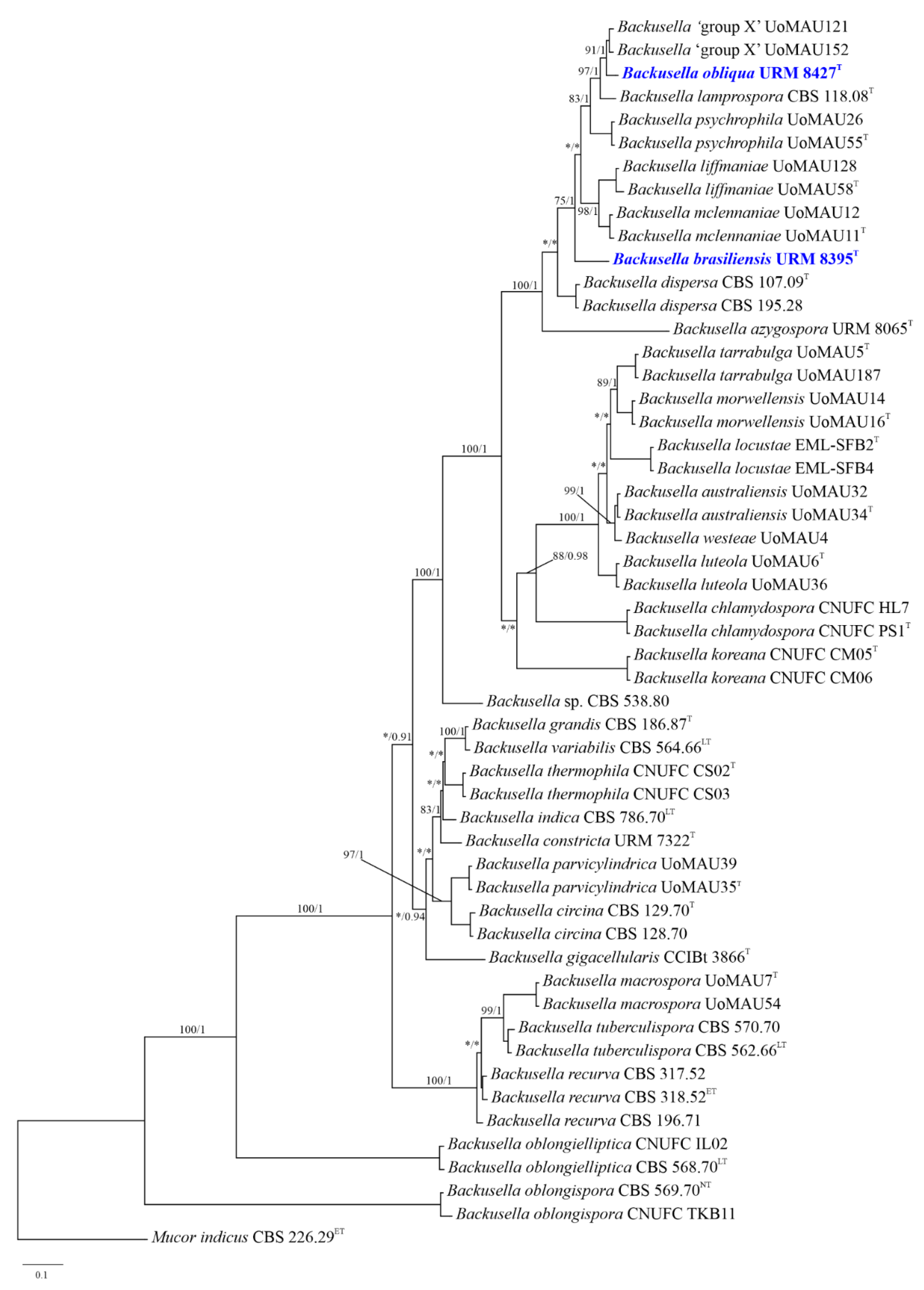
3.1. Phylogenetic Analyses

Phylogenetic analyses of the combined ITS and LSU sequence data identified 53 taxa, including the newly generated sequences and outgroup (Figure 1). The sequence dataset consisted of 1828 characters including gaps (ITS, 1098 characters; LSU, 730 characters). Results obtained using BI and maximum likelihood analyses phylogenetically characterized URM 8395 and URM 8427 as new species that forms a distinct clade from other Backusella species with 100% MLBS and 1 BPP support values (Figure 1). URM 8395 is close to B. dispersa and to a clade containing URM 8427, B. azygospora, B. mclennaniae, B. liffmaniae, B. psychrophila, B. lamprospora, and Backusella as ‘group X’. URM 8427 is closer to the not yet described Backusella ‘group X’.

3.2. Taxonomy

Backusella brasiliensis C.L. Lima, Hyang B. Lee & A.L. Santiago, sp. nov. (Figure 2, Table 2).
Index Fungorum: IF559794;
Etymology: brasiliensis, referring to the country where the species was first isolated.
Colony with cottony appearance, initially white and then becoming pale gray (1–1B), colonizing the entire Petri dish (9 cm diameter) after incubation for 4 days on PDA at 25 °C; reverse yellowish-gray (2–2B). Rhizoids well-branched. Sporangiophores occasionally with a slight constriction below the sporangia, hyaline, arising directly from the substrate, recurved when young and erect at maturity, up to 12 µm diameter, unbranched or, rarely, with simple branches. Sterile branches common on long sporangiophores. Both sporangiophores and branches have multispored sporangium at their apex, with a smooth to slightly encrusted wall. One septum is commonly formed near the branching point. The main axes of sporangiophores may support hyaline, slightly curved or circinate pedicels (up to 95 × 45 μm2) with multispored sporangiola at their apical portion and a smooth to slightly encrusted outer wall. Sporangia yellowish, globose to subglobose (up to 60–70 µm diameter), wall deliquescent and slightly echinulate. Columellae of sporangia hyaline, conical (majority), or ellipsoidal with truncate base, globose to subglobose or subglobose to conical, and, rarely, with a slight constriction at center, (12–) 15–25 (–30) × (15–) 17–30 µm; wall lightly encrusted; collar evident. Short, simple or sympodially branched (up to seven times) sporophores bearing only multispored and/or unispored sporangiola (rare) formed near the substrate. Sporangiola hyaline, globose to subglobose, (12–) 15–45 (–50) μm diameter, echiinulate, and with a persistent wall; columellae of sporangiola globose to subglobose or subglobose to conical, 7–15 µm diameter, hyaline, and with a slightly encrusted wall. Sporangiospores hyaline, globose to subglobose (majority), 4–12 (–17) μm diameter, or irregular, 20–30 × 12–17 μm, smooth-walled. Abundant giant cells observed, hyaline, some with yellowish content, 7–17 µm diameter. Chlamydospores abundant, hyaline, 7–35 × 9.5–17 µm, wall smooth to slightly encrusted. No zygospores were observed.
Habitat: Soil
Distribution: Pernambuco, Brazil.
Specimen examined: BRAZIL, Pernambuco, Mimoso (08°39′688″ S 036°90′013″ W), soil, 2019, C.L.F. Lima (holotype URM 94839, ex-type URM 8395).
Media and temperature tests: On PDA, at 10 °C, slow growth (3 cm diameter in 144 h) with poor sporulation; at 15 °C, better growth (6 cm in 120 h) than that at 10 °C with good sporulation; at 20 °C, good growth (7 cm in 120 h) with good sporulation; at 25 °C, better growth (9 cm in 96 h) with good sporulation; at 30 °C, good growth (9 cm in 144 h) and good sporulation; at 35 °C, slow growth (2 cm in 120 h) with lack of sporulation; and at 40 °C, lack of growth. A similar growth pattern was observed on MEA and PDA at all temperatures.
Backusella obliqua C.L. Lima, J.D. Leitão, Hyang B. Lee & A.L. Santiago, sp. nov. (Figure 3, Table 2).
Index Fungorum: IF559793;
Etymology: obliqua, referring to the columellae arranged obliquely on the sporangiophores.
Colony cottony, white (1–1A), colonizing the entire Petri dish (9 cm diameter) after 4 days on PDA at 25 °C; reverse grayish white (1–1B). Rhizoid-like sterile branched hyphae common. Sporangiophores terminating in a multispored sporangium at their apex, hyaline or with greenish contents, occasionally with slight constriction below the sporangia, arising directly from the substrate, recurved when young and erect at maturity, simple or sympodially branched up to three times, up to 12 µm diameter. A septum is commonly formed near the branch point. Sporangia greenish-yellow, globose to subglobose, 15–60 µm diameter, wall slightly echinulate, deliquescent. Columellae of sporangia hyaline or with greenish-yellow contents, globose to subglobose, cylindrical with a truncate base, some with a slight constriction, applanate, obovoid, ellipsoidal, or, rarely, conical, (9.5–) 12–35 × 15–30 µm, smooth walled; collar little evident when present. Some columellae may have one side more swollen than the other and some are arranged obliquely on the sporangiophores. Short sporophores commonly sympodially branched, up to nine times, containing multispored and/or, rarely, unispored sporangiola (some sterile), are formed close to the substrate. Sporangiola greenish gray, globose to subglobose (9–) 12–24 μm diameter, wall echinulate and persistent. Columellae of sporangiola hyaline, globose to subglobose and applanate, 7–12 µm diameter, wall slightly encrusted. Sporangiospores hyaline or with greenish-yellow contents, globose to subglobose 7–12 (–15) μm diameter, smooth-walled. Giant cells and zygospores not observed.
Habitat: Soil
Distribution: Pernambuco, Brazil.
Specimen examined: BRAZIL, Pernambuco, Águas Belas (09°05′441″ S 037°05′438″ W), soil, 2019, C.L.F. Lima (holotype URM 94983, ex-type URM 8427).
Media and temperature tests: On PDA, at 10 °C, slow growth (2 cm in 120 h) with poor sporulation; at 15 °C, better growth (5 cm in 120 h) than that at 10 °C with good sporulation; at 20 °C, good growth (7 cm in 120 h) with good sporulation; at 25 °C, better growth (9 cm in 96 h) with good sporulation; at 30 °C, good growth (9 cm in 144 h) and good sporulation; at 35 °C, slow growth (3 cm in 120 h) with lack of sporulation; and at 40 °C, lack of growth. A similar growth pattern was observed on MEA and PDA at all temperatures.

4. Discussion

Backusella brasiliensis (URM 8395) and B. obliqua (URM 8427) were described in this study based on their morphological and molecular characteristics that make them different from the other closely related species; therefore, they were noted as new species. Phylogenetically, B. brasiliensis formed a clade distant from other Backusella species but closer to B. dispersa and to a clade containing URM 8427, B. azygospora, B. mclennaniae, B. liffmaniae, B. psychrophila, B. lamprospora and Backusella as ‘group X’ (Figure 1). However, these species are morphologically different. Backusella brasiliensis is differentiated from B. azygospora as it does not produce azygospores, which are common in the later species. Besides, chlamydospores and sterile branches in long sporangiophores are not observed in B. azygospora [11]. Backusella dispersa forms smaller sporangia (30.2–47.5 × 28.3–46.1 μm) and larger columellae (19.5–38.7 × 18.5–33.4 μm) than those observed for the new species. In addition, sporangiospores of B. dispersa are globose to broadly ellipsoidal (8–12 × 7–10 μm), and are different from those of the new species, which are globose to subglobose (majority), 4–12 (–17) μm diameter or irregular, 20–30 × 12–17 μm. Although no chlamydospores are formed in B. dispersa [8], they are common in URM 8395. The new species is easily differentiated from B. mclennaniae, B. liffmaniae and B. psychrophila by forming giant cells and chlamydospores. Yet, B. brasiliensis forms columellae that are conical (majority), ellipsoidal with truncate base, globose to subglobose or subglobose to conical, and, rarely, with a slight constriction at center, whereas columellae of B. mclennaniae and B. liffmaniae and B. psychrophila are globose, ellipsoidal or applanate.
Backusella brasiliensis and B. lamprospora present morphological similarities, such as the following: the main axes of their sporangiophores support hyaline, slightly curved or circinate pedicels with multispored sporangiola at their apical portion; short and simple or sympodially branched sporophores bearing only multispored and/or unispored sporangiola (rare) are formed near the substrate [7]. However, the main sporangiophore axes of B. lamprospora are simple or sympodially branched, differing from those of the new species that are unbranched or once-branched. Backusella lamprospora is characterized by forming sporangiophores with hemispherical to globose or ovoid columellae, differing from those found in B. brasiliensis, which are conical, ellipsoidal with a truncate base, globose to subglobose, subglobose to conical, conical, or cylindrical, including some with a slight constriction at the center. Furthermore, the sporangiospores of B. brasiliensis are globose, subglobose, and irregular, whereas those of B. lamprospora are only subglobose [7].
The formation of giant cells and chlamydospores is not common in Backusella species, and as far as we know, in addition to the new species, giant cells have been cited only in B. gigacellularis and B. oblongielliptica, whereas chlamydospores have only been observed in B. chlamydospora. Backusella gigacellularis does not produce unispored sporangiola, and its sporangia are larger (40–131 μm diameter) than those of B. brasiliensis. Moreover, sporangiospores of the new species are globose, subglobose, and irregular, whereas those of B. gigacellularis are ellipsoidal and, rarely, irregular [9]. Backusella oblongielliptica can be easily differentiated from B. brasiliensis by the lack of chlamydospore and sporangiola formation and the production of oblongly ellipsoidal sporangiospores [2,34]. Backusella chlamydospora differs from B. brasiliensis as it produces abundant unispored sporangiola, larger sporangia (35–80 × 35–75 μm), and subglobose, conical, ellipsoidal, cylindrical, hemispherical, or nearly pyriform columellae, which are sometimes bell-shaped, long conical, or constricted at the center [4]. In contrast, B. brasiliensis rarely produces unispored sporangiola and does not form cylindrical or pyriform columellae.
Phylogenetically, B. obliqua (URM 8427) is close to the Backusella species of ‘group X’, which is a group of a putative new species not yet proposed [8], but also close to B. lamprospora and B. psychrophila. Morphologically, Backusella ‘group X’ forms sporangiophores up to 8 µm diameter, unlike B. obliqua, which forms sporangiophores up to 12 µm diameter. The columellae produced by B. obliqua are globose to subglobose, subglobose to irregular, cylindrical with a truncate base (some with slight constriction), flattened, obovoid, ovoid, ellipsoidal, or, rarely, conical, some arranged obliquely on the sporangiophores, and some with one side more swollen than the other, while Backusella ‘group X’ forms only globose, ellipsoidal, or applanate columellae. The sporangiospores of B. obliqua are globose to subglobose, unlike that of Backusella ‘group X’, which forms globose to broadly ellipsoidal sporangiospores. Backusella obliqua can be differentiated from B. lamprospora by forming sporangiophores without lateral pedicellate sporangiola. Columellae of B. lamprospora are hemispherical to globose or ovoid, differing from those of B. obliqua [7]. This new species forms sporangiophores that are simple or sympodially branched up to three times, whereas sporangiophores of B. psychrophila are occasionally branched. In addition, sporangiospores of B. obliqua are globose to subglobose, whereas those of B. psychrophila are broadly ellipsoidal to ellipsoidal [8].
In conclusion, B. obliqua and B. brasiliensis are morphologically and genetically different from other Backusella species, justifying their recognition as novel species. Brazil is a tropical country with areas considered global biodiversity hotspots that support high fungal diversity [12,35,36,37]. Voigt et al. [38] showed that of the 74 Mucoromycota species newly described between 2015 and 2020, 54 (74%) were isolated from Brazil, South Korea, Australia, and China, with 17 new species described in Brazil, confirming the great potential for the discovery of new taxa of Mucoromycota in this country. Regarding Backusella, of the 26 accepted species, only six (B. azygospora, B. brasiliensis, B. constricta, B. gigacellularis, B. lamprospora, and B. variabilis) were isolated from Brazil, including from areas of upland forest [10,11,12,39]. Considering that a high diversity of fungi is expected in tropical hotspots, we believe that this number of known Backusella species in Brazil is underestimated and it could increase with more investments in research and further training of Brazilian taxonomists studying Mucoromycota.

Author Contributions

Material collection: J.D.A.L.L. and C.L.F.d.L.; methodology: T.R.L.C., D.X.L., H.B.L., D.P.d.C., and T.T.T.N.; writing—original draft preparation: C.L.F.d.L., A.L.C.M.d.A.S., and L.M.S.G.; writing—review and editing: A.L.C.M.d.A.S., H.B.L., and C.L.F.d.L. All authors have read and agreed to the published version of the manuscript.

Funding

This research was funded by the “Mucoromycotina diversity in the different ecosystems of the Atlantic Forest of Pernambuco” project (FACEPE - First Projects Program PPP/FACEPE/APQ - 0842-2.12/14). We also thank the Conselho Nacional de Desenvolvimento Científico e Tecnológico (CNPq) for the research grant awarded to André L. C. M. A. Santiago and the Coordenação de Aperfeiçoamento de Pessoal de Nível Superior (CAPES) for the scholarship awarded to Catarina L. F. de Lima. This work was also supported in part by the Graduate Program for the Undiscovered Taxa of Korea funded by NIBR of the Ministry of Environment (MOE) of Korea, and in part by the Basic Science Research Program (2022R1I1A3068645) through the National Research Foundation of Korea (NRF) funded by the Ministry of Education.

Institutional Review Board Statement

Not applicable.

Informed Consent Statement

Not applicable.

Data Availability Statement

The ITS and LSU rDNA sequences generated during the current study were submitted to Genbank (https://www.ncbi.nlm.nih.gov). The newly described species were deposited in Index Fungorum (http://www.indexfungorum.org).

Conflicts of Interest

The authors declare no conflict of interest.

References

  1. Benny, G.L. Zygomycetes. Available online: http://zygomycetes.org/index.php?id=27 (accessed on 25 May 2022).
  2. Walther, G.; Pawłowska, J.; Alastruey-Izquierdo, A.; Wrzosek, M.; Rodriguez-Tudela, J.L.; Dolatabadi, S.; Chakrabarti, A.; de Hoog, G.S. DNA barcoding in Mucorales: An inventory of biodiversity. Persoonia 2013, 30, 11–47. [Google Scholar] [CrossRef] [PubMed]
  3. Wanasinghe, D.N.; Phukhamsakda, C.; Hyde, K.D.; Jeewon, R.; Lee, H.B.; Jones, E.B.G.; Tibpromma, S.; Tennakoon, D.S.; Dissanayake, A.J.; Jayasiri, S.C.; et al. Fungal diversity notes 709–839: Taxonomic and phylogenetic contributions to fungal taxa with an emphasis on fungi on Rosaceae. Fungal Divers. 2018, 89, 1–236. [Google Scholar] [CrossRef]
  4. Nguyen, T.T.T.; Voigt, K.; Santiago, A.L.C.M.A.; Kirk, P.M.; Lee, H.B. Discovery of novel Backusella (Backusellaceae, Mucorales) isolated from invertebrates and toads in Cheongyang, Korea. J. Fungi 2021, 7, 513. [Google Scholar] [CrossRef] [PubMed]
  5. Ellis, J.J.; Hesseltine, C.W. Two new members of the Mucorales. Mycologia 1969, 61, 863–872. [Google Scholar] [CrossRef]
  6. Pidoplichko, N.M.; Milko, A.A. Atlas of Mucoralean Fungi; Academy of Sciences of the Ukrainian SSR: Kiev, Ukraine, 1971. [Google Scholar]
  7. Benny, G.L.; Benjamin, R.K. Observations on Thamnidiaceae (Mucorales). New taxa, new combinations, and notes on selected species. Aliso 1975, 8, 301–351. [Google Scholar] [CrossRef]
  8. Urquhart, A.S.; Douch, J.K.; Heafield, T.A.; Buddie, A.G.; Idnurm, A. Diversity of Backusella (Mucoromycotina) in south-eastern Australia revealed through polyphasic taxonomy. Persoonia 2021, 46, 1–25. [Google Scholar] [CrossRef]
  9. de Souza, J.I.; Marano, A.V.; Pires-Zottarelli, C.L.A.; Chambergo, F.S.; Harakava, R. A new species of Backusella (Mucorales) from a Cerrado reserve in Southeast Brazil. Mycol. Progr. 2014, 13, 981. [Google Scholar] [CrossRef]
  10. Lima, D.X.; Voigt, K.; de Souza, C.A.F.; de Oliveira, R.J.V.; Souza-Motta, C.M.; Santiago, A.L.C.M.A. Description of Backusella constricta sp. nov. (Mucorales, ex Zygomycota) from the Brazilian Atlantic Rainforest, including a key to species of Backusella. Phytotaxa 2016, 289, 059–068. [Google Scholar] [CrossRef]
  11. Crous, P.W.; Carnegie, A.J.; Wingfield, M.J.; Sharma, R.; Mughini, G.; Noordeloos, M.E.; Santini, A.; Shouche, Y.S.; Bezerra, J.D.P.; Dima, B.; et al. Fungal Planet description sheets: 868–950. Persoonia 2019, 42, 291–473. [Google Scholar] [CrossRef]
  12. Flora e Funga do Brasil. Jardim Botânico do Rio de Janeiro. Available online: http://floradobrasil.jbrj.gov.br/reflora/listaBrasil/PrincipalUC/PrincipalUC.do;jsessionid=D8E2833E5478AF52DD9209A15B77387F#CondicaoTaxonCP (accessed on 12 March 2022).
  13. Damasceno, M.L.; Pereira, J.A.S.; Schuler, C.A.B. Análise espaço temporal da cobertura vegetal do município de Arcoverde (Pernambuco). RBSR 2020, 1, 38–50. [Google Scholar]
  14. IBGE. Instituto Brasileiro de Geografia e Estatística. Available online: http://cod.ibge.gov.br/2AT (accessed on 14 April 2022).
  15. Departamento de Ciências Atmosféricas—DCA. Available online: http://www.dca.ufcg.edu.br/clima/dadospe.htm (accessed on 14 April 2022).
  16. Beltrão, B.A.; Mascarenhas, J.C.; Miranda, J.L.F.; Souza Junior, L.C.; Galvão, M.J.T.G.; Pereira, S.N. Projeto Cadastro de Fontes de Abastecimento por Água Subterrânea Estado de Pernambuco. Diagnóstico do Município de Águas Belas. Available online: http://rigeo.cprm.gov.br/xmlui/bitstream/handle/doc/15611 (accessed on 13 July 2022).
  17. Benny, G.L. The methods used by Dr. R.K. Benjamin, and other Mycologists to isolate Zygomycetes. Aliso 2008, 26, 37–61. [Google Scholar] [CrossRef]
  18. Kornerup, A.; Wanscher, J.H. Methuen Handbook of Colour; Eyre Methuen: London, UK, 1978. [Google Scholar]
  19. de Oliveira, R.J.V.; Bezerra, J.L.; Lima, T.E.F.; da Silva, G.A.; Cavalcanti, M.A.Q. Phaeosphaeria nodulispora, a new endophytic coelomycete isolated from tropical palm (Cocos nucifera) in Brazil. Nova Hedwigia 2016, 103, 185–192. [Google Scholar] [CrossRef]
  20. Doyle, J.J.; Doyle, J.L. A rapid DNA isolation procedure for small quantities of fresh leaf tissue. Phytochem. Bull. 1987, 19, 11–15. [Google Scholar]
  21. Doyle, J.J.; Doyle, J.L. Isolation of plant DNA from fresh tissue. Focus 1990, 12, 13–15. [Google Scholar]
  22. White, T.J.; Bruns, T.; Lee, S.; Taylor, J. Amplification and direct sequencing of fungal ribosomal RNA genes for phylogenetics. In PCR Protocols: A Guide to Methods and Applications; Innis, M.A., Gelfand, D.H., Sninsky, J.J., White, T.J., Eds.; Academic Press: San Diego, CA, USA, 1990; pp. 315–322. [Google Scholar]
  23. van Tuinen, D.; Zhao, B.; Gianinazzi-Pearson, V. PCR in studies of AM fungi: From primers to application. In Mycorrhizal Manual; Varma, A.K., Ed.; Springer: Berlin, Germany, 1998; pp. 387–399. [Google Scholar]
  24. Santiago, A.L.C.M.A.; Hoffmann, K.; Lima, D.X.; de Oliveira, R.J.V.; Malosso, E.; Maia, L.C.; Silva, G.A. A new species of Lichtheimia (Mucoromycotina, Mucorales) isolated from Brazilian soil. Mycol. Progr. 2014, 13, 343–352. [Google Scholar] [CrossRef]
  25. Katoh, K.; Standley, D.M. MAFFT multiple sequence alignment software version 7: Improvements in performance and usability. Mol. Biol. Evol. 2013, 30, 772–780. [Google Scholar] [CrossRef]
  26. Katoh, K.; Rozewicki, J.; Yamada, K.D. MAFFT online service: Multiple sequence alignment, interactive sequence choice and visualization. Brief. Bioinform. 2019, 20, 1160–1166. [Google Scholar] [CrossRef]
  27. Kumar, S.; Stecher, G.; Tamura, K. MEGA7: Molecular evolutionary genetics analysis version 7.0 for bigger datasets. Mol. Biol. Evol. 2016, 33, 1870–1874. [Google Scholar] [CrossRef]
  28. Stamatakis, A. RAxML version 8: A tool for phylogenetic analysis and post-analysis of large phylogenies. Bioinformatics 2014, 30, 1312–1313. [Google Scholar] [CrossRef]
  29. Miller, M.A.; Pfeiffer, W.; Schwartz, T. Creating the CIPRES Science Gateway for inference of large phylogenetic trees. 2010 Gateway Computing Environments Workshop (GCE). New Orleans 2010, 1–8. [Google Scholar] [CrossRef]
  30. Ronquist, F.; Teslenko, M.; van der Mark, P.; Ayres, D.L.; Darling, A. MrBayes 3.2: Efficient bayesian phylogenetic inference and model choice across a large model space. Syst. Biol. 2012, 61, 539–542. [Google Scholar] [CrossRef] [PubMed]
  31. Guindon, S.; Gascuel, O. A simple, fast, and accurate algorithm to estimate large phylogenies by maximum likelihood. Syst. Biol. 2003, 52, 696–704. [Google Scholar] [CrossRef] [PubMed]
  32. Darriba, D.; Taboada, G.L.; Doallo, R.; Posada, D. jModelTest 2: More models, new heuristics and parallel computing. Nat. Methods 2012, 9, 772. [Google Scholar] [CrossRef] [PubMed] [Green Version]
  33. Letunic, I.; Bork, P. Interactive Tree of Life (iTOL) v4: Recent updates and new developments. Nucleic Acids Res. 2019, 47, W256–W259. [Google Scholar] [CrossRef] [PubMed]
  34. Schipper, M.A.A. On certain species of Mucor with a key to all accepted species. Studies in Mycology 1978, 17, 1–69. [Google Scholar]
  35. Arnold, A.E.; Lutzoni, F. Diversity and host range of foliar fungal endophytes: Are tropical leaves biodiversity hotspots? Ecology 2007, 88, 541–549. [Google Scholar] [CrossRef]
  36. Carvalho, F.; Souza, F.A.; Carrenho, R.; Moreira, F.M.S.; Jesus, E.C.; Fernandes, G.W. The mosaic of habitats in the high-altitude Brazilian rupestrian fields is a hotspot for arbuscular mycorrhizal fungi. Appl. Soil Ecol. 2012, 52, 9–19. [Google Scholar] [CrossRef]
  37. Maia, L.C.; de Carvalho Júnior, A.A.; de H. Cavalcanti, L.; de M. Gugliotta, A.; Drechsler-Santos, E.R.; de A. Santiago, A.L.M.; da S. Cáceres, M.E.; Gibertoni, T.B.; Aptroot, A.; Giachini, A.J.; et al. Diversity of Brazilian Fungi. Rodriguésia 2015, 66, 1–13. [Google Scholar] [CrossRef]
  38. Voigt, K.; James, T.Y.; Kirk, P.M.; Santiago, A.L.; Waldman, B.; Griffith, G.W.; Fu, M.; Radek, R.; Strassert, J.F.; Wurzbacher, C.; et al. Early-diverging fungal phyla: Taxonomy, species concept, ecology, distribution, anthropogenic impact, and novel phylogenetic proposals. Fungal Divers. 2021, 109, 59–98. [Google Scholar] [CrossRef]
  39. de Melo Alves, A.L.S.; de Freitas, L.W.S.; Sartori Gurgel, L.M.; Alves Leitão, J.D.; Leite Cordeiro, T.R.; de Azevedo Santiago, A.L.C.M. First occurrence of Backusella gigacellularis (Mucorales, Mucoromycota) in a fragment of an Upland Forest within the semi-arid region of northeastern Brazil. Rodriguésia 2021, 72, e01262019. [Google Scholar] [CrossRef]
Figure 1. Phylogenetic tree of Backusella based on maximum likelihood analysis of a combined DNA dataset of internal transcribed spacer and large subunit sequences. Bootstrap values for Bayesian posterior probabilities over 0.90 and maximum likelihood higher than 70% are placed above the branches. Bootstrap values lower than 0.90 and 70% are marked with “*”. The bar indicates the number of substitutions per position. Mucor indicus CBS 226.29 was used as an outgroup. The newly generated sequences of B. obliqua URM 8427 and B. brasiliensis URM 8395 are indicated in blue. Ex-type, ex-epitype, ex-lectotype, and ex-neotype strains are marked with T, ET, LT, and NT, respectively.
Jof 08 01038 g001
Figure 2. Backusella brasiliensis (URM 8395): (A) sporangiophore with sporangium; (B) pedicellate sporangiola arising from the sporangiophore; (C) sporangiophore branch with sporangium; (D) short-branched sporophore bearing only sporangiola; (E,F) sporangiophore with columella; (G) terminal chlamydospores; (H) giant cell; and (I) sporangiospores. Scale bars = 25 μm.
Jof 08 01038 g002
Figure 3. Backusella obliqua (URM 8427): (A) sporangiophore with sporangium; (B) branched sporangiophore with sporangia; (C) branched sporangiophore with sporangium and columella; (D–G) sporangiophore with columella; (H) short-branched sporophore bearing uni- and multispored sporangiola; (I) short-branched sporophore bearing only multispored sporangiola and columella; and (J) sporangiospores. Scale bars: (A,DG,J) = 25 μm; (B,C) = 200 μm; (H,I) = 50 μm.
Jof 08 01038 g003
Table 1. Culture collection accession numbers and voucher numbers of sequences used for the phylogenetic analyses.
Taxon NameCollection No.CountryGenBank Accession Number
ITSLSU
Backusella australiensisUoMAU34AustraliaMK959062MK958800
Backusella australiensisUoMAU32Australia-MK958802
Backusella azygosporaURM 8065BrazilMK625216MK625222
Backusella brasiliensisURM 8395BrazilOM458082.1OM458083.1
Backusella chlamydosporaCNUFC PS1South KoreaMZ171385MZ148709
Backusella chlamydosporaCNUFC HL7South KoreaMZ171386MZ148710
Backusella circinaCBS 128.70USAJN206258JN206529
Backusella circinaCBS 129.70USAJN206257MH871299
Backusella constrictaURM 7322BrazilKT937158KT937156
Backusella dispersaCBS 107.09NorwayJN206269MH866118
Backusella dispersaCBS 195.28USAJN206271JN206530
Backusella gigacellularisCCIBt 3866BrazilKF742415-
Backusella grandisCBS 186.87IndiaJN206252JN206527
Backusella indicaCBS 786.70IndiaJN206255MH871743
Backusella koreanaCNUFC CM05KoreaMZ171387MZ148711
Backusella koreanaCNUFC CM06KoreaMZ171388MZ148712
Backusella lamprosporaCBS 118.08SwitzerlandNR_145291NG_058650
Backusella liffmaniaeUoMAU128Australia-MK958735
Backusella liffmaniaeUoMAU58AustraliaMK959065MK958734
Backusella luteolaUoMAU6AustraliaMK959058MK958795
Backusella luteolaUoMAU36Australia-MK958794
Backusella locustaeEML-SFB4KoreaKY449293KY449290
Backusella locustaeEML-SFB2KoreaKY449291KY449292
Backusella macrosporaUoMAU7AustraliaMK959107MK958628
Backusella macrosporaUoMAU54Australia-MK958629
Backusella mclennaniaeUoMAU12AustraliaMK959087MK958777
Backusella mclennaniaeUoMAU11AustraliaMK959077MK958776
Backusella morwellensisUoMAU16AustraliaMK959059MK958808
Backusella morwellensisUoMAU14Australia-MK958806
Backusella obliquaURM 8427BrazilON858475ON858467
Backusella oblongiellipticaCBS 568.70JapanJN206278JN206533
Backusella oblongiellipticaCNUFC IL02KoreaMZ171391MZ148715
Backusella oblongisporaCBS 569.70JapanJN206251JN206407
Backusella oblongisporaCNUFC TKB11KoreaMZ420786MZ148717
Backusella parvicylindricaUoMAU35AustraliaMK959109MK958727
Backusella parvicylindricaUoMAU39Australia-MK958728
Backusella psychrophilaUoMAU26Australia-MK958748
Backusella psychrophilaUoMAU55AustraliaMK959093MK958749
Backusella recurvaCBS 318.52USAJN206261JN206522
Backusella recurvaCBS 317.52MacedoniaJN206262MH868593
Backusella recurvaCBS 196.71-JN206265JN206523
Backusella tarrabulgaUoMAU187Australia-MK958805
Backusella tarrabulgaUoMAU5AustraliaMK959060MK958804
Backusella thermophilaCNUFC CS02KoreaMZ171389MZ148713
Backusella thermophilaCNUFC CS03KoreaMZ171390MZ148714
Backusella tuberculisporaCBS 562.66IndiaJN206267JN206525
Backusella tuberculisporaCBS 570.70JapanJN206266MH871631
Backusella variabilisCBS 564.66IndiaJN206254JN206528
Backusella westeaeUoMAU4AustraliaMK959061MK958796
Backusella sp.CBS 538.80EgyptHM999964HM849692
Backusella ‘group X’UoMAU121AustraliaMK959103MK958792
Backusella ‘group X’UoMAU152AustraliaMK959102MK958791
Mucor indicusCBS 226.29SwitzerlandMH855050HM849690
The currently isolated species and accession numbers determined in the study are indicated in bold. CBS: Culture collection of the Westerdijk Fungal Biodiversity Institute, The Netherlands; CCIBt: Collection of Algae, Cyanobacteria, and Fungi Cultures of the Botany Institute; URM: URM Culture Collection, Universidade Federal de Pernambuco, Recife, Brazil; U0MAU: National Herbarium of Victoria, Australia; CNUFC: Chonnam National University Fungal Collection, Gwangju, South Korea; EML: Environmental Microbiology Laboratory, Fungarium, Chonnam National University, Gwangju, South Korea; ITS: internal transcribed spacer rDNA; LSU: large subunit rDNA.
Table 2. Identification key for species of Backusella in the Americas.
1. Sporangiola formed2
1. Sporangiola not formedB. oblongielliptica
2. Unispored sporangiola abundantB. circina
2. Unispored sporangiola rare or not formed3
3. Giant cells formed4
3. Giant cells not formed5
4. Columellae of sporangia ellipsoidal, cylindrical, rarely pyriform; chlamydospores absentB. gigacellularis
4. Columellae of sporangia conical (majority), but also ellipsoidal with a truncate base, globose to subglobose, subglobose to conical, or, rarely, conical or cylindrical with slight constriction at the center; chlamydospores abundantB. brasiliensis
5. Azygospores formedB. azygospora
5. Azygospores not formed6
6. Sporangiospores ellipsoidal7
6. Sporangiospores not ellipsoidal8
7. Sporangia up to 150 (–200) μm diam.; sporangiospores 20–26 × 10–12 μmB. recurva
7. Sporangia up to 100 (–125) μm diam.; sporangiospores 11–15 × 7–9 μmB. variabilis
8. Sporangiophores forming a terminal sporangium and few lateral pedicellate sporangiolaB. lamprospora
8. Sporangiophores forming a terminal sporangium with no lateral pedicellate sporangiola9
9. Columellae of sporangia of varied shapes, some arranged obliquely on the sporangiophores, some with one side more swollen than the other; sporangiospores globose to subgloboseB. obliqua
Columellae of sporangia conical and cylindrical, sometimes constricted at the center, never arranged obliquely on the sporangiophores nor with one side more swollen than the other; sporangiospores subglobose to ellipsoidal, some slightly irregularB. constricta
Publisher’s Note: MDPI stays neutral with regard to jurisdictional claims in published maps and institutional affiliations.

