
Sunscreen Ingredient Octocrylene’s Potency to Disrupt Vitamin D Synthesis
 , and
, and
Abstract
:1. Introduction
2. Results
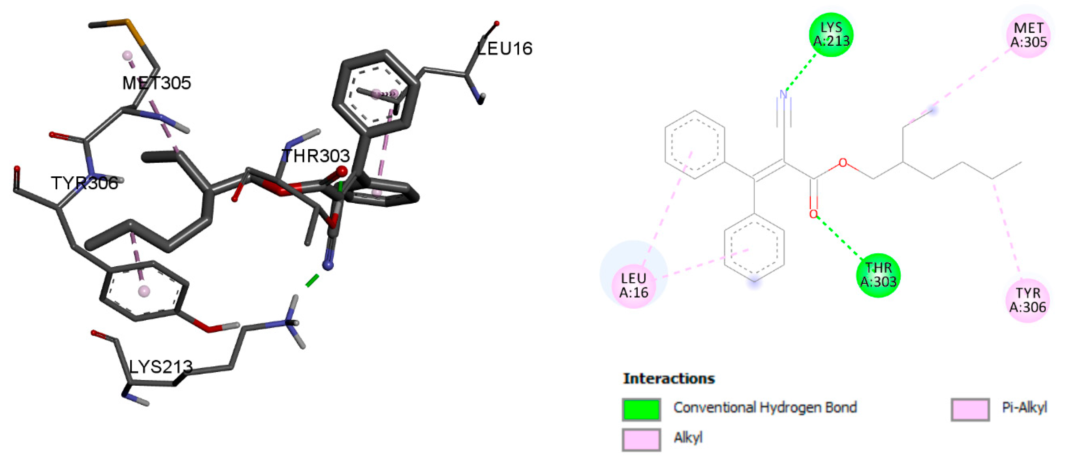
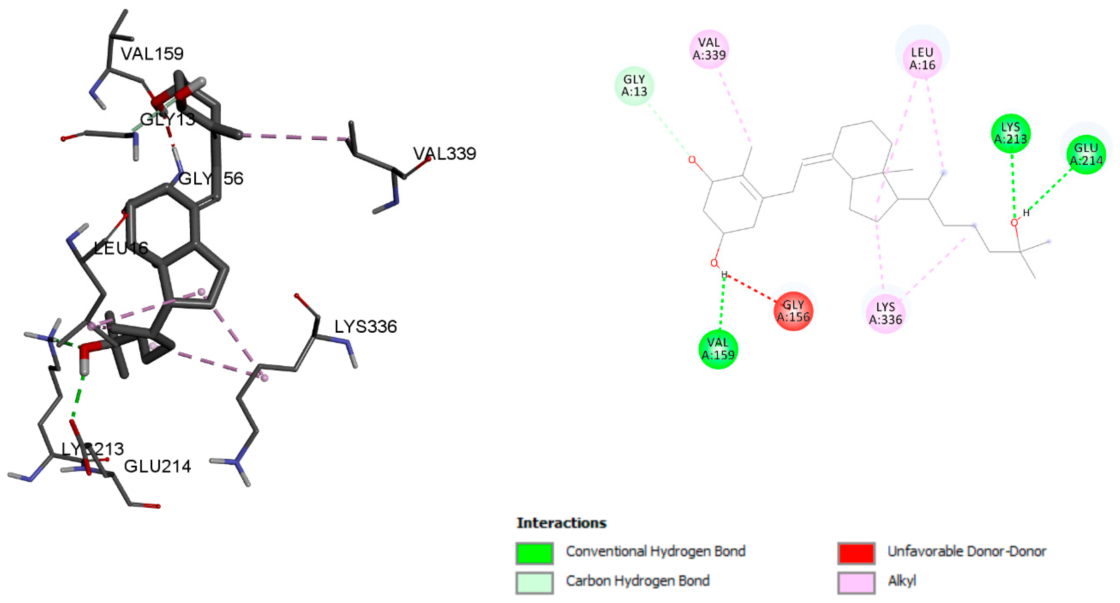
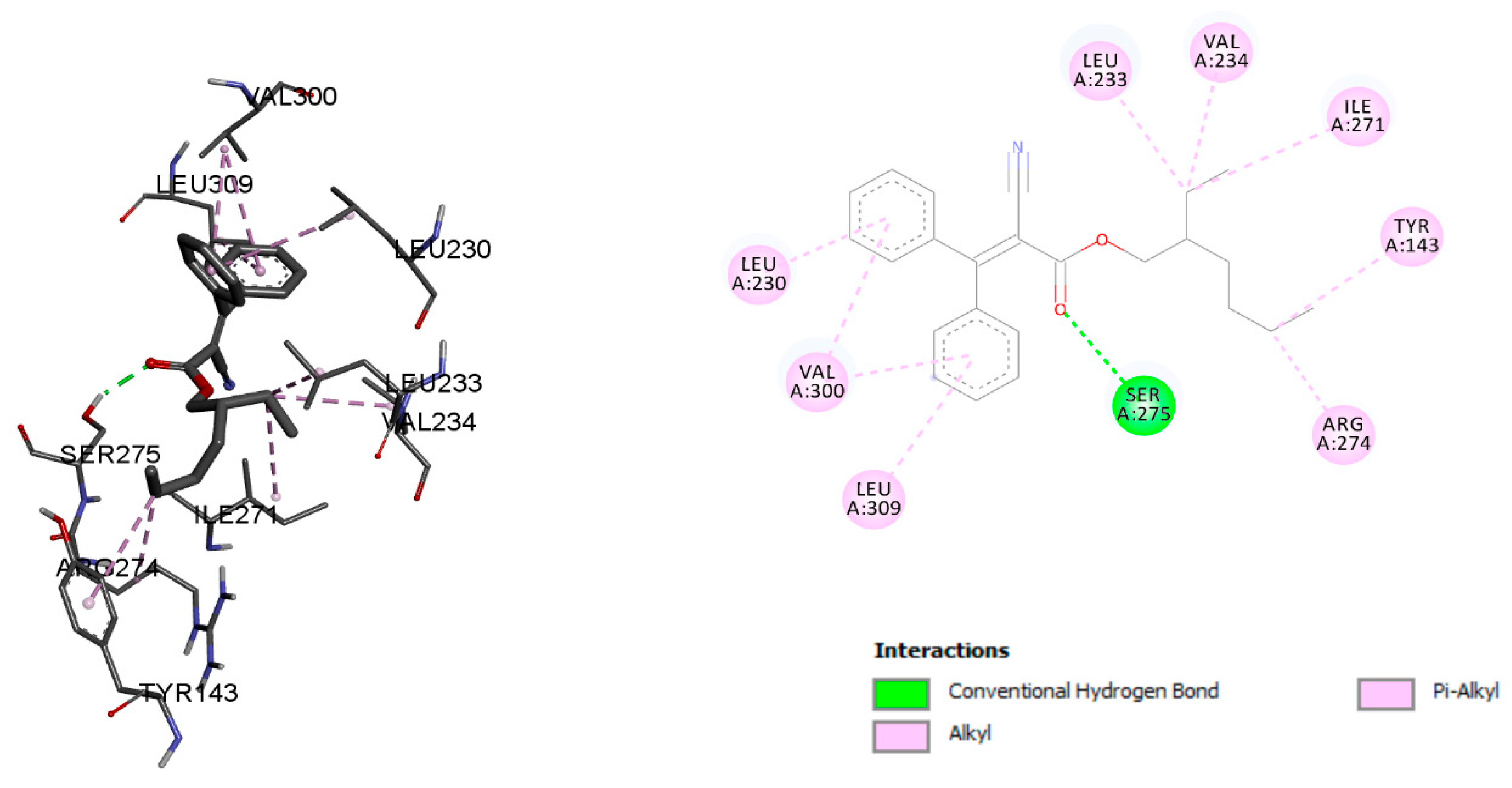
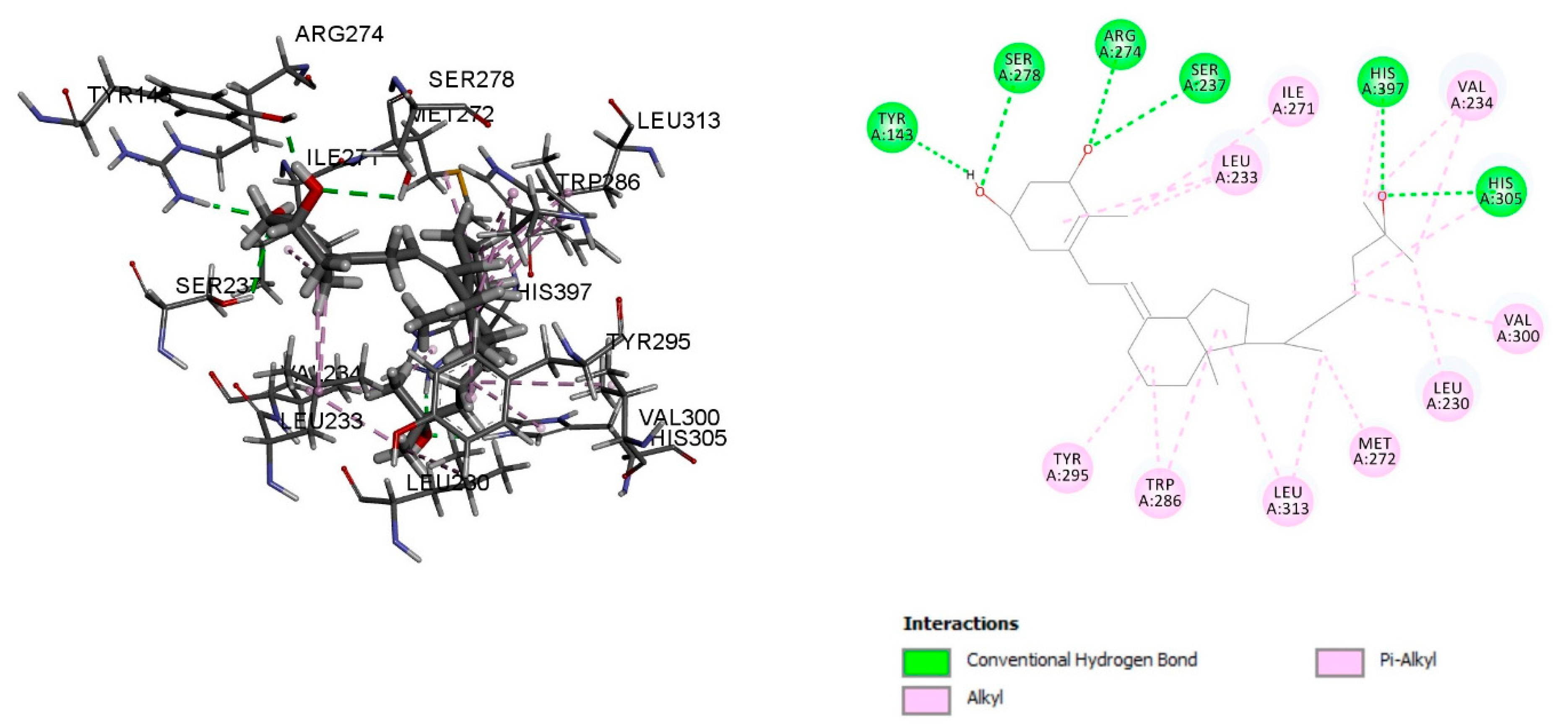
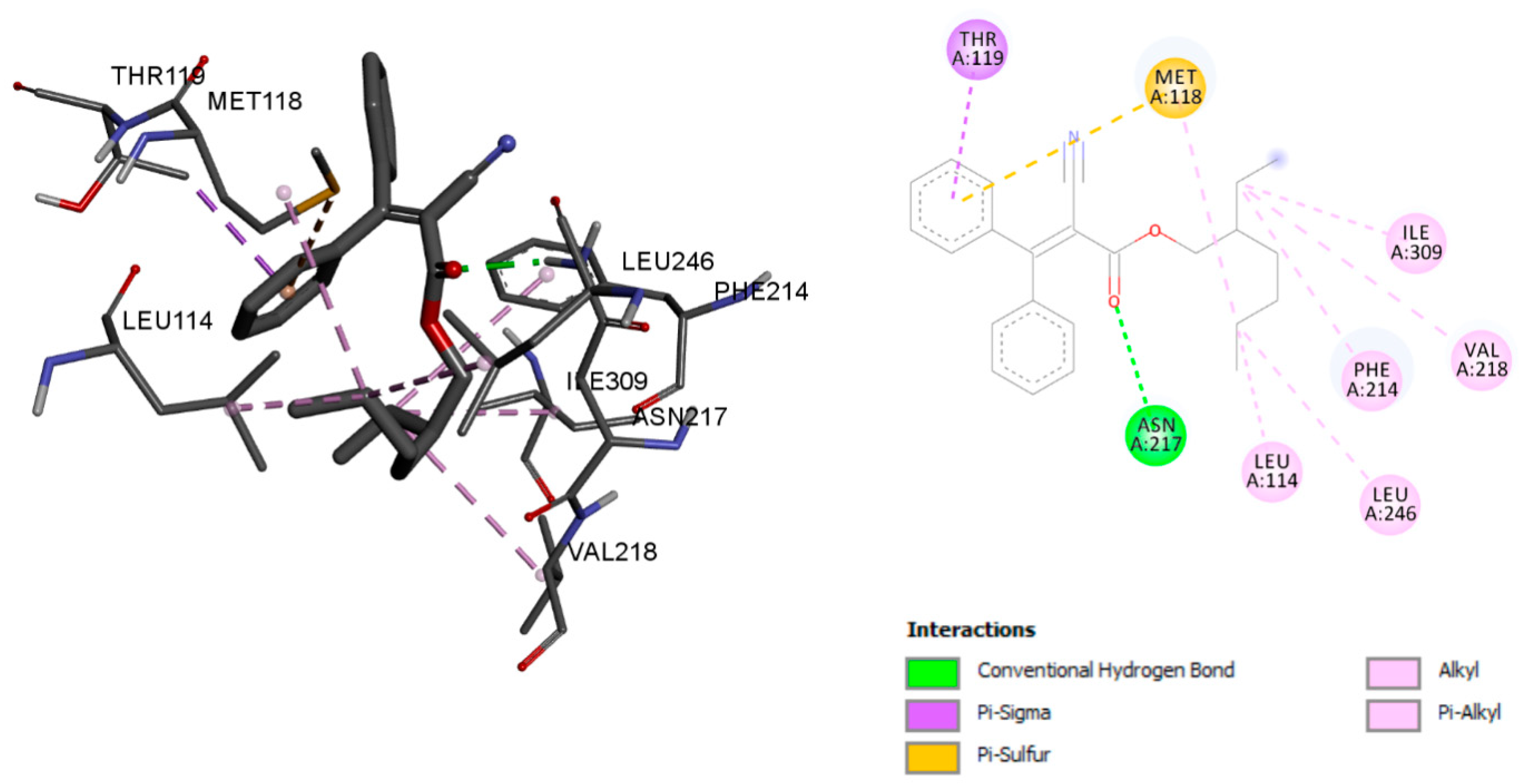
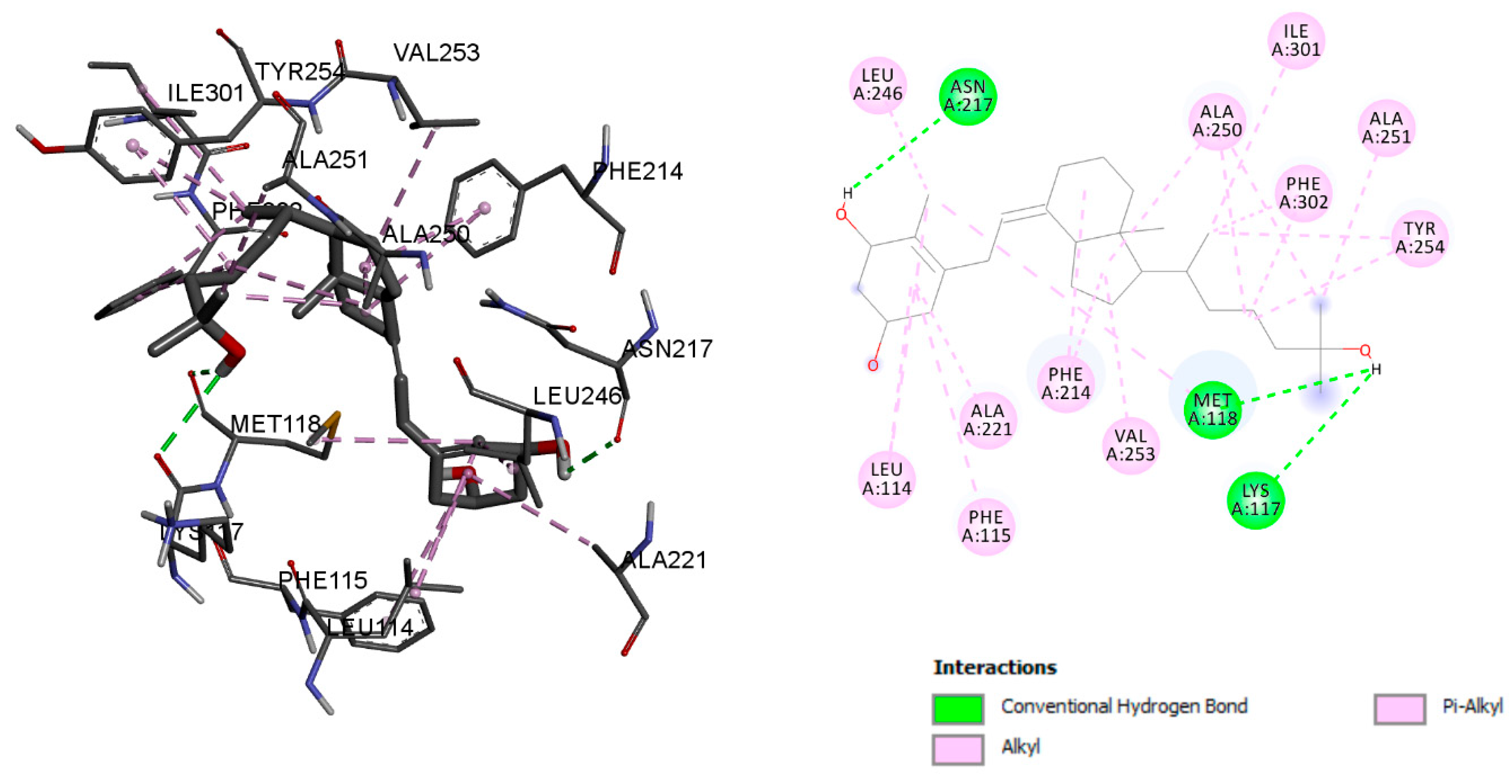
2.1. Molecular Docking Analyses of Octocrylene in Comparison with Calcitriol for the Vitamin D Binding Protein
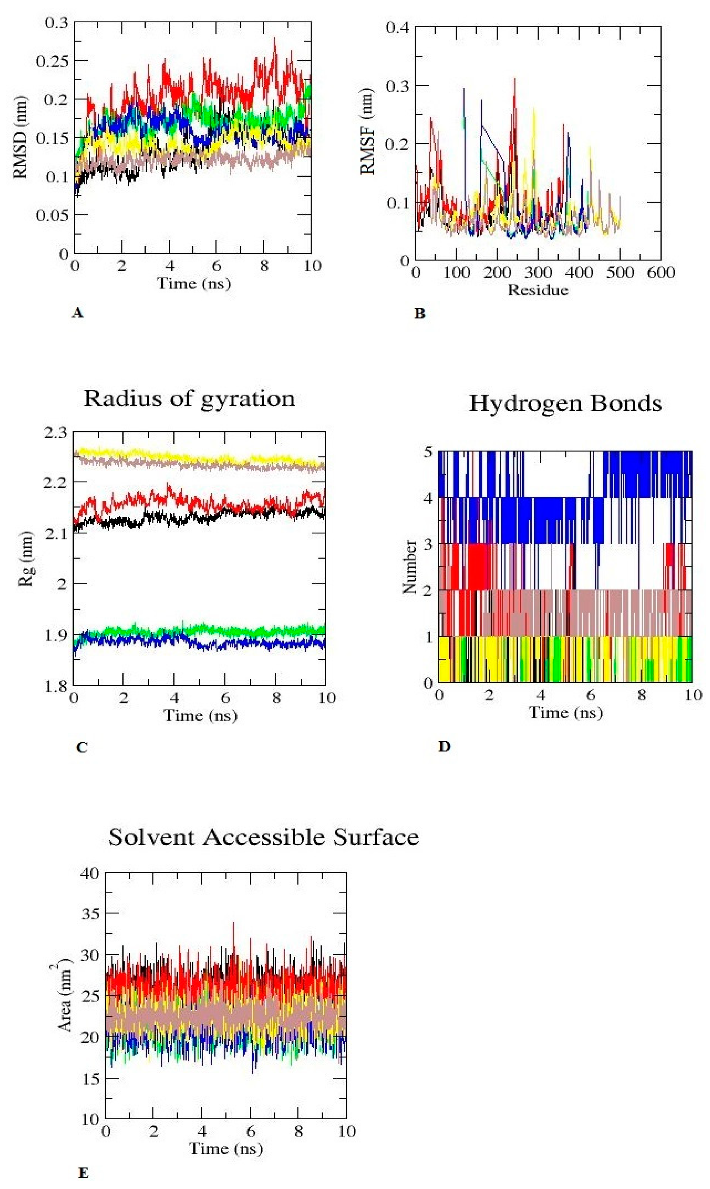
2.2. MD Simulation Analyses
2.3. Molecular Mechanics Poisson–Boltzmann Surface Area (MM-PBSA) and Interaction Energy of Octocrylene and Calcitriol
2.4. ADME (Absorption, Distribution, Metabolism, and Excretion) Analyses
3. Discussion
4. Materials and Methods
4.1. Structural Preparation for Molecular Docking
4.2. Molecular Dynamic (MD) Simulation for Stability Assessment of Octocrylene with Respective Protein
4.3. Analysis of Trajectory
4.4. Interaction Energy of Octocrylene and Calcitriol
4.5. Molecular Mechanics Poisson–Boltzmann Surface Area (MM-PBSA)
4.6. ADME Analyses
5. Conclusions
Author Contributions
Funding
Data Availability Statement
Acknowledgments
Conflicts of Interest
References
- Egambaram, O.P.; Kesavan Pillai, S.; Ray, S.S. Materials Science Challenges in Skin UV Protection: A Review. Photochem. Photobiol. 2020, 96, 779–797. [Google Scholar] [CrossRef]
- Hanson, K.M.; Gratton, E.; Bardeen, C.J. Sunscreen Enhancement of UV-Induced Reactive Oxygen Species in the Skin. Free Radic. Biol. Med. 2006, 41, 1205–1212. [Google Scholar] [CrossRef] [PubMed]
- Matsuoka, L.Y.; Wortsman, J.; Hanifan, N.; Holick, M.F. Chronic Sunscreen Use Decreases Circulating Concentrations of 25-Hydroxyvitamin D. A Preliminary Study. Arch. Dermatol. 1988, 124, 1802–1804. [Google Scholar] [CrossRef] [PubMed]
- Gilaberte, Y.; Carrascosa, J.M. Sun Protection in Children: Realities and Challenges. Actas Dermo-Sifiliográficas 2014, 105, 253–262. [Google Scholar] [CrossRef] [PubMed]
- RPPL No. 10–30: The Responsible Tourism Education Act of 2018 2019. Available online: https://www.palaugov.pw/wp-content/uploads/2018/10/RPPL-No.-10-30-re.-The-Responsible-Tourism-Education-Act-of-2018.pdf (accessed on 14 August 2022).
- Balmer, M.E.; Buser, H.-R.; Müller, M.D.; Poiger, T. Occurrence of Some Organic UV Filters in Wastewater, in Surface Waters, and in Fish from Swiss Lakes. Environ. Sci. Technol. 2005, 39, 953–962. [Google Scholar] [CrossRef] [PubMed]
- Matta, M.K.; Zusterzeel, R.; Pilli, N.R.; Patel, V.; Volpe, D.A.; Florian, J.; Oh, L.; Bashaw, E.; Zineh, I.; Sanabria, C.; et al. Effect of Sunscreen Application Under Maximal Use Conditions on Plasma Concentration of Sunscreen Active Ingredients. JAMA 2019, 321, 2082. [Google Scholar] [CrossRef]
- Hiller, J.; Klotz, K.; Meyer, S.; Uter, W.; Hof, K.; Greiner, A.; Göen, T.; Drexler, H. Systemic Availability of Lipophilic Organic UV Filters through Dermal Sunscreen Exposure. Environ. Int. 2019, 132, 105068. [Google Scholar] [CrossRef]
- Downs, C.A.; DiNardo, J.C.; Stien, D.; Rodrigues, A.M.S.; Lebaron, P. Benzophenone Accumulates over Time from the Degradation of Octocrylene in Commercial Sunscreen Products. Chem. Res. Toxicol. 2021, 34, 1046–1054. [Google Scholar] [CrossRef]
- Blüthgen, N.; Meili, N.; Chew, G.; Odermatt, A.; Fent, K. Accumulation and Effects of the UV-Filter Octocrylene in Adult and Embryonic Zebrafish (Danio rerio). Sci. Total Environ. 2014, 476, 207–217. [Google Scholar] [CrossRef]
- Yan, S.; Liang, M.; Chen, R.; Hong, X.; Zha, J. Reproductive Toxicity and Estrogen Activity in Japanese Medaka (Oryzias latipes) Exposed to Environmentally Relevant Concentrations of Octocrylene. Environ. Pollut. 2020, 261, 114104. [Google Scholar] [CrossRef]
- Pawlowski, S.; Lanzinger, A.C.; Dolich, T.; Füßl, S.; Salinas, E.R.; Zok, S.; Weiss, B.; Hefner, N.; Van Sloun, P.; Hombeck, H.; et al. Evaluation of the Bioaccumulation of Octocrylene after Dietary and Aqueous Exposure. Sci. Total Environ. 2019, 672, 669–679. [Google Scholar] [CrossRef]
- Treffel, P.; Gabard, B. Skin Penetration and Sun Protection Factor of Ultra-Violet Filters from Two Vehicles. Pharm. Res. 1996, 13, 770–774. [Google Scholar] [CrossRef] [PubMed]
- Walters, K.A.; Roberts, M.S. Percutaneous Absorption of Sunscreens. In Topical Absorption of Dermatological Products, 21st ed.; CRC Press: Boca Raton, FL, USA, 2002; ISBN 9780429207556. [Google Scholar]
- Schneider, S.L.; Lim, H.W. Review of Environmental Effects of Oxybenzone and Other Sunscreen Active Ingredients. J. Am. Acad. Dermatol. 2019, 80, 266–271. [Google Scholar] [CrossRef] [PubMed]
- Neale, R.E.; Khan, S.R.; Lucas, R.M.; Waterhouse, M.; Whiteman, D.C.; Olsen, C.M. The Effect of Sunscreen on Vitamin D: A Review. Br. J. Dermatol. 2019, 181, 907–915. [Google Scholar] [CrossRef] [PubMed]
- Cheng, J.B.; Levine, M.A.; Bell, N.H.; Mangelsdorf, D.J.; Russell, D.W. Genetic Evidence That the Human CYP2R1 Enzyme Is a Key Vitamin D 25-Hydroxylase. Proc. Natl. Acad. Sci. USA 2004, 101, 7711–7715. [Google Scholar] [CrossRef] [PubMed]
- Sizar, O.; Khare, S.; Goyal, A.; Givler, A. Vitamin D Deficiency. In StatPearls [Internet]; StatPearls: Treasure Island, FL, USA, 2022. Available online: https://www.ncbi.nlm.nih.gov/books/NBK532266/ (accessed on 18 July 2022).
- Bury, D.; Belov, V.N.; Qi, Y.; Hayen, H.; Volmer, D.A.; Brüning, T.; Koch, H.M. Determination of Urinary Metabolites of the Emerging UV Filter Octocrylene by Online-SPE-LC-MS/MS. Anal. Chem. 2018, 90, 944–951. [Google Scholar] [CrossRef]
- Hernández Leal, L.; Vieno, N.; Temmink, H.; Zeeman, G.; Buisman, C.J.N. Occurrence of Xenobiotics in Gray Water and Removal in Three Biological Treatment Systems. Environ. Sci. Technol. 2010, 44, 6835–6842. [Google Scholar] [CrossRef]
- Liu, H.; Liu, L.; Xiong, Y.; Yang, X.; Luan, T. Simultaneous Determination of UV Filters and Polycyclic Musks in Aqueous Samples by Solid-Phase Microextraction and Gas Chromatography–Mass Spectrometry. J. Chromatogr. A 2010, 1217, 6747–6753. [Google Scholar] [CrossRef]
- Morris, G.M.; Goodsell, D.S.; Halliday, R.S.; Huey, R.; Hart, W.E.; Belew, R.K.; Olson, A.J. Automated Docking Using a Lamarckian Genetic Algorithm and an Empirical Binding Free Energy Function. J. Comput. Chem. 1998, 19, 1639–1662. [Google Scholar] [CrossRef]
- Morris, G.M.; Huey, R.; Lindstrom, W.; Sanner, M.F.; Belew, R.K.; Goodsell, D.S.; Olson, A.J. AutoDock4 and AutoDockTools4: Automated Docking with Selective Receptor Flexibility. J. Comput. Chem. 2009, 30, 2785–2791. [Google Scholar] [CrossRef] [Green Version]
- Dassault Systemes BIOVIA. 2019. Available online: https://www.3dsbiovia.com/about/citations-references (accessed on 14 August 2022).
- Bjelkmar, P.; Larsson, P.; Cuendet, M.A.; Hess, B.; Lindahl, E. Implementation of the CHARMM Force Field in GROMACS: Analysis of Protein Stability Effects from Correction Maps, Virtual Interaction Sites, and Water Models. J. Chem. Theory Comput. 2010, 6, 459–466. [Google Scholar] [CrossRef] [PubMed]
- Lee, H.-C.; Hsu, W.-C.; Liu, A.-L.; Hsu, C.-J.; Sun, Y.-C. Using Thermodynamic Integration MD Simulation to Compute Relative Protein–Ligand Binding Free Energy of a GSK3β Kinase Inhibitor and Its Analogs. J. Mol. Graph. Model. 2014, 51, 37–49. [Google Scholar] [CrossRef] [PubMed]
- Van Der Spoel, D.; Lindahl, E.; Hess, B.; Groenhof, G.; Mark, A.E.; Berendsen, H.J.C. GROMACS: Fast, Flexible, and Free. J. Comput. Chem. 2005, 26, 1701–1718. [Google Scholar] [CrossRef] [PubMed]
- Lemkul, J. From Proteins to Perturbed Hamiltonians: A Suite of Tutorials for the GROMACS-2018 Molecular Simulation Package [Article v1.0]. Living J. Comput. Mol. Sci. 2019, 1, 5068. [Google Scholar] [CrossRef]
- Kuzmanic, A.; Zagrovic, B. Determination of Ensemble-Average Pairwise Root Mean-Square Deviation from Experimental B-Factors. Biophys. J. 2010, 98, 861–871. [Google Scholar] [CrossRef] [PubMed]
- Homeyer, N.; Gohlke, H. Free Energy Calculations by the Molecular Mechanics Poisson−Boltzmann Surface Area Method. Mol. Inform. 2012, 31, 114–122. [Google Scholar] [CrossRef] [PubMed]


| SN | Product Name | Octocrylene Concentration | Information Source |
|---|---|---|---|
| 1 | Nivea Sun Item weight 200 mL | Octocrylene 50 mg | Available online: https://www.amazon.de/Nivea-Feuchtigkeits-Sonnenlotion-200mL-Badartikel/dp/B000PE8B16 (Accessed on 14 August 2022) |
| 2 | Bana Boat Sport performance Item weight 172 gm | Octocrylene 13.76 mg | Available online: https://www.amazon.in/Banana-Boat-Performance-Sunscreen-Lotion/dp/B00UNCZ1SG (Accessed on 14 August 2022) |
| 3 | Bana Boat Item weight 18 gm | Octocrylene 1.33 mg | Available online: https://www.amazon.in/Banana-Boat-Sunscreen-Perfomance-Spectrum/dp (Accessed on 14 August 2022) |
| 4 | Neutrogena Sunscreen SPF 70—6.7 Ounces Lotion 190 gm | Octocrylene 1.9 mg | Available online: https://www.amazon.in/Neutrogena-Defense-Sunscreen-Lotion-Spectrum/dp/B01N1IJA4N (Accessed on 14 August 2022) |
| 5 | Coppertone Sport Clear SPF 30 | Octocrylene 5.98 mg | Available online: https://www.amazon.in/Coppertone-Sport-Continuous-Spray-6- (Accessed on 14 August 2022) |
| 6 | Coppertone Sport Clear SPF 50 | Octocrylene 5.98 mg | Avialble online: https://www.amazon.com/Coppertone-Sunscreen-Lotion (Accessed on 14 August 2022) |
| Ligands | Amino Acid Residues Involved in Hydrogen Bonds | Docking Final Intermolecular Energy (ΔG) = vdW + Hbond + Desolv Energy (kcal/mol) | Inhibition Constant (Ki) | Protein |
|---|---|---|---|---|
| Octocrylene | LYS 213 THR 303 | −11.52 | 365.25 nM | 1KXP |
| Calcitriol | LYS 213 VAL 159 GLU 214 | −11.71 | 117.72 nM | |
| Octocrylene | SER 275 | −11.15 | 979.57 nM | 1DB1 |
| Calcitriol | SER 278 TYR 143 ARG 274 SER 237 HIS 397 HIS 305 | −8.73 | 2.99 µM | |
| Octocrylene | ASN 217 | −11.9 | 271.01 nM | 3CZH |
| Calcitriol | ASN 217 MET 118 LYS 117 | −13.03 | 25.63 nM |
| Parameters | Octocrylene + Vitamin D Binding Protein (1KXP) | Calcitriol + Vitamin D Binding Protein (1KXP) | Octocrylene + Vitamin D Receptor (1DB1) | Calcitriol + Vitamin D Receptor (1DB1) | Octocrylene + Enzyme CYP2R1 (3CZH) | Calcitriol + Enzyme CYP2R1 (3CZH) |
|---|---|---|---|---|---|---|
| van der Waal’s energy | −37.87 ± 0.133 kcal/mol | −43.35 ± 0.16 kcal/mol | −49.28± 0.07 kcal/mol | −55.09 ± 0.09 kcal/mol | −49.62 ± 0.08 kcal/mol | −54.75 ± 0.08 kcal/mol |
| Electrostatic energy | −1.8019 ± 0.03 kcal/mol | −1.699 ± 0.02 kcal/mol | −0.477 ± 0.0094 kcal/mol | −41.82± 0.1 kcal/mol | −0.752 ± 0.0087 kcal/mol | −49.62 ± 0.08 kcal/mol |
| Solvation energy | −1.8019 ± 1.2 kcal/mol | −1.91 ± 0.01 kcal/mol | −5.05 ± 0.009 kcal/mol | −41.82 ± 0.1 kcal/mol | −5.009 ± 0.0093 kcal/mol | −5.11 ± 0.008 kcal/mol |
| Binding free energy | −41.82 ± 0.1 kcal/mol | −46.96 ± 0.17 kcal/mol | −54.81 ± 0.07 kcal/mol | −63.25 ± 0.095 kcal/mol | −55.37 ± 0.086 kcal/mol | −61.06 ± 0.08 kcal/mol |
| Interaction Energy | Coul-SR: −58.22 KJ/mol | Coul-SR: −47.91 KJ/mol | Coul-SR: −21.23 KJ/mol | Coul-SR: −106.515 KJ/mol | Coul-SR: −38.62 KJ/mol | Coul-SR: −33.97 KJ/mol |
| LJ-SR: −141.58 KJ/mol | LJ-SR: −160.619 KJ/mol | LJ-SR: −185.137 KJ/mol | LJ-SR: −204.88 KJ/mol | LJ-SR: −186.002 KJ/mol | LJ-SR: −205.138 KJ/mol |
| SN | ADME Parameters | Results |
|---|---|---|
| 1 | Molecular weight | 361.48 g/mol |
| 2 | GI absorption | High |
| 3 | BBB permeant | Yes |
| 4 | Log Kp (skin permeation) | −3.44 cm/s |
| 5 | CYP1A2 inhibitor | Yes |
| 6 | CYP2C19 inhibitor | Yes |
| 7 | CYP2C9 inhibitor | Yes |
| 8 | CYP2D6 inhibitor | Yes |
| 9 | CYP3A4 inhibitor | Yes |
| Protein Data Bank | Ligand | Control |
|---|---|---|
| Vitamin D binding protein (1KXP) | ||
| Vitamin D receptor (1DB1) | Octocrylene | Calcitriol |
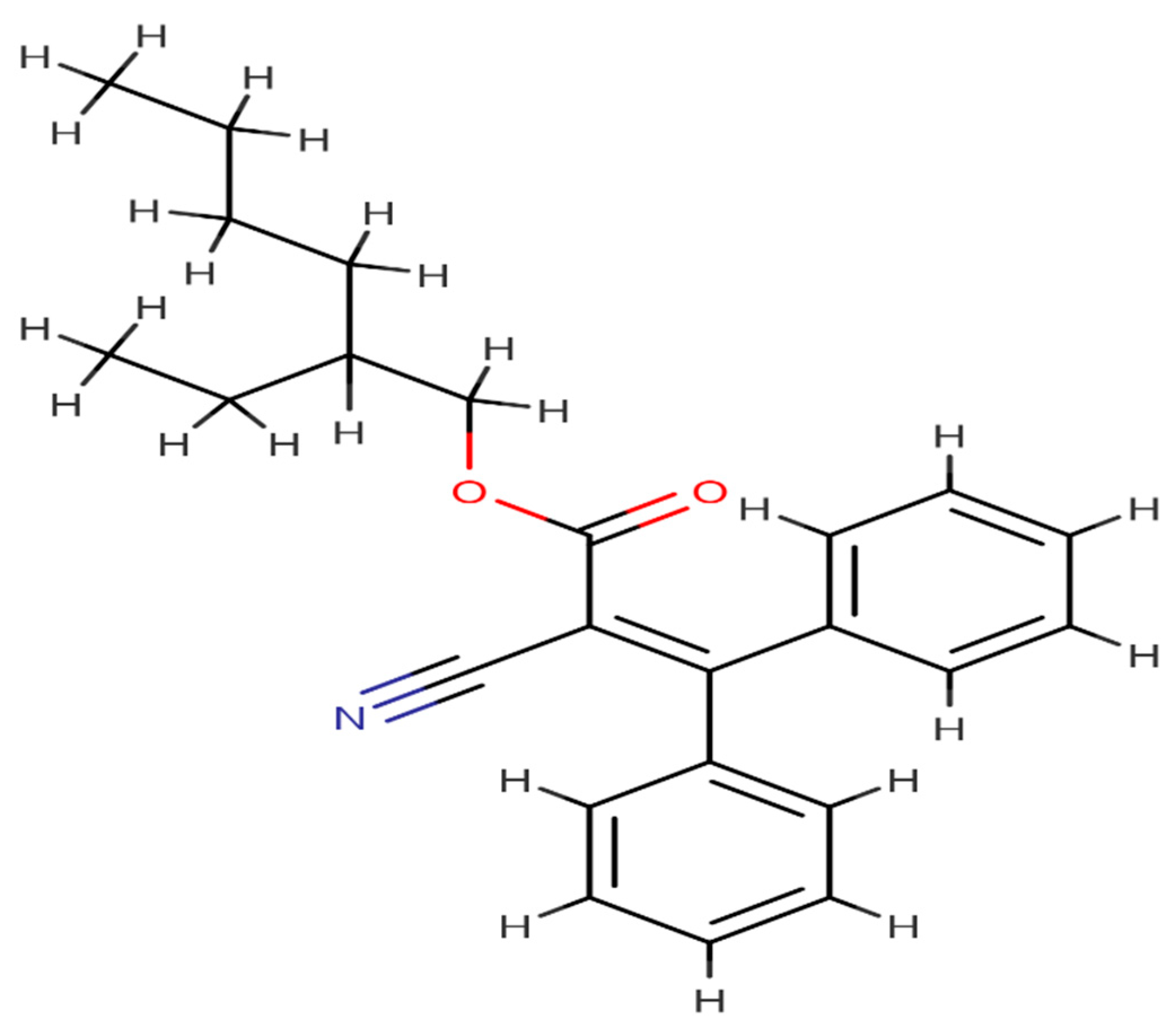
| Enzyme CYP2R1 (3CZH) | PubChem Id 22571 | PubChem Id 5280453 |
Publisher’s Note: MDPI stays neutral with regard to jurisdictional claims in published maps and institutional affiliations. |
© 2022 by the authors. Licensee MDPI, Basel, Switzerland. This article is an open access article distributed under the terms and conditions of the Creative Commons Attribution (CC BY) license (https://creativecommons.org/licenses/by/4.0/).
Share and Cite
Abdi, S.A.H.; Ali, A.; Sayed, S.F.; Nagarajan, S.; Abutahir; Alam, P.; Ali, A. Sunscreen Ingredient Octocrylene’s Potency to Disrupt Vitamin D Synthesis. Int. J. Mol. Sci. 2022, 23, 10154. https://doi.org/10.3390/ijms231710154
Abdi SAH, Ali A, Sayed SF, Nagarajan S, Abutahir, Alam P, Ali A. Sunscreen Ingredient Octocrylene’s Potency to Disrupt Vitamin D Synthesis. International Journal of Molecular Sciences. 2022; 23(17):10154. https://doi.org/10.3390/ijms231710154
Chicago/Turabian StyleAbdi, Sayed Aliul Hasan, Amena Ali, Shabihul Fatma Sayed, Sumathi Nagarajan, Abutahir, Prawez Alam, and Abuzer Ali. 2022. "Sunscreen Ingredient Octocrylene’s Potency to Disrupt Vitamin D Synthesis" International Journal of Molecular Sciences 23, no. 17: 10154. https://doi.org/10.3390/ijms231710154