Article

Large-Scale Phosphoproteomic Study of Arabidopsis Membrane Proteins Reveals Early Signaling Events in Response to Cold

1
United Graduate School of Agricultural Sciences, Iwate University, Morioka 020-8550, Japan
2
Graduate School of Bio-Applications and Systems Engineering, Tokyo University of Agriculture and Technology, Koganei 184-8588, Japan
3
Gene Discovery Research Group, RIKEN Center for Sustainable Resource Science, 3-1-1 Koyadai, Tsukuba 305-0074, Japan
4
Department of Biochemistry, Iwate Medical University, Yahaba 028-3694, Japan
5
Department of Plant-Bioscience, Faculty of Agriculture, Iwate University, Morioka 020-8550, Japan
*
Author to whom correspondence should be addressed.
Int. J. Mol. Sci. 2020, 21(22), 8631; https://doi.org/10.3390/ijms21228631
Submission received: 30 October 2020 / Revised: 12 November 2020 / Accepted: 13 November 2020 / Published: 16 November 2020
(This article belongs to the Collection Feature Papers in Molecular Plant Sciences)

Abstract

:
Cold stress is one of the major factors limiting global crop production. For survival at low temperatures, plants need to sense temperature changes in the surrounding environment. How plants sense and respond to the earliest drop in temperature is still not clearly understood. The plasma membrane and its adjacent extracellular and cytoplasmic sites are the first checkpoints for sensing temperature changes and the subsequent events, such as signal generation and solute transport. To understand how plants respond to early cold exposure, we used a mass spectrometry-based phosphoproteomic method to study the temporal changes in protein phosphorylation events in Arabidopsis membranes during 5 to 60 min of cold exposure. The results revealed that brief cold exposures led to rapid phosphorylation changes in the proteins involved in cellular ion homeostasis, solute and protein transport, cytoskeleton organization, vesical trafficking, protein modification, and signal transduction processes. The phosphorylation motif and kinase–substrate network analysis also revealed that multiple protein kinases, including RLKs, MAPKs, CDPKs, and their substrates, could be involved in early cold signaling. Taken together, our results provide a first look at the cold-responsive phosphoproteome changes of Arabidopsis membrane proteins that can be a significant resource to understand how plants respond to an early temperature drop.

1. Introduction

Throughout the Earth’s history, plants have evolved adaptive mechanisms against multiple abiotic and biotic stresses. Cold stress, which results from both chilling (0–10/15 °C) and freezing (<0 °C) conditions, is one of the major abiotic stresses that happens all over the world and affects every process in the plant life cycle, including growth, development, survival, and productivity [1]. Cold-stress responses vary with plant species. Agronomically important plants, such as rice (Oryza sativa), corn (Zea mays), banana (Musa spp.), and tomato (Lycopersicon esculentum), originated from tropical and subtropical regions and are sensitive to cold stress. [2,3,4,5]. On the other hand, plants native to temperate climatic zones can take advantage of the colder autumn temperature and prepare themselves for the upcoming freezing conditions using an adaptive strategy called cold acclimation [4,5,6]. Therefore, perception of and response to a drop in temperature is essential for plants that originated in tropic and sub-tropic regions to deploy survival mechanisms and for those in temperate climatic zones to start the acclimation processes and prepare for upcoming freezing stress. Responses to cold under acclimating and freezing conditions are studied extensively on both genomic and proteomic levels [7,8,9,10,11,12,13]. However, there is little or no information available on how plants sense temperature drops over a short time (i.e., seconds and minutes) and how they respond and form adaptive signals to the cold. Sensing early changes is an essential step for the plant to initiate the adaptive process and survive under cold temperatures. Furthermore, how plants respond to early temperature drops is necessary to develop cold-resistant/-tolerant plant varieties and secure crop supply in the global climate change era.
The cell wall (CW), the extracellular/apoplastic region, and the plasma membrane (PM) are the outermost layers of a plant cell. Therefore, upon exposure to external environmental stimuli (biotic and abiotic), these layers are the most likely to receive, generate, and transmit signals further downstream and ultimately to the nuclei to form an appropriate response [14,15,16,17]. PM is the outermost living part of the cell and has received the most attention for cold response studies in plants [7,8,10,11,13,18,19]. Few studies have also emerged on CW modifications during prolonged cold acclimation and in freezing response [18,20,21]. These studies indicate that the outer layers work in concert, as the first checkpoint to sense and generate signals from cold and other environmental factors, by changing their physicochemical conformation and composition and utilizing the power of posttranslational modifications (PTMs), such as phosphorylation of receptor-like kinases (RLKs) and other membrane-localized kinases [19,22,23,24,25]. Within 30 min to cold exposure at 4 °C, the PM rigidifies and increases Ca2+ influx into the cell, which in turn activates Ca2+/calmodulin (CaM)-regulated receptor-like kinase1 (CRLK1), leading to mitogen-activated protein kinase (MAPK) pathway activation via a chain of phosphorylation events [23,26]. The MAPK pathway is one of the canonical signaling pathways that enhance freezing tolerance in Arabidopsis [25,27]. A phosphoproteomic study of cold-tolerant and cold-sensitive bananas (Musa spp.) showed that Thr31 phosphorylation of MAPK kinase 2 (MKK2) is an essential component of the cold tolerance mechanism [28]. CRLK1, MEKK1 (MAPK kinase kinase 1), and MKK2 are all reported to be localized in the PM [29,30]. Similarly, another membrane-localized kinase, named cold-responsive protein kinase 1 (CPPK1), was activated upon prolonged cold exposure and directly sent a signal to the nucleus to fine-tune the C-repeated binding factor (CBF) signaling pathway by phosphorylating its 14-3-3 protein substrate [31]. Another membrane-bound receptor-like kinase, receptor-like protein kinase 1 (RPK1), also increase cold tolerance along with tolerance to other abiotic stresses [32]. CW, apoplast, PM-bound and localized RLKs, and transmembrane kinases (TMKs), and their signaling events, are better documented in biotic stresses than abiotic stresses [21,33]. Therefore, further studies are needed to discover cold-stress-responsive RLKs and other protein kinases that are localized in the membranes and membrane-adjacent extracellular spaces.
Furthermore, these examples strongly indicate that protein phosphorylation of membrane-bound kinases and their targets (e.g., transporters, ion channels, and transcription factors) plays a crucial role in cold and other abiotic stress-triggered molecular events. Along with stress signal transduction, protein phosphorylation/dephosphorylation is also involved in regulating diverse biological and molecular functions, such as cell division, differentiation, gene expression, hormone perception, homeostasis, and metabolism. Therefore, protein phosphorylation is one of the most important PTMs in plants [34,35,36,37]. Signals for all the processes mentioned above originate at the PM and its adjacent regions either by a change in its physicochemical properties or by phosphorylation of the kinases associated with these regions. Therefore, studying and profiling the membrane-localized phosphoproteome and kinome on a large scale at earlier stages of cold exposure will help us to understand the plant’s response to cold and other signaling events related to cold response.
Mass spectrometry (MS)-based shotgun phosphoproteomics has emerged as a powerful analytical tool to study protein phosphorylation on a large scale. MS-based phosphoproteomics has become a standard method to study global changes in phosphorylation dynamics [17,38,39,40,41,42]. Although significant technical improvements have been made to study the global or total phosphoproteins, characterization of membrane phosphoproteins is still challenging. A low abundance, short-lived dynamic signaling events under specific conditions, contamination from other non-membrane proteins, and the overabundance of non-phosphopeptides are a few of the major roadblocks in studying membrane phosphoproteins [36,43,44]. Here, to improve the detection of low-abundance membrane phosphopeptides, we used a well-established membrane protein extraction method [45] with minor modifications and enriched phosphopeptides with TiO2. We further analyzed the TiO2-enriched membrane phosphopeptides using a high-resolution mass spectrometer coupled with a long monolithic column to improve the separation capacity. We prepared the microsomal membrane fractions (MMFs) from two-week-old Arabidopsis aerial parts after exposure to 2 °C for short periods (i.e., 5, 15, 30, or 60 min). Using these samples, we successfully identified the phosphorylated proteins from different membrane-localized kinases and their substrates and analyzed their dynamics upon short cold exposure.

2. Results

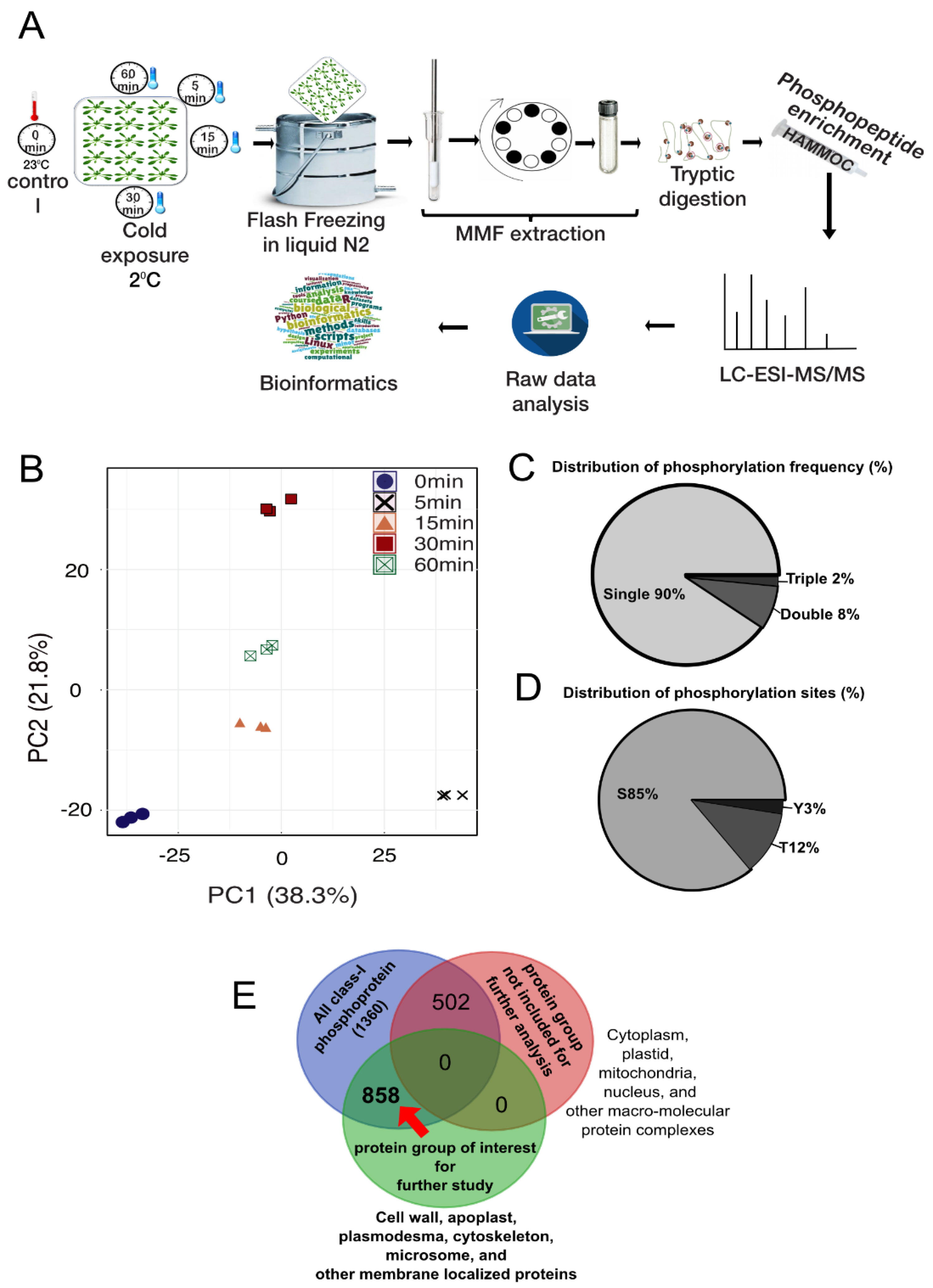
2.1. Description of the Phosphoproteomic Data of Early Cold Treatment in Arabidopsis

The purpose of this study is to elucidate the signaling events in response to cold stress in the model plant Arabidopsis by a time-course (0, 5, 15, 30, and 60 min)-based phosphoproteomic study (Figure 1A). We identified a total of 2795 unique phosphopeptides, which were phosphorylated in at least two biological replicates at all five time points, and these phosphopeptides constitute a total of 1360 proteins (Table S1A,B). We performed a principal component analysis (PCA) to evaluate the quality of the biological replicates at each time point. PCA analysis showed all biological replicates at each time point cluster together, and simultaneously, were separated from each other (Figure 1B). Furthermore, about 90% of these phosphopeptides were singly phosphorylated (Figure 1C). From the identified phosphopeptides, a total of 2930 phosphosites were enriched, where the most frequent phosphorylation was observed in serine (S, 85%), followed by in threonine (T, 12%) and tyrosine (Y, 3%) (Figure 1D). The frequency distribution of the phosphorylation sites was similar to those previously reported in plant phosphoproteomic studies [44,45,46,47,48,49,50].
The MMF used in the present study is a mixture of various cellular membranes and compartments and enriched in lighter membranes such as the PM, endoplasmic reticulum, Golgi body, and vacuole, but it also contains fractions from the extracellular space, cell wall, apoplast, cytoskeleton, plasmodesmata, cytosol, mitochondria, plastid, nucleus, and other cellular compartments. Because the objective of our study was to observe the phosphorylation-level changes in proteins potentially involved in the early cold response in the MMF, we performed an over-representation analysis on the cellular localization of the proteins and divided them into two groups based on significant enrichment (Fisher test, p < 0.05): (A) the protein group of interest, which includes proteins from the PM, endoplasmic reticulum, Golgi body, vacuole, extracellular space, cell wall, apoplast, cytoskeleton, and plasmodesmata; and (B) the sub-portion of proteins from the cytosol, mitochondria, plastid, nucleus, and macromolecular complexes. Out of 1360 phosphoproteins, 858 were from Group A. Our detailed study in the following sections only included group A proteins, and the proteins from Group B were not included in further analyses (Figure 1E). Group A contains 1821 phosphopeptides and 1894 phosphosites. Next, we analyzed the relative changes in the phosphopeptides after cold exposure compared to the control (0 min cold exposure) in a time-course manner (Table S1B).

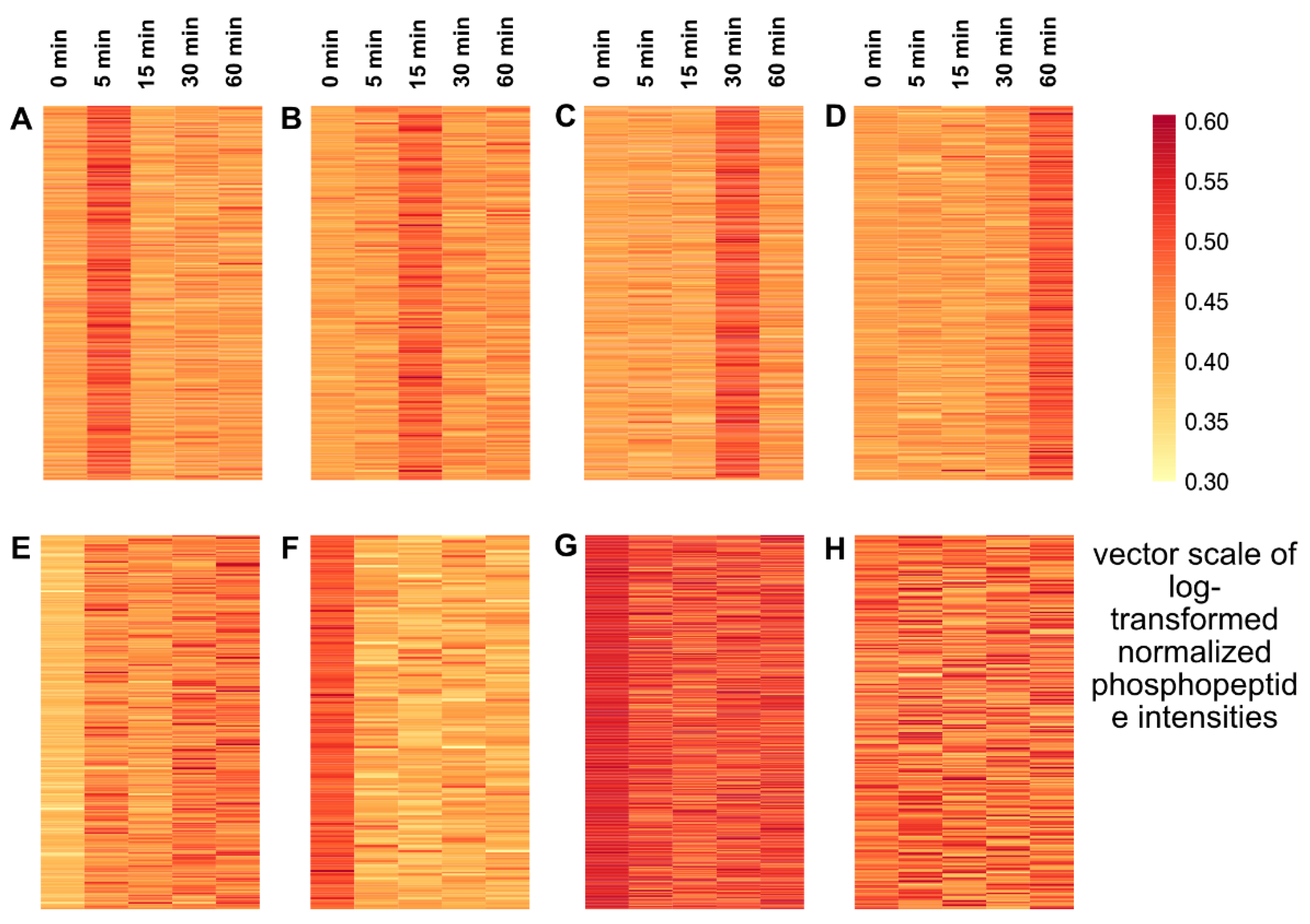
2.2. Temporal Profile of the Phosphopeptides in Response to Cold

Previous phosphoproteomic studies of osmotic stress and systemin treatment reported that the total protein abundance does not change within a time frame of minutes; instead, the peptide abundance comes from the changes in phosphorylation dynamics [42,48,51]. To determine whether this was the case under our experimental conditions, we performed a proteomic study on plants exposed to cold for up to 60 min. We did not see any significant changes in protein abundance (Table S2). Therefore, the changes in peptide abundance we observed in this study were due to changes in phosphorylation itself rather than de novo protein synthesis.
We next studied the phosphorylation dynamics upon cold exposure using K-means clustering of the log-transformed normalized phosphopeptides from the Group A proteins. The temporal dynamics of phosphopeptides created eight characteristic K-means clusters (Figure 2). Upon cold exposure, a large number (481) of peptides were dephosphorylated (Figure 2, Clusters F and G). Among the dephosphorylated peptides, 140 peptides stayed in the dephosphorylated state relative to the control throughout the cold exposure period (Figure 2, Cluster F). Cluster G, which contains 341 peptides, showed a fluctuation in its dephosphorylation pattern, but none of these peptides showed increased phosphorylation relative to the control. Next, four characteristic clusters were found where peptide phosphorylation transiently peaked at each of the four cold exposure time points (Figure 2, Clusters A, B, C, D, and E). Cluster A peaked very early at 5 min with 243 phosphopeptides, and Cluster B with a peak at 15 min contained 177 phosphopeptides. We then grouped these two clusters as “early response” to cold exposure. The two clusters C (285 phosphopeptides) and D (209 phosphopeptides) peaked at later time points, 30 and 60 min, respectively. In Cluster E, peptides were immediately phosphorylated upon cold exposure and maintained phosphorylation states throughout the cold exposure period. Even though the peptides in this group (Cluster E) showed continuous phosphorylation, their peak intensities fluctuated at different time points. In further steps, we redistributed 177 phosphopeptides from Cluster E to Clusters A and B (early response) and Clusters C and D (late response) based on their highest peak intensities at each time point during cold exposure. Cluster H, containing 230 phosphopeptides, did not show any changes upon cold treatment compared to the control; it was classified as “unresponsive” and was excluded from further analysis. For quantitative analysis, we compared significant (Student’s t-test, p-value < 0.05) fold changes (cold exposed/non-exposed: 0.5 ≤ log2FC ≥ 1) for individual phosphorylated/dephosphorylated peptides in each cluster and visualized them in a heatmap (Figure S1). All the significantly (p-value < 0.05) phosphorylated peptides (1009) in the early and late response groups were subjected to gene ontology over-representation analysis (ORA) of molecular function (GOMF) and biological processes (GOBP), and motif analysis.

2.3. Cold Signal Responsive Molecular Functions in Arabidopsis: GOMF

To gain insight into the biological relevance of phosphorylation, we performed an over-representation analysis (Fisher exact test) in the early response and late response group proteins. The overrepresented GO terms revealed characteristic shifts in the molecular function (GOMF) of these phosphoproteins in response to cold exposure. Proteins involved in binding, catalytic activity, signaling, and transport were phosphorylated upon cold exposure (Figure 3). Binding proteins in the early response group were mostly phospholipid, calmodulin, clathrin, calcium, and other ion-binding phosphoproteins. Interestingly, ATP, chloride ions, the cytoskeleton, and microtubule-binding phosphoproteins were highly abundant in the late response clusters, and nucleic acid binding phosphoproteins were over-represented in both clusters (Figure 3A). Phosphoproteins with catalytic/hydrolase activity were significantly over-represented in the late response clusters, except for phosphoproteins related to serine/threonine phosphatase activity (Figure 3B). Proteins related to blue light photoreceptor activity were in the phosphorylated state throughout the cold exposure period. Signal-transducing phosphoproteins were enriched only within 15 min of cold exposure, and soluble N-ethylmaleimide-sensitive factor (NSF) adaptor protein (SNAP) receptors were only phosphorylated in the late response group (Figure 3C). Signal transduction related proteins (kinase/transferase/transducers) were also significantly overrepresented after 30 min of cold stress; however, serine/threonine/tyrosine kinase, palmitoyltransferase, and galactosyltransferase activity-related phosphoproteins were only found in the early response group (Figure 3D). Transport and transmembrane transport-related phosphoproteins were enriched in the early and late response clusters, where transmembrane transporters involved in auxin influx and ion/cation/carbohydrate transportation were only phosphorylated within 15 min of cold exposure (Figure 3D). Transporters and transmembrane transporters involved in auxin efflux, auxin proton symporter, anion channel activity, and ATPase-mediated transportation were highly enriched in the late cold response clusters (Figure 3E). In brief, these results show a large number of proteins involved in important molecular functions are temporally phosphorylated under cold exposure.

2.4. Cold Signal Responsive Biological Processes in Arabidopsis: GOBP

Changes in the molecular function of a gene product, such as protein or a group of proteins, in response to external or internal stimuli, initiate a collection of molecular events in different locations (cellular components) relative to the cell. These changes ultimately lead to a collective response from the cells, tissues, organs, and even the organism itself, contributing to a broader biological response/process [52,53]. To understand how the protein functionally changes due to phosphorylation upon cold exposure, we performed an over-representation analysis of the biological processes (GOBP) in early and late response clusters (Figure 4 and Figure S2). Proteins involved in phosphorylation and signal transduction were over-represented in both early and late response to cold exposure; however, these processes were highly abundant in the late response group compared to the early response group (Figure 4A). A large group of proteins involved in trafficking and transport-related events (ion transport, vesicle-mediated transport, protein/peptide, and carbohydrate transport) was differentially overrepresented in response to cold (Figure 4B), such as ion transport-related proteins phosphorylated in an early responsive manner, except for chloride ion transport. Vesicle-mediated transport-related proteins were over-represented throughout the cold exposure. However, proteins involved in Golgi-to-PM transport, exocyst assembly, exocytosis in vesicle docking, and receptor-mediated endocytosis were only enriched in the early response clusters. Interestingly, exocytosis, vesicle docking, and transport proteins targeting the PM were exclusively found in the late response clusters. Protein and glucose transport-related protein phosphorylation events took place during early cold exposure, but transmembrane transport, intracellular protein transport, peptide transport, and secretory protein transport-related processes were enriched in the late cold exposure. Auxin-related processes were most abundant in the late response clusters, including polar transport, basipetal transport, efflux, and auxin homeostasis. Proteins that play a role in maintaining homeostasis, such as carbohydrate homeostasis and pH regulation, responded to cold early (Figure 4C). Biological processes related to phytohormones, such as indole butyric acid, cytokinin, and gibberellic acid, were exclusive in the late response clusters, whereas the ethylene inhibitor, cobalt-ion-responsive phosphoproteins were only found in the early response clusters. Interestingly, the abscisic acid (ABA)-responsive proteins showed an increase in phosphorylation with cold exposure duration (Figure 4D). Proteins involved in responding to external stimuli, such as biotic stress and abiotic stress, including blue light, cold stress, and osmotic stress response, were significantly over-represented in the early response group. With a few exceptions, such as red light-responsive proteins, proteins involved in cold acclimation, and cellular response to heat, phototropism and gravitropism were found in the late response clusters (Figure 4E and Figure S2). Defense response-related protein phosphorylation was also abundant in the early response clusters, and interestingly cell death, negative response to cell death, and negative regulation of defense response-related proteins were highly enriched in the early response group. However, proteins responsible for CW callose deposition in response to stress were only found in the late response clusters (Figure 4E). Another interesting group of proteins phosphorylated under cold exposure belonged to the cytoskeleton/actin filament/microtubule-based process-related proteins. Most of the proteins from this cellular process were highly abundant in the late response clusters, except for the proteins involved the actin-filament severing process (Figure 4E). Proteins involved in other cellular/metabolic processes were mostly phosphorylated in late response to cold (Figure S2). In summary, brief temporal exposure to cold results in dynamic changes in a wide range of biological processes in Arabidopsis. These biological processes may be involved in early cold perception.

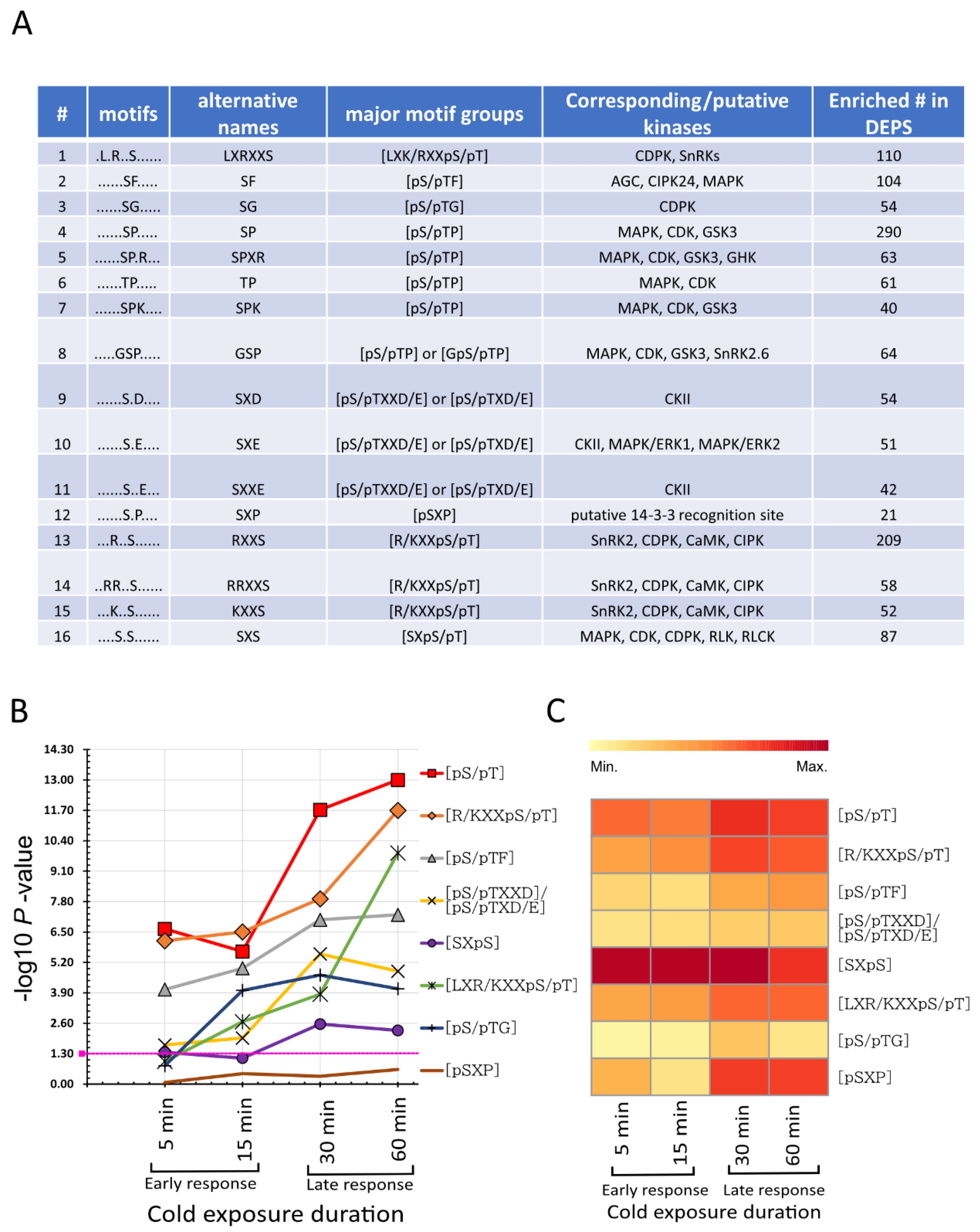
2.5. Cold-Responsive Phosphorylation Kinase Motifs

We performed a motif analysis using all the differentially phosphorylated phosphosites (0.5 ≤ log2FC ≥ 1; p < 0.5) to gain insight into which kinases are involved in the early cold response. The Motif-x algorithm extracted 16 characteristic kinase motifs, where 15 were serine centered, and 1 was threonine centered, but no tyrosine-centered motifs were enriched (Figure 5A). These motifs were further grouped into eight major motif types, according to van Wijk et al. (Figure 5A) [54]. The SP or [pS/pTP]-type motif group in this study consisted of [pSP], [pTP], [GpSP], [pSPXR], and [pSPK] motifs. The RXXS or [R/KXXpS/pT] motif group contained [RXXpS], [KXXpS], and [RRXXpS] motifs. The [pS/pTXD/E] or [pS/pTXXD/E] motif group contained [pSXD], [pSXE], and [pSXXE] motifs. The other individually enriched motifs were [SXpS], [LXR/KXXpS/pT], [pS/pTF], [pS/pTG], and [pSXP]. Most of these motif sequences are well annotated to their up-stream kinases in the literature and databases [54,55,56,57,58,59,60,61,62,63]. Proline-directed motifs [pS/pTP] are a potential substrate for MAPK, cyclin-dependent protein kinase (CDK), CDK-like, and glycogen synthase kinase-3/shaggy-like kinase (GSK3/SLK). [R/KXXpS/pT] is a target motif for SNF1-related kinase II (SnRK2), calcium-dependent protein kinase (CDPK), Ca2+/calmodulin dependent protein kinase (CaMKII), and CBL interacting protein kinase (CIPK); it was also reported to be a binding motif for 14-3-3 proteins [61,62,63]. Another basic motif [LXR/KXXpS/pT] was also found to be targeted by CDPK, SnRK2s, and SnRK3s [58]. It was reported that SnRK2.6 targeted [GpS] motifs [58]. Interestingly, we also found the [GpSP] motif, which is similar to [GpS]. Even though we classified [GpSP] under the general [pS/pT] motif group, it might also be a potential recognition site for SnRK2.6. [SXpS/pT] has been reported to be recognized by MAPK, CDK, RLKs, and receptor-like cytoplasmic protein kinases (RLCKs) [54]. Arabidopsis AGC serine/threonine-protein kinase OXI1 (oxidative signal-inducible 1) and CIPK24/SnRK3.11 kinase SOS2 (salt overly sensitive 2) can recognize [pS/pTF], which is also known as a target for minimal MAPK [58]. The motif group [pS/pTXXD/E] or [pS/pTXD/E] is a target for casein kinase II (CKII), and [pSXE] is also a potential recognition site for plant ERK1 and ERK2 [50,64]. [pS/pTG] is a glycine-rich motif that mostly belongs to the secreted protein group and can be targeted by CDPK and protein phosphatase 2C (PP2C) [54]. Even though the [pSXP] motif has yet to be assigned to a specific kinase, it has been reported as a potential target site for 14-3-3 binding proteins [54]. Enrichment of these diverse groups of kinases indicates that brief exposure to cold significantly perturbed the kinome, and these potential kinases are involved in early cold-sensing and signaling networks.
Next, we targeted each motif/motif group and assigned them to their corresponding source phosphopeptides to study the time-course based dynamics using Fisher’s exact test (Figure 5B). The motifs showed a characteristic response pattern to cold exposure. Even though few motifs were significantly enriched at all time points, their activity indicates the upstream kinase actions during cold exposure, which may affect the change in intensities in the corresponding target motifs (Figure 5C) [50,65]. In our data, we found that phosphopeptides containing [pS/pTP], [R/KXXpS/pT], [pS/pTF], and [pS/pTXXD/E]/[pS/pTXD/E] motifs were significantly enriched at all time points (Figure 5B). The phosphorylation tendency increased with cold exposure time, which was also visible from the increasing abundance of the phosphorylated peptides, indicating a higher level of kinase activity might belong to these motifs (Figure 5C). [SXpS] showed significantly increased enrichment at 30 and 60 min; however, the maximum abundance in the corresponding phosphopeptides was at 5 to 30 min with a decreasing trend at 60 min (Figure 5B,C). [LXR/KXXpS/pT] and [pS/pTG] showed increasing enrichment from 15 min, and the phosphopeptide abundance peaked in the late response clusters with an increase in the cold exposure period (Figure 5B,C). Even though the [pSXP] motif was not significantly enriched under cold exposure, its corresponding peptide abundance peaked at 30 and 60 min (Figure 5B,C).

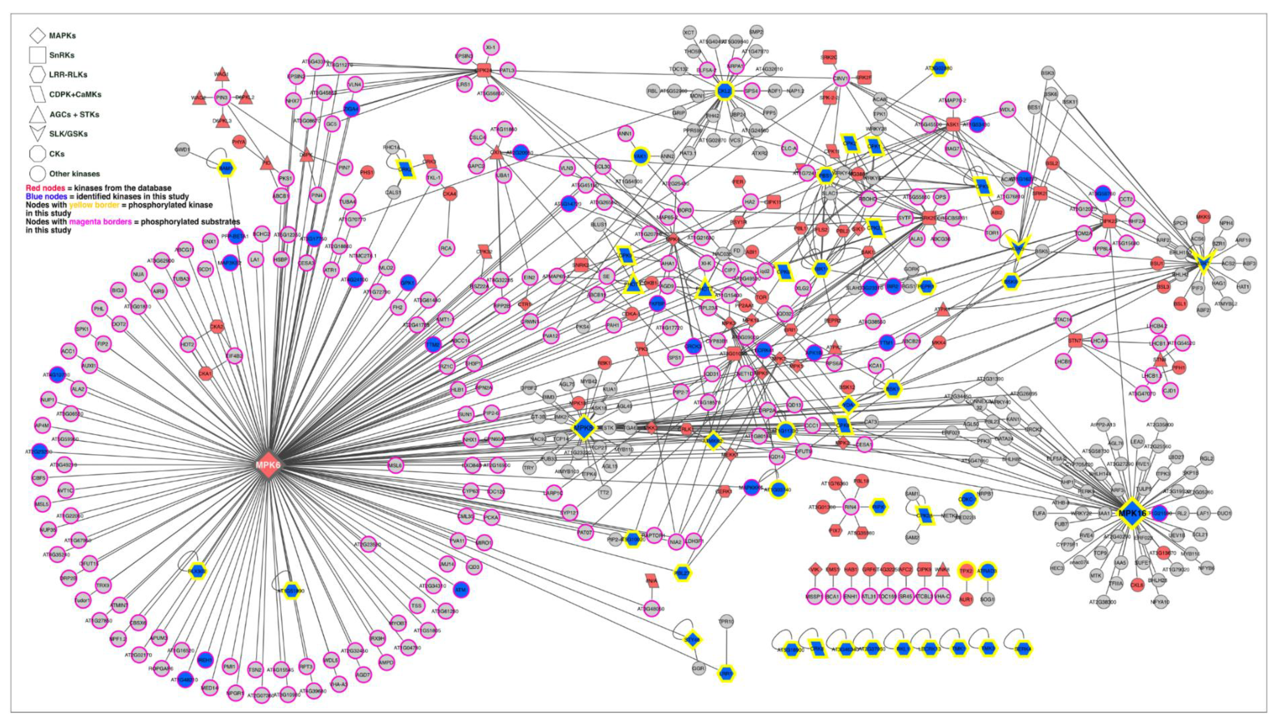
2.6. Kinase–Substrate Interaction Network

We constructed an updated kinase–target network containing 3461 interactions (Figure S3 and Table S3A) based on the relevant databases and a literature search [45,55,56,57,58,65]. Differentially and significantly (p < 0.05) phosphorylated proteins in our study were then mapped into the kinase–substrate network to extract a subnetwork (Figure 6 and Table S3B). The interaction network revealed that early cold signaling involved 695 highly complex kinase–substrate interaction events among the MAPKs, SnRKs, CDPK/CaMKs, LRR-RLKs, SLKs, GSKs, CKs AGCs, and other kinases, and we also found their corresponding substrates in the motif-x analysis. Out of 695 interactions, MAP kinases, including MAPK, MAP2K, and MAP3Ks, formed nearly 350 interactions. ABA signaling-related kinases SnRKs, including SnRK1, SnRK2, and SnRK3, were involved in 95 interactions, followed by 59 interactions by LRR-RLKs, 45 interactions by CDPK and CaMK, 40 interactions by AGC and STKs, 38 interactions by SLK and GSKs, and 36 interactions by CKs and CKLs (Table S3C). The complex interactions between the different kinase–kinase and kinase–substrate counterparts indicate that the early cold response involves different signaling pathways to adjust to a low-temperature environment.

3. Discussion

This report provides dynamic evidence that signaling and molecular events in the model plant Arabidopsis switch by reversible protein phosphorylation after short exposure to cold. In this study, we found early cold response in plants at the posttranslational level associated with a complex network of cold signaling composed of various kinases and their targets. So far, to our knowledge, this is the first large-scale time-course phosphoproteomic study on Arabidopsis membranes and membrane-related proteins under brief cold exposure. This study will provide new insight into cold perception from a reversible phosphorylation point of view.

3.1. Cold-Responsive Kinases and Signaling Proteins Might Play a Crucial Role in Early Cold Perception and Signal Transduction

Changes in phosphorylation to a large number of proteins in response to brief cold exposure indicate a significant perturbation in activity associated with kinases and phosphatases. A large number of enriched kinase target motifs from different major kinase families and direct identification of 152 kinases also imply a strong response from the kinases to cold exposure.
Ca2+ is one of the essential signaling components in plants, and cytosolic Ca2+ changes have been reported as one of the first sensors of cold stress as well as other stresses [66,67,68,69,70]. Ca2+/calmodulin (CaM) binding, CaM/CaM-like proteins, and calcineurin B like (CBL) and CBL-interacting proteins (CIPKs) play significant roles in sensing and interpreting Ca2+ signals [71,72] as well as recruiting and activating other signaling components, such as RLKs, CDPK/CaMKs, MAPKs, and ROS, in response to cold stress [73]. PM-localized Ca2+/CaM-regulated receptor-like kinase 1 (CRLK1), a serine/threonine kinase, in Arabidopsis is reported to be involved in cold-stress response and activated by Ca2+/CaM [26]. Further studies showed that this CRKL1 could interact and phosphorylate MEKK1, a core component of the cold-regulated MAP kinase signaling pathway [25,29]. A phosphoproteomic study of cold stress in rice showed OsCDPK13 is an essential part of the cold response system [74]. CDPK phosphorylation, in response to chilling stress, was also reported in plants, including Broussonetia papyrifera and Jatropha curcas [75,76]. Similar to these studies, we also found several members of CDPK and CaM-domain protein kinase (CPK) (Table 1 and Table S1A) that were not reported to be regulated at the phosphorylation level in response to a brief cold response. In our study, a large number of CDPK/CaMK and CIPK target motifs ([LXR/KXXpS/pT], [R/KXXpS/pT], and [pS/pTG]) were enriched at the phosphorylation sites (Figure 5A) and in the kinase–target interaction network (Figure 6; Table S3B), indicating the role of the Ca2+-mediated signaling pathways, regulated at the phosphorylation level in the early cold response.
Receptor kinases (RKs) or RLKs have been extensively studied at the phosphorylation level in response to pathogens and hormones. However, out of 600 RLKs, only about 60 have been functionally characterized so far [77]. Furthermore, only very few RLKs, for example, CRLK1 and RPK1, are functionally connected to cold response when exposed for more than several hours [25,29,78]. Thus, questions remain about whether any other RLKs respond to cold and how they respond at the PTM level. In this study, we found a large number of RLKs were phosphorylated in response to cold exposure (Table 1 and Table S1A). For instance, multifunctional somatic embryogenesis receptor kinase 4 (SERK4) is involved in brassinosteroid (BR) signaling and regulation of BR-dependent growth, and it also negatively regulates cell-death growth and phytohormone signaling [79,80,81,82]. Another peptide hormone-like receptor, RGF1 insensitive 3 (RGI3), is involved in meristem growth [83,84]. Transmembrane kinase 1 and 3 (TMK1 and TMK3) were reported to be involved in auxin-mediated cell growth and development and ROP GTPase signaling [85,86]. These RLKs might be involved in growth and development under cold stress. Botrytis-induced kinase 1 (BIK1) is one of the vastly studied RLKs that is central to the plant immune response and is also involved in Ca2+-dependent reactive oxygen (ROS) signaling, ethylene, and BR response [87,88,89], but it has never been reported to be phosphorylated in response to cold [87,88,89]. Receptor cytoplasmic kinase PBS1-like protein 27 (PBL27) activates the MAPK signaling cascade in response to chitin [90]. These findings imply that early RLK-mediated cold signaling might share, at least in part, some common pathways with other biotic, abiotic, ROS, and hormone signaling processes. Furthermore, the close phylogenetic relationship (Figure S4) between these cold-responsive RLKs could indicate a coordinated response from related gene family members at the PTM level under cold and could potentially be involved in cold perception and signaling in Arabidopsis.
The most studied cold response pathway in Arabidopsis is the MAP kinase pathway, which is linked to freezing tolerance in cold-acclimated plants [23,27]. We have found four phosphoproteins from the poorly characterized MAP4K members of MAP kinases. Interestingly, three of these MAP4Ks were found to be closely positioned in the phylogenetic tree (Figure 5A). Moreover, MAP4K5 and MAP4K6 were phosphorylated at the SXS motif in the central region of the protein but not inside any kinase domain. It could be possible that upstream RLKs or CDPKs phosphorylate these two proteins in the target SXS motif and regulate their function. It has been reported that some members of MAP4Ks can phosphorylate MAP3Ks or Raf kinases or directly phosphorylate MAP2Ks; others can function as adapters [91,92]. Similar to the MAP4Ks, phylogenetic analysis of the MAP3Ks and MAP3K-Raf (Figure 5B,C) showed most phosphorylated proteins are closely related to their gene family members, with a few exceptions, such as raf22, raf36, and raf47. For example, raf18 in this study, raf20, and raf24 [93], which are closely positioned in the phylogenetic tree, were phosphorylated within 5 to 30 min of cold exposure in the RXXS motif (Figure S5B). Even though there are no known functions ascribed to most Raf kinases, their response at the phosphorylation level to different stresses, including early cold (in this study) and early osmotic stress [42], indicates a potential involvement of Raf kinases in early cold-stress signaling. Other members of MAP3Ks phosphorylated in response to cold are also closely related in their phylogenies, such as M3KE1 and MAP3KE2, YDA/YODA, and MAP3Kγ (Figure S5C). These four MAP3Ks are reported to be involved in plant growth, development [94,95,96], and the MAPK signaling pathway [90,97]. Similar cold response phosphorylation patterns within identical phosphosites in closely related subfamily members of a large gene family could indicate coordinated phosphorylation and may be important for cold perception/signaling. Furthermore, phosphorylation of more downstream kinases, such as MKK2, MAPK6, MAPK8, and MAPK16, suggests that MAP kinases are involved in the early cold response and signal transduction. In summary, reversible phosphorylation of these MAP kinases, the highest enrichment in MAP kinase target motifs, and the highest number of kinase–substrate interactions indicate that the MAP kinase families play an essential role in early cold signaling.
Enrichment of SnRK2 target motifs [R/KXXpS/pT] and [LXR/KXXpS/pT] as well as a large number of SnRK targets from the kinase–substrate network also indicate that the ABA signaling pathway is triggered in response to brief cold exposure. GOBP analysis showed ABA-responsive proteins were phosphorylated throughout the cold exposure (Figure 4D). Although we could not detect any SnRKs in our data, we found several SnRK substrates reported to be involved in different biological processes in response to different abiotic stresses, such as respiratory burst oxidase homolog protein D (RBOHD), ATP-binding cassette (ABC) protein G36 (ABCG36), plasma membrane intrinsic protein 2-7 (PIP2-7), phototropin 2 (PHOT2), syntaxins of plants 121 (SYP121), and myosin motor XI-K (Table 1, Tables S1 and S3).
A few other important kinases and their substrates were also phosphorylated in response to cold exposure (Table 1 and Table S1), such as GSK3/shaggy-like kinase (ASK) brassinosteroid (BR) insensitive 2 (BIN2/ASK7), a positive regulator of BR signaling [98], and GSK3/SHAGGY-like protein kinase 1 (ASK9) and BSK4, negative regulators of BR signaling [99]. Phosphorylation of the BR signaling pathway regulators might indicate an involvement of BRs in early cold response in Arabidopsis. Casein kinase-like 2 (CKL2) was also phosphorylated immediately upon cold exposure. In addition, we found several reported and putative substrates of CK2 in the kinase–substrate network. A recent phosphoproteomic study in tomatoes also showed that CKL2 homologs were phosphorylated in response to prolonged cold stress, where SnRK2E could directly phosphorylate CKL2 [47].

3.2. Early Cold Response-Mediated Protein Phosphorylation Can Activate Phospholipid/Phosphoinositide Signaling Events

Phospholipids are the main building blocks of PM and responsible for maintaining the membrane structure and function. Phosphoinositides (PIs), one of the lipid family members present in small amounts in the membrane, are involved in various crucial functions in plant growth and development, including environmental adaptation. From the gene ontology analysis (Figure 4A), we found that phospholipid, phosphatidylinositol binding, and signaling-related proteins were phosphorylated within 15 min of cold exposure, indicating that phospholipids respond to the temperature drop at a very early stage. We found a total of 44 phosphopeptides from different lipid-related proteins, and 16 were involved in the phosphatidylinositol metabolism pathway (Table 1 and Table S1A). The phosphatidylinositol metabolism pathway members found in this study are involved in the generation and degradation of PIs. It has been reported that PIs are continuously generated and degrade rapidly and dynamically in response to different abiotic stresses such as wounding [100], heat stress [101], and salt stress or osmotic stress [102]. Phosphatidylinositol 4-kinase (PI4Ks) can phosphorylate PIs and generate PI monophosphates PI3P and PI4P. PI monophosphates can further be phosphorylated by forms aploid and binucleate1 (FAB1) and phosphatidylinositol 4-phosphate 5-kinase (PIP5K) into phosphatidylinositol 3,5-bisphosphate (PI(3,5)P2) and phosphatidylinositol 4,5-bisphosphate (PI(4,5)P2), respectively [103]. Vacuolar localization of PI(3,5)P2 under osmotic stress is essential for maintaining the proper structure and function of vacuoles in yeast [104,105,106]. PI(3,5)P2 also accumulates in the early osmotic stress response in Arabidopsis [42]. FAB1 and/or its product PI(3,5)P2 are essential for ABA-mediated stomatal closure, pollen tube growth, development, viability, endosome maturation, and interaction with microtubules [107]. In moss, PI(3,5)P2 are required for polarized cell growth and actin-cytoskeleton arrangement [108]. Endomembrane-localized FAB1 kinase knockout or knockdown results in enlarged vacuole formation in yeast because of impaired membrane trafficking [109,110,111]. The PI4P-5K product PI(4,5)P2 is also well known for its role in exocytosis, actin cytoskeleton organization, vesicle-mediated trafficking, and regulation of ion channels [112,113].
Furthermore, suppressor of actin 1 (SAC1) and phospholipase C/D (PLC/D) can degrade different PI species and generate downstream second messengers [103]. For example, PLC2 is one of the critical enzymes that can catalyze the hydrolysis of PI(4,5)P2 into diacylglycerol (DAG) and inositol 1,4,5-trisphosphate (IP3), which in turn increases Ca2+ influx into the cytoplasm [114,115]. Diacylglycerol kinases (DGKs) can convert DAG into phosphatidic acid (PA) [116], and PA is well known as a signaling molecule under different kinds of abiotic stresses, including cold. Interestingly, in our study, Sec14p-like phosphatidylinositol transfer family protein PATL3, responsible for transferring PIs to the PM [117], was found to be phosphorylated throughout the cold exposure duration (Table 1 and Table S1A). Phosphorylation of these PI kinases, phosphatases, and trafficking-related proteins clearly indicates that PI is involved in early cold perception, signaling, and cytoskeleton organization and might be regulated by posttranslational modifications.

3.3. Early Cold Response Events Can Be Associated with Cytoskeletal and Vesicle Trafficking Systems

The cytoskeleton in plants mainly consists of microtubules and actin filaments. It is essential for maintaining cell shape, and it facilitates organellar and vesicular trafficking inside the cell [118]. Several cytoskeleton-related proteins were phosphorylated in response to early cold exposure, including myosin and myosin-binding (MyoB) proteins myosin 1 (VIII-1), myosin-5 (XI-1), myosin-9 (XI-C), myosin-17 (XI-K), and MyoB7 (Table 1 and Table S1A). MyoB7 phosphorylation peaked at 5 min, and other myosin and myosin family proteins mostly peaked after 30 min of cold exposure. The myosin-binding protein MyoB1 showed increased phosphorylation within 2 min of osmotic stress [42]. In addition, myosin-like protein abundance was also found to be increased under cold stress in rice and perennial grasses [119,120]. Myosin and myosin-binding proteins facilitate cytoplasmic streaming [121]. Even though myosin and myosin-binding proteins respond to cold and osmotic treatments, their exact function still has to be determined. Perhaps, phosphorylation in the myosin-binding and myosin motor proteins could act as an early adaptive response to cold to maintain proper growth via cytoplasmic streaming and maintain cytoskeletal integrity of the cell under prolonged cold.
Microtubule-related proteins, 65 kDa microtubule-associated protein 1 (MAP65-1), microtubule-associated protein 70-2 and 70-3 (MAP70-2 and MAP70-3), tubulin alpha-3 (Tuba-3), and microtubule-associated protein TORTIFOLIA1 (TOR1) were also phosphorylated and categorized in the late response clusters (30 min and 60 min, Table 1 and Table S1A). These proteins are responsible for the bundling and stabilization/destabilization of microtubules, the formation of tubulin dimers, and cell division [17]. Microtubules also act as sensors that perceive mechanical membrane stress derived from cold [122]. Furthermore, the sensory function of the microtubules is determined by dynamic instability and subsequent formation of stable microtubules during stress adaptation [122]. The MAP65-1 protein is phosphorylated in the microtubule-interacting region (pS532), and phosphorylation in this region disrupts the interaction properties [123]. Moreover, in the gene ontology analysis, we also observed that actin filament and microtubule-based processes were enriched in the late response group (Figure 4F). It might be possible that with the increasing duration and intensity of cold exposure, plant cells sense the cold via phosphorylation-mediated microtubule disruption, which acts as a cue for further cold adaptation-related processes.
Endosomal trafficking plays a crucial role in plant growth and development and maintains organellar homeostasis under different stressful conditions. Several studies showed endosomal transcript or protein abundance changes in response to longer durations (several hours to days) of cold stress [7,124,125,126]. However, before this study, it was unclear whether endosomes also respond to early cold exposure at the protein phosphorylation level. In our study, we observed changes in several previously described members of vesicle-mediated transport cargoes in plants at the phosphorylation level (Table 1 and Table S1A). For example, dynamin-like protein 2A and 2B (DRP2A, DRP 2B), sec7 domain-containing protein GN (GNOM), SYP121 and SYP132, soluble n-ethylmaleimide-sensitive factor adaptor protein 33 (SNAP33), sorting nexin 1 and 2A (SNX1, SNX2A), vacuolar protein sorting 41 (VPS41), and other proteins (Table 1 and Table S1A) showed cold-responsive phosphorylation within 60 min of cold exposure, suggesting that their PTM level changes might prepare plants for a longer cold exposure or stress.

3.4. Transporters Respond to Early Cold Stress to Maintain Homeostasis

Under any environmental stress situations, for proper growth, development, and survival, plants consistently need to maintain ionic and cellular homeostasis through different transporter and channel proteins located in the membranes by balancing the influx and efflux of the different ions, toxic byproducts, and carbohydrates [127]. Phosphorylation changes in the transporter proteins are also one of the rapid adaptive responses in plants under environmental stresses [37]. Many proteins related to solute and ion transport were also phosphorylated upon cold exposure (Table 1 and Table S1A). We found that two Ca2+ channel/transporters, the hyperosmolality-gated Ca2+ permeable channel 1.1 and 1.2 (AtOSCA1.1 and 1.2), were phosphorylated in the conserved C-terminal cytoplasmic domain at a close distance to each other at pT750 (at 15 min only) and pS757 (5 min and peaked at 30 and 60 min) (Table 1 and Table S1A). Both of these channels are known to permeate Ca2+ in response to osmotic stress, and AtOSCA1.1 is an essential component of osmosignaling [128,129]. However, these two phosphorylation sites were never functionally characterized in any stress response. Moreover, AtOSCA1.1 is also the first genetically identified Ca2+ channel. It is possible these two phosphosites, similar to osmosensing, play an essential role in sensing cold. Interestingly, we also found two autoinhibited Ca2+ ATPases (ACA), ACA1 and ACA4, were phosphorylated within 5 min and decreased gradually with the cold exposure duration (Table 1 and Table S1A). These ACAs are responsible for Ca2+ efflux out of the cell and maintaining the Ca2+ balance in the cell [130]. Elevation of the Ca2+ concentration inside the cell under cold is a significant signaling event that initiates further downstream canonical signaling pathways [68,69,70,131]. In this study, phosphorylation in both the Ca2+ influx and efflux channels is possibly involved in modulating a balanced Ca2+ level for fine-tuning Ca2+-mediated signaling events under cold.
During the cold acclimation process, plants accumulate various cryoprotectants. Soluble sugars are one of the major cryoprotectants and can protect plants from freezing damage and maintain cellular integrity and functions [132]. In our study, we found several sugar and carbohydrate transmembrane transporters respond to early cold exposure, such as bidirectional sugar transporter SWEET12, polyol/monosaccharide transporter 5 (PLT5), tonoplast monosaccharide transporter 1 and 2 (TMT1, TMT2), sugar transporter ERD6, and ERD6-like 6 (ERDL6) (Table 1 and Table S1A). Reports showed disruption of SWEET12 resulted in thinner stems and reduced freezing tolerance in Arabidopsis [133], and its transcript levels increased under drought stress [134]. However, before this study, there was no report on the phosphoproteome level changes in SWEET12 in response to cold. Vacuolar monosaccharide transporters TMT1 and TMT 2 were reported to be phosphorylated at S385 and S376 under 5 days cold acclimation at 4 °C [135]. In our study, we found TMT1 was phosphorylated at pS277 throughout the cold exposure, and phosphorylation at pS446 immediately decreased at 5 min, then increased four-fold at 15 min and decreased at 30 and 60 min. TMT2 showed increased phosphorylation at pS448 peaked at 5 min and was maintained throughout the cold exposure, where pS287 peaked at 60 min only (Table 1 and Table S1A). All phosphorylation took place between transmembrane domains 5 and 6, and 6 and 7, which generally agrees with in silico prediction studies of the phosphorylation sites in TMTs [136]. Similar to the reports showing that phosphorylation regulates sugar transport under prolonged cold stress and other abiotic stresses [135], our study indicates that, in Arabidopsis, the early cold response also initiates sugar transport-related events in the PM and vacuoles and might be regulated by phosphorylation.
We observed strong phosphorylation in two PM proton-pump members: H+ATApase1 (AHA1) and H+ATPase4 (AHA4) (Table 1 and Table S1A). AHA1 was phosphorylated at multiple sites, including the canonical 14-3-3 protein binding YTV domains such as T881, T948, and the inhibitory S899 site. AHA4 showed constant phosphorylation at pT959 inside the 14-3-3 protein binding YTV domain throughout the cold exposure after peaking at 5 min. The binding of 14-3-3 protein at pT948 and pT959 in the YTV domain activates the proton pump [137,138]. On the other hand, phosphorylation at pS899 inhibits pump activity by an unknown mechanism [139]. The observed phosphorylation at both the 14-3-3 protein binding sites and the pS899 inhibitory site in this study seem to be contradictory to the cold response. Even though AHA1 is phosphorylated at the inhibitory pS899 site, the pumps might be hyperactivated by the phosphorylated interactor isoform 1 (PPI1). It has been reported that PPI1 can hyperactivate AHAs [140]. We observed immediate phosphorylation of PPI1 at 5 min, and increased phosphorylation was maintained at 30 min and 60 min (Table 1 and Table S1A).
One of the most potent transporter groups in plants is the ATP-binding cassette (ABC) transporters. ABC transporters are reported to be involved in sequestration of toxic byproducts and in exchanging compounds like phytohormones (auxin), secondary metabolites, and defense molecules [141,142]. ABC transporters have mostly been studied at genomic and transcriptomic levels; for example, plants with overexpressed ABCG36 show improved resistance to salt and drought stress [143]. ABCC1 acts as a pump for glutathione S-conjugates [144], and ABCB1, ABCB14, ABCB19, ABCG36, and ABCG37 were reported to be involved in auxin transport-mediated plant growth and development [145,146,147]. In addition, a few studies showed their regulation and function at the phosphorylation level; for example, ABCB19 is a direct phosphorylation target of kinase PHOT1, which can transiently deactivate ABCB19 and result in the accumulation of auxin [148], and ABCG36 is phosphorylated in response to pathogen attack. We also found several ABC transporters to be phosphorylated under early cold exposure, and 75% of these were immediately phosphorylated within 5 min of cold exposure (Table 1 and Table S1A). The most notable transporters found in our study were ABCG36/37, ABCB1, and ABCC1, which were involved in detoxification, auxin transport, biotic and abiotic stress responses, and resistance [141]. The PTM or phosphorylation-level regulation of these ABC transporters found in this study might play a crucial role in maintaining homeostasis and detoxification as an adaptive early cold response in Arabidopsis.
Auxin is a key phytohormone involved in different regulatory processes in plants. The spatiotemporal distribution of auxin is critical for auxin-mediated growth and development under normal and stressful conditions, including cold [149,150,151]. Studies reporting the involvement of auxin transport or homeostasis are mostly carried out under prolonged exposure (several hours to days), but not short (within minutes) exposure to stress as we employed in our study. Furthermore, most studies have focused on functional regulation at the genomic or transcriptomic level than the PTM level. Reversible phosphorylation at conserved serine and threonine residues could control the spatiotemporal distribution of auxin efflux carrier family protein 1 (PIN1) and its transport activity [152]. In our study, we found auxin transporter proteins, such as AUX1/LAX (influx) and PIN3, 4, and 7 (efflux), were phosphorylated in response to early cold stress (Table 1 and Table S1A). AUX1 was phosphorylated immediately and maintained its phosphorylation throughout the cold exposure time at two different phosphosites, pS14 and pS27, and inside the target SXXD/E motifs for casein kinase (CKII). On the other hand, PIN3 was phosphorylated at conserved pS366 with a 1.8-fold increase at 5 min and peaked at 60 min. PIN4 was also phosphorylated at conserved pS395 within the SXXD/E motif and maintained its phosphorylation throughout the cold exposure/treatment time and peaked at 60 min. We also found a phosphosite of PIN7 at pS431 in the SXXD/E motif, which maintained a continuous phosphorylation state. CKII was also reported to play an essential role in auxin transport and auxin-mediated growth and development in Arabidopsis [153,154]. Interestingly, PIN7 was also phosphorylated at T242 in the conserved TPRXSN motif. The TPRXSN motif is involved in phosphorylation-dependent control of PIN1 localization [152]. Cold-responsive phosphorylation of PIN and AUX1 at different conserved sites indicates an essential role of protein phosphorylation under a short period of cold exposure, which might be involved in auxin transport and homeostasis for maintaining growth and development for upcoming longer and extreme cold conditions.
Our data also showed that PM intrinsic proteins (PIPs) respond actively and temporally to brief cold exposure (Table 1 and Table S1A). We observed continuous phosphorylation of PIP2-5 (pS279) throughout the cold treatment. A doubly phosphorylated peptide of PIP2-6 (pS279 and pS282) was phosphorylated over two-fold at 60 min, and PIP2-7 (pS276) showed decreasing phosphorylation after peaking at 5 min of cold exposure (Table 1 and Table S1A). PIPs are transmembrane transporters for water and small molecules and operate in a gated, controlled way. Gating properties vary under different abiotic stresses and are controlled by pH in flooding/anoxic conditions [155,156], phosphorylation/dephosphorylation under drought stress and low temperature [157,158], and calcium under salt stress [159]. It was also reported that chilling-induced H2O2 could be diffused by PIPs in a phosphorylation-dependent manner in maize [160]. Previously, it was shown that phosphorylation at pS280 and pS283 of PIP2-1 was associated with gating or hydraulic conductivity [161]. Multiple sequence alignment analysis showed that pS279 of PIP2-5 and PIP2-6, and pS276 of PIP2-7, are conserved with pS280 of PIP2-1, and the pS282 of PIP2-5 is also conserved with pS283 of PIP2-1 (data not shown). Phosphorylation of PIP2s in all these phosphosites suggests that the activity of the PIPs is controlled in a phosphorylation-dependent manner under cold and maintains cellular water and H2O2 in a balanced state. Previous studies also showed that PIP1-4 and PIP2-5 could regulate cold acclimation and freezing tolerance in Arabidopsis [162]. Thus, continuous phosphorylation of PIP2-2 at its regulatory S279 position throughout cold exposure, along with other PIP2s, might play a crucial role in cold response and prepare plants for long-term cold acclimation.

3.5. ROS Signaling under Cold Response

ROS, generating respiratory burst oxidase homolog D (RBOHD), were also phosphorylated, as shown in this study, which has been never reported in response to brief cold exposure (Table 1 and Table S1A). In this study, RBOHD was phosphorylated in pS8, pS148, pS163, and pS347 within 15 min of cold exposure (Table 1 and Table S1A). RBOHD can generate ROS via Ca2+-dependent and -independent phosphorylation by CDPKs/CIPKs and BIK1 kinase, respectively [87,163]. Ca2+-dependent protein kinase 5 (CPK5) and BIK can phosphorylate and regulate RBOHD at the same amino acid positions (pS148, pS163, and pS347) [163,164,165], which are the same positions we observed as the cold-responsive phosphorylated sites in this study. Interestingly, in our data, we detected that both CPK5 and BIK were phosphorylated in response to brief cold (Table 1 and Table S1A). Furthermore, the phosphosites showed increased phosphorylation containing characteristic RXXS/LXRXXS motifs, a potential target motif of CDPKs (Table 1 and Table S1A). This rapid phosphorylation of ROBHD and its kinases indicates ROS signaling involvement in early cold response.

4. Materials and Methods

4.1. Plant Growth Conditions and Cold Exposure

Twenty-five surface-sterilized seeds of Arabidopsis thaliana ecotype Colombia-0 (Lehle Seeds, Round Rock, TX, USA) were sown in 9 cm square petri dishes containing modified Hoagland solution [166] with 0.8% agar (Wako, Tokyo, Japan). The seeds were then cold stratified for 48 h at 4 °C. The seeds were germinated and grown for two weeks at 23 °C and a 16/8 h photoperiod at 80–100 μmolm−2s−1 light intensity in a growth chamber (CLE-303, Tomy Digital Biology, Tokyo, Japan). For cold exposure analysis, agar plates containing two-week-old Arabidopsis seedlings were exposed to 2 °C for 0 min (control: no cold exposure), 5 min, 15 min, 30 min, and 60 min without the lid of the petri dish. Plates were flash-frozen in liquid nitrogen at the end of each cold exposure duration. The frozen plants were used for protein extraction or stored at −80 °C for later use. For each time point three independent biological replicates were used.

4.2. Microsomal Membrane Fraction (MMF) Extraction

MMF was extracted according to the method of Wu et al. [45], with modifications. Around 75 two-week-old pre-frozen seedlings (~1–1.5 g) were broken into small pieces and homogenized in 10 mL of homogenization buffer consisting of 500 mM sorbitol, 50 mM 3-(n-morpholino) propanesulfonic acid-KOH (MOPS-KOH) (pH 7.6), 5 mM ethylenediaminetetraacetic acid (EDTA), 5 mM dithiothreitol (DTT), 1 mM phenylmethylsulfonyl fluoride (PMSF), 2 mM salicylhydroxamic acid (SHAM), 25 mM sodium fluoride (NaF), 0.5 × protease inhibitor cocktail (p9599, Sigma-Aldrich, St. Louis, MO, USA), and 1 × phosphatase inhibitor cocktail 2 and 3 (P5726 and P0044, Sigma-Aldrich, St. Louis, MO, USA), using a 30 mL motorized glass homogenizer (81-0487, Sansyo Co., Ltd., Tokyo, Japan) on ice. For proper homogenization, the rotatory pastel was moved up and down slowly for at least 3 min. The homogenate was filtered through six layers of cheesecloth and centrifuged at 7500× g for 15 min at 4 °C. The supernatant was collected and ultracentrifuged at 100,000× g for 50 min at 4 °C. The pellet was washed twice with homogenization buffer by centrifugation at 100,000× g for 20 min at 4 °C. The final washed pellet was collected as MMF and resuspended in a minimum volume of Tris-UTU buffer (100 mM tris aminomethane, 6 M urea, 2 M thiourea, pH 8.5). The protein concentration was measured using a Bradford (Bio-Rad, Hercules, CA, USA) assay [167]. The MMF samples were used for peptide processing immediately or stored at −80 °C for later use.

4.3. Phosphoproteomic Analysis

4.3.1. In-Solution Trypsin Digestion

The phosphoproteomic analysis was carried out according to methods previously reported [44,46,49,50,168,169], with minor modifications. For trypsin digestion, aliquots of MMF (1 mg protein) were used. The digested MMF was diluted four times with sterile distilled water, reduced with 10 mM DTT for 60 min, and then alkylated in the dark with 55 mM iodoacetamide for 30 min at room temperature. After the alkylation, the proteins were digested overnight at room temperature with trypsin gold (1:100, w/w; Promega, Madison, WI, USA). After digestion, the sample tubes were centrifuged for 5 min at 21,000× g at room temperature. The supernatant was transferred to a fresh tube and used for phosphopeptide enrichment.

4.3.2. Phosphopeptide Enrichment and Desalting

Phosphopeptides were enriched at room temperature using hydroxy acid-modified metal oxide chromatography (HAMMOC), with minor modifications [168]. Titansphere® Phos-TiO columns (syringe barrel type SPE cartridge, cat. No. 5010-21291, GL Sciences Inc., Tokyo, Japan) were used according to the manufacturer’s instructions. Enriched phosphopeptides were desalted using two-step washing with a combination of GL-Tip SDB and GC columns (GL Sciences Inc., Tokyo, Japan), according to the manufacturer’s instructions. Desalted phosphopeptides were dried in a vacuum evaporator (Tomy, Tokyo, Japan), resuspended in 10 µL of 0.1% (v/v) TFA, and stored at −80 °C for later LC-MS/MS analysis.

4.3.3. LC-ESI-MS/MS Analysis

Desalted phosphopeptides were analyzed in the TripleTOF5600 system (AB SCIEX, Framingham, MA, USA) equipped with Autosampler-2 1D plus (Eksigent, Framingham, MA, USA) and NanoLC Ultra (Eksigent, Framingham, MA, USA) using a MonoCap C18 High-Resolution 2000 column (GL Sciences Inc., Tokyo, Japan) and PicoTip emitter SilicaTip (New Objective Inc., Woburn, MA, USA). Peptides were eluted at a flow rate of 500 nL/min−1 with a four-step gradient using 0.5% (v/v) acetic acid: 0.5% (v/v) and 80% (v/v) acetic acid = 98:2 (0 min), 60:40 (300 min), 10:90 (20 min), and 98:2 (40 min). The eluate was sprayed into the mass spectrometer by electrospray ionization (ESI). The mass spectrometry (MS) scan range was 400–1250 m/z, and the MS/MS scan range was 100–1600 m/z.

4.3.4. Phosphopeptide Identification and Quantification

The spectrum files (wiff and wiff.scan) generated from the mass spectrometer were converted to peak lists in mascot generic format (mgf) using Protein Pilot software (v5.0.0.4769; AB-SCIEX, Framingham, MA, USA). The mgf files were searched against the Arabidopsis protein database Araport11 (11 July 2019) [170] obtained from the Arabidopsis information resource (TAIR) (https://www.Arabidopsis.org/) [171] in the Mascot ion search platform (v2.6, Matrix Science, London, UK). The search parameters were applied as follows: a precursor mass tolerance of 3 ppm, a fragment ion mass tolerance of 0.8 Da, and a cut-off value of 0.95, allowing for up to two missed cleavage from trypsin digestion. Fixed modification of carbamidomethylation of cysteine and variable modifications of oxidation of methionine and phosphorylation of serine, threonine, and tyrosine were used [44,46,50,169,172]. The false discovery rate (FDR) was calculated using the Benjamini–Hochberg method and set to 5%. The phosphorylation site localization confidence was assessed by site localization probability scores calculated from the mascot delta score, and the threshold level was set to > 0.75 to identify class-I phosphorylation sites [46,173].
Skyline-Daily software (v20.1.1.83; https://skyline.ms/project/home/software/Skyline/daily/begin.view) [174] was used to quantify the peak areas/intensities. The search parameters in the Skyline-Daily were the same as those in the Mascot search parameters. A minimum of two peptides identified per protein was considered as a confident identification of that protein. The peak intensities, from both the phosphorylated and proteotypic non-phosphorylated peptides generated from Skyline-Daily, were normalized using the EigenMS normalization method [175,176,177] in the ProteoMM R-Bioconductor package [178]. All raw data files were deposited in the Japan Proteome Standard Repository/Database (jPOST; JPST000993, Kyoto, Japan).

4.4. Bioinformatic Analysis

Identified phosphopeptides and proteotypic non-phosphopeptides from all biological replicates from each time point (0, 5, 15, 30, and 60 min) were compared using unsupervised principal component analysis (PCA). The phosphopeptides identified in all five conditions were selected for further analysis. The changes in phosphorylation dynamics were studied by K-means clustering of the log-transformed normalized phosphopeptide intensities in MeV (MultiExperiment Viewer), using Euclidean distances [179]. Comparative analysis between the untreated (0 min) and treated (cold exposed) groups was performed in each characteristic K-means clusters; increased phosphorylation was assigned if the log2 fold change (Log2FC) of the treated/untreated ration was ≥ 1 in at least two of the three biological replicates for each time point, and a decreased phosphorylation was when the Log2FC was ≤ 0.5 in at least two of three replicates. Individual phosphopeptides were compared using pairwise t-tests between the average intensities at 0 min and each cold exposure time point. The relative time profile for significant phosphopeptide changes in each cluster was also created based on the Log2FCs between 0 min and all other cold exposure time points. All time-profile clusters were visualized using a heatmap in the online platform ClustVis (https://biit.cs.ut.ee/clustvis/) [180].

4.4.1. Gene Ontology Analysis

Gene Ontology (GO) annotation (GO-Slim, last update: 1 May 2020) for all Arabidopsis proteins was obtained from the latest TAIR [171]. The GO annotation database was created by excluding genes with gene ontology-consortium evidence code IEA (Inferred from Electronic Annotation) and ND (No biological Data available) for further analysis. Over-representative GO term analysis was carried out in AgriGO (v2.0: http://systemsbiology.cau.edu.cn/agriGOv2/) [181,182], uploading the GO annotation database as background. The Singular Enrichment Analysis (SEA) tool was used in the AgriGO platform combined with Fisher’s exact test and Yekutieli’s test (FDR under dependency) for the multi-test p-value (0.05) adjustment method; the minimum number of mapping entries was five, and plant GO-Slim was the Gene Ontology type. To decrease the chance of false discovery, GO cross-validation was also carried out in DAVID: BP_Direct (v6.8: https://david.ncifcrf.gov/) [183,184] and CluterProfiler: groupGO function [185] using the same updated GO annotation database used in AgriGO as background, in g:Profiler g:GOSt using Arabidopsis as background and g:SCS threshold 0.05 [186], and finally matched with SUBA4 localization [187]. Significantly (adjusted P-value ≤ 0.05) enriched GO cellular component (GO_CC) terms related to MMF, such as plasma membrane (PM), endoplasmic reticulum (ER), Golgi (G), vacuole (V), cell wall (W), apoplast (A), cytoskeleton (CS), plasmodesma (PD), and other membrane-related proteins, were selected as the protein group of interest for further analysis. Proteins that belong to the cytosol (C), plastid (P), mitochondria (M), and nucleus (N) were excluded from further analysis.
For the over-representation analysis of GO biological processes (GOBP) and molecular function (GOMF), only the protein group of interest was used in process similar to that for GOCC. Significantly over-represented GO terms (BH adjusted p < 0.05 after Fisher’ exact test) were grouped in similar categories using REViGO (http://revigo.irb.hr/) [188]. Settings used in REViGO were as follows: medium (0.7) similarity, UniProt Arabidopsis database (https://www.uniprot.org), and SimRel semantic measure. The negative log10-transformed, adjusted p-value < 0.05 for each GO term in the REVIGO groups was scaled with vector scaling and visualized in a heatmap in the online platform ClustVis (https://biit.cs.ut.ee/clustvis/) [180].

4.4.2. Phosphorylation Motif Dynamics Analysis

Phosphorylation motifs were predicted using the motif-x [189] algorithm in rmotifx (https://github.com/omarwagih/rmotifx) [190] in the R package. All confident phosphopeptides were used for motif prediction. Phosphopeptide sequences were centered around the phosphorylation sites (STY), and 13 amino acids (±6) around the phosphorylated residues were extracted and submitted to the rmotifx. In cases where the phosphorylation sites were near the N/C terminal, the motif sequences were filled in using 13 amino acids with the required number of “X”, which denotes any amino acid [65]. The settings for rmotifx were as follows: the minimum number of central amino acid residues was set to 30 for Ser (S) and 20 for Thr (T) and Tyr (Y), the significance threshold was set to p < 10−6, and background database was extracted from Araport11 [170]. Any enriched motif was attributed to a kinase or kinase group if the identical motif was reported in the literature or matched in the PhosPhAt database [55,56,57]. Similar motifs were grouped according to van Wijk et al. [54].
To predict kinase activity and kinase regulation dynamics, each enriched motif was assessed at each time point. The activity of upstream kinases affects the phosphorylation intensity of the downstream target motifs. To reveal the effect of kinases on targets, we computed the total average intensities from the phosphopeptides that belong to each enriched motif or motif groups at each time point and calculated the fold change between 0 min and any other cold exposure time point [50]. Motif dynamics were assessed using Fisher’s exact test using a 2 × 2 contingency table, as described by Lin et al. [65].

4.4.3. Kinase–Substrate Network Construction

An updated kinase–substrate network was built by collecting experimental data from the literature [58] and the PhosPhAt database [55,56,57]. The constructed network was used as a benchmark for mapping and visualizing the identified kinases and putative substrates in this study by Cytoscape [191].

5. Conclusions

In summary, to understand phosphoregulation in the earliest stage of cold response (within minutes), we performed a large-scale mass spectrometry-based phosphoproteomic analysis of the Arabidopsis microsomal membrane proteins. We detected nearly 1900 phosphosites from over 850 proteins, where around 1000 phosphosites showed temporal phosphorylation in response to cold. From an in-depth analysis of the phosphoproteomic data, we revealed that a plant’s response to a brief drop in temperature is a rather complicated process. Phosphorylation of ion channel or transporter proteins, such as Ca2+ (OSCAs, ACAs), K+ (POTs), or Na+/H+ (NHXs), which are involved in maintaining ionic homeostasis inside the cell, indicated that plant cells try to maintain ionic balance in a steady state under cold conditions. Our data also indicated that the plant kinome is perturbed by cold exposure and kinases, such that RLKs, MAPKs, CDPKs/CaMKs, CKs, and GSKs play active roles in cold sensing and signaling processes. Ca2+, phosphoinositide, ABA, auxin, BR, and ROS mediated signaling pathways were also found to be involved in the early cold response. Protein transport, trafficking, and cytoplasmic streaming may also be involved in early cold response events. We further found that plant cells tried to maintain cytoskeletal integrity as an adaptive response to cold, which was seen by the rapid phosphorylation of proteins involved in actin-/microtubule-based processes. We expect that this first large-scale membrane phosphoproteomic study to elucidate early cold response in Arabidopsis will provide valuable information to understand cold perception in plants.

Supplementary Materials

Supplementary Materials can be found at https://www.mdpi.com/1422-0067/21/22/8631/s1.

Author Contributions

M.M.K., Y.K., and M.U. conceived and designed the study; M.M.K. performed the experiments, analyzed data, and took lead in manuscript writing; S.I. and T.U. assisted the analysis and interpretation of data; K.S., M.K., F.T., and K.S. supported the mass spectrometry analysis; F.T. assisted in the mass spectrometry-based phosphoproteomic experiments. All authors have read and agreed to the published version of the manuscript.

Funding

This work was in part supported by the Japan Society for Promotion of Science (JSPS) Kakenhi No. JP19H03255 (to F.T.) and 17H03961 (to M.U. and Y.K.).

Acknowledgments

We are grateful to Hirofumi Nakagami (Max Planck Institute for Plant Breeding Research) for his suggestions and comments on the phosphoproteomic experiments. We thank Daisuke Takahashi (Saitama University) for discussing the proteomic data analysis. We are also grateful to Yuko Suzuki, Michiko Saito, Nozomi Yokota, Momo Otake, Aoi Sawada, and Kei Onodera for their technical help to prepare the plants.

Conflicts of Interest

The authors declare no conflict of interest. The funders had no role in the design of the study; in the collection, analyses, or interpretation of data; in the writing of the manuscript, or in the decision to publish the results.

Abbreviations

MMFMicrosomal membrane fraction
LC-ESI-MS/MSLiquid chromatography–electrospray ionization mass spectrometry/mass spectrometry

References

  1. Levitt, J. Chilling, Freezing and High Temperature Stresses; Academic Press: Cambridge, MA, USA, 1980; Volume 1, pp. 27–64. [Google Scholar]
  2. Zhu, J.; Dong, C.-H.; Zhu, J.-K. Interplay between cold-responsive gene regulation, metabolism and RNA processing during plant cold acclimation. Curr. Opin. Plant Biol. 2007, 10, 290–295. [Google Scholar] [CrossRef]
  3. Chinnusamy, V.; Zhu, J.; Zhu, J.-K. Cold stress regulation of gene expression in plants. Trends Plant Sci. 2007, 12, 444–451. [Google Scholar] [CrossRef]
  4. Miura, K.; Furumoto, T. Cold Signaling and Cold Response in Plants. Int. J. Mol. Sci. 2013, 14, 5312–5337. [Google Scholar] [CrossRef] [Green Version]
  5. Thomashow, M.F. Plant Cold Acclimation: Freezing Tolerance Genes and Regulatory Mechanisms. Annu. Rev. Plant Biol. 1999, 50, 571–599. [Google Scholar] [CrossRef] [Green Version]
  6. Gilmour, S.J.; Hajela, R.K.; Thomashow, M.F. Cold Acclimation in Arabidopsis thaliana. Plant Physiol. 1988, 87, 745–750. [Google Scholar] [CrossRef] [Green Version]
  7. Miki, Y.; Takahashi, D.; Kawamura, Y.; Uemura, M. Temporal proteomics of Arabidopsis plasma membrane during cold- and de-acclimation. J. Proteom. 2019, 197, 71–81. [Google Scholar] [CrossRef]
  8. Takahashi, D.; Kawamura, Y.; Uemura, M. Changes of Detergent-Resistant Plasma Membrane Proteins in Oat and Rye during Cold Acclimation: Association with Differential Freezing Tolerance. J. Proteome Res. 2013, 12, 4998–5011. [Google Scholar] [CrossRef]
  9. Li, B.; Takahashi, D.; Kawamura, Y.; Uemura, M. Comparison of Plasma Membrane Proteomic Changes of Arabidopsis Suspension-Cultured Cells (T87 Line) after Cold and ABA Treatment in Association with Freezing Tolerance Development. Plant Cell Physiol. 2012, 53, 543–554. [Google Scholar] [CrossRef] [Green Version]
  10. Takahashi, D.; Imai, H.; Kawamura, Y.; Uemura, M. Lipid profiles of detergent resistant fractions of the plasma membrane in oat and rye in association with cold acclimation and freezing tolerance. Cryobiology 2016, 72, 123–134. [Google Scholar] [CrossRef] [Green Version]
  11. Takahashi, D.; Kawamura, Y.; Uemura, M. Cold acclimation is accompanied by complex responses of glycosylphosphatidylinositol (GPI)-anchored proteins in Arabidopsis. J. Exp. Bot. 2016, 67, 5203–5215. [Google Scholar] [CrossRef] [Green Version]
  12. Medina, J.; Catalá, R.; Salinas, J. The CBFs: Three arabidopsis transcription factors to cold acclimate. Plant Sci. 2011, 180, 3–11. [Google Scholar] [CrossRef] [Green Version]
  13. Etakahashi, D.; Eli, B.; Enakayama, T.; Kawamura, Y.; Uemura, M. Plant plasma membrane proteomics for improving cold tolerance. Front. Plant Sci. 2013, 4, 90. [Google Scholar] [CrossRef] [Green Version]
  14. Osakabe, Y.; Yamaguchi-Shinozaki, K.; Shinozaki, K.; Tran, L.S.P. Sensing the environment: Key roles of membrane-localized kinases in plant perception and response to abiotic stress. J. Exp. Bot. 2013, 64, 445–458. [Google Scholar] [CrossRef] [Green Version]
  15. Kim, T.-W.; Guan, S.; Sun, Y.; Deng, Z.; Tang, W.; Shang, J.-X.; Sun, Y.; Burlingame, A.L.; Wang, Z.-Y. Brassinosteroid signal transduction from cell-surface receptor kinases to nuclear transcription factors. Nat. Cell Biol. 2009, 11, 1254–1260. [Google Scholar] [CrossRef]
  16. Marshall, A.; Aalen, R.B.; Audenaert, D.; Beeckman, T.; Broadley, M.R.; Butenko, M.A.; Caño-Delgado, A.I.; De Vries, S.; Dresselhaus, T.; Felix, G.; et al. Tackling Drought Stress: Receptor-like kinases present new approaches. Plant Cell 2012, 24, 2262–2278. [Google Scholar] [CrossRef]
  17. Mattei, B.; Spinelli, F.; Pontiggia, D.; De Lorenzo, G. Comprehensive Analysis of the Membrane Phosphoproteome Regulated by Oligogalacturonides in Arabidopsis thaliana. Front. Plant Sci. 2016, 7, 7. [Google Scholar] [CrossRef] [Green Version]
  18. Takahashi, D.; Gorka, M.; Erban, A.; Graf, A.; Kopka, J.; Zuther, E.; Hincha, D.K. Both cold and sub-zero acclimation induce cell wall modification and changes in the extracellular proteome in Arabidopsis thaliana. Sci. Rep. 2019, 9, 2289. [Google Scholar] [CrossRef] [Green Version]
  19. Yoshida, S.; Uemura, M. Protein and Lipid Compositions of Isolated Plasma Membranes from Orchard Grass (Dactylis glomerata L.) and Changes during Cold Acclimation. Plant Physiol. 1984, 75, 31–37. [Google Scholar] [CrossRef] [Green Version]
  20. Willick, I.R.; Takahashi, D.; Fowler, D.B.; Uemura, M.; Tanino, K. Tissue-specific changes in apoplastic proteins and cell wall structure during cold acclimation of winter wheat crowns. J. Exp. Bot. 2018, 69, 1221–1234. [Google Scholar] [CrossRef] [Green Version]
  21. Novaković, L.; Guo, T.; Bacic, A.; Sampathkumar, A.; Johnson, K.L. Hitting the Wall—Sensing and Signaling Pathways Involved in Plant Cell Wall Remodeling in Response to Abiotic Stress. Plants 2018, 7, 89. [Google Scholar] [CrossRef] [Green Version]
  22. Schulze, W.X. Proteomics approaches to understand protein phosphorylation in pathway modulation. Curr. Opin. Plant Biol. 2010, 13, 279–286. [Google Scholar] [CrossRef] [PubMed]
  23. Furuya, T.; Matsuoka, D.; Nanmori, T. Membrane rigidification functions upstream of the MEKK1-MKK2-MPK4 cascade during cold acclimation in Arabidopsis thaliana. FEBS Lett. 2014, 588, 2025–2030. [Google Scholar] [CrossRef] [PubMed]
  24. Zhu, J.-K. Abiotic Stress Signaling and Responses in Plants. Cell 2016, 167, 313–324. [Google Scholar] [CrossRef] [Green Version]
  25. Furuya, T.; Matsuoka, D.; Nanmori, T. Phosphorylation of Arabidopsis thaliana MEKK1 via Ca2+ signaling as a part of the cold stress response. J. Plant Res. 2013, 126, 833–840. [Google Scholar] [CrossRef]
  26. Yang, T.; Chaudhuri, S.; Yang, L.; Du, L.; Poovaiah, B. A Calcium/Calmodulin-regulated Member of the Receptor-like Kinase Family Confers Cold Tolerance in Plants. J. Biol. Chem. 2010, 285, 7119–7126. [Google Scholar] [CrossRef] [Green Version]
  27. Teige, M.; Scheikl, E.; Eulgem, T.; Dóczi, R.; Ichimura, K.; Shinozaki, K.; Dangl, J.L.; Hirt, H. The MKK2 Pathway Mediates Cold and Salt Stress Signaling in Arabidopsis. Mol. Cell 2004, 15, 141–152. [Google Scholar] [CrossRef]
  28. Gao, J.; Zhang, S.; He, W.-D.; Shao, X.-H.; Li, C.-Y.; Wei, Y.-R.; Deng, G.-M.; Kuang, R.-B.; Hu, C.-H.; Yi, G.-J.; et al. Comparative Phosphoproteomics Reveals an Important Role of MKK2 in Banana (Musa spp.) Cold Signal Network. Sci. Rep. 2017, 7, srep40852. [Google Scholar] [CrossRef]
  29. Yang, T.; Ali, G.S.; Yang, L.; Du, L.; Reddy, A.; Poovaiah, B. Calcium/calmodulin-regulated receptor-like kinase CRLK1 interacts with MEKK1 in plants. Plant Signal. Behav. 2010, 5, 991–994. [Google Scholar] [CrossRef]
  30. Benschop, J.J.; Mohammed, S.; O’Flaherty, M.; Heck, A.J.R.; Slijper, M.; Menke, F.L.H. Quantitative Phosphoproteomics of Early Elicitor Signaling inArabidopsis. Mol. Cell. Proteomics 2007, 6, 1198–1214. [Google Scholar] [CrossRef] [Green Version]
  31. Liu, Z.; Jia, Y.; Ding, Y.; Shi, Y.; Li, Z.; Guo, Y.; Gong, Z.; Yang, S. Plasma Membrane CRPK1-Mediated Phosphorylation of 14-3-3 Proteins Induces Their Nuclear Import to Fine-Tune CBF Signaling during Cold Response. Mol. Cell 2017, 66, 117–128.e5. [Google Scholar] [CrossRef] [Green Version]
  32. Osakabe, Y.; Mizuno, S.; Tanaka, H.; Maruyama, K.; Osakabe, K.; Todaka, D.; Fujita, Y.; Kobayashi, M.; Shinozaki, K.; Yamaguchi-Shinozaki, K. Overproduction of the Membrane-bound Receptor-like Protein Kinase 1, RPK1, Enhances Abiotic Stress Tolerance in Arabidopsis. J. Biol. Chem. 2010, 285, 9190–9201. [Google Scholar] [CrossRef] [PubMed] [Green Version]
  33. Tenhaken, R. Cell wall remodeling under abiotic stress. Front. Plant Sci. 2015, 5, 771. [Google Scholar] [CrossRef] [PubMed] [Green Version]
  34. Tichy, A.; Salovska, B.; Rehulka, P.; Klimentova, J.; Vavrova, J.; Stulik, J.; Hernychova, L. Phosphoproteomics: Searching for a needle in a haystack. J. Proteomics 2011, 74, 2786–2797. [Google Scholar] [CrossRef] [PubMed]
  35. Kersten, B.; Agrawal, G.K.; Iwahashi, H.; Rakwal, R. Plant phosphoproteomics: A long road ahead. Proteomics 2006, 6, 5517–5528. [Google Scholar] [CrossRef]
  36. Chen, Y.; Weckwerth, W. Mass Spectrometry Untangles Plant Membrane Protein Signaling Networks. Trends Plant Sci. 2020, 25, 930–944. [Google Scholar] [CrossRef]
  37. Chen, Y.; Hoehenwarter, W. Changes in the Phosphoproteome and Metabolome Link Early Signaling Events to Rearrangement of Photosynthesis and Central Metabolism in Salinity and Oxidative Stress Response in Arabidopsis1. Plant Physiol. 2015, 169, 3021–3033. [Google Scholar] [CrossRef] [Green Version]
  38. Nühse, T.S.; Stensballe, A.; Jensen, O.N.; Peck, S.C. Large-scale Analysis of in Vivo Phosphorylated Membrane Proteins by Immobilized Metal Ion Affinity Chromatography and Mass Spectrometry. Mol. Cell. Proteomics 2003, 2, 1234–1243. [Google Scholar] [CrossRef] [Green Version]
  39. Niittylä, T.; Fuglsang, A.T.; Palmgren, M.G.; Frommer, W.B.; Schulze, W.X. Temporal Analysis of Sucrose-induced Phosphorylation Changes in Plasma Membrane Proteins of Arabidopsis. Mol. Cell. Proteom. 2007, 6, 1711–1726. [Google Scholar] [CrossRef] [Green Version]
  40. Engelsberger, W.R.; Schulze, W.X. Nitrate and ammonium lead to distinct global dynamic phosphorylation patterns when resupplied to nitrogen-starved Arabidopsis seedlings. Plant J. 2012, 69, 978–995. [Google Scholar] [CrossRef] [Green Version]
  41. Xue, L.; Wang, P.; Wang, L.; Renzi, E.; Radivojac, P.; Tang, H.; Arnold, R.; Zhu, J.-K.; Tao, W.A. Quantitative Measurement of Phosphoproteome Response to Osmotic Stress inArabidopsisBased on Library-Assisted eXtracted Ion Chromatogram (LAXIC). Mol. Cell. Proteomics 2013, 12, 2354–2369. [Google Scholar] [CrossRef] [Green Version]
  42. Stecker, K.E.; Minkoff, B.B.; Sussman, M.R. Phosphoproteomic Analyses Reveal Early Signaling Events in the Osmotic Stress Response. Plant Physiol. 2014, 165, 1171–1187. [Google Scholar] [CrossRef] [PubMed] [Green Version]
  43. Savas, J.N.; Stein, B.D.; Wu, C.C.; Yates, J.R. Mass spectrometry accelerates membrane protein analysis. Trends Biochem. Sci. 2011, 36, 388–396. [Google Scholar] [CrossRef] [PubMed] [Green Version]
  44. Nakagami, H.; Sugiyama, N.; Mochida, K.; Daudi, A.; Yoshida, Y.; Toyoda, T.; Tomita, M.; Ishihama, Y.; Shirasu, K. Large-Scale Comparative Phosphoproteomics Identifies Conserved Phosphorylation Sites in Plants. Plant Physiol. 2010, 153, 1161–1174. [Google Scholar] [CrossRef] [PubMed] [Green Version]
  45. Na Wu, X.; Xi, L.; Pertl-Obermeyer, H.; Liang-Cui, C.; Chu, L.-C.; Schulze, W.X. Highly Efficient Single-Step Enrichment of Low Abundance Phosphopeptides from Plant Membrane Preparations. Front. Plant Sci. 2017, 8, 1673. [Google Scholar] [CrossRef]
  46. Ishikawa, S.; Barrero, J.M.; Takahashi, F.; Peck, S.C.; Gubler, F.; Shinozaki, K.; Umezawa, T. Comparative Phosphoproteomic Analysis of Barley Embryos with Different Dormancy during Imbibition. Int. J. Mol. Sci. 2019, 20, 451. [Google Scholar] [CrossRef] [Green Version]
  47. Hsu, C.-C.; Zhu, Y.; Arrington, J.V.; Paez, J.S.; Wang, P.; Zhu, P.; Chen, I.-H.; Zhu, J.-K.; Tao, W.A. Universal Plant Phosphoproteomics Workflow and Its Application to Tomato Signaling in Response to Cold Stress. Mol. Cell. Proteom. 2018, 17, 2068–2080. [Google Scholar] [CrossRef] [Green Version]
  48. Ahmad, F.H.; Na Wu, X.; Stintzi, A.; Schaller, A.; Schulze, W.X. The Systemin Signaling Cascade As Derived from Time Course Analyses of the Systemin-responsive Phosphoproteome. Mol. Cell. Proteom. 2019, 18, 1526–1542. [Google Scholar] [CrossRef] [Green Version]
  49. Ishikawa, S.; Takahashi, F.; Nakagami, H.; Peck, S.C.; Gubler, F.; Shinozaki, K.; Umezawa, T.; Barrero, J.M.; Barrero, J.M. Comparative Phosphoproteomic Analysis Reveals a Decay of ABA Signaling in Barley Embryos during After-Ripening. Plant Cell Physiol. 2019, 60, 2758–2768. [Google Scholar] [CrossRef]
  50. Umezawa, T.; Sugiyama, N.; Takahashi, F.; Anderson, J.C.; Ishihama, Y.; Peck, S.C.; Shinozaki, K. Genetics and Phosphoproteomics Reveal a Protein Phosphorylation Network in the Abscisic Acid Signaling Pathway in Arabidopsis thaliana. Sci. Signal. 2013, 6, rs8. [Google Scholar] [CrossRef]
  51. Wu, X.; Sklodowski, K.; Encke, B.; Schulze, W.X. A Kinase-Phosphatase Signaling Module with BSK8 and BSL2 Involved in Regulation of Sucrose-Phosphate Synthase. J. Proteome Res. 2014, 13, 3397–3409. [Google Scholar] [CrossRef]
  52. Balakrishnan, R.; Harris, M.A.; Huntley, R.P.; Van Auken, K.; Cherry, J.M. A guide to best practices for Gene Ontology (GO) manual annotation. Database 2013, 2013, bat054. [Google Scholar] [CrossRef] [PubMed]
  53. Thomas, P.D. The Gene Ontology and the Meaning of Biological Function. In The Gene Ontology Handbook; Springer: New York, NY, USA, 2017; Volume 1446, pp. 15–24. [Google Scholar]
  54. Van Wijk, K.J.; Friso, G.; Walther, D.; Schulze, W.X. Meta-Analysis of Arabidopsis thaliana Phospho-Proteomics Data Reveals Compartmentalization of Phosphorylation Motifs. Plant Cell 2014, 26, 2367–2389. [Google Scholar] [CrossRef] [PubMed] [Green Version]
  55. Zulawski, M.; Braginets, R.; Schulze, W.X. PhosPhAt goes kinases—Searchable protein kinase target information in the plant phosphorylation site database PhosPhAt. Nucleic Acids Res. 2012, 41, D1176–D1184. [Google Scholar] [CrossRef] [PubMed] [Green Version]
  56. Heazlewood, J.L.; Durek, P.; Hummel, J.; Selbig, J.; Weckwerth, W.; Walther, D.; Schulze, W.X. PhosPhAt: A database of phosphorylation sites in Arabidopsis thaliana and a plant-specific phosphorylation site predictor. Nucleic Acids Res. 2007, 36, D1015–D1021. [Google Scholar] [CrossRef] [PubMed]
  57. Durek, P.; Schmidt, R.; Heazlewood, J.L.; Jones, A.; MacLean, D.; Nagel, A.; Kersten, B.; Schulze, W.X. PhosPhAt: The Arabidopsis thaliana phosphorylation site database. An update. Nucleic Acids Res. 2010, 38, D828–D834. [Google Scholar] [CrossRef]
  58. Wang, P.; Hsu, C.-C.; Du, Y.; Zhu, P.; Zhao, C.; Fu, X.; Zhang, C.; Paez, J.S.; Macho, A.P.; Tao, W.A.; et al. Mapping proteome-wide targets of protein kinases in plant stress responses. Proc. Natl. Acad. Sci. USA 2020, 117, 3270–3280. [Google Scholar]
  59. Khan, M.; Rozhon, W.; Bigeard, J.; Pflieger, D.; Husar, S.; Pitzschke, A.; Teige, M.; Jonak, C.; Hirt, H.; Poppenberger, B. Brassinosteroid-regulated GSK3/Shaggy-like kinases phosphorylate mitogen-activated protein (MAP) kinase kinases, which control stomata development in Arabidopsis thaliana. J. Biol. Chem. 2013, 288, 7519–7527. [Google Scholar]
  60. Lv, D.; Li, X.; Zhang, M.; Gu, A.; Zhen, S.; Wang, C.; Li, X.; Yan, Y. Large-scale phosphoproteome analysis in seedling leaves of Brachypodium distachyon L. BMC Genom. 2014, 15, 1–18. [Google Scholar] [CrossRef] [Green Version]
  61. Schwartz, D.; Gygi, S.P. An iterative statistical approach to the identification of protein phosphorylation motifs from large-scale data sets. Nat. Biotechnol. 2005, 23, 1391–1398. [Google Scholar] [CrossRef]
  62. Zhang, S.H.; Kobayashi, R.; Graves, P.R.; Piwnica-Worms, H.; Tonks, N.K. Serine phosphorylation-dependent association of the band 4.1-related protein-tyrosine phosphatase PTPH1 with 14-3-3beta protein. J. Biol. Chem. 1997, 272, 27281–27287. [Google Scholar]
  63. Ku, N.-O.; Liao, J.; Omary, M.B. Phosphorylation of human keratin 18 serine 33 regulates binding to 14-3-3 proteins. EMBO J. 1998, 17, 1892–1906. [Google Scholar] [CrossRef] [PubMed] [Green Version]
  64. Pi, E.; Zhu, C.; Fan, W.; Huang, Y.; Qu, L.; Li, Y.; Zhao, Q.; Ding, F.; Qiu, L.; Wang, H.; et al. Quantitative Phosphoproteomic and Metabolomic Analyses Reveal GmMYB173 Optimizes Flavonoid Metabolism in Soybean under Salt Stress. Mol. Cell. Proteom. 2018, 17, 1209–1224. [Google Scholar] [CrossRef] [PubMed] [Green Version]
  65. Lin, L.-L.; Hsu, C.-L.; Hu, C.-W.; Ko, S.-Y.; Hsieh, H.-L.; Huang, H.-C.; Juan, H.-F. Integrating Phosphoproteomics and Bioinformatics to Study Brassinosteroid-Regulated Phosphorylation Dynamics in Arabidopsis. BMC Genom. 2015, 16, 1–17. [Google Scholar] [CrossRef] [PubMed] [Green Version]
  66. Knight, H.; Knight, M.R. Imaging spatial and cellular characteristics of low temperature calcium signature after cold acclimation in Arabidopsis. J. Exp. Bot. 2000, 51, 1679–1686. [Google Scholar] [CrossRef] [Green Version]
  67. Martí, M.C.; Stancombe, M.A.; Webb, A.A. Cell- and Stimulus Type-Specific Intracellular Free Ca2+ Signals in Arabidopsis. Plant Physiol. 2013, 163, 625–634. [Google Scholar] [CrossRef] [Green Version]
  68. Hiraki, H.; Uemura, M.; Kawamura, Y. Calcium Signaling-LinkedCBF/DREB1Gene Expression was Induced Depending on the Temperature Fluctuation in the Field: Views from the Natural Condition of Cold Acclimation. Plant Cell Physiol. 2018, 60, 303–317. [Google Scholar] [CrossRef]
  69. Yuan, P.; Yang, T.; Poovaiah, B. Calcium Signaling-Mediated Plant Response to Cold Stress. Int. J. Mol. Sci. 2018, 19, 3896. [Google Scholar] [CrossRef] [Green Version]
  70. Knight, H.; Trewavas, A.J.; Knight, M.R. Cold calcium signaling in Arabidopsis involves two cellular pools and a change in calcium signature after acclimation. Plant Cell 1996, 8, 489–503. [Google Scholar]
  71. Mao, J.; Manik, S.M.N.; Shi, S.; Chao, J.; Jin, Y.; Wang, Q.; Liu, H. Mechanisms and Physiological Roles of the CBL-CIPK Networking System in Arabidopsis thaliana. Genes 2016, 7, 62. [Google Scholar] [CrossRef] [Green Version]
  72. Luan, S. The CBL–CIPK network in plant calcium signaling. Trends Plant Sci. 2009, 14, 37–42. [Google Scholar] [CrossRef]
  73. Zeng, H.; Xu, L.; Singh, A.; Wang, H.; Du, L.; Poovaiah, B. Involvement of calmodulin and calmodulin-like proteins in plant responses to abiotic stresses. Front. Plant Sci. 2015, 6, 600. [Google Scholar] [CrossRef] [PubMed] [Green Version]
  74. Khan, M.; Takasaki, H.; Komatsu, S. Comprehensive Phosphoproteome Analysis in Rice and Identification of Phosphoproteins Responsive to Different Hormones/Stresses. J. Proteome Res. 2005, 4, 1592–1599. [Google Scholar] [CrossRef] [PubMed]
  75. Pi, Z.; Zhao, M.-L.; Peng, X.-J.; Shen, S. Phosphoproteomic Analysis of Paper Mulberry Reveals Phosphorylation Functions in Chilling Tolerance. J. Proteome Res. 2017, 16, 1944–1961. [Google Scholar] [CrossRef] [PubMed]
  76. Liu, H.; Wang, F.-F.; Peng, X.; Huang, J.; Shen, S. Global Phosphoproteomic Analysis Reveals the Defense and Response Mechanisms of Jatropha Curcas Seedling under Chilling Stress. Int. J. Mol. Sci. 2019, 20, 208. [Google Scholar] [CrossRef] [Green Version]
  77. Wu, Y.; Xun, Q.; Guo, Y.; Zhang, J.; Cheng, K.; Shi, T.; He, K.; Hou, S.; Gou, X.; Li, J. Genome-Wide Expression Pattern Analyses of the Arabidopsis Leucine-Rich Repeat Receptor-Like Kinases. Mol. Plant 2016, 9, 289–300. [Google Scholar] [CrossRef] [Green Version]
  78. Lee, B.-H.; Henderson, D.A.; Zhu, J.-K. The Arabidopsis Cold-Responsive Transcriptome and Its Regulation by ICE1. Plant Cell 2005, 17, 3155–3175. [Google Scholar] [CrossRef] [Green Version]
  79. Albrecht, C.; Russinova, E.; Kemmerling, B.; Kwaaitaal, M.; De Vries, S.C. Arabidopsis somatic embryogenesis receptor kinase proteins serve brassinosteroid-dependent and -independent signaling pathways. Plant Physiol. 2008, 148, 611–619. [Google Scholar] [CrossRef] [Green Version]
  80. He, K.; Gou, X.; Yuan, T.; Lin, H.; Asami, T.; Yoshida, S.; Russell, S.D.; Li, J. BAK1 and BKK1 Regulate Brassinosteroid-Dependent Growth and Brassinosteroid-Independent Cell-Death Pathways. Curr. Biol. 2007, 17, 1109–1115. [Google Scholar] [CrossRef] [Green Version]
  81. Oh, M.-H.; Wang, X.; Kota, U.; Goshe, M.B.; Clouse, S.D.; Huber, S.C. Tyrosine phosphorylation of the BRI1 receptor kinase emerges as a component of brassinosteroid signaling in Arabidopsis. Tyrosine phosphorylation of the BRI1 receptor kinase emerges as a component of brassinosteroid signaling in Arabidopsis. Proc. Natl. Acad. Sci. USA 2009, 106, 658–663. [Google Scholar]
  82. Kong, Q.; Sun, T.; Qu, N.; Ma, J.; Li, M.; Cheng, Y.-T.; Zhang, Q.; Wu, D.; Zhang, Z.; Zhang, Y. Two redundant receptor-like cytoplasmic kinases function downstream of pattern recognition receptors to regulate activation of SA biosynthesis in Arabidopsis. Plant Physiol. 2016, 171, 1344–1354. [Google Scholar] [CrossRef] [Green Version]
  83. Ou, Y.; Lu, X.; Zi, Q.; Xun, Q.; Zhang, J.; Wu, Y.; Shi, H.; Wei, Z.; Zhao, B.; Zhang, X.; et al. RGF1 INSENSITIVE 1 to 5, a group of LRR receptor-like kinases, are essential for the perception of root meristem growth factor 1 in Arabidopsis thaliana. Cell Res. 2016, 26, 686–698. [Google Scholar] [CrossRef] [PubMed] [Green Version]
  84. Song, W.; Liu, L.; Wang, J.; Wu, Z.; Zhang, H.; Tang, J.; Lin, G.; Wang, Y.; Wenyang, L.; Li, W.; et al. Signature motif-guided identification of receptors for peptide hormones essential for root meristem growth. Cell Res. 2016, 26, 674–685. [Google Scholar] [CrossRef] [PubMed] [Green Version]
  85. Xu, T.; Dai, N.; Chen, J.; Nagawa, S.; Cao, M.; Li, H.; Zhou, Z.; Chen, X.; De Rycke, R.; Rakusová, H.; et al. Cell Surface ABP1-TMK Auxin-Sensing Complex Activates ROP GTPase Signaling. Science 2014, 343, 1025–1028. [Google Scholar] [CrossRef] [PubMed] [Green Version]
  86. Cao, M.; Chen, R.; Li, P.; Yu, Y.; Zheng, R.; Ge, D.; Zheng, W.; Wang, X.; Gu, Y.; Gelová, Z.; et al. TMK1-mediated auxin signalling regulates differential growth of the apical hook. Nat. Cell Biol. 2019, 568, 240–243. [Google Scholar] [CrossRef] [PubMed]
  87. Li, L.; Li, M.; Yu, L.; Zhou, Z.; Liang, X.; Liu, Z.; Cai, G.; Gao, L.; Zhang, X.; Wang, Y.; et al. The FLS2-Associated Kinase BIK1 Directly Phosphorylates the NADPH Oxidase RbohD to Control Plant Immunity. Cell Host Microbe 2014, 15, 329–338. [Google Scholar] [CrossRef] [PubMed] [Green Version]
  88. Liu, Z.; Wu, Y.; Yang, F.; Zhang, Y.; Chen, S.; Xie, Q.; Tian, X.; Zhou, J.-M. BIK1 interacts with PEPRs to mediate ethylene-induced immunity. Proc. Natl. Acad. Sci. USA 2013, 110, 6205–6210. [Google Scholar] [CrossRef] [Green Version]
  89. Lin, W.; Lu, D.; Gao, X.; Jiang, S.; Ma, X.; Wang, Z.; Mengiste, T.; He, P.; Shan, L. Inverse modulation of plant immune and brassinosteroid signaling pathways by the receptor-like cytoplasmic kinase BIK1. Proc. Natl. Acad. Sci. USA 2013, 110, 12114–12119. [Google Scholar] [CrossRef] [Green Version]
  90. Yamada, K.; Yamaguchi, K.; Shirakawa, T.; Nakagami, H.; Mine, A.; Ishikawa, K.; Fujiwara, M.; Narusaka, M.; Narusaka, Y.; Ichimura, K.; et al. The Arabidopsis CERK 1-associated kinase PBL 27 connects chitin perception to MAPK activation. EMBO J. 2016, 35, 2468–2483. [Google Scholar] [CrossRef]
  91. Champion, A.; Picaud, A.; Henry, Y. Reassessing the MAP3K and MAP4K relationships. Trends Plant Sci. 2004, 9, 123–129. [Google Scholar] [CrossRef]
  92. Sun, H.; King, A.J.; Diaz, H.; Marshall, M.S. Regulation of the protein kinase Raf-1 by oncogenic Ras through phosphatidylinositol 3-kinase, Cdc42/Rac and Pak. Curr. Biol. 2000, 10, 281–284. [Google Scholar] [CrossRef] [Green Version]
  93. Miki, Y. Shotgun Proteomics and Phosphoproteomics Analysis of Cold Acclimation and De-Acclimation in Arabidopsis Thaliana. Master’s Thesis, Iwate University, Iwate, Japan, 2015. [Google Scholar]
  94. Chaiwongsar, S.; Strohm, A.K.; Su, S.-H.; Krysan, P.J. Genetic analysis of the Arabidopsis protein kinases MAP3Kε1 and MAP3Kε2 indicates roles in cell expansion and embryo development. Front. Plant Sci. 2012, 3, 228. [Google Scholar] [CrossRef] [PubMed] [Green Version]
  95. Chaiwongsar, S.; Otegui, M.S.; Jester, P.J.; Monson, S.S.; Krysan, P.J. The protein kinase genes MAP3K epsilon 1 and MAP3K epsilon 2 are required for pollen viability in Arabidopsis thaliana. Plant. J. 2006, 48, 193–205. [Google Scholar] [CrossRef] [PubMed]
  96. Bergmann, D.C.; Lukowitz, W.; Somerville, C.R. Stomatal Development and Pattern Controlled by a MAPKK Kinase. Science 2004, 304, 1494–1497. [Google Scholar] [CrossRef] [PubMed] [Green Version]
  97. Meng, X.; Wang, H.; He, Y.; Liu, Y.; Walker, J.C.; Torii, K.U.; Zhang, S. A MAPK Cascade Downstream of ERECTA Receptor-Like Protein Kinase Regulates Arabidopsis Inflorescence Architecture by Promoting Localized Cell Proliferation. Plant Cell 2012, 24, 4948–4960. [Google Scholar] [CrossRef] [Green Version]
  98. Li, J.; Nam, K.H.; Vafeados, D.; Chory, J. BIN2, a New Brassinosteroid-Insensitive Locus in Arabidopsis. Plant Physiol. 2001, 127, 14–22. [Google Scholar] [CrossRef] [Green Version]
  99. Sreeramulu, S.; Mostizky, Y.; Sunitha, S.; Shani, E.; Nahum, H.; Salomon, D.; Ben Hayun, L.; Gruetter, C.; Rauh, D.; Ori, N.; et al. BSKs are partially redundant positive regulators of brassinosteroid signaling in Arabidopsis. Plant J. 2013, 74, 905–919. [Google Scholar] [CrossRef]
  100. Mosblech, A.; König, S.; Stenzel, I.; Grzeganek, P.; Feussner, I.; Heilmann, I. Phosphoinositide and Inositolpolyphosphate Signalling in Defense Responses of Arabidopsis thaliana Challenged by Mechanical Wounding. Mol. Plant 2008, 1, 249–261. [Google Scholar] [CrossRef] [Green Version]
  101. Mishkind, M.; Vermeer, J.E.M.; Darwish, E.; Munnik, T. Heat stress activates phospholipase D and triggers PIP2accumulation at the plasma membrane and nucleus. Plant J. 2009, 60, 10–21. [Google Scholar] [CrossRef]
  102. Dewald, D.B.; Torabinejad, J.; Jones, C.A.; Shope, J.C.; Cangelosi, A.R.; Thompson, J.E.; Prestwich, G.D.; Hama, H. Rapid Accumulation of Phosphatidylinositol 4,5-Bisphosphate and Inositol 1,4,5-Trisphosphate Correlates with Calcium Mobilization in Salt-Stressed Arabidopsis. Plant Physiol. 2001, 126, 759–769. [Google Scholar] [CrossRef] [Green Version]
  103. Heilmann, I. Phosphoinositide signaling in plant development. Development 2016, 143, 2044–2055. [Google Scholar] [CrossRef] [Green Version]
  104. Dove, S.K.; Cooke, F.T.; Douglas, M.R.; Sayers, L.G.; Parker, P.J.; Michell, R.H. Osmotic stress activates phosphatidylinositol-3,5-bisphosphate synthesis. Nat. Cell Biol. 1997, 390, 187–192. [Google Scholar] [CrossRef] [PubMed]
  105. Bonangelino, C.J.; Nau, J.J.; Duex, J.E.; Brinkman, M.; Wurmser, A.E.; Gary, J.D.; Emr, S.D.; Weisman, L.S. Osmotic stress–induced increase of phosphatidylinositol 3,5-bisphosphate requires Vac14p, an activator of the lipid kinase Fab1p. J. Cell Biol. 2002, 156, 1015–1028. [Google Scholar] [CrossRef] [PubMed] [Green Version]
  106. Ho, C.Y.; Alghamdi, T.A.; Botelho, R.J. Phosphatidylinositol-3,5-Bisphosphate: No Longer the Poor PIP2. Traffic 2011, 13, 1–8. [Google Scholar] [CrossRef] [PubMed]
  107. Hirano, T.; Munnik, T.; Sato, M.H. Phosphatidylinositol 3-phosphate 5-kinase, fab1/pikfyve kinase mediates endosome maturation to establish endosome-cortical microtubule interaction in Arabidopsis. Plant Physiol. 2015, 169, 1961–1974. [Google Scholar] [CrossRef] [Green Version]
  108. Van Gisbergen, P.A.; Li, M.; Wu, S.-Z.; Bezanilla, M. Class II formin targeting to the cell cortex by binding PI(3,5)P2 is essential for polarized growth. J. Cell Biol. 2012, 198, 235–250. [Google Scholar] [CrossRef] [Green Version]
  109. Hirano, T.; Matsuzawa, T.; Takegawa, K.; Sato, M.H. Loss-of-Function and Gain-of-Function Mutations in FAB1A/B Impair Endomembrane Homeostasis, Conferring Pleiotropic Developmental Abnormalities in Arabidopsis. Plant Physiol. 2010, 155, 797–807. [Google Scholar] [CrossRef] [Green Version]
  110. Gary, J.D.; Wurmser, A.E.; Bonangelino, C.J.; Weisman, L.S.; Emr, S.D. Fab1p Is Essential for PtdIns(3)P 5-Kinase Activity and the Maintenance of Vacuolar Size and Membrane Homeostasis. J. Cell Biol. 1998, 143, 65–79. [Google Scholar] [CrossRef]
  111. Cooke, F.T.; Dove, S.K.; McEwen, R.K.; Painter, G.; Holmes, A.B.; Hall, M.N.; Michell, R.H.; Parker, P.J. The stress-activated phosphatidylinositol 3-phosphate 5-kinase Fab1p is essential for vacuole function in S. cerevisiae. Curr. Biol. 1998, 8, 1219–S2. [Google Scholar] [CrossRef] [Green Version]
  112. Suh, B.-C.; Hille, B. Regulation of ion channels by phosphatidylinositol 4,5-bisphosphate. Curr. Opin. Neurobiol. 2005, 15, 370–378. [Google Scholar] [CrossRef]
  113. Yao, H.-Y.; Xue, H.-W. Signals and mechanisms affecting vesicular trafficking during root growth. Curr. Opin. Plant Biol. 2011, 14, 571–579. [Google Scholar] [CrossRef]
  114. Hunt, L.; Otterhag, L.; Lee, J.C.; Lasheen, T.; Hunt, J.; Seki, M.; Shinozaki, K.; Sommarin, M.; Gilmour, D.J.; Pical, C.; et al. Gene-specific expression and calcium activation of Arabidopsis thaliana phospholipase C isoforms. New Phytol. 2004, 162, 643–654. [Google Scholar] [CrossRef]
  115. Kim, D.H.; Eu, Y.J.; Yoo, C.M.; Kim, Y.W.; Pih, K.T.; Jin, J.B.; Kim, S.J.; Stenmark, H.; Hwang, I. Trafficking of phosphatidylinositol 3-phosphate from the trans-Golgi network to the lumen of the central vacuole in plant cells. Plant Cell 2001, 13, 287–301. [Google Scholar] [CrossRef] [PubMed] [Green Version]
  116. Munnik, T. Phosphatidic acid: An emerging plant lipid second messenger. Trends Plant Sci. 2001, 6, 227–233. [Google Scholar] [CrossRef]
  117. Di Paolo, G.; De Camilli, P. Phosphoinositides in cell regulation and membrane dynamics. Nature 2006, 443, 651–657. [Google Scholar] [CrossRef]
  118. Wang, C.; Zhang, L.-J.; Huang, R.-D. Cytoskeleton and plant salt stress tolerance. Plant Signal. Behav. 2011, 6, 29–31. [Google Scholar] [CrossRef] [Green Version]
  119. Xuan, J.; Song, Y.; Zhang, H.; Liu, J.; Guo, Z.-R.; Hua, Y. Comparative Proteomic Analysis of the Stolon Cold Stress Response between the C4 Perennial Grass Species Zoysia japonica and Zoysia metrella. PLoS ONE 2013, 8, e75705. [Google Scholar] [CrossRef]
  120. Yan, S.-P.; Zhang, Q.-Y.; Tang, Z.-C.; Su, W.-A.; Sun, W.-N. Comparative Proteomic Analysis Provides New Insights into Chilling Stress Responses in Rice. Mol. Cell. Proteomics 2006, 5, 484–496. [Google Scholar] [CrossRef] [Green Version]
  121. Peremyslov, V.V.; Morgun, E.A.; Kurth, E.G.; Makarova, K.S.; Koonin, E.V.; Dolja, V.V. Identification of Myosin XI Receptors in Arabidopsis Defines a Distinct Class of Transport Vesicles. Plant Cell 2013, 25, 3022–3038. [Google Scholar] [CrossRef] [Green Version]
  122. Nick, P. Microtubules, signalling and abiotic stress. Plant J. 2013, 75, 309–323. [Google Scholar] [CrossRef]
  123. Smertenko, A.; Chang, H.Y.; Sonobe, S.; Fenyk, S.I.; Weingartner, M.; Bögre, L.; Hussey, P.J. Control of the AtMAP65-1 interaction with microtubules through the cell cycle. J. Cell Sci. 2006, 119, 3227–3237. [Google Scholar] [CrossRef] [Green Version]
  124. Li, B.; Takahashi, D.; Kawamura, Y.; Uemura, M. Plasma membrane proteome analyses of Arabidopsis thaliana suspension-cultured cells during cold or ABA treatment: Relationship with freezing tolerance and growth phase. J. Proteom. 2019, 211, 103528. [Google Scholar] [CrossRef]
  125. Ashraf, M.A.; Rahman, A. Cold stress response in Arabidopsis thaliana is mediated by GNOM ARF-GEF. Plant J. 2018, 97, 500–516. [Google Scholar] [CrossRef] [Green Version]
  126. Minami, A.; Fujiwara, M.; Furuto, A.; Fukao, Y.; Yamashita, T.; Kamo, M.; Kawamura, Y.; Uemura, M. Alterations in Detergent-Resistant Plasma Membrane Microdomains in Arabidopsis thaliana During Cold Acclimation. Plant Cell Physiol. 2008, 50, 341–359. [Google Scholar] [CrossRef] [PubMed] [Green Version]
  127. Conde, A.; Chaves, M.M.; Gerós, H. Membrane Transport, Sensing and Signaling in Plant Adaptation to Environmental Stress. Plant Cell Physiol. 2011, 52, 1583–1602. [Google Scholar] [CrossRef] [PubMed]
  128. Yuan, F.; Yang, H.; Xue, Y.; Kong, D.; Ye, R.; Li, C.; Zhang, J.; Theprungsirikul, L.; Shrift, T.; Krichilsky, B.; et al. OSCA1 mediates osmotic-stress-evoked Ca2+ increases vital for osmosensing in Arabidopsis. Nat. Cell Biol. 2014, 514, 367–371. [Google Scholar] [CrossRef]
  129. Hou, C.; Tian, W.; Kleist, T.; He, K.; Garcia, V.; Bai, F.; Hao, Y.; Luan, S.; Li, L. DUF221 proteins are a family of osmosensitive calcium-permeable cation channels conserved across eukaryotes. Cell Res. 2014, 24, 632–635. [Google Scholar] [CrossRef] [Green Version]
  130. Bose, J.; Pottosin, I.I.; Shabala, S.S.; Palmgren, M.G.; Shabala, S. Calcium Efflux Systems in Stress Signaling and Adaptation in Plants. Front. Plant Sci. 2011, 2, 85. [Google Scholar] [CrossRef] [Green Version]
  131. Carpaneto, A.; Ivashikina, N.; Levchenko, V.; Krol, E.; Jeworutzki, E.; Zhu, J.-K.; Hedrich, R. Cold Transiently Activates Calcium-Permeable Channels in Arabidopsis Mesophyll Cells. Plant Physiol. 2006, 143, 487–494. [Google Scholar] [CrossRef] [Green Version]
  132. Guy, C.L.; Huber, J.L.A.; Huber, S.C. Sucrose Phosphate Synthase and Sucrose Accumulation at Low Temperature. Plant Physiol. 1992, 100, 502–508. [Google Scholar] [CrossRef] [Green Version]
  133. Le Hir, R.; Spinner, L.; Klemens, P.A.; Chakraborti, D.; De Marco, F.; Vilaine, F.; Wolff, N.; Lemoine, R.; Porcheron, B.; Géry, C.; et al. Disruption of the Sugar Transporters AtSWEET11 and AtSWEET12 Affects Vascular Development and Freezing Tolerance in Arabidopsis. Mol. Plant 2015, 8, 1687–1690. [Google Scholar] [CrossRef] [Green Version]
  134. Durand, M.; Porcheron, B.; Hennion, N.; Maurousset, L.; Lemoine, R.; Pourtau, N. Water Deficit Enhances C Export to the Roots in Arabidopsis thaliana Plants with Contribution of Sucrose Transporters in Both Shoot and Roots. Plant Physiol. 2016, 170, 1460–1479. [Google Scholar] [CrossRef] [PubMed] [Green Version]
  135. Schulze, W.X.; Schneider, T.; Starck, S.; Martinoia, E.; Trentmann, O. Cold acclimation induces changes in Arabidopsis tonoplast protein abundance and activity and alters phosphorylation of tonoplast monosaccharide transporters. Plant J. 2012, 69, 529–541. [Google Scholar] [CrossRef] [PubMed]
  136. Wingenter, K.; Trentmann, O.; Winschuh, I.; Hörmiller, I.I.; Heyer, A.G.; Reinders, J.; Schulz, A.; Geiger, D.; Hedrich, R.; Neuhaus, H.E. A member of the mitogen-activated protein 3-kinase family is involved in the regulation of plant vacuolar glucose uptake. Plant J. 2011, 68, 890–900. [Google Scholar] [CrossRef] [PubMed]
  137. Olsson, A.; Svennelid, F.; Ek, B.; Sommarin, M.; Larsson, C. A Phosphothreonine Residue at the C-Terminal End of the Plasma Membrane H+-ATPase Is Protected by Fusicoccin-Induced 14–3–3 Binding. Plant Physiol. 1998, 118, 551–555. [Google Scholar] [CrossRef] [Green Version]
  138. Axelsen, K.B.; Venema, K.; Jahn, T.; Baunsgaard, L.; Palmgren, M.G. Molecular Dissection of the C-Terminal Regulatory Domain of the Plant Plasma Membrane H+-ATPase AHA2: Mapping of Residues that When Altered Give Rise to an Activated Enzyme. Biochemistry 1999, 38, 7227–7234. [Google Scholar] [CrossRef]
  139. Haruta, M.; Sabat, G.; Stecker, K.; Minkoff, B.B.; Sussman, M.R. A Peptide Hormone and Its Receptor Protein Kinase Regulate Plant Cell Expansion. Science 2014, 343, 408–411. [Google Scholar] [CrossRef] [Green Version]
  140. Viotti, C.; Luoni, L.; Morandini, P.; De Michelis, M.I. Characterization of the interaction between the plasma membrane H+-ATPase of Arabidopsis thaliana and a novel interactor (PPI1). FEBS J. 2005, 272, 5864–5871. [Google Scholar] [CrossRef]
  141. Hwang, J.U.; Song, W.Y.; Hong, D.; Ko, D.; Yamaoka, Y.; Jang, S.; Yim, S.; Lee, E.; Khare, D.; Kim, K.; et al. Plant ABC transporters enable many unique aspects of a terrestrial plant’s lifestyle. Mol. Plant 2016, 9, 338–355. [Google Scholar]
  142. Kang, J.; Park, J.; Choi, H.; Burla, B.; Kretzschmar, T.; Lee, Y.; Martinoia, E. Plant ABC Transporters. Arab. Book 2011, 9, e0153. [Google Scholar] [CrossRef] [Green Version]
  143. Kim, D.Y.; Jin, J.Y.; Alejandro, S.; Martinoia, E.; Lee, Y.S. Overexpression of AtABCG36 improves drought and salt stress resistance in Arabidopsis. Physiol. Plant. 2010, 139, 170–180. [Google Scholar] [CrossRef] [Green Version]
  144. Lu, Y.-P.; Li, Z.-S.; Rea, P.A. AtMRP1 gene of Arabidopsis encodes a glutathione S-conjugate pump: Isolation and functional definition of a plant ATP-binding cassette transporter gene. Proc. Natl. Acad. Sci. USA 1997, 94, 8243–8248. [Google Scholar] [CrossRef] [PubMed] [Green Version]
  145. Cecchetti, V.; Brunetti, P.; Napoli, N.; Fattorini, L.; Altamura, M.M.; Costantino, P.; Cardarelli, M. ABCB1 and ABCB19 auxin transporters have synergistic effects on early and late Arabidopsisanther development. J. Integr. Plant Biol. 2015, 57, 1089–1098. [Google Scholar] [CrossRef] [PubMed] [Green Version]
  146. Lewis, D.R.; Wu, G.; Ljung, K.; Spalding, E.P. Auxin transport into cotyledons and cotyledon growth depend similarly on the ABCB19 Multidrug Resistance-like transporter. Plant J. 2009, 60, 91–101. [Google Scholar] [CrossRef] [PubMed]
  147. Růžička, K.; Strader, L.C.; Bailly, A.; Yang, H.; Blakeslee, J.; Łangowski, Ł.; Nejedlá, E.; Fujita, H.; Itoh, H.; Syōno, K.; et al. Arabidopsis PIS1 encodes the ABCG37 transporter of auxinic compounds including the auxin precursor indole-3-butyric acid. Proc. Natl. Acad. Sci. USA 2010, 107, 10749–10753. [Google Scholar]
  148. Christie, J.M.; Yang, H.; Richter, G.L.; Sullivan, S.; Thomson, C.E.; Lin, J.; Titapiwatanakun, B.; Ennis, M.; Kaiserli, E.; Lee, O.R.; et al. phot1 Inhibition of ABCB19 Primes Lateral Auxin Fluxes in the Shoot Apex Required For Phototropism. PLoS Biol. 2011, 9, e1001076. [Google Scholar] [CrossRef]
  149. Shibasaki, K.; Uemura, M.; Tsurumi, S.; Rahman, A. Auxin Response in Arabidopsis under Cold Stress: Underlying Molecular Mechanisms. Plant Cell 2009, 21, 3823–3838. [Google Scholar] [CrossRef] [Green Version]
  150. Bielach, A.; Hrtyan, M.; Tognetti, V.B. Plants under Stress: Involvement of Auxin and Cytokinin. Int. J. Mol. Sci. 2017, 18, 1427. [Google Scholar] [CrossRef] [Green Version]
  151. Grunewald, W.; Friml, J. The march of the PINs: Developmental plasticity by dynamic polar targeting in plant cells. EMBO J. 2010, 29, 2700–2714. [Google Scholar] [CrossRef]
  152. Huang, F.; Zago, M.K.; Abas, L.; Van Marion, A.; Galván-Ampudia, C.S.; Offringa, R. Phosphorylation of Conserved PIN Motifs Directs Arabidopsis PIN1 Polarity and Auxin Transport. Plant Cell 2010, 22, 1129–1142. [Google Scholar] [CrossRef] [Green Version]
  153. Mulekar, J.J.; Bu, Q.; Chen, F.; Huq, E. Casein kinase II α subunits affect multiple developmental and stress-responsive pathways in Arabidopsis. Plant J. 2012, 69, 343–354. [Google Scholar] [CrossRef]
  154. Moreno-Romero, J.; Espunya, M.C.; Platara, M.; Ariño, J.; Martínez, M.C. A role for protein kinase CK2 in plant development: Evidence obtained using a dominant-negative mutant. Plant J. 2008, 55, 118–130. [Google Scholar] [CrossRef] [PubMed]
  155. Fischer, M.; Kaldenhoff, R. On the pH Regulation of Plant Aquaporins. J. Biol. Chem. 2008, 283, 33889–33892. [Google Scholar] [CrossRef] [PubMed] [Green Version]
  156. Tournaire-Roux, C.; Sutka, M.; Javot, H.; Gout, E.; Gerbeau, P.; Luu, D.-T.; Bligny, R.; Maurel, C. Cytosolic pH regulates root water transport during anoxic stress through gating of aquaporins. Nat. Cell Biol. 2003, 425, 393–397. [Google Scholar] [CrossRef] [PubMed]
  157. Shelden, M.C.; Howitt, S.M.; Kaiser, B.N.; Tyerman, S.D. Identification and functional characterisation of aquaporins in the grapevine, Vitis vinifera. Funct. Plant Biol. 2009, 36, 1065–1078. [Google Scholar] [CrossRef] [Green Version]
  158. Lee, S.H.; Chung, G.C.; Jang, J.Y.; Ahn, S.J.; Zwiazek, J.J. Overexpression of PIP2;5 Aquaporin Alleviates Effects of Low Root Temperature on Cell Hydraulic Conductivity and Growth in Arabidopsis. Plant Physiol. 2012, 159, 479–488. [Google Scholar] [CrossRef] [Green Version]
  159. Prak, S.; Hem, S.; Boudet, J.; Viennois, G.; Sommerer, N.; Rossignol, M.; Maurel, C.; Santoni, V. Multiple phosphorylations in the C-terminal tail of plant plasma membrane aquaporins: Role in subcellular trafficking of AtPIP2;1 in response to salt stress. Mol. Cell Proteom. 2008, 7, 1019–1030. [Google Scholar] [CrossRef] [Green Version]
  160. Aroca, R.F.; Amodeo, G.; Fernández-Illescas, S.; Herman, E.M.; Chaumont, F.; Chrispeels, M.J. The Role of Aquaporins and Membrane Damage in Chilling and Hydrogen Peroxide Induced Changes in the Hydraulic Conductance of Maize Roots12. Plant Physiol. 2005, 137, 341–353. [Google Scholar] [CrossRef] [Green Version]
  161. Prado, K.; Boursiac, Y.; Tournaire-Roux, C.; Monneuse, J.-M.; Postaire, O.; Da Ines, O.; Schäffner, A.R.; Hem, S.; Santoni, V.; Maurel, C. Regulation of Arabidopsis Leaf Hydraulics Involves Light-Dependent Phosphorylation of Aquaporins in Veins[C][W]. Plant Cell 2013, 25, 1029–1039. [Google Scholar] [CrossRef] [Green Version]
  162. Rahman, A.; Kawamura, Y.; Maeshima, M.; Rahman, A.; Uemura, M. Plasma Membrane Aquaporin Members PIPs Act in Concert to Regulate Cold Acclimation and Freezing Tolerance Responses in Arabidopsis thaliana. Plant Cell Physiol. 2020, 61, 787–802. [Google Scholar] [CrossRef]
  163. Kadota, Y.; Sklenar, J.; Derbyshire, P.; Stransfeld, L.; Asai, S.; Ntoukakis, V.; Jones, J.D.; Shirasu, K.; Menke, F.; Jones, A.; et al. Direct Regulation of the NADPH Oxidase RBOHD by the PRR-Associated Kinase BIK1 during Plant Immunity. Mol. Cell 2014, 54, 43–55. [Google Scholar] [CrossRef] [Green Version]
  164. Kadota, Y.; Shirasu, K.; Zipfel, C. Regulation of the NADPH Oxidase RBOHD During Plant Immunity. Plant Cell Physiol. 2015, 56, 1472–1480. [Google Scholar] [CrossRef] [PubMed] [Green Version]
  165. Boudsocq, M.; Willmann, M.R.; McCormack, M.P.; Lee, H.; Shan, L.; He, P.; Bush, J.; Cheng, S.-H.; Sheen, J. Differential innate immune signalling via Ca2+ sensor protein kinases. Nat. Cell Biol. 2010, 464, 418–422. [Google Scholar] [CrossRef] [PubMed] [Green Version]
  166. Uemura, M.; Joseph, R.A.; Steponkus, P.L. Cold Acclimation of Arabidopsis thaliana (Effect on Plasma Membrane Lipid Composition and Freeze-Induced Lesions). Plant Physiol. 1995, 109, 15–30. [Google Scholar] [CrossRef] [PubMed] [Green Version]
  167. Bradford, M.M. A rapid and sensitive method for the quantitation of microgram quantities of protein utilizing the principle of protein-dye binding. Anal. Biochem. 1976, 72, 248–254. [Google Scholar] [CrossRef]
  168. Sugiyama, N.; Masuda, T.; Shinoda, K.; Nakamura, A.; Tomita, M.; Ishihama, Y. Phosphopeptide Enrichment by Aliphatic Hydroxy Acid-modified Metal Oxide Chromatography for Nano-LC-MS/MS in Proteomics Applications. Mol. Cell. Proteom. 2007, 6, 1103–1109. [Google Scholar] [CrossRef] [Green Version]
  169. Choudhary, M.K.; Nomura, Y.; Wang, L.; Nakagami, H.; Somers, D.E. Quantitative Circadian Phosphoproteomic Analysis of Arabidopsis Reveals Extensive Clock Control of Key Components in Physiological, Metabolic, and Signaling Pathways. Mol. Cell. Proteom. 2015, 14, 2243–2260. [Google Scholar] [CrossRef] [Green Version]
  170. Cheng, C.-Y.; Krishnakumar, V.; Chan, A.P.; Schobel, S.; Town, C. Araport11: A complete reannotation of the Arabidopsis thaliana reference genome. Plant J. 2017, 89, 047308. [Google Scholar] [CrossRef] [Green Version]
  171. Huala, E. The Arabidopsis Information Resource (TAIR): A comprehensive database and web-based information retrieval, analysis, and visualization system for a model plant. Nucleic Acids Res. 2001, 29, 102–105. [Google Scholar] [CrossRef] [Green Version]
  172. Olsen, J.V.; Ong, S.-E.; Mann, M. Trypsin Cleaves Exclusively C-terminal to Arginine and Lysine Residues. Mol. Cell. Proteom. 2004, 3, 608–614. [Google Scholar] [CrossRef] [Green Version]
  173. Savitski, M.M.; Lemeer, S.; Boesche, M.; Lang, M.; Mathieson, T.; Bantscheff, M.; Kuster, B. Confident Phosphorylation Site Localization Using the Mascot Delta Score. Mol. Cell. Proteom. 2011, 10, 003830. [Google Scholar] [CrossRef] [Green Version]
  174. MacLean, B.; Tomazela, D.M.; Shulman, N.; Chambers, M.; Finney, G.L.; Frewen, B.; Kern, R.; Tabb, D.L.; Liebler, D.C.; MacCoss, M.J. Skyline: An open source document editor for creating and analyzing targeted proteomics experiments. Bioinformatics 2010, 26, 966–968. [Google Scholar] [CrossRef] [PubMed] [Green Version]
  175. Karpievitch, Y.V.; Dabney, A.R.; Smith, R.D. Normalization and missing value imputation for label-free LC-MS analysis. BMC Bioinform. 2012, 13, S5. [Google Scholar] [CrossRef] [Green Version]
  176. Karpievitch, Y.V.; Taverner, T.; Adkins, J.N.; Callister, S.J.; Anderson, G.A.; Smith, R.D.; Dabney, A.R. Normalization of peak intensities in bottom-up MS-based proteomics using singular value decomposition. Bioinformatics 2009, 25, 2573–2580. [Google Scholar] [CrossRef] [PubMed]
  177. Stetson, L.C.; Ostrom, Q.T.; Schlatzer, D.; Liao, P.; Devine, K.; Waite, K.; Couce, M.E.; Harris, P.L.R.; Kerstetter-Fogle, A.; Berens, M.E.; et al. Proteins inform survival-based differences in patients with glioblastoma. Neurooncol. Adv. 2020, 2. [Google Scholar] [CrossRef] [PubMed]
  178. Karpievitch, Y.V.; Stuart, T.; Mohamed, S. ProteoMM: Multi-dataset model-based differential expression proteomics analysis platform, R package version 1.8.0. Bioconductor 2020. [Google Scholar] [CrossRef]
  179. Howe, E.; Holton, K.; Nair, S.; Schlauch, D.; Sinha, R.; Quackenbush, J. MeV: MultiExperiment Viewer. In Biomedical Informatics for Cancer Research; Ochs, M.F., Casagrande, J.T., Davuluri, R.V., Eds.; Springer: Boston, MA, USA, 2010; pp. 267–277. [Google Scholar]
  180. Metsalu, T.; Vilo, J. ClustVis: A web tool for visualizing clustering of multivariate data using Principal Component Analysis and heatmap. Nucleic Acids Res. 2015, 43, W566–W570. [Google Scholar] [CrossRef]
  181. Du, Z.; Zhou, X.; Ling, Y.; Zhang, Z.; Su, Z. agriGO: A GO analysis toolkit for the agricultural community. Nucleic Acids Res. 2010, 38, W64–W70. [Google Scholar] [CrossRef] [Green Version]
  182. Tian, T.; Liu, Y.; Yan, H.; You, Q.; Yi, X.; Du, Z.; Xu, W.; Su, Z. agriGO v2.0: A GO analysis toolkit for the agricultural community, 2017 update. Nucleic Acids Res. 2017, 45, W122–W129. [Google Scholar] [CrossRef]
  183. Huang Da, W.; Sherman, B.T.; Lempicki, R.A. Systematic and integrative analysis of large gene lists using DAVID bioinformatics resources. Nat. Protoc. 2009, 4, 44–57. [Google Scholar] [CrossRef]
  184. Huang Da, W.; Sherman, B.T.; Lempicki, R.A. Bioinformatics enrichment tools: Paths toward the comprehensive functional analysis of large gene lists. Nucleic Acids Res. 2009, 37, 1–13. [Google Scholar] [CrossRef] [Green Version]
  185. Yu, G.; Wang, L.-G.; Han, Y.; He, Q.-Y. clusterProfiler: An R Package for Comparing Biological Themes Among Gene Clusters. OMICS: A J. Integr. Biol. 2012, 16, 284–287. [Google Scholar] [CrossRef] [PubMed]
  186. Raudvere, U.; Kolberg, L.; Kuzmin, I.; Arak, T.; Adler, P.; Peterson, H.; Vilo, J. g: Profiler: A web server for functional enrichment analysis and conversions of gene lists (2019 update). Nucleic Acids Res. 2019, 47, W191–W198. [Google Scholar] [CrossRef] [PubMed] [Green Version]
  187. Hooper, C.M.; Castleden, I.R.; Tanz, S.K.; Aryamanesh, N.; Harvey Millar, A. SUBA4: The interactive data analysis centre for Arabidopsis subcellular protein locations. Nucl. Acids Res. 2017, 45, 1064–1074. [Google Scholar] [CrossRef] [PubMed] [Green Version]
  188. Supek, F.; Bošnjak, M.; Škunca, N.; Šmuc, T. REVIGO Summarizes and Visualizes Long Lists of Gene Ontology Terms. PLoS ONE 2011, 6, e21800. [Google Scholar] [CrossRef] [Green Version]
  189. Chou, M.F.; Schwartz, D. Biological Sequence Motif Discovery Usingmotif-x. Curr. Protoc. Bioinform. 2011, 35, 13.15.1–13.15.24. [Google Scholar] [CrossRef] [Green Version]
  190. Wagih, O.; Sugiyama, N.; Ishihama, Y.; Beltrao, P. Uncovering Phosphorylation-Based Specificities through Functional Interaction Networks. Mol. Cell. Proteomics 2016, 15, 236–245. [Google Scholar] [CrossRef] [Green Version]
  191. Shannon, P.; Markiel, A.; Ozier, O.; Baliga, N.S.; Wang, J.T.; Ramage, D.; Amin, N.; Schwikowski, B.; Ideker, T. Cytoscape: A Software Environment for Integrated Models of Biomolecular Interaction Networks. Genome Res. 2003, 13, 2498–2504. [Google Scholar] [CrossRef]
Figure 1. Analysis of the cold-responsive phosphoproteins in Arabidopsis thaliana. (A) Experimental design and analytical workflow. (BE) Quantitative analysis of the phosphopeptides: (B) Principal component analysis (PCA) of all the identified peptides (including phosphorylated and non-phosphorylated peptides). The times indicate the cold exposure duration: 5 to 60 min. (C) Frequency distribution of all the identified phosphopeptides based on the number of phosphorylation sites and phosphorylated amino acids. (D) Frequency of phosphosites per peptide: S, serine; T, threonine; Y, tyrosine. (E) Gene-ontology enrichment of cellular localization and the distribution of the proteins of interest. Class-I phosphoprotein: phosphopeptides with a mascot variable modification confidence score ≥ 0.75. The red arrow indicates the number of proteins comes from the MMFs.
Ijms 21 08631 g001
Figure 2. K-means clusters in the time-course analysis of phosphopeptide intensities in response to cold. Clusters are positioned based on their similar time-based characteristics. (A) Phosphorylation peaked at 5 min. (B) Phosphorylation peaked at 15 min. (C) Phosphorylation peaked at 30 min. (D) Phosphorylation peaked at 60 min. (E) Continuous phosphorylation under cold exposure. (F) Continuous dephosphorylation in response to cold. (G) Phosphopeptides dephosphorylate under cold exposure, but not continuously. (H) Unresponsive to cold exposure. The scale represents the vector transformation of the log2-transformed normalized phosphopeptide intensities.
Ijms 21 08631 g002
Figure 3. Gene ontology overrepresentation of molecular function (GOMF) for the phosphorylated proteins under cold exposure. Negative log 10 p-values of the GO terms enriched in the K-means clusters mentioned in Figure 2 were vector-scaled and visualized. Sub-clusters (AE) are based on the similarity of GO terms extracted from ReViGO. Early, GO terms enriched within 5 to 15 min of cold exposure; Late, GO terms enriched within 30 to 60 min of cold exposure.
Ijms 21 08631 g003
Figure 4. Gene ontology overrepresentation of biological processes (GOBP) for the phosphorylated proteins under cold exposure. Negative log 10 p-values of the GO terms enriched in the K-means clusters mentioned in Figure 2 were vector scaled and visualized. Sub-clusters (AF) are based on the similarity of the GO terms extracted from ReViGO. Early, GO terms enriched within 5 to 15 min of cold exposure; Late, GO terms enriched within 30 to 60 min of cold exposure.
Ijms 21 08631 g004
Figure 5. Cold-responsive phosphorylation kinase motifs. (A) Kinase motifs and major motif groups and their corresponding/putative upstream kinases. The numbers of corresponding motifs from the differentially expressed phosphopeptides (DEPS) are listed. (B) Temporal changes in the motif dynamics in response to cold. The p-values were obtained by Fisher’s exact test using enriched motif counts in DEPS and the major motif groups mentioned in (A). (C) Time-dependent change in the relative abundance in the corresponding phosphopeptides of each major motif.
Ijms 21 08631 g005
Figure 6. Cold-responsive kinase–substrate network in Arabidopsis thaliana. The network was constructed based on the identified kinase and substrates from this study and their corresponding interactions from the database-derived kinase–substrate interactions. The unique shapes of the nodes in this figure indicate a specific/unique group of kinases involved in the early cold response. Blue nodes indicate the kinases identified in this study; red nodes indicate the upstream kinases of the substrates identified in this study. Yellow and magenta borders indicate the phosphorylated kinases and substrates identified in this study, respectively.
Ijms 21 08631 g006
Table 1. The phosphopeptides described in the Results and Discussion sections.
TAIR AccessionTAIR SymbolProtein DescriptionLocalizationBin Names of Functional CategoriesPhosphopeptidesPhospho-residuePhospho-sitesKMC Clusters
AT1G59610DRP2Bdynamin-like 3PMVesicle traffickingRYS[+80]DPAQNGEDSSGSGGSSRS883E,C *
AT1G10290DRP2Adynamin-like protein 6G,PMVesicle traffickingRYS[+80]DPAQNGDAASPGSGSNRRS877A *
AT1G13980GNsec7 domain-containing proteinG,PMVesicle traffickingFSQLLS[+80]LDTEEPRS961C *
AT5G06140SNX1sorting nexin 1C,PMVesicle traffickingNISGSMQS[+80]PRS16C *
AT3G11820SYP121syntaxin of plants 121PMVesicle traffickingTLDRLIS[+80]TGESERS190C *
AT5G08080SYP132syntaxin of plants 132PMVesicle traffickingGQS[+80]SREGDVELGEQQGGDQGLEDFFKKS16D *
AT5G61210SNAP33soluble N-ethylmaleimide-sensitive factor adaptor protein 33PMVesicle traffickingTTS[+80]EPSLADMTNPFGGERS47E,C *
AT5G58440SNX2Asorting nexin 2AG,EMVesicle traffickingSPS[+80]SSSSDYIKS146A *
AT1G08190VPS41vacuolar protein sorting 41G,EMVesicle traffickingEDNNRSS[+80]FSQRS860B *
AT5G06560MYOB7myosin-binding protein (Protein of unknown function 2C DUF593)EMunknownFKNDTADGYAMS[+80]PRS385E,A *
AT4G27500PPI1proton pump interactor 1ER,PMunknownKKTGGNTETETEEVPEAS[+80]EEEIEAPVQEEKPQKS540E,C *
AT1G72160PATL3Sec14p-like phosphatidylinositol transfer family proteinPMSolute transportSMIPQNLGS[+80]FKEESSKLSDLSNSEKS108E,C *
AT2G41560ACA4autoinhibited Ca2+-ATPase 2C isoform 4V,PMSolute transportSSVS[+80]IVKNRS28A *
AT1G27770ACA1autoinhibited Ca2+-ATPase 1PMSolute transportFTANLS[+80]KRS46A *
AT4G04340AtOSCA1.1ERD (early-responsive to dehydration stress) family proteinPMSolute transportRNT[+80]PAPSRT750B *
AT4G22120AtOSCA1.2ERD (early-responsive to dehydration stress) family proteinPMSolute transportNTPAPSIIS[+80]GDDSPSLPFSGKS757C *
AT5G23660SWEET12bidirectional sugar transporter SWEET12-like proteinPMSolute transportLGTLTS[+80]PEPVAITVVRS248A *
AT1G08930ERD6Major facilitator superfamily proteinER,PMSolute transportSLS[+80]IRERS17A *
AT3G18830PLT5polyol/monosaccharide transporter 5PMSolute transportTVPNPEVEIGS[+80]NKQWKEGDTQSSS527D *
AT1G20840TMT1tonoplast monosaccharide transporter1V,PMSolute transportLYGTHENQSYLARPVPEQNS[+80]SLGLRS277E,A *
AT1G20840TMT1tonoplast monosaccharide transporter1V,PMSolute transportYYLKEDGAES[+80]RS446B*
AT4G35300TMT2tonoplast monosaccharide transporter2V,PMSolute transportIYLHQEGFPGS[+80]RRS448E,A *
AT4G35300TMT2tonoplast monosaccharide transporter2V,PMSolute transportHGS[+80]TMSRS287D *
AT1G75220ERDL6Major facilitator superfamily proteinV,PMSolute transportRPFIHTGS[+80]WYRS23D *
AT2G18960AHA1H[+]-ATPase 1PMSolute transportT[+80]LHGLQPKEDVNIFPEKGSYRT881D *
AT2G18960AHA1H[+]-ATPase 1PMSolute transportGLDIDTAGHHYT[+80]VT948D *
AT2G18960AHA1H[+]-ATPase 1PMSolute transportEDVNIFPEKGS[+80]YRELSEIAEQAKS899E,D *
AT3G47950AHA4H[+]-ATPase 4PMSolute transportGLDIETIQQAYT[+80]VT959E,A *
AT3G54820PIP2-5plasma membrane intrinsic protein 2%3B5PMSolute transportALGS[+80]FRSQPHVS279E,D *
AT2G39010PIP2-6plasma membrane intrinsic protein 2EPMSolute transportAYGS[+80]VRS[+80]QLHELHAS;S279;282D *
AT4G35100PIP2-7plasma membrane intrinsic protein 3PMSolute transportALGSFRS[+80]NATNS276C *
AT2G36910ABCB1ATP binding cassette subfamily B1PMSolute transportNSVSS[+80]PIMTRS634A *
AT1G30400ABCC1multidrug resistance-associated protein 1V,PDSolute transportSIT[+80]LENKRT1485A *
AT1G59870ABCG36ABC-2 and Plant PDR ABC-type transporter family proteinPMSolute transportSLS[+80]TADGNRRGEVAMGRS825B *
AT3G53480ABCG37pleiotropic drug resistance 9PMSolute transportMNLS[+80]YWRS1187C *
AT5G01240LAX1like AUXIN RESISTANT 1PMSolute transportQAEESIVVS[+80]GEDEVAGRS14E,A *
AT5G01240LAX1like AUXIN RESISTANT 1PMSolute transportKVEDS[+80]AAEEDIDGNGGNGFSMKS27B *
AT5G47910RBOHDrespiratory burst oxidase homologue DPMRedox homeostasisVFS[+80]RRPSPAVRS148B *
AT5G47910RBOHDrespiratory burst oxidase homologue DPMRedox homeostasisTSS[+80]AAIHALKGLKS163C *
AT5G47910RBOHDrespiratory burst oxidase homologue DPMRedox homeostasisILSQMLS[+80]QKS347B *
AT3G15220AT3G15220Protein kinase superfamily proteinMT, CTProtein modificationRQEVS[+80]PNRISQRS364D *
AT5G14720AT5G14720Protein kinase superfamily proteinPMProtein modificationYLEQTSAKQPGS[+80]PETNVDDLLQTPPATSRS613E,C *
AT4G24100AT4G24100Protein kinase superfamily proteinPMProtein modificationSDS[+80]NGNVEPVASERERS654E,D *
AT4G10730AT4G10730Protein kinase superfamily proteinPMProtein modificationKSAS[+80]VGNWILDSKS579C *
AT1G63700YDAProtein kinase superfamily proteinPMProtein modificationS[+80]LPCLDSEDATNYQQKS692D *
AT5G66850MAPKKK5mitogen-activated protein kinase kinase kinase 5PMProtein modificationSPS[+80]AFTAVPRS90C *
AT3G07980MAP3KE2mitogen-activated protein kinase kinase kinase 6VProtein modificationKIS[+80]GQLDYVKS925E,D *
AT3G13530M3KE1mitogen-activated protein kinase kinase kinase 7PMProtein modificationS[+80]GQLDPNNPIFGQNETSSLSMIDQPDVLKS788C,A *
AT5G19010MPK16mitogen-activated protein kinase 16PMProtein modificationVAFNDTPTAIFWTDY[+80]VATRY189D,A *
AT1G18150MPK8Protein kinase superfamily proteinPMProtein modificationAAAAVASTLESEEADNGGGYS[+80]ARS539D *
AT2G31010Raf13Protein kinase superfamily proteinPMProtein modificationKLSNTSHS[+80]EPNVATVFWRS335D *
AT1G16270Raf18kinase superfamily with octicosapeptidePMProtein modificationNT[+80]LVSGGVRGTLPWMAPELLNGSSSKVSEKT1024E *,D *
AT2G24360Raf22Protein kinase superfamily proteinPMProtein modificationHYS[+80]LSVGQSVFRPGRS81A *
AT5G50000Raf33Protein kinase superfamily proteinPMProtein modificationLLDWGEEGHRS[+80]EAEIVSLRS120C *
AT5G58950Raf36Protein kinase superfamily proteinPMProtein modificationSVS[+80]PSPQMAVPDVFKS101C *
AT3G58760Raf47Integrin-linked protein kinase familyPMProtein modificationSSGS[+80]FNRS468A *
AT4G35310CPK5Calcium-dependent protein kinase 5PMProtein modificationNSLNIS[+80]MRDAS552A *
AT1G49580CRK8Calcium-dependent protein kinase (CDPK) family proteinPMProtein modificationTES[+80]GIFRS360A *
AT4G04720CPK21calcium-dependent protein kinase 21PMProtein modificationT[+80]MFANIDTDKT387A *
AT2G17290CPK6Calcium-dependent protein kinase family proteinC,PMProtein modificationNSLNIS[+80]MRDVS540E,D *
AT5G12480CPK7calmodulin-domain protein kinase 7PMProtein modificationFNSLS[+80]LKLMRS520E,C *
AT3G10660CPK2calmodulin-domain protein kinase cdpk isoform 2ER,EMProtein modificationVSSAGLRT[+80]ESVLQRKT171C *
AT3G19100CRK2Protein kinase superfamily proteinPMProtein modificationDAVLQNDDSTPAHPGKS[+80]PVRS37D *
AT5G58380CIPK10SOS3-interacting protein 1C,PMProtein modificationKS[+80]NGDTLEYQKS421B *
AT3G09830PCRK1Protein kinase superfamily proteinPMProtein modificationIVEASSGNGS[+80]PQLVPLNSVKS377A *
AT2G01820TMK3Leucine-rich repeat protein kinase family proteinPMProtein modificationLAPDGKYS[+80]IETRS745A *
AT5G18610PBL27Protein kinase superfamily proteinPMProtein modificationLGPVGDKTHVS[+80]TRS244B *
AT1G66150TMK1transmembrane kinase 1PMProtein modificationEASFKKAIDT[+80]T[+80]IDLDEET[+80]LASVHTVAELAGHCCART;T;T825;826;833C *
AT2G39660BIK1botrytis-induced kinase1PMProtein modificationLDTQYLPEEAVRMASVAVQCLS[+80]FEPKSRPTMDQVVRS333C *
AT3G53380LECRK81Concanavalin A-like lectin protein kinase family proteinPMProtein modificationQIEHDKS[+80]PEATVAAGTMGYLAPEYLLTGRS530C *
AT4G23250EMB1290cysteine-rich receptor-like protein kinase 17PMProtein modificationIFGVDQTVANT[+80]ART518D *
AT2G37710LECRK41receptor lectin kinasePMProtein modificationLYDHGSDPQTT[+80]HVVGTLGYLAPEHTRT506D *
AT5G16590LRR1Leucine-rich repeat protein kinase family proteinPMProtein modificationWVSSITEQQS[+80]PSDVFDPELTRS562E,B *
AT5G41210GSTT1glutathione S-transferase THETA 1PerProtein modificationREMGTLSKPGLQS[+80]KIS243E,C *
AT1G72710CKL2casein kinase 1-like protein 2PMProtein modificationNSGQIFNS[+80]GSLAKS353C *
AT4G26540RGI3Leucine-rich repeat receptor-like protein kinase family proteinPMPhytohormone actionNLTS[+80]ANVIGTGSSGVVYRS762B *
AT2G13790SERK4somatic embryogenesis receptor-like kinase 4PMPhytohormone actionLMNYNDS[+80]HVTTAVRS451B *
AT5G38990AT5G38990Malectin/receptor-like protein kinase family proteinPMPhytohormone actionVGPTSAS[+80]QTHVSTVVKS683C *
AT5G59700AT5G59700Protein kinase superfamily proteinPMPhytohormone actionANPKSQQGLAEFRT[+80]EIEMLSQFRT525D *
AT1G06390ASK9GSK3/SHAGGY-like protein kinase 1PMPhytohormone actionVLVKGEPNIS[+80]YICSRS229B *
AT1G01740BSK4kinase with tetratricopeptide repeat domain-containing proteinPMPhytohormone actionSYS[+80]TNLAFTPPEYLRS210E,C *
AT4G18710ASK7Protein kinase superfamily proteinPMPhytohormone actionQLVKGEANISY[+80]ICSRY200D *
AT1G70940PIN3Auxin efflux carrier family proteinPMPhytohormone actionELHMFVWSSNGS[+80]PVSDRS366D *
AT2G01420PIN4Auxin efflux carrier family proteinPMPhytohormone actionMVVSDQPRKS[+80]NARS395E,C *
AT2G01420PIN4Auxin efflux carrier family proteinPMPhytohormone actionKS[+80]GGDDIGGLDSGEGEREIEKS395C *
AT1G23080PIN7Auxin efflux carrier family proteinPMPhytohormone actionLRCNS[+80]TAELNPKS431E,C *
AT1G23080PIN7Auxin efflux carrier family proteinPMPhytohormone actionSFYGGGGTNMTPRPSNLTGAEIYSLNTT[+80]PRT242C *
AT4G33240FAB1A1-phosphatidylinositol-3-phosphate 5-kinase FAB1AG,EMMulti-process regulationNVS[+80]LEKLSDEKVKS1143C *
AT1G71010FAB1CFORMS APLOID AND BINUCLEATE CELLS 1CC,EMMulti-process regulationVQS[+80]FDSAIRS1184A *
AT1G49340PI4KA1Phosphatidylinositol 3- and 4-kinase family proteinPMMulti-process regulationLIS[+80]GAFSQAPQPEDDSFNEMLIARS1111D *
AT1G60890AT1G60890Phosphatidylinositol-4-phosphate 5-kinase family proteinPMMulti-process regulationAFS[+80]VGEKEVDLILPGTARS662C *
AT3G01310AT3G01310Phosphoglycerate mutase-like family proteinG,PMMulti-process regulationQGS[+80]GIIGTFGQSEELRS358B *
AT1G22620SAC1Phosphoinositide phosphatase family proteinG,EMMulti-process regulationASQLSHANTAREPS[+80]LRDLRS456A *
AT2G18730DGK3diacylglycerol kinase 3PMLipid metabolismFVAS[+80]RPSTADSKTMRS18A *
AT3G08510PLC2phospholipase C 2PMLipid metabolismRLS[+80]LSEEQLEKS346D
AT5G60900RLK1receptor-like protein kinase 1PMEnzyme classificationGAFGIVYKGYLEVAGGSEVT[+80]VAVKKT559D *
AT1G17580XI-1myosin 1C,CTCytoskeleton organizationQQTLTIS[+80]PTTRS1052E,D *
AT3G19960ATM1myosin 1PMCytoskeleton organizationS[+80]LPADYRFDGSPVSDRLENSSGASVRS14E,C *
AT5G20490XI-Kmyosin family protein with DilG,EMCytoskeleton organizationENS[+80]GFGFLLTRKS1516C *
AT1G08730XI-CMyosin family protein with Dil domain-containing proteinC,CTCytoskeleton organizationKLHVASLVVQT[+80]GLRT823C *
AT1G24764ATMAP70-2microtubule-associated proteins 70-2MTCytoskeleton organizationGTSKS[+80]FDGGTRS464C *
AT4G27060TOR1ARM repeat superfamily proteinC,MTCytoskeleton organizationEASDGSTLS[+80]PDSASKGKS370E,A *
AT2G01750MAP70-3microtubule-associated proteins 70-3PMCytoskeleton organizationMS[+80]EKLKLTENLLDS[+80]KS;S125;137E,C *
AT4G29810ATMKK2MAP kinase kinase 2PMCell cycle organizationIISQLEPEVLS[+80]PIKPADDQLSLSDLDMVKS56D *
AT5G55230ATMAP65-1microtubule-associated proteins 65-1MTCell cycle organizationRLS[+80]LNANQNGSRS532C *
Bin names are extracted from the MAPMAN4.0 functional classification of proteins. K-means Cluster E represents peptides phosphorylated during at least three time points of cold exposure; the cluster number next to E represents the clusters where the phosphopeptide intensity was highest. Asterisk (*) indicates statistical significance (p < 0.05) by Student’s t-test. Localizations—PM: plasma membrane; G: Golgi; ER: endoplasmic reticulum; V: vacuole; Per: peroxisome; CT: cytoskeleton; MT: microtubule; EM: endomembrane system; PD: plasmodesmata; C: cytosol. For more detailed information, see Table S1. Detailed explanations on the K-means cluster names are in Figure 2. TAIR: the Arabidopsis information resource.
Publisher’s Note: MDPI stays neutral with regard to jurisdictional claims in published maps and institutional affiliations.

