Review

Multiple Sulfatase Deficiency: A Disease Comprising Mucopolysaccharidosis, Sphingolipidosis, and More Caused by a Defect in Posttranslational Modification

by
Lars Schlotawa
1,*,†,
Laura A. Adang
2,†,
Karthikeyan Radhakrishnan
3,‡ and
Rebecca C. Ahrens-Nicklas
4,*,‡
1
Department of Paediatrics and Adolescent Medicine, University Medical Centre Goettingen, 37075 Goettingen, Germany
2
Division of Child Neurology, The Children’s Hospital of Philadelphia, Philadelphia, PA 19104, USA
3
Department of Chemistry, Bielefeld University, Biochemistry I, 33615 Bielefeld, Germany
4
Division of Human Genetics and Metabolism, The Children’s Hospital of Philadelphia, Philadelphia, PA 19104, USA
*
Authors to whom correspondence should be addressed.
These authors contributed equally to this work, respectively.
Int. J. Mol. Sci. 2020, 21(10), 3448; https://doi.org/10.3390/ijms21103448
Submission received: 13 March 2020 / Revised: 6 May 2020 / Accepted: 6 May 2020 / Published: 13 May 2020
(This article belongs to the Special Issue Mucopolysaccharidoses: Diagnosis, Treatment, and Management)

Abstract

:
Multiple sulfatase deficiency (MSD, MIM #272200) is an ultra-rare disease comprising pathophysiology and clinical features of mucopolysaccharidosis, sphingolipidosis and other sulfatase deficiencies. MSD is caused by impaired posttranslational activation of sulfatases through the formylglycine generating enzyme (FGE) encoded by the sulfatase modifying factor 1 (SUMF1) gene, which is mutated in MSD. FGE is a highly conserved, non-redundant ER protein that activates all cellular sulfatases by oxidizing a conserved cysteine in the active site of sulfatases that is necessary for full catalytic activity. SUMF1 mutations result in unstable, degradation-prone FGE that demonstrates reduced or absent catalytic activity, leading to decreased activity of all sulfatases. As the majority of sulfatases are localized to the lysosome, loss of sulfatase activity induces lysosomal storage of glycosaminoglycans and sulfatides and subsequent cellular pathology. MSD patients combine clinical features of all single sulfatase deficiencies in a systemic disease. Disease severity classifications distinguish cases based on age of onset and disease progression. A genotype- phenotype correlation has been proposed, biomarkers like excreted storage material and residual sulfatase activities do not correlate well with disease severity. The diagnosis of MSD is based on reduced sulfatase activities and detection of mutations in SUMF1. No therapy exists for MSD yet. This review summarizes the unique FGE/ sulfatase physiology, pathophysiology and clinical aspects in patients and their care and outlines future perspectives in MSD.

1. Introduction

Multiple sulfatase deficiency (MSD; MIM #272200) is an ultra-rare disease caused by defective activation of cellular sulfatases. Despite being the result of a single enzyme deficiency, MSD leads to a failure of a group of enzymes involved in different cellular processes affecting lysosomes and beyond [1,2,3] (Figure 1). Encoded by sulfatase modifying factor 1 gene (SUMF1), formylglycine generating enzyme (FGE), the deficient enzyme in MSD, drives a unique posttranslational modification in newly synthesized sulfatases that is necessary for full catalytic activity.
MSD affects 1 in 1.4 million newborns, and approximately 50 living individuals have been identified [4]. From isolated case reports and publication of small cohorts of individuals, we have an incomplete understanding of the complex phenotype and pathophysiology of MSD.

2. From a Variant Form of MLD to a Unique Posttranslational Modification and the Discovery of the SUMF1 Gene

One of the sulfatases that MSD affects is arylsulfatase A, the defective enzyme in metachromatic leukodystrophy (MLD). Not surprisingly, in 1965, MSD was initially described as a variant form of MLD. In addition to low arylsulfatase A activities, the first described cases also demonstrated reduced activities of different other sulfatases [5]. This was later defined to be a key diagnostic feature of MSD [6,7].
Early attempts to improve the pathophysiology of MSD by genetic complementation of defective or inactive sulfatases were unsuccessful [8,9,10,11]. This led to the hypothesis that a co-factor for sulfatases or a posttranslational modification was missing in MSD. Evidence for a defect of posttranslational modification arose from experiments demonstrating that expressed sulfatases introduced into MSD cells by retroviral transduction were inactive [12]. In 1995, in a breakthrough study, Kurt von Figura and colleagues discovered that sulfatases contain a crucial cysteine in the active site that needs posttranslational oxidation to C-alpha formylglycine for full catalytic activity and that this modification is missing in sulfatases expressed in MSD fibroblasts [13]. The identification of this unique and sulfatase-specific modification initiated the search for the responsible enzyme which resulted in the simultaneous, independent discovery of the formylglycine generating enzyme (FGE) and the sulfatase modifying factor 1 (SUMF1) gene in 2003 by two groups using different experimental approaches. The group of Kurt von Figura purified FGE from microsomal fractions of bovine testis. The success of this approach largely relied on the development of a mass spectrometry based FGE activity assay, in which a synthetic peptide, encompassing residues derived from arylsulfatase A active site, served as substrate. FGE activity in elution fractions was monitored for a shift in the mass of the peptide resulting from the conversion of cysteine to formylglycine, until a pure protein could be referred to peptide mass fingerprint analysis identifying bovine FGE leading to its human orthologue. Concurrently, the group of Andrea Ballabio used a microsome-mediated chromosome transfer of human chromosomes into an immortalized MSD fibroblast cell line, narrowing down the genomic location of the gene and identified SUMF1 through mutation analysis in MSD fibroblasts. Both groups’ results were published in 2003 [1,3].

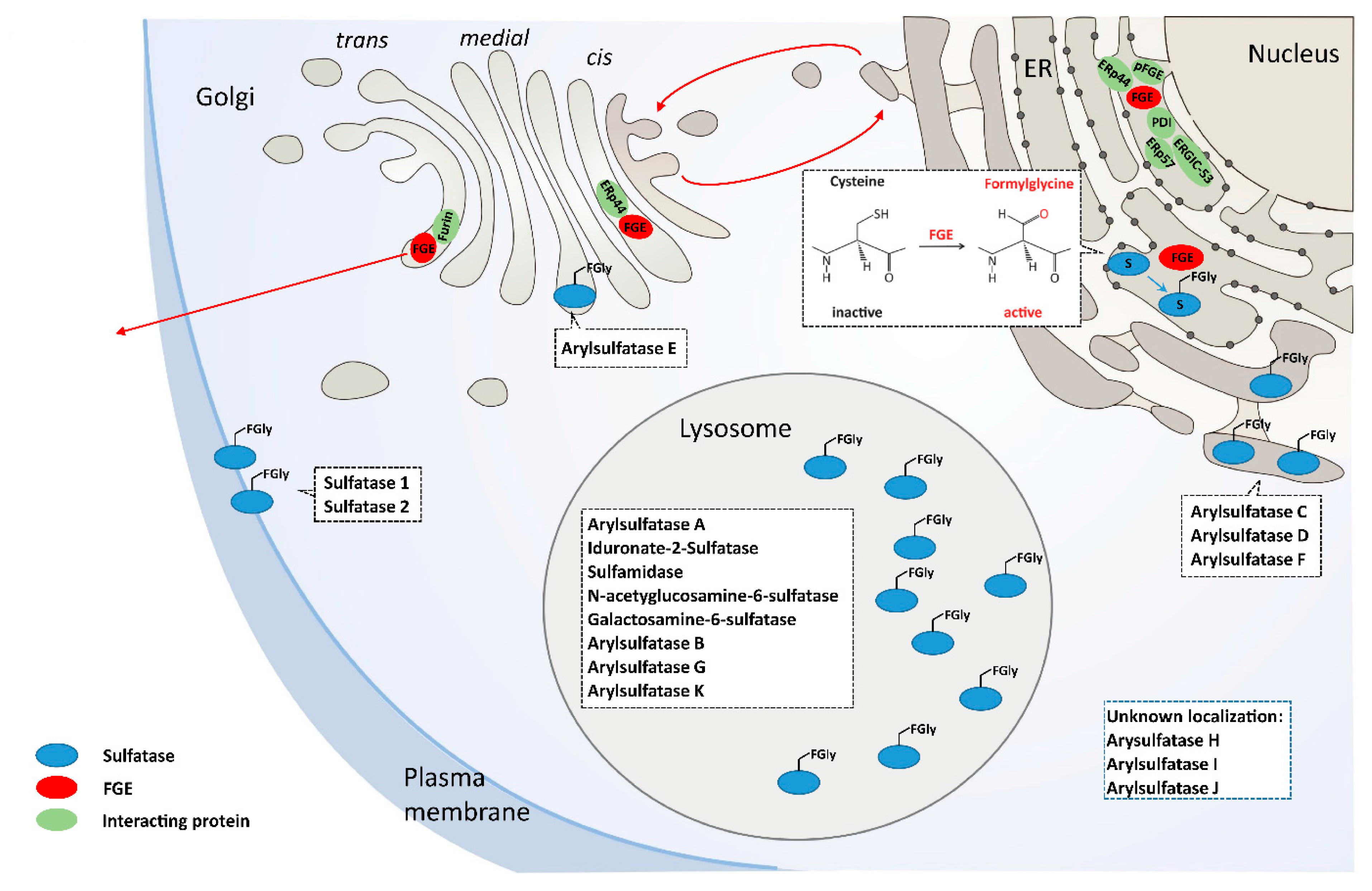
3. FGE the MSD Protein

SUMF1, located on chromosome 3p26, is 106 kb long and contains 9 exons, and is ubiquitously expressed with highest levels in kidneys and pancreas. Human FGE is a 40 kDa (374 residues) glycoprotein localized in the endoplasmic reticulum (ER). Residues 1–32 constitute the cleavable ER-signal sequence and the mature protein (residues 33–374) is N-glycosylated at asparagine N141. The mature protein is organized into a core domain (residues 73–374) that harbors the active site and an N-terminal extension (residues 33–72). FGE contains 8 conserved cysteines forming 2 disulfide bridges, a pair of catalytically active cysteines in the active site, and a pair of cysteines in the N-terminal extension [14,15]. In cultured cells, FGE is secreted and a major fraction of the secreted form is devoid of this N-terminal extension. The N-terminal extension (residues 33–72) encoded by exon 1 of SUMF1 is a unique feature found only in FGE from higher eukaryotes and was shown to impart two important functions via its two cysteins C50 and C52. Mutating these cysteines to alanine led to loss of activation of sulfatases and loss of interaction with ERp44 for FGE retrieval back to the ER. For unknown reasons, the N-terminal extension is cleaved off by the trans-Golgi-resident furin protease. Other interacting partners ERGIC-53, ERp57 and PDI mediate ER retention, export and retrieval [16]. PDI is of special interest because it facilitates sulfatase activation through gatekeeping of proper FGE folding (see below) [17]. FGE has an interacting paralogue protein pFGE, encoded by the SUMF2 gene that shares 47% percent of homology. pFGE is co-expressed with FGE in many tissues and binds sulfatases but lacks catalytic function. A proposed function of pFGE is to help FGE activate sulfatase; however, more evidence is needed to support this role [18,19,20]. The intracellular localization and trafficking of FGE along the secretory pathway is considered to be a highly regulated process spatially and temporally controlled by its interacting partners (Figure 1).
The FGE core protein exerts its enzymatic function on newly synthesized sulfatases co- or post-translationally. FGE recognizes sulfatases by a linear sequence motif CxPxR as part of the putative sulfatase signature I (C-STACG-P-STA-R-x(2)-LIVMFW) in the N-terminal catalytic domain. This motif is evolutionary highly conserved and specific for recognition by FGE that converts the cysteine into a sulfatase-specific amino acid, Cα-formylglycine (FGly) [14,21,22,23]. Introducing the CxPxR motif into engineered proteins in cellular expression systems lead to FGly conversion by FGE and is used in a variety of biotechnology applications [24,25].
Sulfatases are a group of enzymes that degrade or remodel sulfate esters. There are 17 sulfatases encoded by the human genome, 13 of which have been characterized biochemically. The majority of sulfatases are localized to the lysosome, while others are located in the ER, Golgi, or on the cell surface (Table 1).
Sulfatase substrates comprise glycosaminoglycans (GAGs), sulfolipids, and steroid hormones [26,27]. In addition, sulfatases exert important regulatory roles on heparan sulfate-dependent cellular signaling pathways [28,29]. High specificity for substrates leads to low redundancy by different sulfatases, as such single sulfatase deficiencies result in severe disorders, most of which are lysosomal disorders [26]. No redundant FGly generating mechanism exists in mammals, and FGE is a limiting factor for sulfatase activation. A complete loss of SUMF1 or FGE function results in decreased activity of all sulfatases rendering MSD a monogenetic disease [1,3]. The requirement of FGly generation by FGE for catalytic activity of sulfatases has prompted the co-expression of FGE in the production process of recombinant sulfatases for enzyme replacement therapy as well as gene-therapy approaches for single sulfatase deficiencies [30,31,32,33,34].
The role of FGEs secreted form is unknown. A paracrine function has been discussed because of its cellular uptake followed by FGE reaching the ER and exerting sulfatase activation [35,36]. This renders FGE to be one exceptional example of a protein reaching the ER after endocytosis, a mechanism known from toxins or viruses [37]. Further experimental evidence is needed before recombinant FGE could eventually be used for ERT as a therapy for MSD.
Recently, another mechanism of regulation for FGE activity has been proposed: miRNA-95 depletes FGE and results in decreased sulfatase activities leading to impaired lysosomal function, substrate accumulation and a block of autophagy. Lowering the levels of miRNA-95 in MSD patient cells restored sulfatase function exerting a therapeutic potential [38].
The crystal structure of FGE has been established in 2005 and allows insight into FGEs mode of action as well as modelling changes on its structure by MSD causing SUMF1 mutations [14,39]. FGE has a unique fold with low amount of secondary structure (13% α-helices, 20% β-sheets). Stabilization occurs via disulphide bridges (see above) and two calcium ions. The crystal structure suggested FGE to be a mono-oxygenase as FGly generation consumed equimolar levels of molecular oxygen although no redox active metal ions could be detected [14]. For over a decade, FGE was regarded as a metal-independent mono- oxygenase.
However, an increase in the activity of FGE after in vitro reconstitution with Cu2+, led to the proposal that FGE utilizes copper as a cofactor [40,41,42]. Recently, the crystal structures of two prokaryotic FGE-holoenzymes reconstituted with Cu from Streptomycis coelicolor and Thermomonospora curvata were reported [43,44]. Both structures revealed that a Cu(I) atom is coordinated by two active site cysteine residues in a nearly linear geometry, unusual for copper-dependent oxidases. Based on structural and biochemical data, a structural basis for oxygen activation by the catalytic copper center via a Cu(II) superoxo-intermediate was proposed, thus providing mechanistic insights into FGly generation by FGE and activation of sulfatases.

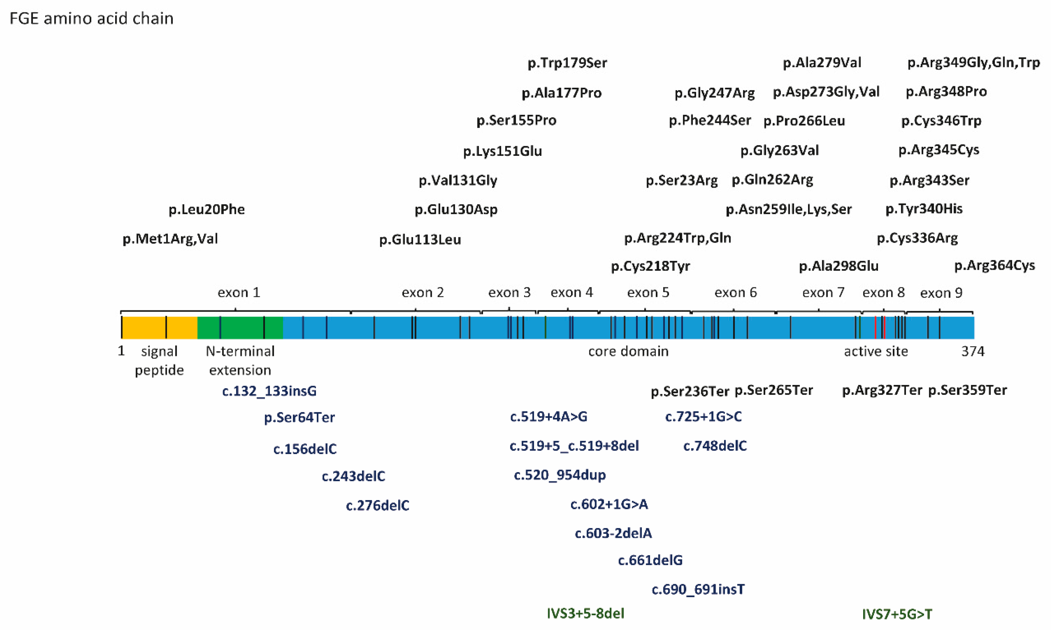
4. SUMF1 Mutations and Functional Consequences

Fifty-three SUMF1 mutations have been published in the literature since the discovery of the SUMF1 gene including 21 nonsense mutations (frameshift, stop-gain, deletions), and 32 missense mutations distributed over the entire length of the protein (Figure 2).
Functional consequences of SUMF1 mutations on the FGE protein have been analyzed for a subset of mutations using a combination of in silico and in vitro biochemical methods. Crystallization of the FGE protein facilitated in silico modelling of amino acid exchanges and prediction of FGE stability and activity especially when variants affect the active site of FGE [14,39]. In vitro experimental data were generated for a subset of mutations using different experimental approaches. Analysis of endogenous FGE levels in patient derived fibroblasts provided information on protein stability and subcellular localization. Pulse-chase experiments of cell lines expressing variant FGE constructs have been used to determine protein half-life for a variety of SUMF1 variants [45].
In addition to impaired protein stability, FGE enzymatic activity is affected by clinically relevant SUMF1 mutations. FGE activity is best assessed through the measurement of sulfatase activities at endogenous levels in patient cells or by co- expression of indicative sulfatases and sulfatase activity assays in cell models [45,46,47,48,49,50]. A well-established assay uses bi-directional co-expression of steroid sulfatase and FGE from an inducible vector proven to yield highly reproducible and comparable sulfatase activities referring to FGE activity [51]. Apart from the mass spectrometry-based assay described above, currently there is no assay available for direct FGE activity measurement. Although this approach requires partially purified variant FGE, it has been used for FGE activity determination for a subset of variant FGE proteins [45,50].
The majority of SUMF1 mutations are of hypomorphic nature resulting in expression of FGE with residual activity [46]. Only nonsense mutations completely abrogate FGE function [52]. All previously analyzed mutations demonstrated reduced intracellular protein levels and excretion supporting the hypothesis that most variants result in early degradation and/or reduced stability of FGE [45,46,47,48,50,52]. In fact, the majority of FGE variants analyzed demonstrate a half-life of less than 2 h [45,49,50,51].
Residual FGE activity of variant proteins was variable ranging from complete loss of activity in active site mutations to 50% of reference activity [45,49,50]. Of note, 1/3 of all published SUMF1 mutations lack experimental data on stability and activity. Disease causing variants of FGE likely disrupt function through overall instability and decreased enzymatic activity.
Data are beginning to come up on the intracellular fate of variant FGE species. Protein disulfide isomerase (PDI) was shown to interact with wild type FGE and co-expression of PDI improved activation of sulfatases. This led to the suggestion that PDI could play a role in improving proper folding of wildtype FGE [16]. However, PDI was shown to preferentially interact with misfolded variant FGE, which established misfolded variant FGE as a physiological substrate for PDI [17]. Moreover, the crucial function of PDI as a disease modifier in MSD emerged when it was shown to play a role in variant FGE stability and residual activity. In vitro, overexpression of PDI reduces residual sulfatase activities, while PDI silencing rescues sulfatase activities by increasing variant FGE stability.
Downstream effects of FGE deficiency and sulfatase dysfunction are only partially understood in MSD, and only a limited number of studies have explored the pathophysiology of disease. As part of the MSD phenotype, glycosaminoglycans (GAGs) and sulfatides accumulate, as is seen in mucopolysaccharidoses (MPS) and MLD. This abnormal storage results in lysosomal dysfunction, impacting several cellular processes including autophagy [53]. Many pathophysiological aspects proposed for lysosomal dysfunction, e.g., membrane turnover, calcium storage or mTORC1 function, have not been explored in MSD [54]. Effects caused by the remaining uncharacterized sulfatases are yet to be determined.

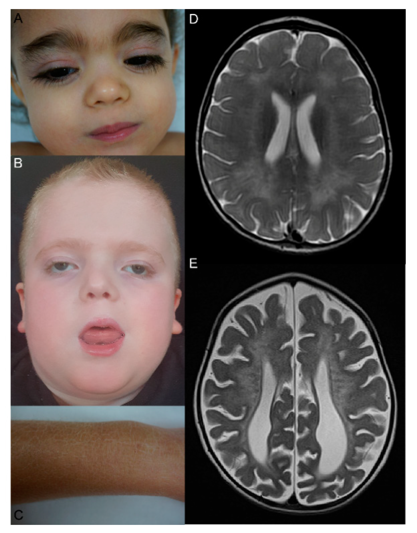
5. Clinical, MRI, and Ultrastructural Features, Disease Classifications, Genotype-Phenotype Correlation

MSD is a complex disease because it combines symptoms of single sulfatase deficiencies. It is a progressive, systemic, neurodegenerative disorder of early childhood. The majority of individuals with MSD present with systemic features and psychomotor retardation followed by loss of motor skills, speech, hearing, and vision. Many individuals also have characteristic facial features consistent with MPS disorders, chondrodysplasia punctata, hepatosplenomegaly, and ichthyosis (Figure 3A–C). While all individuals with MSD have neurologic impairment, the other systemic features can be variable and are often not present at birth [45,48,50]. Additional symptoms include cardiac defects, pulmonary involvement, recurrent ear infections, sleeping disorders, hydrocephalus, corneal clouding, retinitis pigmentosa, and hydrops fetalis. Sleep disturbance, feeding difficulties, constipation, spasticity, and hip dislocation are frequent symptoms that affect quality of life. Some patients failed in newborn hearing screening tests. MRI findings can also be variable with demyelination resembling MLD, brain atrophy, corpus callosum hypoplasia, subcerebellar cysts, and hydrocephalus [55] (Figure 3D,E). Ultrastructural findings comprise pleomorphic lysosomal inclusions in Schwann cells detected in skin biopsies, and reduced germ size and hypomineralized enamel in teeth. Brain tissue analysis revealed pleomorphic extracellular, intraneural, and intraglial inclusions and white matter showed gliosis and metachromasia [56,57,58].
Similar to other lysosomal storage disorders, MSD can be divided into neonatal, late infantile and juvenile forms based on age at onset [59]. Revisions of this classification added gradations of disease severity to the age of onset subtypes. While early onset of disease is associated with the most severe forms, it is unknown if how well these subtypes associate with overall clinical outcomes.
Children with early onset disease, the neonatal form, are characterized by presence of multiple MSD-related symptoms at birth. Late infantile cases, which are divided into severe and mild forms, present symptoms in infancy. The latter shows a reduced number of symptoms and a later onset, especially of neurodegeneration and neurological symptoms, and slow acquisition of additional symptoms [45,48,50]. Juvenile MSD is the most rare and the most attenuated form [60].
While these classifications are still in use, there are several limitations to their clinical utility. Upon deeper medical history review, many individuals with an attenuated form of MSD presented with a limited number of symptoms in the neonatal period. These early symptoms often include failure to thrive with feeding difficulties, inguinal hernias, and hypotonia. Based on the existing classification system, these individuals would qualify as neonatal form MSD by onset, but as juvenile or late infantile by clinical course. Clearly, there is a need to define clinically coherent subtypes of MSD that are based on our modern, deeper understanding of the clinical course of MSD [45,48,50].
Sulfatase activities in patient fibroblasts have been used for an alternative, historical disease classification in MSD: Group I fibroblasts exhibit residual activity below 15% of control activity measured in fibroblasts from non-MSD patients. Group II fibroblasts show more than 15% of residual activity, sometimes reaching normal values. Group I fibroblasts derive from severe MSD cases whereas group II fibroblasts originate from attenuated MSD cases [52,56,61,62,63]. Interestingly, and in contrast to group I fibroblasts, group II fibroblasts were reported to show variable sulfatase activity over time in the same cell line. Such cells are likely influenced by cell culture conditions and culture time [61,64]. The mechanisms underlying sulfatase activity variation are unknown. The number of MSD fibroblast cell lines that have been investigated are limited, impairing our full understanding of sulfatase activity fluctuations. However, subsequent publications have supported that a subset of patients could have variable sulfatase activities overtime [48].
Despite the difficulties in establishing a reliable disease classification, the clinical course of MSD falls into two categories: Severe and attenuated. Further work is needed to associate these clinical forms with other features of the disease. There is emerging evidence for a genotype–phenotype correlation in the homozygous form of the disease. Patients with biallelic nonsense mutations have the most severe symptoms and often have absent sulfatase activities (severe or neonatal very severe form) [52]. Attenuated patients often harbor variants that result in a highly unstable variant FGE with low residual activity. Milder attenuated patients can have unstable FGE variants with high residual activity or stable FGE variants with low residual activity. This preliminary genotype–phenotype correlation allows for a rough prediction of the course of disease in cases with homozygous SUMF1 mutations that have been experimentally assessed [45,48,50]. A genotype–phenotype correlation in compound heterozygous patients has not been fully investigated and is an area of active interest.

6. Biomarkers and Diagnosis

Disease specific biomarkers for MSD exploit the biomarkers developed for the related lysosomal disorders. This includes measurements of sulfatase activities, urinary sulfatides, and glycosaminoglycan accumulation. The simultaneous excretion of heparan sulfate, dermatan sulfate, keratan sulfate and chondroitin sulfate is strongly suggestive of MSD. Interestingly, increased GAG excretion is absent in some MSD patients [48] and the factors contributing to such negative GAG measurements in MSD patients are poorly understood.
Sulfatase activities are low in MSD patients, but levels can vary over time, even in the same individual. Also, since sulfatase activities are very dependent on the biochemical assay design, comparing results obtained in different laboratories is difficult [63,65]. In short, a detailed understanding of the association between specific SUMF1 variants and biochemical phenotypes are lacking. Historically, the clinical diagnosis of MSD was confirmed by measuring simultaneous deficiency of more than one sulfatase activity in patient leukocytes or fibroblasts. Nowadays, SUMF1 genetic testing supports the diagnosis of MSD. With the advancement of broad based, non-targeted sequencing (next-generation sequencing panels, exome and genome sequencing), it is likely that novel SUMF1 mutations will be found and the phenotypic spectrum will also evolve.
However, it has to be noted that both biochemical and genetic confirmation have a risk of false negative results. MSD could be missed on panels that measure limited sulfatases, for example, when a test measures only two sulfatases, one could be abnormal and the other normal. In theory, cases could be misdiagnosed as MSD due to arylsulfatase A pseudodeficiency, which is a biochemical artefact of in vitro testing. Sanger sequencing of genomic SUMF1 could miss large deletions expanding into or spanning the complete SUMF1 gene. Also, as MSD is so rare, it is possible that pathogenic variants could be mislabeled as variants of unknown significance. Given these diagnostic challenges, we recommend a combination of genetic and biochemical testing with the measurement of at least three sulfatases.

7. Care of MSD Patients

No curative therapy is currently available for MSD. MSD patients often face multiple health problems due to the complexity of the disease. As a result from the first International Conference on MSD (Dublin, July 2017) a consensus statement on the complex care and management of MSD patients was published providing the first clinical guidelines for MSD [55]. The consensus statement outlines a standardized framework for comprehensively addressing clinical problems and the management of symptoms in MSD. These guidelines summarize a list of initial evaluations that should be considered in every newly-diagnosed MSD patient to assess for the major clinical complications that have been reported on this rare disease. This includes evaluation of 10 different organ systems, each with a variety of potential complications (Table 2). This comprehensive list of potential interventions needs to be individualized based on the patient and institutional experience. An expert-driven multidisciplinary approach with goal of a high quality-of-life for the affected individuals and also the care-givers should be the primary objective of care [55].

8. MSD Animal Disease Models

A SUMF1 gene-trap knock-out mouse (SUMF1gt) was the first MSD animal model. Homozygous SUMF1gt mice displayed absence of eight sulfatase activities tested and storage of glycosaminoglycans in macrophages, kidney and liver. Mice showed signs of head tremor, seizures, skeletal abnormalities and a flat facial appearance. All had congenital growth retardation and more than 90% of SUMF1gt mice died prior to 3 months of age. The mice demonstrated significant systemic and neuroinflammation, indicated by activated microglia, astrocytosis and neuronal loss [66]. Furthermore, fibroblast growth factor signaling was constitutively activated in SUMF1 knock-out mice derived hematopoietic stem cells and hematopoietic stem progenitor cells resulting in a block in erythroid differentiation, disruption of B lymphocyte differentiation, reduction in mature myeloid cells and aberrant T lymphocyte development [67]. Astrocyte specific knock-out of SUMF1 in a Cre/Lox MSD mouse model revealed lysosomal storage of substrates and autophagy substrates impairing astrocyte function. This led to neuronal loss thereby highlighting the importance of astrocyte dysfunction in the pathophysiology of MSD [68].
The SUMF1gt mouse model has also been used for the first MSD treatment approach: Combined intraventricular and systemic administration of a recombinant adeno-associated virus type 9 (rAAV9) encoding FGE resulted in a widespread transduction of different tissues followed by an increase of sulfatase activities, clearance of glycosaminoglycans and decrease of inflammation. rAAV9SUMF1 treated mice also showed improved behavioral issues. These results suggest that gene therapy might be a treatment option in MSD [69]. Additional mouse models harboring common human SUMF1 variants are currently being generated and characterized. These tools can be used to evaluate novel therapies aimed at correcting misfolded FGE species or increasing lysosomal performance and clearance abilities. The high evolutionary conservation of SUMF1/FGE has allowed for the establishment of additional animal models. A drosophila melanogaster SUMF1 knock out fly and SUMF1 knock out zebrafish lines are currently being characterized. Both models can be used to study early embryonic development in MSD and could also prove useful to future drug screening efforts.

9. Patient Organizations and Research Towards a Therapy for MSD

Independently two patient organizations were founded in 2015 by parents with children affected by MSD. MSD Action foundation in Ireland and United MSD Foundation in the US have become drivers in collaborative efforts to improve the clinical care and research options for children with MSD. Since the founding work of these important organizations, two more national patient organizations were founded in Spain and Argentina. Patient organizations have a major impact on basic and clinical research for rare diseases by connecting researchers, clinicians and patients, offering grants for research towards therapy and running patient registries [70]. Social networks connect patients and families with rare diseases globally and facilitate efforts to make rare diseases known to the general public [71,72]. Like other rare diseases, comprehensive natural disease history data on MSD is lacking. Current efforts are underway to better define the clinical course of MSD around the world. Results from these projects will serve as a basis for defining natural disease history and clinical endpoints in anticipation of clinical trials in the near future.

10. Future Perspectives

Given the growing interest in the science of MSD and awareness of the disease in general, we anticipate that our understanding of MSD will also continue to develop. The search for additional interacting partners of FGE that regulate enzymatic function, folding, trafficking and degradation will elucidate novel aspects of cell biology. All newly discovered interacting proteins and pathways have the potential of serving as targets for the development of MSD therapy.
In terms of clinical knowledge, definitions of MSD will be informed by ongoing and future natural disease history studies. Such studies may further our understanding of factors that determine disease severity, clarify genotype-phenotype relationships, and reveal biomarkers that can be used to track disease progression. Finally, as newborn screening for single sulfatase disorders and genomic sequencing are more widely utilized, new MSD patients will be identified that will likely expand the MSD phenotype.
The exciting recent progress in MSD research represents the synthesis of basic science and clinical medicine with patient advocacy. As this foundation of knowledge grows, our hope is that potential treatment options will be developed. The growing understanding of the pathophysiology of the disease and generation of the MSD animal models, will set the stage for exploring potential treatment options, design potential therapeutic agents and serve as tools for pre-clinical therapy evaluation and testing. Also, one can expect that meaningful clinical endpoints derived from natural disease history data will aid clinically-relevant trial design. It is crucial that for an ultra-rare and orphan disease like MSD, any therapeutic strategy that ameliorates the disease condition would greatly improve the lives of MSD patients and their families.

Author Contributions

L.S., L.A.A., K.R., and R.C.A.-N. did a literature search, corrected and edited the manuscript. L.S. wrote the first draft and prepared figures and tables. All authors have read and agreed to the published version of the manuscript.

Funding

L.S., K.R. and R.C.A.-N. received research grants from MSDAF, and L.S. received research grants from MRCG/HRB, Ireland, for research on treatment development for MSD. Publication fees are granted by Lower Saxony State- and University- Library Göttingen, Germany.

Acknowledgments

We would like to thank Amber Olsen, United MSD Foundation (UMSDF), USA, and Alan Finglas, MSD Action Foundation (MSDAF), Ireland, for initiation of research projects on MSD and their contribution in connecting MSD patients, families, doctors and researchers. We are grateful to Jutta Gärtner, Department of Pediatrics and Adolescent Medicine, University Medical Center Goettingen, Germany, Thomas Dierks, Biochemistry I, Bielefeld University, Germany, and Adeline Vanderver, The Children’s Hospital of Philadelphia for ongoing support. We thank all MSD patients and their parents for their participation in actual research projects on MSD.

Conflicts of Interest

Authors declare no conflicts of interest.

References

  1. Cosma, M.P.; Pepe, S.; Annunziata, I.; Newbold, R.F.; Grompe, M.; Parenti, G.; Ballabio, A. The Multiple Sulfatase Deficiency Gene Encodes an Essential and Limiting Factor for the Activity of Sulfatases. Cell 2003, 113, 445–456. [Google Scholar] [CrossRef]
  2. Dierks, T.; Schlotawa, L.; Frese, M.-A.; Radhakrishnan, K.; Von Figura, K.; Schmidt, B. Molecular basis of multiple sulfatase deficiency, mucolipidosis II/III and Niemann–Pick C1 disease—Lysosomal storage disorders caused by defects of non-lysosomal proteins. Biochim. Biophys. Acta (BBA) Bioenerg. 2009, 1793, 710–725. [Google Scholar] [CrossRef] [PubMed] [Green Version]
  3. Dierks, T.; Schmidt, B.; Borissenko, L.V.; Peng, J.; Preusser, A.; Mariappan, M.; Von Figura, K. Multiple Sulfatase Deficiency Is Caused by Mutations in the Gene Encoding the Human Cα-Formylglycine Generating Enzyme. Cell 2003, 113, 435–444. [Google Scholar] [CrossRef] [Green Version]
  4. Meikle, P.J.; Hopwood, J.J.; Clague, A.E.; Carey, W.F. Prevalence of lysosomal storage disorders. JAMA 1999, 281, 249–254. [Google Scholar] [CrossRef]
  5. Austin, J.; Armstrong, D.; Shearer, L. Metachromatic form of diffuse cerebral sclerosis. V. The nature and significance of low sulfatase activity: A controlled study of brain, liver and kidney in four patients with metachromatic leukodystrophy (MLD). Arch. Neurol. 1965, 13, 593–614. [Google Scholar] [CrossRef]
  6. Austin, J.H. Studies in Metachromatic Leukodystrophy. Arch. Neurol. 1973, 28, 258. [Google Scholar] [CrossRef]
  7. Eto, Y.; Wiesmann, U.N.; Carson, J.H.; Herschkowitz, N.N. Multiple sulfatase deficiencies in cultured skin fibroblasts. Occurrence in patients with a variant form of metachromatic leukodystrophy. Arch. Neurol. 1974, 30, 153–156. [Google Scholar] [CrossRef]
  8. Horwitz, A.L. Genetic complementation studies of multiple sulfatase deficiency. Proc. Natl. Acad. Sci. USA 1979, 76, 6496–6499. [Google Scholar] [CrossRef] [Green Version]
  9. Chang, P.L.; Davidson, R.G. Complementation of arylsulfatase A in somatic hybrids of metachromatic leukodystrophy and multiple sulfatase deficiency disorder fibroblasts. Proc. Natl. Acad. Sci. USA 1980, 77, 6166–6170. [Google Scholar] [CrossRef] [Green Version]
  10. Fedde, K.; Horwitz, A.L. Complementation of multiple sulfatase deficiency in somatic cell hybrids. Am. J. Hum. Genet. 1984, 36, 623–633. [Google Scholar]
  11. Ballabio, A.; Parenti, G.; Napolitano, E.; Di Natale, P.; Andria, G. Genetic complementation of steroid sulphatase after somatic cell hybridization of X-linked ichthyosis and multiple sulphatase deficiency. Qual. Life Res. 1985, 70, 315–317. [Google Scholar] [CrossRef] [PubMed]
  12. Rommerskirch, W.; Von Figura, K. Multiple sulfatase deficiency: Catalytically inactive sulfatases are expressed from retrovirally introduced sulfatase cDNAs. Proc. Natl. Acad. Sci. USA 1992, 89, 2561–2565. [Google Scholar] [CrossRef] [PubMed] [Green Version]
  13. Schmidt, B.; Selmer, T.; Ingendoh, A.; Von Figurat, K. A novel amino acid modification in sulfatases that is defective in multiple sulfatase deficiency. Cell 1995, 82, 271–278. [Google Scholar] [CrossRef] [Green Version]
  14. Dierks, T.; Dickmanns, A.; Preusser-Kunze, A.; Schmidt, B.; Mariappan, M.; Von Figura, K.; Ficner, R.; Rudolph, M.G. Molecular Basis for Multiple Sulfatase Deficiency and Mechanism for Formylglycine Generation of the Human Formylglycine-Generating Enzyme. Cell 2005, 121, 541–552. [Google Scholar] [CrossRef] [PubMed] [Green Version]
  15. Mariappan, M.; Gande, S.L.; Radhakrishnan, K.; Schmidt, B.; Dierks, T.; Von Figura, K. The Non-catalytic N-terminal Extension of Formylglycine-generating Enzyme Is Required for Its Biological Activity and Retention in the Endoplasmic Reticulum. J. Boil. Chem. 2008, 283, 11556–11564. [Google Scholar] [CrossRef] [Green Version]
  16. Fraldi, A.; Zito, E.; Annunziata, F.; Lombardi, A.; Cozzolino, M.; Monti, M.; Spampanato, C.; Ballabio, A.; Pucci, P.; Sitia, R.; et al. Multistep, sequential control of the trafficking and function of the multiple sulfatase deficiency gene product, SUMF1 by PDI, ERGIC-53 and ERp44. Hum. Mol. Genet. 2008, 17, 2610–2621. [Google Scholar] [CrossRef]
  17. Schlotawa, L.; Wachs, M.; Bernhard, O.; Mayer, F.J.; Dierks, T.; Schmidt, B.; Radhakrishnan, K. Recognition and ER Quality Control of Misfolded Formylglycine-Generating Enzyme by Protein Disulfide Isomerase. Cell Rep. 2018, 24, 27–37. [Google Scholar] [CrossRef] [Green Version]
  18. Mariappan, M.; Preusser-Kunze, A.; Balleininger, M.; Eiselt, N.; Schmidt, B.; Gande, S.L.; Wenzel, D.; Dierks, T.; von Figura, K. Expression, localization, structural, and functional characterization of pFGE, the paralog of the Calpha-formylglycine-generating enzyme. J. Biol. Chem. 2005, 280, 15173–15179. [Google Scholar] [CrossRef] [Green Version]
  19. Gande, S.L.; Mariappan, M.; Schmidt, B.; Pringle, T.H.; Von Figura, K.; Dierks, T. Paralog of the formylglycine-generating enzyme—Retention in the endoplasmic reticulum by canonical and noncanonical signals. FEBS J. 2008, 275, 1118–1130. [Google Scholar] [CrossRef]
  20. Zito, E.; Fraldi, A.; Pepe, S.; Annunziata, I.; Kobinger, G.; Di Natale, P.; Ballabio, A.; Cosma, M.P. Sulphatase activities are regulated by the interaction of sulphatase-modifying factor 1 with SUMF2. EMBO Rep. 2005, 6, 655–660. [Google Scholar] [CrossRef] [Green Version]
  21. Roeser, D.; Preusser-Kunze, A.; Schmidt, B.; Gasow, K.; Wittmann, J.G.; Dierks, T.; Von Figura, K.; Rudolph, M.G. A general binding mechanism for all human sulfatases by the formylglycine-generating enzyme. Proc. Natl. Acad. Sci. USA 2005, 103, 81–86. [Google Scholar] [CrossRef] [PubMed] [Green Version]
  22. Dierks, T.; Lecca, M.; Schlotterhose, P.; Schmidt, B.; Von Figura, K. Sequence determinants directing conversion of cysteine to formylglycine in eukaryotic sulfatases. EMBO J. 1999, 18, 2084–2091. [Google Scholar] [CrossRef] [PubMed] [Green Version]
  23. Recksiek, M.; Selmer, T.; Dierks, T.; Schmidt, B.; Von Figura, K. Sulfatases, Trapping of the Sulfated Enzyme Intermediate by Substituting the Active Site Formylglycine. J. Boil. Chem. 1998, 273, 6096–6103. [Google Scholar] [CrossRef] [PubMed] [Green Version]
  24. Carrico, I.S.; Carlson, B.L.; Bertozzi, C.R. Introducing genetically encoded aldehydes into proteins. Nat. Methods 2007, 3, 321–322. [Google Scholar] [CrossRef] [PubMed]
  25. Rupniewski, I.; Rabuka, D. Site-Specific Labeling of Proteins Using the Formylglycine-Generating Enzyme (FGE). Breast Cancer 2019, 2012, 63–81. [Google Scholar] [CrossRef]
  26. Diez-Roux, G.; Ballabio, A. Sulfatases and Human Disease. Annu. Rev. Genom. Hum. Genet. 2005, 6, 355–379. [Google Scholar] [CrossRef] [PubMed]
  27. Sardiello, M.; Annunziata, I.; Roma, G.; Ballabio, A. Sulfatases and sulfatase modifying factors: An exclusive and promiscuous relationship. Hum. Mol. Genet. 2005, 14, 3203–3217. [Google Scholar] [CrossRef] [Green Version]
  28. Wang, S.; Ai, X.; Freeman, S.D.; Pownall, M.E.; Lu, Q.; Kessler, D.S.; Emerson, C.P. QSulf1, a heparan sulfate 6-O-endosulfatase, inhibits fibroblast growth factor signaling in mesoderm induction and angiogenesis. Proc. Natl. Acad. Sci. USA 2004, 101, 4833–4838. [Google Scholar] [CrossRef] [Green Version]
  29. Dhoot, G.K. Regulation of Wnt Signaling and Embryo Patterning by an Extracellular Sulfatase. Science 2001, 293, 1663–1666. [Google Scholar] [CrossRef]
  30. Muenzer, J.; Wraith, J.E.; Beck, M.; Giugliani, R.; Harmatz, P.; Eng, C.M.; Vellodi, A.; Martin, R.; Ramaswami, U.; Gucsavas-Calikoglu, M.; et al. A phase II/III clinical study of enzyme replacement therapy with idursulfase in mucopolysaccharidosis II (Hunter syndrome). Genet. Med. 2006, 8, 465–473. [Google Scholar] [CrossRef] [Green Version]
  31. Takakusaki, Y.; Hisayasu, S.; Hirai, Y.; Shimada, T. Coexpression of formylglycine-generating enzyme is essential for synthesis and secretion of functional arylsulfatase A in a mouse model of metachromatic leukodystrophy. Hum. Gene Ther. 2005, 16, 929–936. [Google Scholar] [CrossRef] [PubMed]
  32. Fraldi, A.; Hemsley, K.M.; Crawley, A.; Lombardi, A.; Lau, A.A.; Sutherland, L.; Auricchio, A.; Ballabio, A.; Hopwood, J.J. Functional correction of CNS lesions in an MPS-IIIA mouse model by intracerebral AAV-mediated delivery of sulfamidase and SUMF1 genes. Hum. Mol. Genet. 2007, 16, 2693–2702. [Google Scholar] [CrossRef] [PubMed] [Green Version]
  33. Sevin, C.; Verot, L.; Benraiss, A.; Van Dam, D.; Bonnin, D.; Nagels, G.; Fouquet, F.; Gieselmann, V.; Vanier, M.T.; De Deyn, P.P.; et al. Partial cure of established disease in an animal model of metachromatic leukodystrophy after intracerebral adeno-associated virus-mediated gene transfer. Gene Ther. 2006, 14, 405–414. [Google Scholar] [CrossRef] [PubMed] [Green Version]
  34. Tomatsu, S.; Montaño, A.M.; Ohashi, A.; Gutierrez, M.A.; Oikawa, H.; Oguma, T.; Dung, V.C.; Nishioka, T.; Orii, T.; Sly, W.S. Enzyme replacement therapy in a murine model of Morquio A syndrome. Hum. Mol. Genet. 2007, 17, 815–824. [Google Scholar] [CrossRef] [Green Version]
  35. Preusser-Kunze, A.; Mariappan, M.; Schmidt, B.; Gande, S.L.; Mutenda, K.; Wenzel, D.; Von Figura, K.; Dierks, T. Molecular Characterization of the Human Cα-formylglycine-generating Enzyme. J. Boil. Chem. 2005, 280, 14900–14910. [Google Scholar] [CrossRef] [Green Version]
  36. Zito, E.; Buono, M.; Pepe, S.; Settembre, C.; Annunziata, I.; Surace, E.M.; Dierks, T.; Monti, M.; Cozzolino, M.; Pucci, P.; et al. Sulfatase modifying factor 1 trafficking through the cells: From endoplasmic reticulum to the endoplasmic reticulum. EMBO J. 2007, 26, 2443–2453. [Google Scholar] [CrossRef] [Green Version]
  37. Spooner, R.A.; Smith, D.C.; Easton, A.J.; Roberts, L.M.; Lord, J.M. Retrograde transport pathways utilised by viruses and protein toxins. Virol. J. 2006, 3, 26. [Google Scholar] [CrossRef] [Green Version]
  38. Frankel, L.B.; Di Malta, C.; Wen, J.; Eskelinen, E.-L.; Ballabio, A.; Lund, A.H. A non-conserved miRNA regulates lysosomal function and impacts on a human lysosomal storage disorder. Nat. Commun. 2014, 5, 5840. [Google Scholar] [CrossRef] [Green Version]
  39. Paul, D.M.; Chadah, T.; Senthilkumar, B.; Sethumadhavan, R.; Rajasekaran, R. Structural distortions due to missense mutations in human formylglycine-generating enzyme leading to multiple sulfatase deficiency. J. Biomol. Struct. Dyn. 2017, 36, 3575–3585. [Google Scholar] [CrossRef]
  40. Holder, P.G.; Jones, L.C.; Drake, P.M.; Barfield, R.M.; Bañas, S.; De Hart, G.W.; Baker, J.; Rabuka, D. Reconstitution of Formylglycine-generating Enzyme with Copper(II) for Aldehyde Tag Conversion. J. Boil. Chem. 2015, 290, 15730–15745. [Google Scholar] [CrossRef] [Green Version]
  41. Knop, M.; Lemnaru, R.; Seebeck, F.P. Mutation of Conserved Residues Increases in Vitro Activity of the Formylglycine-Generating Enzyme. ChemBioChem 2017, 18, 1755–1761. [Google Scholar] [CrossRef]
  42. Meury, M.; Knop, M.; Seebeck, F.P. Structural Basis for Copper-Oxygen Mediated C−H Bond Activation by the Formylglycine-Generating Enzyme. Angew. Chem. 2017, 56, 8115–8119. [Google Scholar] [CrossRef] [PubMed] [Green Version]
  43. Appel, M.; Meier, K.K.; Lafrance-Vanasse, J.; Lim, H.; Tsai, C.-L.; Hedman, B.; Hodgson, K.O.; Tainer, J.; Solomon, E.I.; Bertozzi, C.R. Formylglycine-generating enzyme binds substrate directly at a mononuclear Cu(I) center to initiate O2activation. Proc. Natl. Acad. Sci. USA 2019, 116, 5370–5375. [Google Scholar] [CrossRef] [PubMed] [Green Version]
  44. Miarzlou, D.A.; Leisinger, F.; Joss, D.; Häussinger, D.; Seebeck, F.P. Structure of formylglycine-generating enzyme in complex with copper and a substrate reveals an acidic pocket for binding and activation of molecular oxygen. Chem. Sci. 2019, 10, 7049–7058. [Google Scholar] [CrossRef] [PubMed] [Green Version]
  45. Schlotawa, L.; Ennemann, E.C.; Radhakrishnan, K.; Schmidt, B.; Chakrapani, A.; Christen, H.-J.; Moser, H.; Steinmann, B.; Dierks, T.; Gärtner, J. SUMF1 mutations affecting stability and activity of formylglycine generating enzyme predict clinical outcome in multiple sulfatase deficiency. Eur. J. Hum. Genet. 2011, 19, 253–261. [Google Scholar] [CrossRef] [PubMed] [Green Version]
  46. Annunziata, I.; Bouché, V.; Lombardi, A.; Settembre, C.; Ballabio, A. Multiple sulfatase deficiency is due to hypomorphic mutations of theSUMF1 gene. Hum. Mutat. 2007, 28, 928. [Google Scholar] [CrossRef]
  47. Cosma, M.P.; Pepe, S.; Parenti, G.; Settembre, C.; Annunziata, I.; Wade-Martins, R.; Di Domenico, C.; Di Natale, P.; Mankad, A.; Cox, B.; et al. Molecular and functional analysis ofSUMF1 mutations in multiple sulfatase deficiency. Hum. Mutat. 2004, 23, 576–581. [Google Scholar] [CrossRef]
  48. Sabourdy, F.; Mourey, L.; Le Trionnaire, E.; Bednarek, N.; Caillaud, C.; Chaix, Y.; Delrue, M.-A.; Dusser, A.; Froissart, R.; Garnotel, R.; et al. Natural disease history and characterisation of SUMF1 molecular defects in ten unrelated patients with multiple sulfatase deficiency. Orphanet J. Rare Dis. 2015, 10, 31. [Google Scholar] [CrossRef] [Green Version]
  49. Schlotawa, L.; Radhakrishnan, K.; Baumgartner, M.R.; Schmid, R.; Schmidt, B.; Dierks, T.; Gärtner, J. Rapid degradation of an active formylglycine generating enzyme variant leads to a late infantile severe form of multiple sulfatase deficiency. Eur. J. Hum. Genet. 2013, 21, 1020–1023. [Google Scholar] [CrossRef] [Green Version]
  50. Schlotawa, L.; Steinfeld, R.; Von Figura, K.; Dierks, T.; Gärtner, J. Molecular analysis ofSUMF1 mutations: Stability and residual activity of mutant formylglycine-generating enzyme determine disease severity in multiple sulfatase deficiency. Hum. Mutat. 2007, 29, 205. [Google Scholar] [CrossRef]
  51. Jaszczuk, L.; Schlotawa, L.; Dierks, T.; Ohlenbusch, A.; Koppenhöfer, D.; Babicz, M.; Lejman, M.; Radhakrishnan, K.; Ługowska, A. Expanding the genetic cause of multiple sulfatase deficiency: A novel SUMF1 variant in a patient displaying a severe late infantile form of the disease. Mol. Genet. Metab. 2017, 121, 252–258. [Google Scholar] [CrossRef] [PubMed]
  52. Schlotawa, L.; Dierks, T.; Christoph, S.; Cloppenburg, E.; Ohlenbusch, A.; Korenke, G.C.; Gärtner, J. Severe neonatal multiple sulfatase deficiency presenting with hydrops fetalis in a preterm birth patient. JIMD Rep. 2019, 49, 48–52. [Google Scholar] [CrossRef] [PubMed] [Green Version]
  53. Settembre, C.; Fraldi, A.; Jahreiss, L.; Spampanato, C.; Venturi, C.; Medina, D.L.; De Pablo, R.; Tacchetti, C.; Rubinsztein, D.C.; Ballabio, A. A block of autophagy in lysosomal storage disorders. Hum. Mol. Genet. 2007, 17, 119–129. [Google Scholar] [CrossRef] [PubMed]
  54. Platt, F.M.; d’Azzo, A.; Davidson, B.L.; Neufeld, E.F.; Tifft, C.J. Lysosomal storage diseases. Nat. Rev. Dis. Primers 2018, 4, 27. [Google Scholar] [CrossRef] [PubMed]
  55. Ahrens-Nicklas, R.; Schlotawa, L.; Ballabio, A.; Brunetti-Pierri, N.; De Castro, M.; Dierks, T.; Eichler, F.; Ficicioglu, C.; Finglas, A.; Gaertner, J.; et al. Complex care of individuals with multiple sulfatase deficiency: Clinical cases and consensus statement. Mol. Genet. Metab. 2018, 123, 337–346. [Google Scholar] [CrossRef]
  56. Guerra, W.F.; Verity, M.A.; Philippart, M.; Fluharty, A.L.; Nguyen, H.T. Multiple Sulfatase Deficiency: Clinical, Neuropathological, Ultrastructural and Biochemical Studies. J. Neuropathol. Exp. Neurol. 1990, 49, 406–423. [Google Scholar] [CrossRef]
  57. Zilberman, U.; Bibi, H.; Baumgartner, M.R.; Patterson, M.; Rahman, S.; Peters, V.; Morava, E.; Zschocke, J. The Effect of Multiple Sulfatase Deficiency (MSD) on Dental Development: Can We Use the Teeth as an Early Diagnostic Tool? JIMD Rep. 2016, 30, 95–101. [Google Scholar] [CrossRef] [Green Version]
  58. Prasad, C.; Rupar, C.A.; Campbell, C.; Napier, M.; Ramsay, D.; Tay, K.; Sharan, S.; Prasad, A.N. Case of Multiple Sulfatase Deficiency and Ocular Albinism: A Diagnostic Odyssey. Can. J. Neurol. Sci. J. Can. Sci. Neurol. 2014, 41, 626–631. [Google Scholar] [CrossRef] [Green Version]
  59. Eto, Y.; Gomibuchi, I.; Umezawa, F.; Tsuda, T. Pathochemistry, Pathogenesis and Enzyme Replacement in Multiple-Sulfatase Deficiency. Enzyme 1987, 38, 273–279. [Google Scholar] [CrossRef]
  60. Schlotawa, L.; Adang, L.; De Castro, M.; Ahrens-Nicklas, R. Multiple Sulfatase Deficiency. In GeneReviews((R)); Adam, M.P., Ardinger, H.H., Pagon, R.A., Wallace, S.E., Bean, L.J.H., Stephens, K., Amemiya, A., Eds.; NCBI Bookshelf: Seattle, WA, USA, 1993. [Google Scholar]
  61. Burch, M.; Fensom, A.H.; Jackson, M.; Pitts-Tucker, T.; Congdon, P.J. Multiple sulphatase deficiency presenting at birth. Clin. Genet. 1986, 30, 409–415. [Google Scholar] [CrossRef]
  62. Steckel, F.; Hasilik, A.; Figura, K. Synthesis and stability of arylsulfatase A and B in fibroblasts from multiple sulfatase deficiency. JBIC J. Boil. Inorg. Chem. 1985, 151, 141–145. [Google Scholar] [CrossRef] [PubMed]
  63. Yutaka, T.; Okada, S.; Kato, T.; Inui, K.; Yabuuchi, H. Properties of sulfatases in cultured skin fibroblasts of multiple sulfatase deficient patients. Clin. Genet. 1981, 20, 296–303. [Google Scholar] [CrossRef] [PubMed]
  64. Fluharty, A.L.; Stevens, R.L.; De La Flor, S.D.; Shapiro, L.J.; Kihara, H. Arysulfatase A modulation with pH in multiple sulfatase deficiency disorder fibroblasts. Am. J. Hum. Genet. 1979, 31, 574–580. [Google Scholar] [PubMed]
  65. Chang, P.L.; Rosa, N.E.; Ballantyne, S.R.; Davidson, R.G. Biochemical variability of arylsulphatases-A,-B and-C in cultured fibroblasts from patients with multiple sulphatase deficiency. J. Inherit. Metab. Dis. 1983, 6, 167–172. [Google Scholar] [CrossRef] [PubMed]
  66. Settembre, C.; Annunziata, I.; Spampanato, C.; Zarcone, D.; Cobellis, G.; Nusco, E.; Zito, E.; Tacchetti, C.; Cosma, M.P.; Ballabio, A. Systemic inflammation and neurodegeneration in a mouse model of multiple sulfatase deficiency. Proc. Natl. Acad. Sci. USA 2007, 104, 4506–4511. [Google Scholar] [CrossRef] [Green Version]
  67. Buono, M.; Visigalli, I.; Biffi, A.; Bergamasco, R.; Cosma, M.P. Sulfatase modifying factor 1-mediated fibroblast growth factor signaling primes hematopoietic multilineage development. J. Cell Boil. 2010, 190, 1647–1660. [Google Scholar] [CrossRef]
  68. Di Malta, C.; Fryer, J.; Settembre, C.; Andrea, B. Astrocyte dysfunction triggers neurodegeneration in a lysosomal storage disorder. Mol. Genet. Metab. 2013, 108, S35. [Google Scholar] [CrossRef] [Green Version]
  69. Spampanato, C.; De Leonibus, E.; Dama, P.; Gargiulo, A.; Fraldi, A.; Sorrentino, N.C.; Russo, F.; Nusco, E.; Auricchio, A.; Surace, E.M.; et al. Efficacy of a Combined Intracerebral and Systemic Gene Delivery Approach for the Treatment of a Severe Lysosomal Storage Disorder. Mol. Ther. 2011, 19, 860–869. [Google Scholar] [CrossRef]
  70. Raffai, F.; Timmis, O. Building the patient community. Gene Ther. 2017, 24, 547–550. [Google Scholar] [CrossRef]
  71. DeWard, S.J.; Wilson, A.; Bausell, H.; Volz, A.S.; Mooney, K. Practical Aspects of Recruitment and Retention in Clinical Trials of Rare Genetic Diseases: The Phenylketonuria (PKU) Experience. J. Genet. Couns. 2013, 23, 20–28. [Google Scholar] [CrossRef]
  72. Milne, C.-P.; Ni, W. The Use of Social Media in Orphan Drug Development. Clin. Ther. 2017, 39, 2173–2180. [Google Scholar] [CrossRef] [PubMed] [Green Version]
Figure 1. Illustration of the mode of action of formylglycine generating enzyme (FGE). All cellular sulfatases, most of which are localized in the lysosome, need posttranslational activation by FGE in the endoplasmic reticulum (ER) through the conversion of conserved cysteines to formylglycine in their active sites. Few proteins that interact with FGE in the ER and during its secretion upon overexpression in the Golgi are known.
Ijms 21 03448 g001
Figure 2. Fifty-three published SUMF1 mutations. Nonsense mutations (lower panel) and missense mutations displayed as amino acid changes in FGE (upper panel) and their localization on FGE. Exons encoding respective parts of the amino acid chain as well as regions forming the signal peptide, the N-terminal extension and the core domain with the active site are labelled. Mutations are distributed all over the entire length of the FGE amino acid chain without obvious hotspots.
Ijms 21 03448 g002
Figure 3. (A,B): Facial appearance of multiple sulfatase deficiency (MSD) patients. (C): Ichthyosis in a MSD patient. (D,E): T2-weighted cMRI pictures from two different MSD patients displaying typical signs of a leukodystorphy with radiating stripes (D) and additional progressive cerebral atrophy (E).
Ijms 21 03448 g003
Table 1. Sulfatases affected in MSD, associated information and disease.
SulfataseAliasChromosomal RegionGeneLocalizationSubstrateDisease or SyndromeAbbreviationMIM No.
Arylsulfatase ACerebroside-3-sulfatase22q13.33ARSALysosomeCerebroside-3-sulfateMetachromatic LeukodystrophyMLD250,100
Iduronate-2-Sulfatase Xq28IDSLysosomeHS, DS, HHunter MPS II309,900
SulfamidaseN-Sulfoglucosamine-sulfohydrolase17q25.3SGSHLysosomeHS, HSanfilippo IIIaMPS IIIa252,900
N-acetyglucosamine-6-sulfatase 12q14.3GNSLysosomeHS, HSanfilippo IIIdMPS IIId252,940
Galactosamine-6-sulfatase 16q24.3GALNSLysosomeCS, KSMorquio AMPS IVa253,000
Arylsulfatase BN-acetylgalactosamine-4-sulfatase5q14.1ARSBLysosomeCS, DSMaroteaux-LamyMPS VI253,200
Arylsulfatase GN-sulfoglucosamine-3-sulfatase17q24.2ARSGLysosomeHSUsher syndrome type 4USH4618,144
Arylsulfatase KGlucuronate-2-sulfatase5q15ARSKLysosomeHS, DSunknown
Arylsulfatase CSteroidsulfataseXp22.31STSERSteroid sulfatesX-linked ichthyosisXLI308,100
Arylsulfatase D Xp22.33ARSDER unknown
Arylsulfatase F Xp22.33ARSFER unknown
Arylsulfatase E Xp22.33ARSEGolgi Chondrodysplasia punctata type ICDPXI302,950
Sulfatase 1Sulf18q13.2-q.13.3SULF1Cell surfaceHSunknown
Sulfatase 2Sulf220q13.12SULF2Cell surfaceHSunknown
Arylsulfatase H Xp22.33ARSHunknown unknown
Arylsulfatase ISulf55q32ARSIunknown unknown
Arylsulfatase JSulf44q26ARSJunknown unknown
HS: Heparansulfate, DS: Dermatansulfate, KS: Keratansulfate, CS: Chondroitinsulfate, H like component.
Table 2. Clinical care of MSD patients-systems-based approach.
System Clinical Concerns
Cardiac and vascularArrythmias
Cardiac hypertrophy
Cardiac valve issues
Hypertension
DermatologicHirsutism
Ichthyosis
MusculoskeletalCord compression
Dysostosis multiplex
Poor bone health
Tone abnormalities
NeurologicPeripheral neuropathy
Hydrocephalus
Intracranial pressure
Seizures
Nutrition and gastroenterologicFeeding intolerance
Constipation
Hepatosplenomegaly
Gastroesophageal reflux
Gallbladder issues
OphthalmicCataracts
Corneal clouding
Glaucoma
Retinopathy
Retinitis pigmentosa
Optic nerve abnormalities
Strabismus
OralDental complications
Hyperplastic gums
Poor oral- motor coordination
Tooth enamel abnormalities
OtolaryngologicAirway obstruction
Airway narrowing
Oral and pharyngeal obstruction
Hearing disorders
Recurrent otitis media
RespiratoryObstructive and recessive lung disease
Sleep issues
Apnea (central and peripheral)
Recurrent pneumonia

