Article

Genome Mining Revealed a High Biosynthetic Potential for Antifungal Streptomyces sp. S-2 Isolated from Black Soot

Institute of Biology, Biotechnology and Environmental Protection, Faculty of Natural Sciences, University of Silesia in Katowice, 40-032 Katowice, Poland
*
Author to whom correspondence should be addressed.
Int. J. Mol. Sci. 2020, 21(7), 2558; https://doi.org/10.3390/ijms21072558
Submission received: 2 March 2020 / Revised: 2 April 2020 / Accepted: 3 April 2020 / Published: 7 April 2020
(This article belongs to the Section Molecular Microbiology)

Abstract

:
The increasing resistance of fungal pathogens has heightened the necessity of searching for new organisms and compounds to combat their spread. Streptomyces are bacteria that are well-known for the production of many antibiotics. To find novel antibiotic agents, researchers have turned to previously neglected and extreme environments. Here, we isolated a new strain, Streptomyces sp. S-2, for the first time, from black soot after hard coal combustion (collected from an in-use household chimney). We examined its antifungal properties against plant pathogens and against fungi that potentially pose threat to human health (Fusarium avenaceum, Aspergillus niger and the environmental isolates Trichoderma citrinoviridae Cin-9, Nigrospora oryzae sp. roseF7, and Curvularia coatesieae sp. junF9). Furthermore, we obtained the genome sequence of S-2 and examined its potential for secondary metabolites production using anti-SMASH software. The S-2 strain shows activity against all of the tested fungi. Genome mining elucidated a vast number of biosynthetic gene clusters (55), which distinguish this strain from closely related strains. The majority of the predicted clusters were assigned to non-ribosomal peptide synthetases or type 1 polyketide synthetases, groups known to produce compounds with antimicrobial activity. A high number of the gene clusters showed no, or low similarity to those in the database, raising the possibility that S-2 could be a producer of novel antibiotics. Future studies on Streptomyces sp. S-2 will elucidate its full biotechnological potential.

1. Introduction

Streptomyces, a genus of Actinobacteria, includes over 800 species described so far [1]. These bacteria are Gram-positive, have a large genome (8–10 Mbp) with high GC content, have a complex morphology, and are widespread in soil, where they play important roles in the degradation of organic matter and nutrient circulation (for a review, see [2]).
The members of this genus are known producers of a wide variety of secondary metabolites that are important in medicine and industry [2,3,4]. Streptomyces are a major source for antibiotics used by humans, as they produce over two-thirds of the clinically useful antibiotics that are of natural origin [1,5,6,7,8,9]. Moreover, it has been estimated that Streptomyces potentially produce approximately 100,000 antimicrobial metabolites, of which only a small portion have been identified [5,7]. Furthermore, they produce many other ecologically and biotechnologically important molecules such as pigments, biologically active compounds, and exoenzymes [3,10,11]. In nature, microbial secondary metabolites are an important part of ecological and physiological interactions with the environment and within microbial communities. They allow Streptomyces to outcompete other microorganisms as well as overcome environmental stressors [12,13]—roles that are crucial especially in extreme environments [13,14,15]. Advancements in sequencing and genome mining methods have shown that Streptomyces have greater than previously predicted potential to produce secondary metabolites, as a large number of biosynthetic gene clusters (BGCs) have been revealed [5,16,17,18,19]. Often, more than 15% of the Streptomyces genome is dedicated to secondary metabolism [20]. Most BGCs remain uncharacterized, and the peptides that they encode may synthesize new antibiotics [17]. The expression of BGCs is tightly regulated, and many remain in a dormant stage [2,4,18,19,21,22]. Recently, the activation of BGCs from Streptomyces sclerotialus NRLP ISP-5269 using genetic engineering has led to the discovery of scleric acid, a new compound that is active against Mycobacterium tuberculosis [23].
Extensive antibiotic resistance among bacteria and fungi is one of the major challenges in medicine and agriculture. The need to constantly battle new pathogens has led to searches of previously neglected and extreme environments for new strains that show antibiotic activity [1,13,24,25]. For instance, several Streptomyces strains with antibiotic activity have been isolated from a marine sponge [26]. A thermotolerant strain showing antimicrobial potential, Streptomyces sp. TM32, has been recently reported [17]. Furthermore, the novel alkaliphilic strain, Streptomyces sp. McG1, has been shown to inhibit the growth of ESKAPE pathogens (Enterococcus feacium, Staphylococcus aureus, Klebsiella pneumoniae, Acinetobacter baumani, Pseudomonas aeruginosa, and Enterobacter spp.) [27]. Streptomyces strains with antimicrobial activity have been found in insect microbiomes [25] as well as in Antarctica [13]. Many members of Streptomyces have antifungal properties [2,3,25,28,29,30,31,32,33]. Streptomyces varsoviensis produces hygrobafilomycin, an antifungal macrolide with a unique molecular structure [34]. A new species, Streptomyces fodineus, has shown antifungal activity against a broad range of filamentous fungi. The strain was first isolated from acidic soil collected from an abandoned coal mine area in Korea [1]. This points to mines and environments related to fossils as sources of new strains with antibiotic activity.
A number of filamentous fungi are common pathogens of crops and other plants important for agriculture and other industries [35,36,37]. Members of the genus Fusarium are examples of such pathogens. Fusarium graminearum infects wheat, barley, and maize, causing losses for food and brewing industries [38]. Another species, Fusarium oxysporum, is a pathogen with numerous potential hosts [36]. It causes Fusarium wilt of banana disease, one of the most destructive plant diseases of the modern era [35]. Fusarium avenaceum, another pathogen with numerous hosts, can cause infections in crops, peas, or other plants [39,40,41]. Nigrospora is another genus whose species are common pathogens of plants [42,43,44,45]. Fighting fungal infections is thus of high importance. The use of synthetic fungicides can place additional pressure on the environment, and their accumulation can cause unpredicted toxic effects. Furthermore, their efficiency and the possibility of using them is influenced by seasonal weather conditions. Therefore, researchers are trying to find natural biocontrol agents and natural antifungal products [2,28,32,37]. The biological control of Fusarium wilt of banana has been investigated using both bacteria and other fungi (for a review, see Reference [35]). Actinobacteria have also been studied for their ability to protect plants from fungal infections [2,28,35,37,46,47]. One example is S. griseorubiginouse, which has been shown to be effective against Fusarium wilt of banana [48]. However, this research was limited to in vitro conditions and no field experiments were conducted [35]. S. lydicus WYEC 108 is part of a bio fungicide that has been approved by the Environmental Protection Agency (EPA) for use in the United States (EPA registration number 73314-1) [49]. S. violaceusniger YCED9 produces three antifungal compounds that are effective against plant pathogens [50]. Nevertheless, a continuous search for new antibiotic-producing strains and new compounds with biological activity against plant pathogens is important within the scope of constant development of resistance to existing agents. Genome mining can be used to reveal gene clusters, leading to the synthesis of a large plethora of new compounds and advancing the discovery of new antibiotics. Moreover, newly discovered strains and compounds might be beneficial to the health of humans as well as to livestock and household animals. A number of filamentous fungi pose a health hazard as they release mycotoxins that can cause severe diseases and death [32,51,52]. This group includes members of Aspergillus and Trichoderma, [32,51,53,54] and the genus Fusarium, which can cause systemic infections [55]. New, natural fungicides can potentially be of use for the reduction of exposure to mycotoxins, or could lead to new fungicides that can help treat humans or animals.
The aim of this study was to examine the antifungal potential of a new strain, Streptomyces sp. S-2, which was isolated from black soot after coal combustion (collected from a household chimney). The influence of this strain on filamentous fungi, including plant pathogens, was investigated. The genome of the new strain was sequenced and mined to elucidate the strain biosynthetic potential of secondary metabolites. Streptomyces sp. S-2 exhibited strong antifungal activity against all tested fungi. Genome mining revealed a large number of biosynthetic gene clusters suggesting that the strain has the potential to produce novel compounds with biological activity.

2. Results

2.1. The Isolation of the Streptomyces sp. S-2 Strain From Black Soot

Here, 1 g of the black soot sample was spread onto a minimal salt medium (MSM) agar plate supplemented with 1% hard coal dust (MSM-C2). After two weeks, a thin, white circlet appeared around the piece of soot. This was collected and spread onto a fresh MSM-C2 agar plate. After 3 days, colonies with a compact structure and creamy-white protrusions appeared, growing within the media. Single colonies were spread onto nutrient agar (NA) and Mueller—Hinton agar (MHA) plates and were used for Gram staining (Figure 1). On each medium, the colonies exhibited a typical Streptomyces morphology that was compact and white on the MSM-C2 and slightly brown on the NA and MHA. There were creamy-white protrusions on the MSM-C2 and creamy-white, wrinkled plaque on the NA and MHA plates (Figure 1A). Gram staining showed Gram-positive bacteria with a mycelial-like morphology (Figure 1B). Additionally, after 3 days, the colonies emitted an earthy/forest-like odor, which is another distinct feature of Streptomyces. The strain also stained culture media releasing a dark brown pigment. This occurred even when it was kept in pure culture. The strain was used for further characterization.

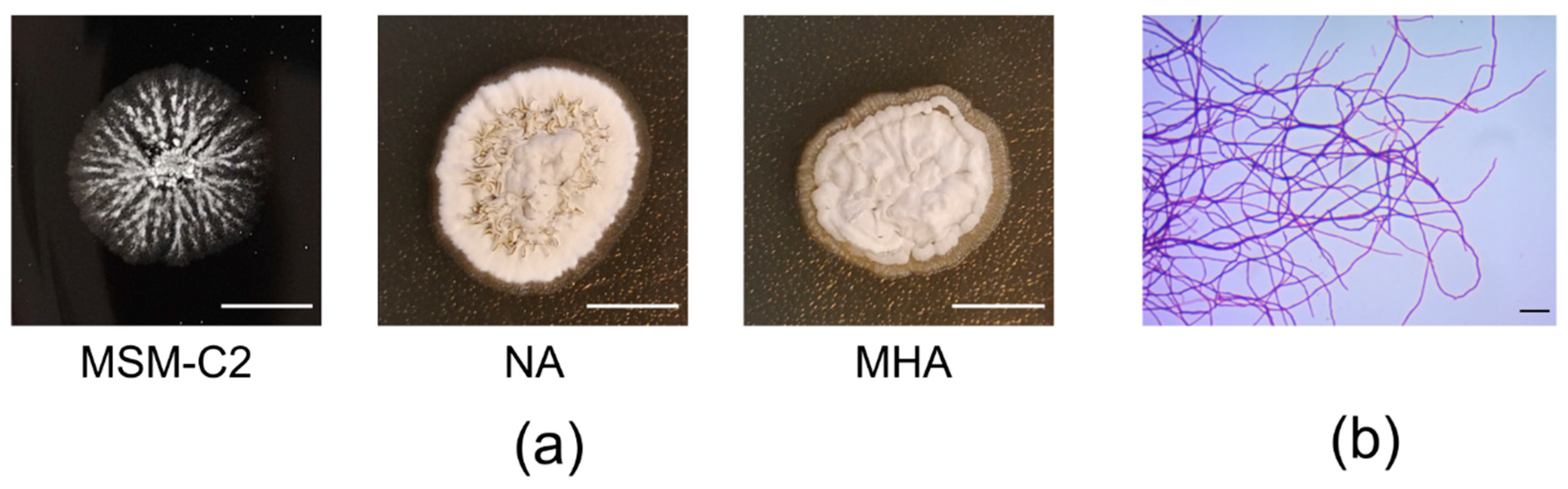
2.2. Phenotypic Fingerprint of Streptomyces sp. S-2

To characterize Streptomyces sp. S-2, 8-day-old cell cultures were scraped from the MSM-C2 plates and inoculated on GENIII microplate assay plates. The growth of the bacteria was measured 48 h post-inoculation (Table 1 and Table S1). The strain is able to utilize 42 out of the 71 compounds in the assay as a source of carbon and energy. These compounds included basic sugars, disaccharides dextrins, and polyols (glycerol and D-mannitol). The strain is also capable of utilizing several amino acids and a number of organic acids, including polycarboxylic acids. It has a proteolytic feature (the utilization of gelatin) and the ability to degrade polysorbates (Tween 40). The Streptomyces sp. S-2 tolerates slight-to-moderate halophilic conditions (growth at an NaCl concentration of 1% to 8% with a reverse correlation to salt concentration) and slightly acidic conditions (growth at pH 6 but not pH 5). It is susceptible to all of the tested antibiotics except for nalidixic acid and Aztreonam. The strain is able to grow in the presence of denaturing agents (guanidine HCl) and desiccants (lithium chloride), features that could be connected to adaptation to extreme and arid environments, such as the black soot from the household chimney.

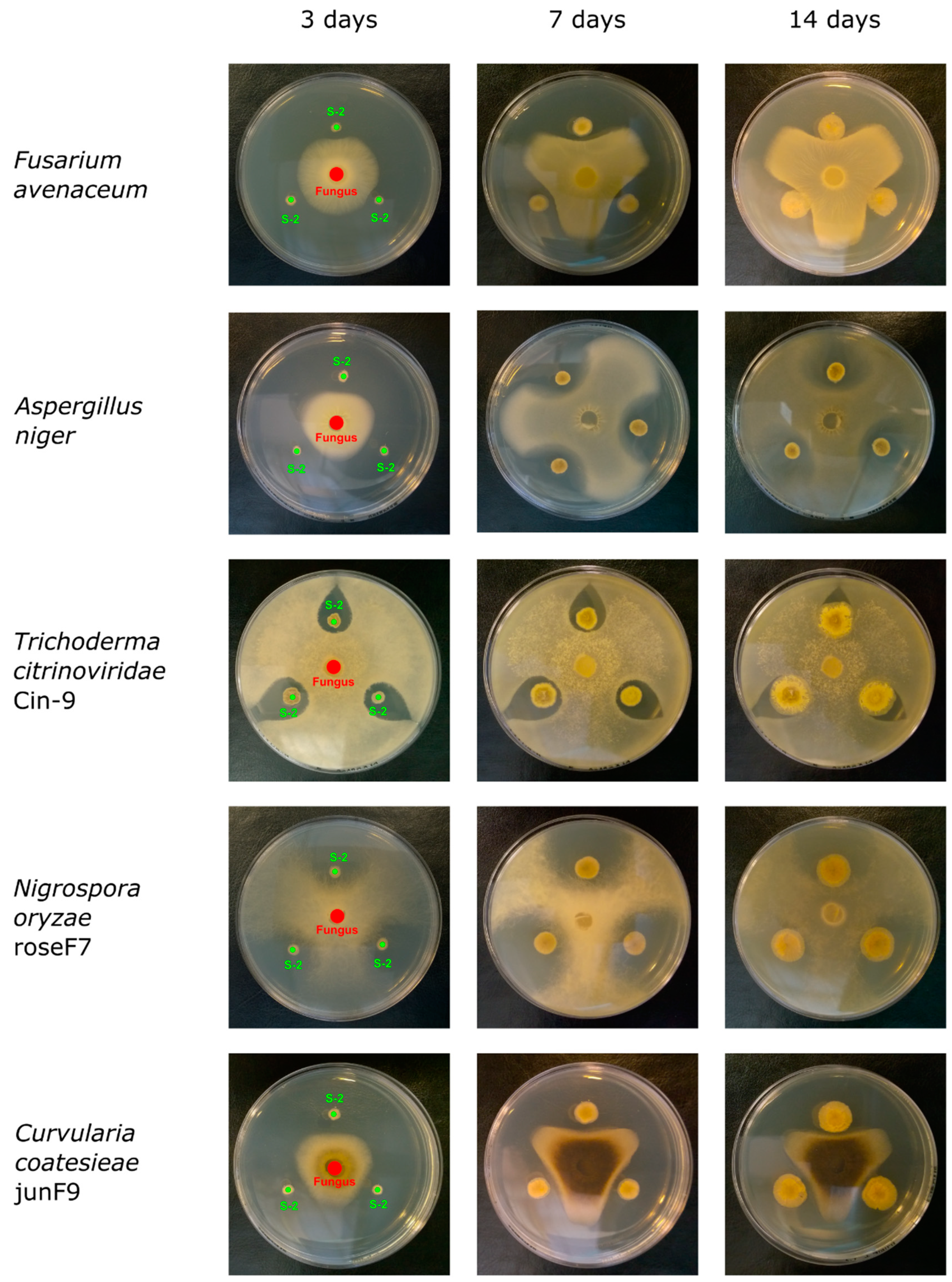
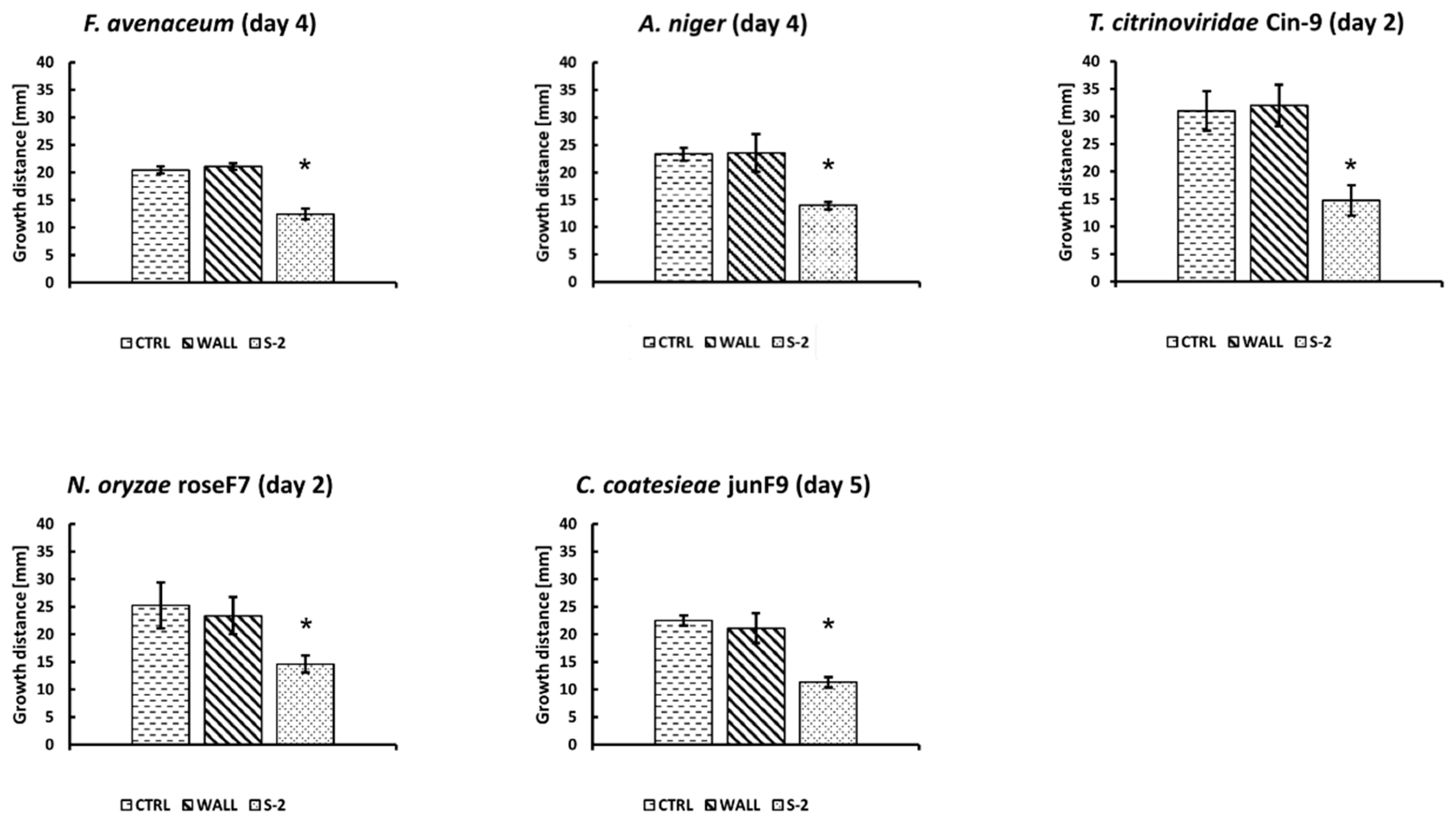
2.3. Antifungal Activity of Streptomyces sp. S-2

The antifungal activity of Streptomyces sp. S-2 was tested against two fungi from culture collections—Aspergillus niger and Fusarium avenaceum—and three fungi from the environment, including two pathogens that were isolated from a garden rose and a juniper. Fungal strains isolated from the environment were selected for the study based on their different morphologies in order to obtain diverse genera. The selected fungi were identified by sequencing the internal transcribe spacer (ITS) region and searching the National Center for Biotechnology Information NCBI database using a Blast tool. All sequences were deposited in the NCBI database. The fungi used in the study were from a cinder sample: Trichoderma citrinoviridae Cin-9 (MN756678), from a garden rose: Nigrospora oryzae roseF7 (MN756677), and from a juniper: Curvularia coatesieae junF9 (MN756676).
To test the antifungal activity, the S-2 strain was simultaneously co-cultured with fungi on MHA or NA plates. The plates were cultured at 25 °C, a temperature optimal for fungi growth, except for co-cultures with T. citrinoviridae Cin-9, which were cultured at 30 °C. Fungi growth was investigated every day for 14 days. Representative pictures of the co-cultures are shown in Figure 2. Antifungal activity was measured by comparing the inhibition of mycelial growth towards the center of the S-2 colonies to growth towards the border of the plates. The S-2 strain exhibited antifungal activity against all of the tested fungi, but at different levels of inhibition (Figure 3; data not shown). The effect was visible early in the co-cultures with the fast-growing fungi (T. citrinoviridae Cin-9 and N. oryzae roseF7). The highest antifungal activity was observed against N. oryzae roseF7, where, over time, a visual inspection of plates indicated that compactness of the mycelium decreased (data not shown), suggesting lysis of the fungi. A strong inhibitory effect was also observed for C. coatesieae junF9. A moderate effect was observed for F. avenaceum. For T. citrinoviridae Cin-9 and A. niger, even though there was a high level of inhibition observed at the beginning of the co-culture, during the course of the experiment, both fungi were able to nearly completely cover the plates with mycelia (Figure 2). Importantly, when the fungal mycelia reached the S-2 colonies, further growth could not be measured even though the fungi could have been viable. Thus, this was a situation in which measuring growth distances did not reflect actual effects. Therefore, to better compare the effects of S-2 on the tested fungi, the relative growth inhibition was measured on the day when the mycelia that were growing towards the plate border reached a distance of S-2 inoculation spots, 20 mm from the plate center (Figure 3). For N. oryzae roseF7 and T. citrinoviridae Cin-9 this was on day 2 post-inoculation, for A. niger and F. avenaceum this occurred on day 4 post-inoculation, and for C. coatesieae junF9 this was on day 5 post-inoculation. At the point when the mycelia reached that distance, the strongest inhibitory effect was observed against T. citrinoviridae Cin-9 (53.84% ± 7.17%; n = 15), followed by C. coatesieae junF9 (45.97% ± 4.67%; n = 18), then F. avenaceum (40.81% ± 5.97%; n = 15), and A. niger (39.86% ± 7.86%; n = 14), and finally N. oryzae roseF7 (35.54% ± 14.17%; n = 16) (Figure 3).
To further investigate antifungal activity and overcome the issues with the co-cultures that were mentioned above, Streptomyces sp. S-2 was inoculated in the same way as before, but with pre-incubation for 3, 7, and 14 days. Next, an agar puck with mycelia was placed at the center of the plates and incubated for an additional 7 days. After that, fungal growth was classified as “-” (no growth), “+” (growth up to 7 mm), “++” (growth between 7 and 15 mm), or “+++” (growth above 15 mm) (Table 2). On the plates pre-incubated for 3 days, all fungi were able to grow, with T. citrinoviridae Cin-9 showing the fastest growth. However, growth was diminished compared to plates with simultaneous co-cultures. On the plates pre-incubated for 7 days, N. oryzae roseF7 was not able to grow, and the growth of the other fungi was reduced. Finally, on the plates pre-incubated for 14 days, none of the fungi used in the test were able to grow (Table 2). This experiment confirmed the antifungal activity of Streptomyces sp. S-2 against a variety of fungi. Further, it indicated that one or more products secreted by bacteria in the late phase of growth were responsible for the antifungal activity observed.

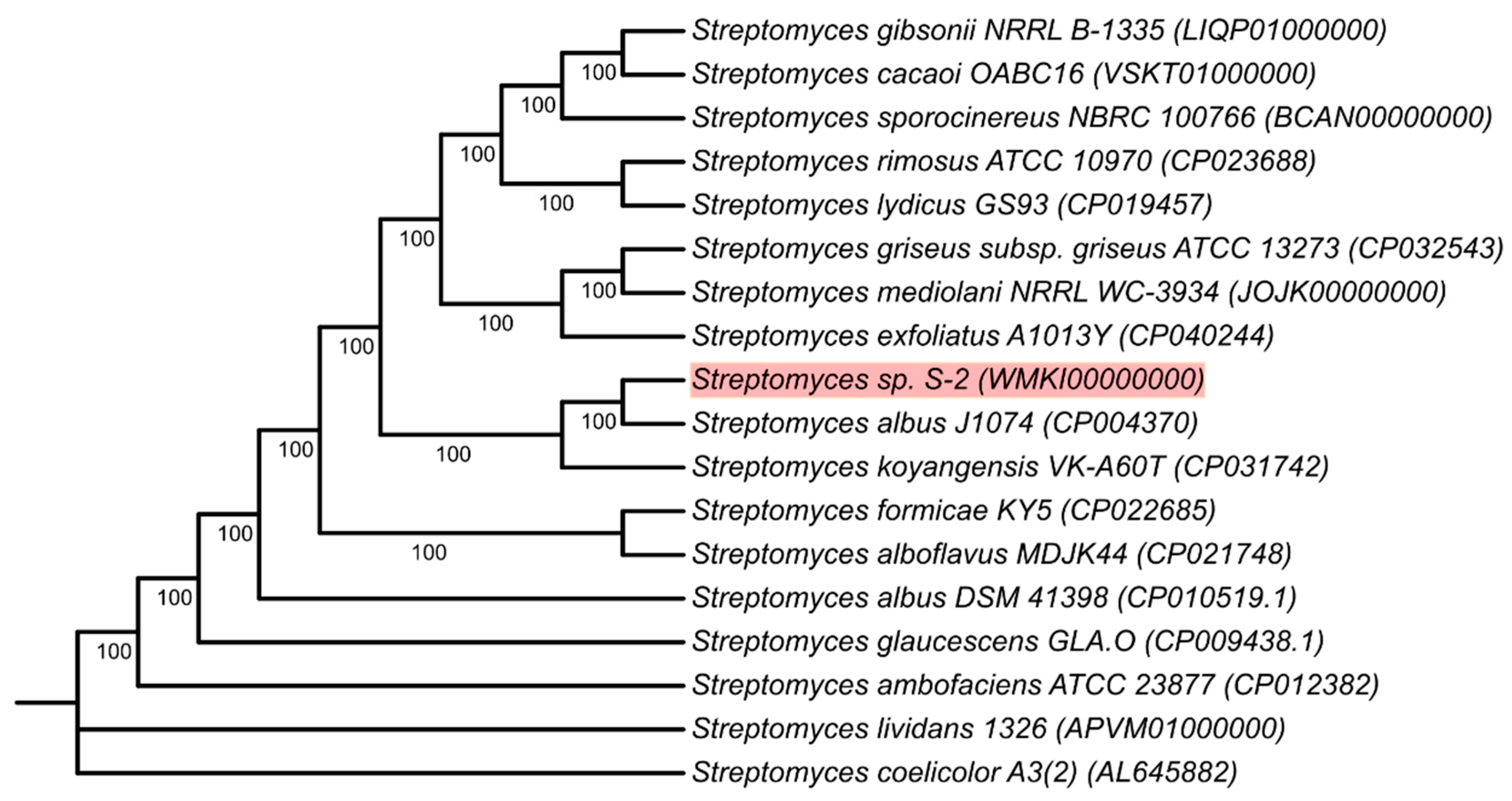
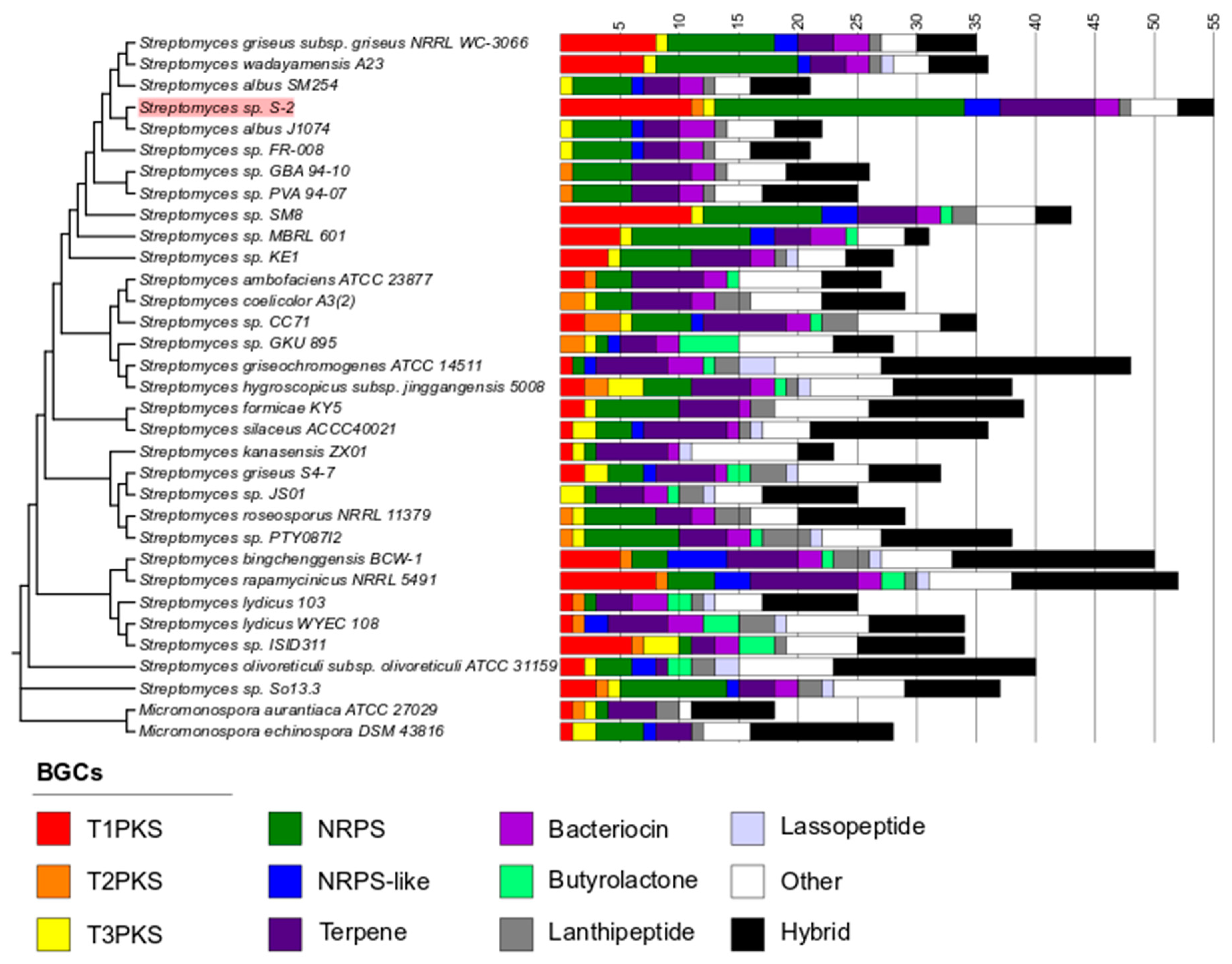
2.4. Genome Sequencing and Phylogeny of Streptomyces sp. S-2

Whole-genome sequencing of Streptomyces sp. S-2 was performed using the Illumina MiSeq platform. The assembled sequence was deposited in GenBank under accession no. WMKI00000000. The total assembled genome of strain S-2 was 6.97 Mbp with a GC content of 73.1% and 6680 CDS. Statistical data for the genome sequence are presented in Table 3. A maximum-likelihood phylogenetic analysis was performed using the core proteomes of 18 Streptomyces strains. It showed the closest relation of the S-2 strain to be S. albus J1074 (Figure 4). Therefore, we suggest that the isolate is a new strain of the Streptomyces genus and is closely related to S. albus.

2.5. Prediction of Secondary Metabolites Biosynthetic Gene Clusters (BGCs) in the Streptomyces sp. S-2 Genome

The Streptomyces sp. S-2 genome was subjected to a prediction of secondary metabolites biosynthetic gene clusters using the anti-SMASH database. The prediction revealed that the genome encompasses a total number of 55 BGCs. Moreover, 32 of the predicted BGCs show no similarity to any of the clusters in the database (as of December 2019). Multiple clusters encode non-ribosomal peptide synthetases (NRPS) (21 clusters) and NRPS-like clusters (3); type I polyketide synthases (T1PKS) (11 clusters); type II polyketide synthases (T2PKS) (1 cluster); type III polyketide synthases (T3PKS) (1 cluster); bacteriocins (2 clusters); lanthipeptides (1 cluster); and clusters with a hybrid character (3). All of these cluster types are known to encode genes responsible for the production of compounds with biological activity, including antibiotics. Among these groups, the BGCs with no similarity to known biosynthetic clusters included 17 NRPS and NRPS-like clusters, 8 T1PKS clusters, 2 bacteriocins clusters, and 1 lanthipeptide cluster. This means that there is a heightened chance that Streptomyces sp. S-2 is able to produce novel antibiotics. Among the other types of BGCs with no similarity to known products were the 2 siderophores and 2 terpenes. A detailed analysis of the anti-SMASH results, which are reported in Table 4, showed the presence of BGCs typical for the Streptomyces genus: those used for the synthesis of geosmin, a compound responsible for the earthy odor, and desferrioxamine B, a siderophore involved in iron chelation. A BGC similar to that used for the synthesis of osmolyte ectoine (75% similarity) is present, which is not surprising given the arid environment of the chimney’s black soot. There is also a number of BGCs with moderate-to-low similarity to those that encode antibiotics, fungicides, and other bioactive substances (Table 4).
The number of BGCs from Streptomyces sp. S-2 was compared to the number of BGCs from other members of the genus (Figure 5 and Table S2). The comparison showed that Streptomyces sp. S-2 excels in terms of the number of BGCs. What is worth noticing, is that the genome of strain S-2 is enriched in NRPS and T1PKS clusters in comparison to other Streptomyces, especially phylogenetically close companions. At the same time, only three clusters have low similarity to the hopene synthesis pathway, the compound that might play a role in adaptation to environments with high temperatures. Interestingly, the number of BGCs in the S-2 genome exceeds more than twice the number of BGCs identified in its close relatives, S. albus J1074 (22) and S. albus SM254 (21) (Figure 5). This number is also higher by more than 20 than the number of BGCs in S. wadayamensis A23 (36), another closely related Streptomyces strain.

3. Discussion

We isolated a new Streptomyces sp. S-2 strain from black soot after the combustion of hard coal. The black soot sample was collected from an in-use household chimney. To our knowledge, this is the first case of a microorganism being isolated from black soot. Phenotype fingerprinting using a GEN III microplate assay revealed that the S-2 strain can use a variety of compounds as a source of carbon and energy. This is not surprising, as high metabolic potential is a characteristic feature of the Streptomyces genus [2]. The assay also showed that S-2 is moderately halotolerant (with the ability to grow at 8% NaCl concentration) and can grow in the presence of desiccants. This suggests the strain has the ability to be viable in environments with low water potential, such as in chimney soot. A number of other halophilic or halotolerant Streptomyces strains have been previously isolated, and clearly this feature depends on environmental conditions [26,56,57]. The S-2 strain shows strong antifungal activity against plant pathogens and filamentous fungi potentially dangerous to humans. We observed the inhibition of fungal growth by S-2 early in the simultaneous co-culture with tested fungi, although with different levels for different fungal genera. Notably, Streptomyces produce antibiotics mainly in the later phase of growth, when the sporulation process takes place [58,59]. Moreover, both T. citrinoviridae Cin-9 and A. niger may respond to S-2 secondary metabolites through the secretion of enzymes which counteract the inhibitory effect of compounds found in the environment. Members of the Trichoderma genus are also used as biocontrol agents, and as such have antimicrobial activity and are resistant to a plethora of adverse compounds [60,61,62]. In our experiment, both bacteria and fungi were inoculated simultaneously. This could have potentially influenced the degree of the observed inhibitory effect, especially in the case of fast-growing fungi (i.e. members of the Trichoderma, Aspergillus, or Nigrospora genera). Nevertheless, significant inhibition on day 2 of the co-culture indicated that Streptomyces sp. S-2 has strong antifungal properties. In co-cultures with pre-cultured S-2, the effect was even more pronounced, resulting in the complete inhibition of fungal growth on plates with bacteria pre-cultured for 7 and 14 days for N. oryzae roseF7 and for all tested fungi, respectively. Recently, there have been a number of reports of new Streptomyces strains isolated from extreme and non-conventional environments that are showing antimicrobial activity [1,13,24,25]. Streptomyces strains have also been previously studied for their potential to protect plants against fungal infections [2,35]. One example is S. lydicus WYEC 108, a bioactive compound in antifungal product sold in the United States, for agricultural use [49]. Another example is S. violaceusniger YCED9, which produces antifungal agents able to combat various plant pathogens, including members of Fusarium, Phythium, and Phytophthora genera [50]. On the other hand, field trials to use Streptomyces strains as a biocontrol agent in banana wilt disease are have not yet been reported [35].
Sequencing has shown that the size of the S-2 genome (<7 Mbp) as well as number of coding sequences (CDS) (<7000) are on the lower end for Streptomyces genus whose members’ genomes are 8.6 Mbp on average and contain more than 7000 CDS [5,13]. At the same time, the genome mining of S-2 revealed 55 BGCs. A higher-than-average number of BGCs (20-30) that has been reported for mesophilic Streptomyces [13,63]. It is also more BGCs than identified in Streptomyces isolated from extreme environments, like Artic soil, caves or coal mine soil [1,13,24]. Interestingly, the number of BGCs in the S-2 genome exceeds those identified in genomes of closely related strains. Recent work by Chevrette et al. [25] has shown that the ecology of a microorganism influences its metabolic potential. This could be the explanation for the high number of BGCs in the S-2 genome, giving the strain an advantage in the competition for scarce nutrients in a black soot environment. Similarly, two strains of Streptomyces, sp. ICC1 and ICC4, isolated from the Iron Curtains Cave have been reported to have a higher number of BGCs than their closest terrestrial counterpart, S. lavendulae, has [24]. To the best of our knowledge, only two strains with a higher number of BGCs than S-2 have been described: S. griseochromogenes ATCC-14511 [13] and Streptomyces sp. GMR22 [64], with 59 and 63 BGCs respectively. However, those analyses were performed with an older version of the anti-SMASH database. A re-analysis with anti-SMASH version 5.1.0 revealed the presence of a lower number of BGCs in S. griseochromogenes ATCC-14511 than before, 47 versus 59. Due to not being able to access the genome of Streptomyces sp. GMR22, we were unable to assess the number of BGCs in this strain. Moreover, in our study, 32 out of 55 identified BGCs were classified as either NRPS (21 clusters) or T1PKS (11 clusters). Both groups of BGCs are known to encode proteins involved in biosynthesis and the processing of secondary metabolites with various antibiotic activities [65]. In other microorganisms, NRPS are responsible for synthesis of compounds such as actinomycin, vancomycin, and coelibactin [66,67,68], and T1PKS produce compounds such as rapamycin, cyphomycin, and glutaramides [18,25,69]. On the other hand, phenotype fingerprinting using a microplate assay showed that the S-2 strain was susceptible to several antibacterial antibiotics targeting protein synthesis. Streptomyces sp. myrophorea McG1, which has antibacterial activity, has been shown to be resistant to a number of antibiotics, including those targeting protein synthesis [27]. This might suggest a lack of antibacterial activity in Streptomyces sp. S-2. However, in that study, the resistance to some antibiotics of the McG1 strain was established after 6 days of culture, and the test was performed using different methodology [27]. The high number of BGCs, as well as the prevalence of NRPS and T1PKS clusters, distinguish the S-2 strain from other Streptomyces strains, especially considering its relatively small genome size. Finally, 32 of the identified BGCs show no similarity to any clusters present in the database, and 10 show only low similarity (below 20%). This seems particularly interesting, as it may lead to the production of novel compounds. The same has been suggested for BGCs with low overall similarities to known compounds in other Streptomyces [24].
The results reported in this study point to the capability of Streptomyces sp. S-2 to produce a large number of secondary metabolites. The high numbers of clusters in the NRPS and T1PKS groups indicate that many of its produced metabolites may have antibiotic activity. A number of them might be novel compounds and may be beneficial in medicine and industry. Secondary metabolites produced by Streptomyces sp. S-2, aside from antibiotic activity, might display antitumor, anti-inflammatory, or antioxidant activities, or function as insecticides. Such activities have been previously reported in the Streptomyces genus [2,10,70,71,72]. Considering the biosynthetic potential of the S-2 strain, future research focusing on investigation of these activities is ensured.
It should be noted that some biosynthetic clusters might be dormant and might only be expressed under specific conditions or upon activation by external factors. In members of Streptomyces, it has been shown that a co-culture with other microorganisms might stimulate the production of antibiotics [22]. A two-component signaling cascade is known to be present in these bacteria and might induce the expression of secondary metabolites under specific environmental conditions, e.g. nutrient deprivation [21]. Additionally, the activation of dormant BGCs through genetic manipulation resulting in the production of a new antimicrobial compound has recently been reported [23]. The results from co-cultures with pre-cultured S-2 suggest a constitutive production of antifungal agents by the strain. Nevertheless, a detailed examination of BGCs expression and the activities of the compounds produced by S-2 should be the focus of future research, as this could elucidate the full biotechnological potential of the strain.

4. Materials and Methods

4.1. Microbial Culture Media

Minimal salt medium (MSM) was prepared according to Malik et al. [73] to isolate the microorganisms from the black soot and cinder samples. For media supplementation, hard coal dust was obtained by grinding hard coal pieces using a ball mill. For media solidification ultra-pure agar was added for a final concentration of 1.5% (Select Agar, ThermoFisher Scientific, Waltham, MA, USA). For the fungal cultures potato dextrose agar (PDA) medium was used. For antifungal activity, nutrient agar (NA) and Mueller—Hinton agar (MHA) media were used. For the selection of transformants Luria–Bertani (LB) medium supplemented with 1.5% agar, 50 μg/mL ampicillin (Sigma-Aldrich, Darmstadt, Germany), 40 μg/mL X-gal and 0.1 mM isopropyl β-D-1-thiogalactopyranoside (IPTG) was used (both from A&A Biotechnology, Gdynia, Poland). For liquid cultures, nutrient broth (NB) and LB media were used for S-2 and transformed E. coli DH5α, respectively. All media were purchased from BTL sp. z o.o., Łódź, Poland, unless stated otherwise.

4.2. Sampling and Strains Isolation

The black soot sample was collected from a household chimney under active use (50.191846 N, 19.108628 E) on 19 December 2017 in the city of Mysłowice, Poland. At the time of collection, the chimney interior had a temperature of 30 °C and was exhausting smoke from hard coal combustion. The soot sample was scraped off the chimney wall with a sterilized spoon into a sterile 50 mL tube and transferred to the laboratory. A sample of cinder after hard coal combustion was collected from under the household’s hearth on the same day and in the same location as the black soot sample. The top layer of cinder was scraped off the pile, and the exposed cinder was collected into a sterile 50 mL tube with a sterilized spoon and transferred to the laboratory. 1 g of each sample was spread onto an MSM agar plate supplemented with 1% (w/v) hard coal dust. Plates were incubated at 30 °C. After 2 weeks, the plates were investigated, and bacterial and fungal strains were passed onto MSM agar plates supplemented with 2% (MSM-C2) hard coal dust, in order to isolate pure cultures. Strains S-2 from the black-soot-inoculated plate and Cin-9 from the cinder-inoculated-plate were chosen for subsequent experiments. S-2 resembled the morphology of Actinobacteria, and Cin-9 resembled the morphology of Trichoderma.
The microscopic morphology of isolated bacteria was investigated by using Gram staining. A colony was suspended in a drop of 0.85% NaCl and smeared on a microscope slide, air-dried, and heat fixed. Then, the specimen was stained through flooding, as follows: crystal violet for 3 min, Gram’s iodine for 2 min, 20% ethanol for 30 s, and carbol fuchsin for 30 s, followed by air-drying. A microscopic observation of the specimen was performed using a PrimoStar microscope (Carl Zeiss, Jena, Germany) with a Primo Plan-ACHROMAT 100x NA1.25 objective (Carl Zeiss, Jena, Germany) under immersion. Pictures were recorded using an AxioCam ERc 5s (Carl Zeiss, Jena, Germany) and processed using Fiji software, (version 1.52p) [74]
On 18 August 2018, garden rose leaves showing signs of fungal infection were collected into a sterile 50 mL tube and transferred to the laboratory to isolate fungi. The leaves were surface sterilized in a microbiological flow cabinet by washing them in 70% ethanol, followed by washing them in 5% commercial bleach solution (ACE, Procter & Gamble, Cincinnati, OH, USA) and then washing them twice in deionized water. Next, the leaves were placed on PDA plates and incubated at 22 °C. After 7 days of incubation, the colonies growing from the leaves were placed on fresh PDA plates and incubated at 22 °C in order to isolate the pure cultures. The strain isolate N. oryzae roseF7 was used for subsequent experiments.
Juniper branches with “slime mold” were collected into a sterile 50 mL tube and transferred to the laboratory. Pieces of branches with “slime mold” were placed on PDA plates in a microbiological flow cabinet and incubated at 22 °C. After 7 days of incubation, the colonies growing from the inoculated material were placed onto fresh PDA plates incubated at 22 °C in order to isolate the pure cultures. The C. coatesieae junF9 strain was used for subsequent experiments.

4.3. Microorganism Strains

The Aspergillus niger used in this study was obtained from the Institute of Biology, Biotechnology and Environmental Protection, University of Silesia in Katowice. The Fusarium avenaceum strain isolated from the environmental sample was obtained from the Institute of Biotechnology for Agriculture-Food Industry, Poland. For the transformation with ITS, amplicon Escherichia coli DH5α strain was used.

4.4. DNA Isolation and Sequencing

After 8 days of culture on the MSM-C2 agar plate, the S-2 strain was loop inoculated in 5 mL of NB medium and incubated at 30 °C, with shaking at 120 rpm for 72 h. Next, the cells were pelleted through centrifugation at 10,000 rcf and used for DNA extraction. Genomic DNA was extracted using a PowerSoil DNeasy Kit (MoBio, Carlsbad, CA, USA) according to the manufacturer’s protocol, with the exception that instead of soil, cell pellet was used as the sample. The integrity of the DNA was examined by agarose gel electrophoresis. The whole genome sequencing was performed by MicrobesNG (Birmingham, United Kingdom) using an Illumina MiSeq platform with 2 × 250-bp paired-end reads. The results of the sequencing were put through a standard MicrobesNG analysis pipeline.
Isolated fungal strains were cultured on PDA plates at 22 °C for 8 days, except for the Cin-9 strain, which was cultured at 30 °C. Next, the mycelia were harvested and homogenized using a sterile ceramic mortar and pestle. The homogenates were used for DNA extraction, which was performed with a DNeasy PowerLyzer PowerSoil Kit (MoBio, Carlsbad, CA, USA) according to the manufacturer’s protocol. For fungal strain identification, the ITS region was amplified using an ITS1F primer (5’-TCCGTAGGTGAACCTGCGG-3’) and ITS4R primer (5’-TCCTCCGCTTATTGATATGC-3’). In each sample, the reaction mix was composed of DreamTaq polymerase, 0.25 μL (1.25 U), 10x DreamTaq buffer, 5 μL, dNTPs mix, 5 μL (2 mM of each dNTP), primers, 0.25 μL of each (0.5 μM each), template, and nuclease-free water to fill to 50 μL. All reagents were purchased from Thermo Fisher, Waltham, MA, USA, except for the primers, which were ordered from Sigma–Aldrich, Darmstadt, Germany. Between 10 pg and 1 μg of fungal DNA was used as a template. PCR was performed as follows: initial denaturation at 95 °C for 10 min, followed by 35 cycles of denaturation at 95 °C for 30 s, primers annealing at 57 °C for 30 s, elongation at 72 °C for 1 min, and final elongation at 72 °C for 15 min. PCR products were purified using a Clean Up Ax Kit (A&A Biotechnology, Gdynia, Poland). Purified products were ligated into a pJET1.2 vector using a Clone JET PCR Cloning Kit (Thermo Fisher, Waltham, MA, USA) and subsequently transformed into chemically competent E. coli DH5α. Positive transformants were selected using a blue-white screen on LB plates supplemented with ampicillin (50 μg/mL), X-gal (40 μg/mL), and IPTG (0.1 mM). Vectors from positive clones were isolated using a GeneJet Plasmid MiniPrep Kit (Thermo Fisher Scientific, Waltham, MA, USA) according to the manufacturer’s protocol. Insert sequencing was performed by Macrogen (Amsterdam, Netherlands) using the Sanger method with a pJET1.2 forward primer (5′-CGACTCACTATAGGGAGAGCGGC-3′) and a pJET1.2 reverse primer (5′-AAGAACATCGATTTTCCATGGCAG-3′). Fungal strains were identified using by retrieving sequences of ITS1, 5.8S rRNA, ITS2, and partial large subunit rRNA fragment of closely related strains using a Blast tool [75].

4.5. Phylogenetic Analysis of Streptomyces sp. S-2

The phylogenetic tree was created by extracting and aligning the core proteomes of the S-2 and 18 Streptomyces strains (list of strains available in Table S3) using M1CR0B1AL1Z3R [76]. Poorly aligned regions were removed using Gblocks (version 0.91b) [77], which yielded 176,196 amino acids for 18 genomes. A maximum-likelihood phylogenetic tree was inferred using IQ-TREE Galaxy (version 1.5.5.3) with default settings and 1000 bootstraps [78]. Finally, the phylogenetic tree was visualized using the iTOL Interactive tree of life tool (version 5.5) [79], and is available at https://itol.embl.de/tree/19348172168181091583040528.

4.6. Secondary Metabolite Gene Clusters Prediction

The prediction of biosynthetic gene clusters for secondary metabolite synthesis was performed using Antibiotic and Secondary Metabolite Analysis Shell (anti-SMASH) version 5.1.0 [80,81]. For the characteristic of the cluster types predicted to be present in each genome, clusters assigned to more than one group were pooled in the hybrid group. The strains used in the anti-SMASH analysis were close relatives of S-2, based on a Blast search, or were strains with high a number of BGCs, as reported in literature. A tree of 32 high-quality bacterial genomes (Table S4) was generated by retrieving 31 single-copy, housekeeping genes from each genome using AmphoraNet [82]. The sequences of each individual marker gene were aligned in Geneious Prime software (version 2019.1.1, Geneious, Auckland, New Zealand) with default parameters and the alignments were concatenated. Poorly aligned positions were removed utilizing Gblocks software (version 0.91b). A maximum-likelihood phylogenetic tree was inferred using MEGA 7 software [83]. To support the phylogenetic tree, a bootstrap test with 1000 replicates was performed. Finally, the phylogenetic tree was visualized using the iTOL Interactive tree of life tool and is available at https://itol.embl.de/tree/1551582516147091575216186#.

4.7. Phenotype Fingerprinting

For phenotype fingerprinting, Streptomyces sp. S-2 was cultured on an MSM-C2 agar plate for 8 days. Next, cells were scraped and suspended in IFIB solution (BiOLOG, Hayward, CA, USA) until a transmittance of 98% was achieved. The solution was mixed, and a GENIII plate (BiOLOG, Hayward, CA, USA) was inoculated with 100 μL of cell suspension per well. The absorbance was measured at λ = 590 nm for each well, and the plate was incubated at 30 °C for 48 h. After incubation, the absorbance of each well was measured again. The readings were corrected for the absorbance of the negative control by subtracting the value of the A1 well from the rest of the wells. For negative results, the value was assumed to be zero. Subsequently, readings after 48 h were corrected for the initial absorbance measured after inoculation. Wells with negative values were assumed to be zero. The experiment was repeated three times, the values for each well were averaged, and the standard deviation was calculated.

4.8. Antifungal Activity Tests

To test antifungal activity, Streptomyces sp. S-2 cultured for 8 days on an MSM-C2 agar plate was spot-inoculated at three equally distributed spots, 20 mm from the test plate center. Next, an 8 mm agar puck with mycelia was drilled out of the PDA plates with fungi cultured for 7 days at 22 °C and placed in the center of the test plate (Figure S1). For controls, plates were inoculated in the same fashion, but were prepared with either S-2 or fungi. For A. niger, F. avenceum, N. oryzae roseF7, and C. coatesieae junF9, MHA plates were used. For T. citrinoviridae Cin-9, instead of MHA, NA plates were used, as the growth of this fungi is suppressed on MHA plates. Three control plates for each fungus and the S-2 were prepared, and at least 15 co-culture plates for each pair were prepared. Plates were incubated at 25 °C, except for T. citrinoviridae Cin-9, which was incubated at 30 °C. The plates were investigated every day for 14 days, and pictures were taken. Antifungal activity was analyzed by measuring mycelium growth towards the plate border between colonies and towards the S-2 colonies (Figure S1). Growth distances were measured using Fiji software [74]. A one-way ANOVA test was carried out to determine the significance of differences in the growth distances, followed by Tukey’s post hoc test (p ≤ 0.05). Statistical analyses were conducted using Statistica version 13 (Statsoft, Kraków, Poland). For the second antifungal activity experiment, the plates were prepared in a similar way, with the exception that inoculated S-2 was precultured for 3, 7, and 14 days before the placement of an agar puck with mycelia in the center of the plate. The plates were then incubated for 7 days and classified based on the growth of the mycelia. For each co-culture, the experiment was repeated three times.

5. Conclusions

We have isolated the Streptomyces sp. S-2 strain from black soot after hard coal combustion collected from an in-use household chimney. This is the first report of microorganism isolation from black soot. The isolated strain features strong antifungal activity and inhibits the growth of plant pathogens and filamentous fungi potentially dangerous to humans. It will be interesting to see if the strain can protect plants from fungal infections, a study that is currently ongoing. Genome sequencing and anti-SMASH analysis revealed that although this is not a new species, it contains a very high number of BGCs present in its genome, a feature that distinguishes it from closely related species. This indicates the high metabolic potential of S-2 and might increase the strain fitness in harsh environments e.g. black soot. Many of the detected BGCs show no, or low (below 20%) similarity to the clusters present in the database, increasing the possibility that S-2 might be a source of novel compounds for biomedicine and industry. Future, detailed studies on the biosynthetic abilities of Streptomyces sp. S-2 will elucidate its full biotechnological potential. This work shows that studying previously neglected and extreme environments might be a source of new strains and compounds, with a variety of biotechnological implications.

Supplementary Materials

Supplementary materials can be found at https://www.mdpi.com/1422-0067/21/7/2558/s1. Figure S1: Schematic representation of the co-culture plate inoculation used for examination of the antifungal activity of the S-2 strain. The S-2 strain was inoculated at three equally separated points 20 mm from the center of the plate (green spots), and an agar puck with fungal mycelium was placed at the center of the plate (red spot). The double arrow lines indicate the measured distance of mycelial growth toward the plate wall (violet line) and toward the S-2 colony (blue line). For each plate, three sets of measurements were taken. Table S1: Complete Streptomyces sp. S-2 phenotypic fingerprint based on GEN III microplate assay after 48 h of incubation. Table S2: Anti-SMASH results for all of the strains used for the BGCs analysis. Table S3: List of strains used in the phylogenetic analysis, with basic statistics for their genomes and description of isolation sources. Table S4: List of strains used for the phylogenetic analysis in anti-SMASH analysis with basic statistics for their genomes and description of isolation sources.

Author Contributions

P.S. was involved in sample collection, strain isolation, designing and performing the experiments, as well as analyzing the data. A.P. was involved in phylogenetic and bioinformatic analyses as well as statistical analyses. D.B. was involved in performing the experiments and data collection. P.S., A.P., and Z.P.-S. were involved in manuscript preparation. All authors have read and agreed to the published version of the manuscript.

Funding

The study has been supported by the rector of the University of Silesia in Katowice, Poland.

Acknowledgments

The authors would like to acknowledge technicians Celina Jamrozy and Paulina Biniecka, MSc., for their technical help and Dr. Małgorzata Pawlik, Institute of Biology, Biotechnology, and Environmental Protection, Faculty of Natural Sciences, University of Silesia in Katowice, Poland for provision of the Fusarium avenanceum strain used in the study.

Conflicts of Interest

The authors declare no conflicts of interest.

Abbreviations

BGCsBiosynthetic gene clusters
NRPSNon-ribosomal peptide synthase
T1PKSType 1 polyketide synthase

References

  1. Kim, M.K.; Kang, H.J.; Roh, S.G.; Park, J.S.; Kim, S.B. Streptomyces fodineus sp. nov., an actinobacterium with antifungal activity isolated from mine area soil. Int. J. Syst. Evol. Microbiol. 2019, 69, 1350–1354. [Google Scholar] [CrossRef] [PubMed]
  2. Barka, E.A.; Vatsa, P.; Sanchez, L.; Gaveau-Vaillant, N.; Jacquard, C.; Meier-Kolthoff, J.P.; Klenk, H.-P.; Clément, C.; Ouhdouch, Y.; van Wezel, G.P. Taxonomy, physiology, and natural products of Actinobacteria. Microbiol. Mol. Biol. Rev. 2016, 80, 1–43. [Google Scholar] [CrossRef] [Green Version]
  3. Chater, K.F.; Biró, S.; Lee, K.J.; Palmer, T.; Schrempf, H. The complex extracellular biology of Streptomyces: Review article. FEMS Microbiol. Rev. 2010, 34, 171–198. [Google Scholar] [CrossRef] [PubMed] [Green Version]
  4. Liu, G.; Chater, K.F.; Chandra, G.; Niu, G.; Tan, H. Molecular regulation of antibiotic biosynthesis in Streptomyces. Microbiol. Mol. Biol. Rev. 2013, 77, 112–143. [Google Scholar] [CrossRef] [PubMed] [Green Version]
  5. Stulberg, E.R.; Lozano, G.L.; Morin, J.B.; Park, H.; Baraban, E.G.; Mlot, C.; Heffelfinger, C.; Phillips, G.M.; Rush, J.S.; Phillips, A.J.; et al. Genomic and secondary metabolite analyses of Streptomyces sp. 2AW provide insight into the evolution of the cycloheximide pathway. Front. Microbiol. 2016, 7, 573. [Google Scholar] [CrossRef] [Green Version]
  6. Bérdy, J. Thoughts and facts about antibiotics: Where we are now and where we are heading. J. Antibiot. (Tokyo) 2012, 65, 385–395. [Google Scholar] [CrossRef] [Green Version]
  7. Watve, M.G.; Tickoo, R.; Jog, M.M.; Bhole, B.D. How many antibiotics are produced by the genus streptomyces? Arch. Microbiol. 2001, 176, 386–390. [Google Scholar] [CrossRef]
  8. Nakashima, T.; Takahashi, Y.; Ōmura, S. Search for new compounds from Kitasato microbial library by physicochemical screening. Biochem. Pharmacol. 2017, 134, 42–55. [Google Scholar] [CrossRef]
  9. Tiwari, K.; Gupta, R.K. Rare actinomycetes: A potential storehouse for novel antibiotics. Crit. Rev. Biotechnol. 2012, 32, 108–132. [Google Scholar] [CrossRef]
  10. Kumar, P.S.; Al-Dhabi, N.A.; Duraipandiyan, V.; Balachandran, C.; Kumar, P.P.; Ignacimuthu, S. In vitro antimicrobial, antioxidant and cytotoxic properties of Streptomyces lavendulae strain SCA5. BMC Microbiol. 2014, 14, 291. [Google Scholar]
  11. Dastager, S.G.; Li, W.J.; Dayanand, A.; Tang, S.K.; Tian, X.P.; Zhi, X.Y.; Xu, L.H.; Jiang, C.L. Seperation, identification and analysis of pigment (melanin) production in Streptomyces. African J. Biotechnol. 2006, 5, 1131–1134. [Google Scholar]
  12. Fajardo, A.; Martínez, J.L. Antibiotics as signals that trigger specific bacterial responses. Curr. Opin. Microbiol. 2008, 11, 161–167. [Google Scholar] [CrossRef] [PubMed]
  13. Núñez-Montero, K.; Lamilla, C.; Abanto, M.; Maruyama, F.; Jorquera, M.A.; Santos, A.; Martinez-Urtaza, J.; Barrientos, L. Antarctic Streptomyces fildesensis So13.3 strain as a promising source for antimicrobials discovery. Sci. Rep. 2019, 9, 1–13. [Google Scholar] [CrossRef] [PubMed]
  14. Lo Giudice, A.; Bruni, V.; Michaud, L. Characterization of Antarctic psychrotrophic bacteria with antibacterial activities against terrestrial microorganisms. J. Basic Microbiol. 2007, 47, 496–505. [Google Scholar] [CrossRef] [PubMed]
  15. Gulder, T.A.; Moore, B.S. Chasing the treasures of the sea—Bacterial marine natural products. Curr. Opin. Microbiol. 2009, 12, 252–260. [Google Scholar] [CrossRef] [Green Version]
  16. Zhang, G.; Yu, D.; Sang, B.; Feng, J.; Han, L.; Zhang, X. Genome-wide analysis reveals the secondary metabolome in Streptomyces kanasensis ZX01. Genes 2017, 8, 346. [Google Scholar] [CrossRef] [Green Version]
  17. Nakaew, N.; Lumyong, S.; Sloan, W.T.; Sungthong, R. Bioactivities and genome insights of a thermotolerant antibiotics-producing Streptomyces sp. TM32 reveal its potentials for novel drug discovery. Microbiologyopen 2019, 8, e842. [Google Scholar] [CrossRef] [Green Version]
  18. Low, Z.J.; Pang, L.M.; Ding, Y.; Cheang, Q.W.; Hoang, K.L.M.; Tran, H.T.; Li, J.; Liu, X.W.; Kanagasundaram, Y.; Yang, L.; et al. Identification of a biosynthetic gene cluster for the polyene macrolactam sceliphrolactam in a Streptomyces strain isolated from mangrove sediment. Sci. Rep. 2018, 8, 1–13. [Google Scholar] [CrossRef]
  19. Ikeda, H.; Shin-Ya, K.; Omura, S. Genome mining of the Streptomyces avermitilis genome and development of genome-minimized hosts for heterologous expression of biosynthetic gene clusters. J. Ind. Microbiol. Biotechnol. 2014, 41, 233–250. [Google Scholar] [CrossRef]
  20. Chevrette, M.G.; Currie, C.R. Emerging evolutionary paradigms in antibiotic discovery. J. Ind. Microbiol. Biotechnol. 2019, 46, 257–271. [Google Scholar] [CrossRef]
  21. Rodríguez, H.; Rico, S.; Díaz, M.; Santamaría, R.I. Two-component systems in Streptomyces: Key regulators of antibiotic complex pathways. Microb. Cell Fact. 2013, 12, 127. [Google Scholar] [CrossRef] [PubMed] [Green Version]
  22. Hoshino, S.; Onaka, H.; Abe, I. Activation of silent biosynthetic pathways and discovery of novel secondary metabolites in actinomycetes by co-culture with mycolic acid-containing bacteria. J. Ind. Microbiol. Biotechnol. 2019, 46, 363–374. [Google Scholar] [CrossRef] [PubMed]
  23. Alberti, F.; Leng, D.J.; Wilkening, I.; Song, L.; Tosin, M.; Corre, C. Triggering the expression of a silent gene cluster from genetically intractable bacteria results in scleric acid discovery. Chem. Sci. 2019, 10, 453–463. [Google Scholar] [CrossRef] [Green Version]
  24. Gosse, J.T.; Ghosh, S.; Sproule, A.; Overy, D.; Cheeptham, N.; Boddy, C.N. Whole genome sequencing and metabolomic study of cave Streptomyces isolates ICC1 and ICC4. Front. Microbiol. 2019, 10, 1020. [Google Scholar] [CrossRef] [PubMed]
  25. Chevrette, M.G.; Carlson, C.M.; Ortega, H.E.; Thomas, C.; Ananiev, G.E.; Barns, K.J.; Book, A.J.; Cagnazzo, J.; Carlos, C.; Flanigan, W.; et al. The antimicrobial potential of Streptomyces from insect microbiomes. Nat. Commun. 2019, 10, 516. [Google Scholar] [CrossRef] [Green Version]
  26. Kennedy, J.; Baker, P.; Piper, C.; Cotter, P.D.; Walsh, M.; Mooij, M.J.; Bourke, M.B.; Rea, M.C.; O’Connor, P.M.; Ross, R.P.; et al. Isolation and analysis of bacteria with antimicrobial activities from the marine sponge haliclona simulans collected from irish waters. Mar. Biotechnol. 2009, 11, 384–396. [Google Scholar] [CrossRef]
  27. Terra, L.; Dyson, P.J.; Hitchings, M.D.; Thomas, L.; Abdelhameed, A.; Banat, I.M.; Gazze, S.A.; Vujaklija, D.; Facey, P.D.; Francis, L.W.; et al. A novel alkaliphilic Streptomyces inhibits ESKAPE pathogens. Front. Microbiol. 2018, 9, 2458. [Google Scholar] [CrossRef]
  28. Olanrewaju, O.S.; Babalola, O.O. Streptomyces: Implications and interactions in plant growth promotion. Appl. Microbiol. Biotechnol. 2019, 103, 1179–1188. [Google Scholar] [CrossRef] [Green Version]
  29. Wang, X.; Zhang, M.; Gao, J.; Pu, T.; Bilal, M.; Wang, Y.; Zhang, X. Antifungal activity screening of soil actinobacteria isolated from Inner Mongolia, China. Biol. Control 2018, 127, 78–84. [Google Scholar] [CrossRef]
  30. Wang, C.; Wang, Z.; Qiao, X.; Li, Z.; Li, F.; Chen, M.; Wang, Y.; Huang, Y.; Cui, H. Antifungal activity of volatile organic compounds from Streptomyces alboflavus TD-1. FEMS Microbiol. Lett. 2013, 341, 45–51. [Google Scholar] [CrossRef] [Green Version]
  31. Matsumoto, A.; Takahashi, Y.; Omura, S. Takanawaenes, novel antifungal antibiotics produced by Streptomyces sp. K99-5278. J. Antibiot. (Tokyo) 2003, 56, 448–453. [Google Scholar]
  32. Yang, M.; Lu, L.; Pang, J.; Hu, Y.; Guo, Q.; Li, Z.; Wu, S.; Liu, H.; Wang, C. Biocontrol activity of volatile organic compounds from Streptomyces alboflavus TD-1 against Aspergillus flavus growth and aflatoxin production. J. Microbiol. 2019, 57, 396–404. [Google Scholar] [CrossRef] [PubMed]
  33. Qi, D.; Zou, L.; Zhou, D.; Chen, Y.; Gao, Z.; Feng, R.; Zhang, M.; Li, K.; Xie, J.; Wang, W. Taxonomy and broad-spectrum antifungal activity of Streptomyces sp. SCA3-4 isolated from rhizosphere soil of opuntia stricta. Front. Microbiol. 2019, 10, 1390. [Google Scholar] [CrossRef] [PubMed]
  34. Tchize Ndejouong, B.L.S.; Sattler, I.; Maier, A.; Kelter, G.; Menzel, K.D.; Fiebig, H.H.; Hertweck, C. Hygrobafilomycin, a cytotoxic and antifungal macrolide bearing a unique monoalkylmaleic anhydride moiety, from Streptomyces varsoviensis. J. Antibiot. (Tokyo) 2010, 63, 359–363. [Google Scholar] [CrossRef] [Green Version]
  35. Bubici, G.; Kaushal, M.; Prigigallo, M.I.; Cabanás, C.G.L.; Mercado-Blanco, J. Biological control agents against Fusarium wilt of banana. Front. Microbiol. 2019, 10, 616. [Google Scholar] [CrossRef] [Green Version]
  36. Michielse, C.B.; Rep, M. Pathogen profile update: Fusarium oxysporum. Mol. Plant Pathol. 2009, 10, 311–324. [Google Scholar] [CrossRef]
  37. Law, J.W.F.; Ser, H.L.; Khan, T.M.; Chuah, L.H.; Pusparajah, P.; Chan, K.G.; Goh, B.H.; Lee, L.H. The potential of Streptomyces as biocontrol agents against the rice blast fungus, Magnaporthe oryzae (Pyricularia oryzae). Front. Microbiol. 2017, 8, 3. [Google Scholar] [CrossRef] [Green Version]
  38. Laitila, A. Toxigenic Fungi and Mycotoxins in the Barley-to-Beer Chain; Elsevier Ltd.: Amsterdam, The Netherlands, 2015; ISBN 9781782423492. [Google Scholar]
  39. Feng, J.; Hwang, R.; Chang, K.F.; Hwang, S.F.; Strelkov, S.E.; Gossen, B.D.; Conner, R.L.; Turnbull, G.D. Genetic variation in Fusarium avenaceum causing root rot on field pea. Plant Pathol. 2010, 59, 845–852. [Google Scholar] [CrossRef]
  40. Hershenhorn, J.; Park, S.H.; Stierle, A.; Strobel, G.A. Fusarium avenaceum as a novel pathogen of spotted knapweed and its phytotoxins, acetamido-butenolide and enniatin B. Plant Sci. 1992, 86, 155–160. [Google Scholar] [CrossRef]
  41. Satyaprasad, K.; Bateman, G.L.; Ward, E. Comparisons of isolates of Fusarium avenaceum from white lupin and other crops by pathogenicity tests, DNA analyses and vegetative compatibility tests. J. Phytopathol. 2000, 148, 211–219. [Google Scholar] [CrossRef]
  42. Abass, M.H.; Hameed, M.A.; Ahmed, A.N. First report of Nigrospora sphaerica (Sacc.) Mason as a potential pathogen on date palm (Phoenix dactylifera L.). Can. J. Plant Pathol. 2013, 35, 75–80. [Google Scholar] [CrossRef]
  43. Wang, M.; Liu, F.; Crous, P.W.; Cai, L. Phylogenetic reassessment of Nigrospora: Ubiquitous endophytes, plant and human pathogens. Pers. Mol. Phylogeny Evol. Fungi 2017, 39, 118–142. [Google Scholar] [CrossRef] [PubMed]
  44. Park, G.J.; Kim, J.C.; Shon, M.J.; Kim, H.T.; Cho, K.Y. Phytotoxin production of nigrospora sphaerica pathogenic on turfgrasses. Plant Pathol. J. 2000, 16, 137–141. [Google Scholar]
  45. Fisher, P.J.; Petrini, O. Fungal saprobes and pathogens as endophytes of rice (Oryza sativa L.). New Phytol. 1992, 120, 137–143. [Google Scholar] [CrossRef]
  46. Beom, J.Y.; Jung, J.A.; Lee, K.T.; Hwangbo, A.; Song, M.C.; Lee, Y.; Lee, S.J.; Oh, J.H.; Ha, S.J.; Nam, S.J.; et al. Biosynthesis of nonimmunosuppressive FK506 analogues with antifungal activity. J. Nat. Prod. 2019, 82, 2078–2086. [Google Scholar] [CrossRef] [Green Version]
  47. Hong, S.W.; Kim, D.R.; Kwon, Y.S.; Kwak, Y.S. Genome-wide screening antifungal genes in Streptomyces griseus S4-7, a Fusarium wilt disease suppressive microbial agent. FEMS Microbiol. Lett. 2019, 366, fnz133. [Google Scholar] [CrossRef]
  48. Getha, K.; Vikineswary, S.; Wong, W.H.; Seki, T.; Ward, A.; Goodfellow, M. Evaluation of Streptomyces sp. strain g10 for suppression of Fusarium wilt and rhizosphere colonization in pot-grown banana plantlets. J. Ind. Microbiol. Biotechnol. 2005, 32, 24–32. [Google Scholar] [CrossRef]
  49. Mahadevan, B.; Crawford, D.L. Properties of the chitinase of the antifungal biocontrol agent Streptomyces lydicus WYEC108. Enzym. Microb. Technol. 1997, 20, 489–493. [Google Scholar] [CrossRef]
  50. Trejo-Estrada, S.R.; Paszczynski, A.; Crawford, D.L. Antibiotics and enzymes produced by the biocontrol agent Streptomyces violaceusniger YCED-9. J. Ind. Microbiol. Biotechnol. 1998, 21, 81–90. [Google Scholar] [CrossRef]
  51. Castagnoli, E.; Marik, T.; Mikkola, R.; Kredics, L.; Andersson, M.A.; Salonen, H.; Kurnitski, J. Indoor Trichoderma strains emitting peptaibols in guttation droplets. J. Appl. Microbiol. 2018, 125, 1408–1422. [Google Scholar] [CrossRef] [Green Version]
  52. Pasqualotto, A.C. Differences in pathogenicity and clinical syndromes due to Aspergillus fumigatus and Aspergillus flavus. Med. Mycol. 2009, 47, 261–270. [Google Scholar] [CrossRef] [PubMed] [Green Version]
  53. Karkowska-Kuleta, J.; Rapala-Kozik, M.; Kozik, A. Fungi pathogenic to humans: Molecular bases of virulence of Candida albicans, Cryptococcus neoformans and Aspergillus fumigatus. Acta Biochim. Pol. 2009, 56, 211–224. [Google Scholar] [CrossRef] [PubMed]
  54. Kumar, P.; Mahato, D.K.; Kamle, M.; Mohanta, T.K.; Kang, S.G. Aflatoxins: A global concern for food safety, human health and their management. Front. Microbiol. 2017, 7, 2170. [Google Scholar] [CrossRef] [PubMed] [Green Version]
  55. Nelson, P.E.; Dignani, M.C.; Anaissie, E.J. Taxonomy, biology, and clinical aspects of Fusarium species. Clin. Microbiol. Rev. 1994, 7, 479–504. [Google Scholar] [CrossRef]
  56. Ballav, S.; Kerkar, S.; Thomas, S.; Augustine, N. Halophilic and halotolerant actinomycetes from a marine saltern of Goa, India producing anti-bacterial metabolites. J. Biosci. Bioeng. 2015, 119, 323–330. [Google Scholar] [CrossRef]
  57. Zenova, G.M.; Oborotov, G.V.; Norovsuren, Z.; Fedotova, A.V.; Yakovleva, L.V. Halophilic and alkaliphilic streptomycetes in salt-affected soils. Eurasian Soil Sci. 2007, 40, 1203–1207. [Google Scholar] [CrossRef]
  58. Bibb, M.J. Regulation of secondary metabolism in streptomycetes. Curr. Opin. Microbiol. 2005, 8, 208–215. [Google Scholar] [CrossRef]
  59. Van Wezel, G.P.; McDowall, K.J. The regulation of the secondary metabolism of Streptomyces: New links and experimental advances. Nat. Prod. Rep. 2011, 28, 1311–1333. [Google Scholar] [CrossRef]
  60. Vizcaíno, J.A.; Sanz, L.; Basilio, A.; Vicente, F.; Gutiérrez, S.; Hermosa, M.R.; Monte, E. Screening of antimicrobial activities in Trichoderma isolates representing three Trichoderma sections. Mycol. Res. 2005, 109, 1397–1406. [Google Scholar] [CrossRef]
  61. Sadykova, V.S.; Kurakov, A.V.; Kuvarina, A.E.; Rogozhin, E.A. Antimicrobial activity of fungi strains of Trichoderma from Middle Siberia. Appl. Biochem. Microbiol. 2015, 51, 355–361. [Google Scholar] [CrossRef]
  62. Shi, M.; Wang, H.N.; Xie, S.T.; Luo, Y.; Sun, C.Y.; Chen, X.L.; Zhang, Y.Z. Antimicrobial peptaibols, novel suppressors of tumor cells, targeted calcium-mediated apoptosis and autophagy in human hepatocellular carcinoma cells. Mol. Cancer 2010, 9, 26. [Google Scholar] [CrossRef] [PubMed] [Green Version]
  63. Remali, J.; Sarmin, N.I.M.; Ng, C.L.; Tiong, J.J.L.; Aizat, W.M.; Keong, L.K.; Zin, N.M. Genomic characterization of a new endophytic Streptomyces kebangsaanensis identifies biosynthetic pathway gene clusters for novel phenazine antibiotic production. PeerJ 2017, 2017, e3738. [Google Scholar] [CrossRef] [PubMed] [Green Version]
  64. Herdini, C.; Mubarika, S.; Hariwiyanto, B.; Wijayanti, N.; Hosoyama, A.; Yamazoe, A.; Nojiri, H.; Widada, J. Secondary bioactive metabolite gene clusters identification of anticandida-producing Streptomyces Sp. GMR22 isolated from Wanagama forest as revealed by Genome mining approach. Indones. J. Pharm. 2017, 28, 26–33. [Google Scholar] [CrossRef] [Green Version]
  65. Challis, G.L. Mining microbial genomes for new natural products and biosynthetic pathways. Microbiology 2008, 154, 1555–1569. [Google Scholar] [CrossRef] [PubMed]
  66. Samel, S.A.; Marahiel, M.A.; Essen, L.O. How to tailor non-ribosomal peptide products-new clues about the structures and mechanisms of modifying enzymes. Mol. Biosyst. 2008, 4, 387–393. [Google Scholar] [CrossRef]
  67. Zhao, B.; Moody, S.C.; Hider, R.C.; Lei, L.; Kelly, S.L.; Waterman, M.R.; Lamb, D.C. Structural analysis of cytochrome P450 105N1 involved in the biosynthesis of the zincophore, coelibactin. Int. J. Mol. Sci. 2012, 13, 8500–8513. [Google Scholar] [CrossRef] [Green Version]
  68. Weissbach, H.; Redfield, B.G.; Beaven, V.; Katz, E. Actinomycin synthesis in washed cells of Streptomyces antibioticus. J. Biol. Chem. 1965, 240, 4377–4381. [Google Scholar]
  69. Schwecke, T.; Aparicio, J.F.; Molnár, I.; König, A.; Khaw, L.E.; Haydock, S.F.; Oliynyk, M.; Caffrey, P.; Cortés, J.; Lester, J.B.; et al. The biosynthetic gene cluster for the polyketide immunosuppressant rapamycin. Proc. Natl. Acad. Sci. USA 1995, 92, 7839–7843. [Google Scholar] [CrossRef] [Green Version]
  70. Dholakiya, R.N.; Kumar, R.; Mishra, A.; Mody, K.H.; Jha, B. Antibacterial and antioxidant activities of novel actinobacteria strain isolated from Gulf of Khambhat, Gujarat. Front. Microbiol. 2017, 8, 2420. [Google Scholar] [CrossRef]
  71. Tan, L.T.H.; Chan, K.G.; Khan, T.M.; Bukhari, S.I.; Saokaew, S.; Duangjai, A.; Pusparajah, P.; Lee, L.H.; Goh, B.H. Streptomyces sp. MUM212 as a source of antioxidants with radical scavenging and metal chelating properties. Front. Pharmacol. 2017, 8, 276. [Google Scholar] [CrossRef]
  72. Waldron, C.; Matsushima, P.; Rosteck, P.R.; Broughton, M.C.; Turner, J.; Madduri, K.; Crawford, K.P.; Merlo, D.J.; Baltz, R.H. Cloning and analysis of the spinosad biosynthetic gene cluster of Saccharopolyspora spinosa. Chem. Biol. 2001, 8, 487–499. [Google Scholar] [CrossRef] [Green Version]
  73. Malik, A.Y.; Ali, M.; Jamal, A.; Ali, M.I. Isolation and characterization of coal solubilizing aerobic microorganisms from salt range coal mines, Pakistan. Geomicrobiol. J. 2017, 34, 109–118. [Google Scholar] [CrossRef]
  74. Schindelin, J.; Arganda-Carreras, I.; Frise, E.; Kaynig, V.; Longair, M.; Pietzsch, T.; Preibisch, S.; Rueden, C.; Saalfeld, S.; Schmid, B.; et al. Fiji: An open-source platform for biological-image analysis. Nat. Methods 2012, 9, 676–682. [Google Scholar] [CrossRef] [PubMed] [Green Version]
  75. Altschul, S.F.; Gish, W.; Miller, W.; Myers, E.W.; Lipman, D.J. Basic local alignment search tool. J. Mol. Biol. 1990, 215, 403–410. [Google Scholar] [CrossRef]
  76. Avram, O.; Rapoport, D.; Portugez, S.; Pupko, T. M1CR0B1AL1Z3R-a user-friendly web server for the analysis of large-scale microbial genomics data. Nucleic Acids Res. 2019, 47, W88–W92. [Google Scholar] [CrossRef] [PubMed] [Green Version]
  77. Talavera, G.; Castresana, J. Improvement of phylogenies after removing divergent and ambiguosly aligned blocks from protein sequence alignments. Syst. Biol. 2007, 56, 564–577. [Google Scholar] [CrossRef] [Green Version]
  78. Nguyen, L.T.; Schmidt, H.A.; Von Haeseler, A.; Minh, B.Q. IQ-TREE: A fast and effective stochastic algorithm for estimating maximum-likelihood phylogenies. Mol. Biol. Evol. 2015, 32, 268–274. [Google Scholar] [CrossRef]
  79. Letunic, I.; Bork, P. Interactive Tree Of Life (iTOL) v4: Recent updates and new developments. Nucleic Acids Res. 2019, 47, W256–W259. [Google Scholar] [CrossRef] [Green Version]
  80. Blin, K.; Medema, M.H.; Kottmann, R.; Lee, S.Y.; Weber, T. The antiSMASH database, a comprehensive database of microbial secondary metabolite biosynthetic gene clusters. Nucleic Acids Res. 2017, 45, D555–D559. [Google Scholar] [CrossRef] [Green Version]
  81. Blin, K.; Pascal Andreu, V.; De Los Santos, E.L.C.; Del Carratore, F.; Lee, S.Y.; Medema, M.H.; Weber, T. The antiSMASH database version 2: A comprehensive resource on secondary metabolite biosynthetic gene clusters. Nucleic Acids Res. 2019, 47, D625–D630. [Google Scholar] [CrossRef] [Green Version]
  82. Kerepesi, C.; Bánky, D.; Grolmusz, V. AmphoraNet: The webserver implementation of the AMPHORA2 metagenomic workflow suite. Gene 2014, 533, 538–540. [Google Scholar] [CrossRef] [PubMed]
  83. Kumar, S.; Stecher, G.; Tamura, K. MEGA7: Molecular evolutionary genetics analysis version 7.0 for bigger datasets. Mol. Biol. Evol. 2016, 33, 1870–1874. [Google Scholar] [CrossRef] [PubMed] [Green Version]
Figure 1. Morphology of Streptomyces sp. S-2 colonies. (a) Growth in solid cultures on 1% hard coal dust (MSM-C2), nutrient agar (NA), and Mueller—Hinton agar (MHA) plates. For the initial culture Streptomyces sp. S-2 was streak-inoculated on agar plates. The plates were incubated at 30 °C for 3 days, after which pictures of the colonies were taken. The scale bar represents 5 mm (b) Bacterial mycelia stained using the Gram method. After 3 days, the colonies on the MSM-C2 agar plates were scraped using inoculation loop and suspended in 0.85% NaCl, dried, and stained using the Gram method, followed by observation with a light microscope under immersion, using a Plan-ACHROMAT 100× 1.25NA objective (Carl Zeiss, Germany). Pictures were recorded using an AxioCam ERc 5s (Carl Zeiss, Germany). The scale bar represents 10 μm. All pictures were processed using Fiji software.
Ijms 21 02558 g001
Figure 2. The inhibition of fungal growth by Streptomyces sp. S-2. Representative pictures of the S-2 strain and each fungus co-culture showing the inhibition of fungal mycelia growth taken at 3rd, 5th, and 14th day of the cultures. For each co-culture, MHA plates were used except for the co-culture with Trichoderma citroviridae Cin-9, which was performed on NA plates. The S-2 strain was spot-inoculated at the 20 mm distance from the center of plate, followed by placing a puck of agar with fungal mycelium at the center of the plate. Plates were incubated at 25 °C, except for the co-culture with T. citrinoviridae Cin-9, which was incubated at 30 °C. Co-cultures were investigated daily and pictures of the co-cultures were taken. For each co-culture, at least 14 plates were analyzed.
Ijms 21 02558 g002
Figure 3. Inhibition of fungal growth by Streptomyces sp. S-2. Charts comparing the growth of fungal mycelia towards the plate border (WALL) or toward the S-2 strain colonies (S-2) on co-culture plates and fungal mycelia growth toward the border of the plates on control plates (CTRL). For each co-culture, MHA plates were used except for the co-culture with T. citroviridae Cin-9 which was performed on NA plates. The S-2 strain was spot-inoculated at a distance 20 mm from the center of the plate, followed by the placement of a puck of agar with fungal mycelia at the center of the plate. Plates were incubated at 25 °C, except for the co-culture with T. citrinoviridae Cin-9 which was incubated at 30 °C. For each co-culture, at least 14 plates were analyzed. Three control plates for fungal growth control for each experimental set were also analyzed. A growth comparison for each fungus on the day when fungal mycelia grew ≥ 20 mm (the distance at which the S-2 strain was inoculated) is indicated in brackets above the charts. Statistically significant inhibitions of growth are represented by an asterisk (*) (one-way ANOVA followed by Tukey’s post hoc test, p ≤ 0.05).
Ijms 21 02558 g003
Figure 4. Maximum-likelihood phylogenetic tree of the Streptomyces sp. S-2 strain. The tree shows the phylogenetic relationships between the S-2 strain and closely related Streptomyces strains. The phylogenetic tree was inferred using IQ-TREE Galaxy software from an alignment of the core proteomes of 19 Streptomyces strains. The numbers at the nodes represent bootstrap values (% of 1000 repeats). The position of Streptomyces sp. S-2 strain has been highlighted in red for better visibility.
Ijms 21 02558 g004
Figure 5. Comparison of the number and types of BGCs found in the genomes of closely related strains and strains with a high number of BGCs reported in the literature. The BGCs in each genome were predicted using the anti-SMASH database. The “Hybrid” type clusters that were predicted to belong to more than one type of BGC were pooled. The BGCs that were predicted to belong to the siderophore, ectoine, thiopeptide, indole, melanin, or linardin types which were pooled in “Other”. The position of Streptomyces sp. S-2 has been highlighted in red for better visibility.
Ijms 21 02558 g005
Table 1. Streptomyces sp. S-2 phenotypic fingerprint based on a GEN III microplate assay after 48 h of incubation.
Carbon Source Assay
CompoundOD590CompoundOD590CompoundOD590CompoundOD590
Saccharides
α-D-Glucose0.214 ± 0.064D-Galactose0.089 ± 0.046D-Mannose0.120 ± 0.063D-Fructose0.196 ± 0.047
D-Cellobiose0.145 ± 0.037Gentiobiose0.320 ± 0.151D-Turanose0.036 ± 0.026N-Acetylo-D-Glucosamine0.082 ± 0.44
Dextrin0.155 ± 0.088
Saccharide derivatives
N-Acetyl-β-D-Mannosamine0.077 ± 0.023D-Glucose-6-Phosphate0.086 ± 0.033D-Fructose-6-Phosphate0.063 ± 0.053N-Acetyl- Neuraminic Acid0.183 ± 0.021
Mucic Acid0.065 ± 0.041
Glycosides and polyols
β-D-Methyl-D-Glucoside0.098 ± 0.049D-Salicin0.175 ± 0.030Glycerol0.112 ± 0.048D-Mannitol0.067 ± 0.049
Organic Acids
Formic Acid0.291 ± 0.082Acetic Acid0.249 ± 0.029Propionic Acid0.237 ± 0.021Citric Acid0.104 ± 0.076
L-Lactic Acid0.315 ± 0.224D-Gluconic Acid0.595 ± 0.127D-Malic Acid0.133 ± 0.024L-Malic Acid0.420 ± 0.083
Acetoacetic Acid0.114 ± 0.031Bromo-Succinic Acid0.193 ± 0.084Methyl Pyruvate0.218 ± 0.021α-Hydroxy-Butiric Acid0.318 ± 0.082
β-Hydroxy-D,L-Butyric Acid0.267 ± 0.024α-Keto-Butyric Acid0.248 ± 0.042γ-Amino-Butyric Acid0.102 ± 0.003D-Lactic Acid Methyl Ester0.176 ± 0.041
Amino Acids
L-Alanine0.115 ± 0.087L-Histidine0.199 ± 0.047L-Aspartic Acid0.134 ± 0.098L-Glutamic Acid0.125 ± 0.069
Glycyl-L-Proline0.095 ± 0.020
Others
Gelatin0.220 ± 0.009Inosine0.102 ± 0.026Tween 400.266 ± 0.102
CHEMICAL SENSITIVITY ASSAY
pH 60.487 ± 0.1071% NaCl1.018 ± 0.2944% NaCl0.636 ± 0.1518% NaCl0.349 ± 0.103
1% Sodium Lactate0.508 ± 0.146Guanidine HCl0.364 ± 0.039Nalidixic Acid0.456 ± 0.107Aztreonam0.250 ± 0.085
Sodium Butyrate0.467 ± 0.160
The values represent bacterial growth measured as optical density (OD) at a 590 nm wavelength. The values are the mean and standard deviation error calculated from three individual biological repeats.
Table 2. Inhibition of fungal growth on plates with pre-cultured Streptomyces sp. S-2.
Fungus speciespre-cultured Streptomyces sp. S-2
3 days7 days14 days
Fusarium avenaceum+++-
Aspergillus niger+++-
Trichoderma citrinoviridae Cin-9+++++-
Nigrospora oryzae RoseF7++--
Curvularia coatesieae JunF9+++-
An agar puck with mycelia was placed in the middle of plates with three colonies of Streptomyces sp. S-2. The plates were incubated for 7 days, and the growth of the fungi was measured as the radius between the puck and center of the S-2 colony: “-” (no growth), “+” (growth up to 7 mm), “++” (growth between 7 and 15 mm), and “+++” (growth above 15 mm).
Table 3. Next Generation Sequencing (NGS) statistical data based on a draft genome of S-2.
Streptomyces sp. S-2 Draft Genome
Number of contigs1799
Contigs sum (bp)6.971375
N506745
Longest contig (bp)62.211
Shortest contig (bp)407
Average length (bp)3875
% GC content73.1
Number of CDS6680
Table 4. The distribution of BGCs from Streptomyces sp. S-2.
Gene TypeSpan [nt]Most Similar Biosynthetic Gene Cluster
FromTo
Bacteriocin18813-
Bacteriocin11875-
Ectoine14689Ectoine (75%)
Lanthipeptide16359-
NRPS114,680-
NRPS17664-
NRPS17000-
NRPS15847-
NRPS13863-
NRPS13784-
NRPS13409-
NRPS13289-
NRPS13179-
NRPS13061-
NRPS12796-
NRPS12701-
NRPS12511-
NRPS11858-
NRPS852841,043Diisonitrile entibiotic SF2768 (66%)
NRPS120,858Mannopetimycin (37%)
NRPS127,653Surugamide A / Surugamide D (19%)
NRPS16554Curacomycin (18%)
NRPS130,250Dechlorocuracomycin (16%)
NRPS17538Desotamide (9%)
NRPS110,029Mannopeptimycin (7%)
NRPS-like17313-
NRPS-like14785-
NRPS-like11417-
Hybrid
(NRPS-like, T1PKS)
122,016Levorin A3/C06690/FR-008-III/candicidin A/UNII-AP5PEF5W7U (66%)
siderophore400410,744-
siderophore11673-
siderophore110,733Desferrioxamine B (100%)
T1PKS118,159-
T1PKS16047-
T1PKS16025-
T1PKS15494-
T1PKS14600-
T1PKS13778-
T1PKS11980-
T1PKS11646-
T1PKS115,272ECO-02301 (32%)
T1PKS114,101Candicidin (23%)
T1PKS116,488Reedsmycins (20%)
Hybrid
(T1PKS, NRPS)
113,542Antimycin (20%)
Hybrid
(T1PKS, NRPS)
113,498Antimycin (13%)
T2PKS116,805Fredericamycin A (45%)
T3PKS18472Herboxidiene (7%)
Terpene14631-
Terpene14141-
Terpene12347Geosmin (100%)
Terpene19567Hopene (53%)
Terpene16983Isorenieratene (25%)
Terpene11604Hopene (15%)
Terpene11104Hopene (15%)
Terpene369713,999Phoshonoglycans (6%)
Biosynthetic gene clusters (BGCs) predicted based on the anti-SMASH database (% indicates the proportion of genes with similarities). TIPKS: type I polyketide synthase; TIIPKS: type II polyketide synthase; TIIIPKS: type III polyketide synthase; NRPS: non-ribosomal peptide synthase.

