Article

Ticagrelor Increases SIRT1 and HES1 mRNA Levels in Peripheral Blood Cells from Patients with Stable Coronary Artery Disease and Chronic Obstructive Pulmonary Disease

1
Department of Medical Sciences, University of Ferrara, 44121 Ferrara, Italy
2
Maria Cecilia Hospital, GVM Care & Research, 48033 Cotignola, Italy
3
Department of Morphology, Surgery and Experimental Medicine and Laboratory for Technologies of Advanced Therapies (LTTA), University of Ferrara, 44121 Ferrara, Italy
4
Cardiovascular Institute, Azienda Ospedaliero-Universitaria di Ferrara, 44124 Cona, Italy
5
University of Hawaii Cancer Center, University of Hawaii, Honolulu, HI 96813, USA
*
Author to whom correspondence should be addressed.
These authors contributed equally to this article.
Int. J. Mol. Sci. 2020, 21(5), 1576; https://doi.org/10.3390/ijms21051576
Submission received: 11 February 2020 / Revised: 21 February 2020 / Accepted: 22 February 2020 / Published: 25 February 2020
(This article belongs to the Section Molecular Pathology, Diagnostics, and Therapeutics)

Abstract

:
Ticagrelor is a powerful P2Y12 inhibitor with pleiotropic effects in the cardiovascular system. Consistently, we have reported that in patients with stable coronary artery disease (CAD) and concomitant chronic obstructive pulmonary disease (COPD) who underwent percutaneous coronary intervention (PCI), 1-month treatment with ticagrelor was superior in improving biological markers of endothelial function, compared with clopidogrel. The objective of this study was to investigate the mechanisms underlying these beneficial effects of ticagrelor by conducting molecular analyses of RNA isolated from peripheral blood cells of these patients. We determined mRNAs levels of markers of inflammation and oxidative stress, such as RORγt (T helper 17 cells marker), FoxP3 (regulatory T cells marker), NLRP3, ICAM1, SIRT1, Notch ligands JAG1 and DLL4, and HES1, a Notch target gene. We found that 1-month treatment with ticagrelor, but not clopidogrel, led to increased levels of SIRT1 and HES1 mRNAs. In patients treated with ticagrelor or clopidogrel, we observed a negative correlation among changes in both SIRT1 and HES1 mRNA and serum levels of Epidermal Growth Factor (EGF), a marker of endothelial dysfunction found to be reduced by ticagrelor treatment in our previous study. In conclusion, we report that in stable CAD/COPD patients ticagrelor positively regulates HES1 and SIRT1, two genes playing a protective role in the context of inflammation and oxidative stress. Our observations confirm and expand previous studies showing that the beneficial effects of ticagrelor in stable CAD/COPD patients may be, at least in part, mediated by its capacity to reduce systemic inflammation and oxidative stress.

1. Introduction

Dual antiplatelet therapy (DAPT), consisting of the co-administration of aspirin and of a P2Y12 inhibitor, is the gold standard treatment for chronic stable coronary artery disease (CAD) patients following percutaneous coronary intervention (PCI). Among antiplatelet drugs, ticagrelor, due to its higher efficiency in inhibiting the P2Y12 receptor, is a more potent platelet inhibitor as compared with clopidogrel [1,2]. In acute coronary syndrome (ACS) patients, treatment with ticagrelor resulted in a reduced number of cardiovascular events and in improved peripheral arterial function compared to clopidogrel [3,4,5], suggesting that ticagrelor, beside its antiplatelet activity, modulates biological processes involved in cardiovascular protection. In patients with stable CAD and concomitant chronic obstructive pulmonary disease (COPD), we have shown that ticagrelor, compared with clopidogrel, is superior in improving biological markers of endothelial function [2,6]. These benefits are consistent with observations in patients with stable CAD and concomitant diabetes in which treatment with ticagrelor led to higher improvement of endothelial dysfunction in comparison to ticagrelor [7]. Lastly, unlike clopidogrel, ticagrelor retained some efficacy to prevent ischemic events over and above those of aspirin during chronic therapy [8].
To date, the molecular mechanisms underlying these pleiotropic effects of ticagrelor have not been elucidated, but there is evidence suggesting that they are related to inhibition of the equilibrative nucleoside transporter-1 (ENT1) leading to increased circulating levels of adenosine and cyclic adenosine monophosphate (cAMP) [9,10]. Adenosine, by binding adenosine receptors (ARs), regulates biological processes involved in CAD and COPD progression including oxidative stress and inflammatory responses [11], and functions of monocytes, neutrophils, CD4 T-helper cells, and CD8 cytotoxic T-cells, all implicated in CAD [12,13]. Specifically, in human monocytes, adenosine reduces inflammation through the inhibition of the expression of adhesion molecules, such as intercellular adhesion molecule 1 (ICAM1) [14]. Furthermore, in human monocyte-derived macrophages, cAMP limits inflammasome activation by promoting LR pyrin domain containing 3 (NLRP3) degradation [15,16], this latter found upregulated in cardiovascular disease [17]. Moreover, in several inflammatory diseases [18,19,20,21], A2aR signaling activation has been associated to the reduction of pro-inflammatory T-helper (Th) 17 cells and the increase of anti-inflammatory T-regulatory (Tregs) cells. Importantly, Th17/Tregs ratio has been found to be a determinant of both CAD and COPD progression [22,23,24,25]. Furthermore, adenosine, by downregulating the expression of Notch1 receptor, suppresses the activity of inflammatory CD8 T-cells [26], which are increased in COPD and stable CAD patients [27,28].
Dysregulation of the Notch signaling, a major regulator of cardiovascular homeostasis and of innate and adaptive immune response, has been observed in cardiovascular diseases [13,29]. Specifically, altered levels of Notch ligands JAGGED-1 (JAG1) and Delta-like 4 (DLL4) have been linked to the progression of atherosclerosis [30,31]. Of interest, altered expression levels of Notch1 and its target gene HES1 have been associated to COPD [32,33]. The Notch pathway is regulated by cross-talks with a plethora of pathways [34,35] including the NAD+-dependent protein deacetylase, sirtuin1 (SIRT1), an oxidative stress sensor [36] and a repressor of inflammatory response [37], whose expression and activity is reduced in peripheral blood mononuclear cells (PBMCs) of stable CAD, ACS [38], and COPD [39] patients.
Many studies have investigated the possible molecular mechanisms underlying the pleiotropic activity of ticagrelor in ACS [40,41,42,43] and SCAD [7] providing evidence of a ticagrelor-mediated increase in circulating levels of adenosine and cAMP [10,44,45,46,47]. Additionally, several studies have also focused on the effects of ticagrelor on circulating inflammation markers [48,49,50,51]. The aim of our study was to expand the comparison between ticagrelor and clopidogrel to include the effects of treatment on markers of inflammation related to endothelial dysfunction (Supplementary Figure S1) never investigated in this context, in order to provide more molecular details that could help to gain a better understanding of the pleiotropic effect of ticagrelor on the vascular system.

2. Results

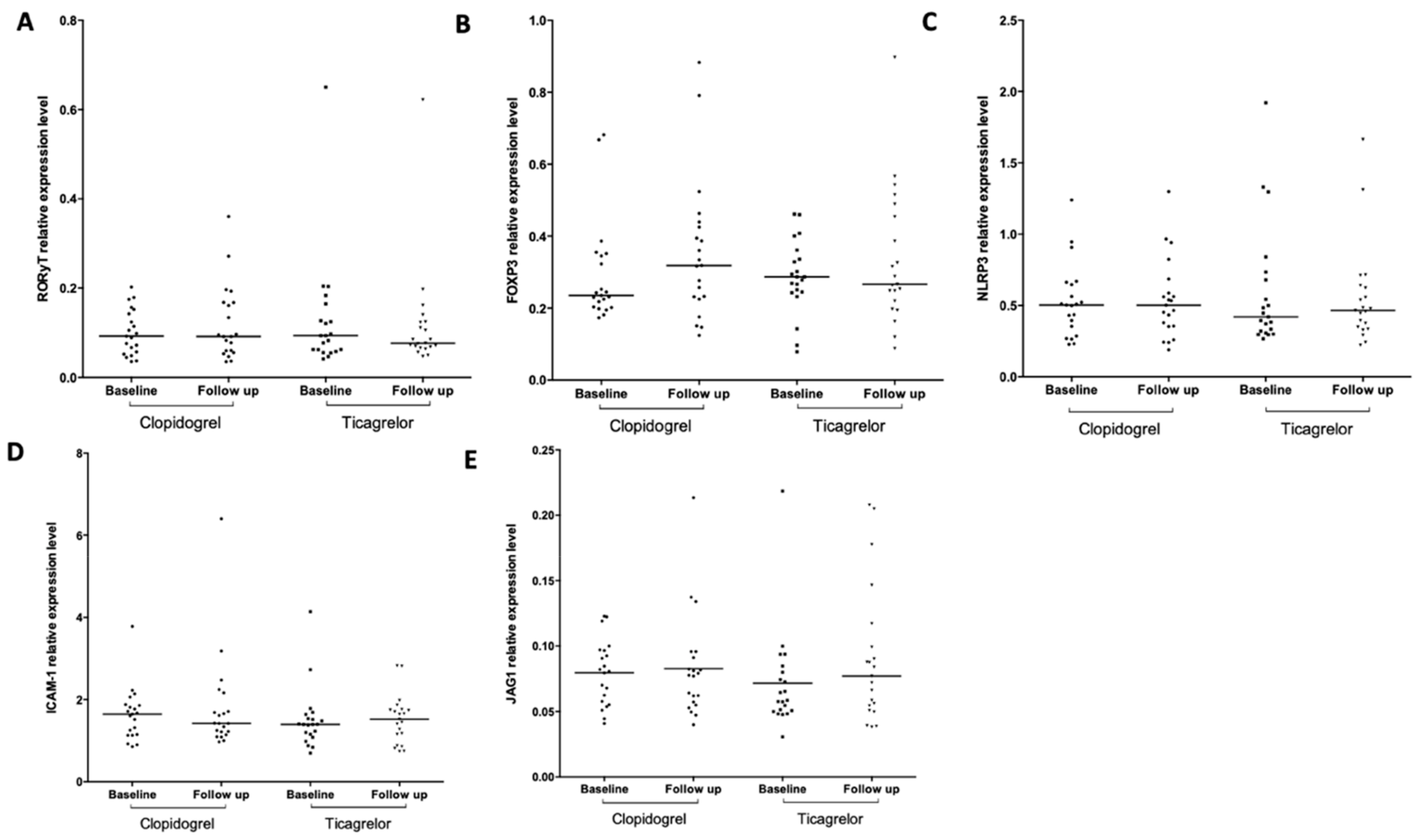
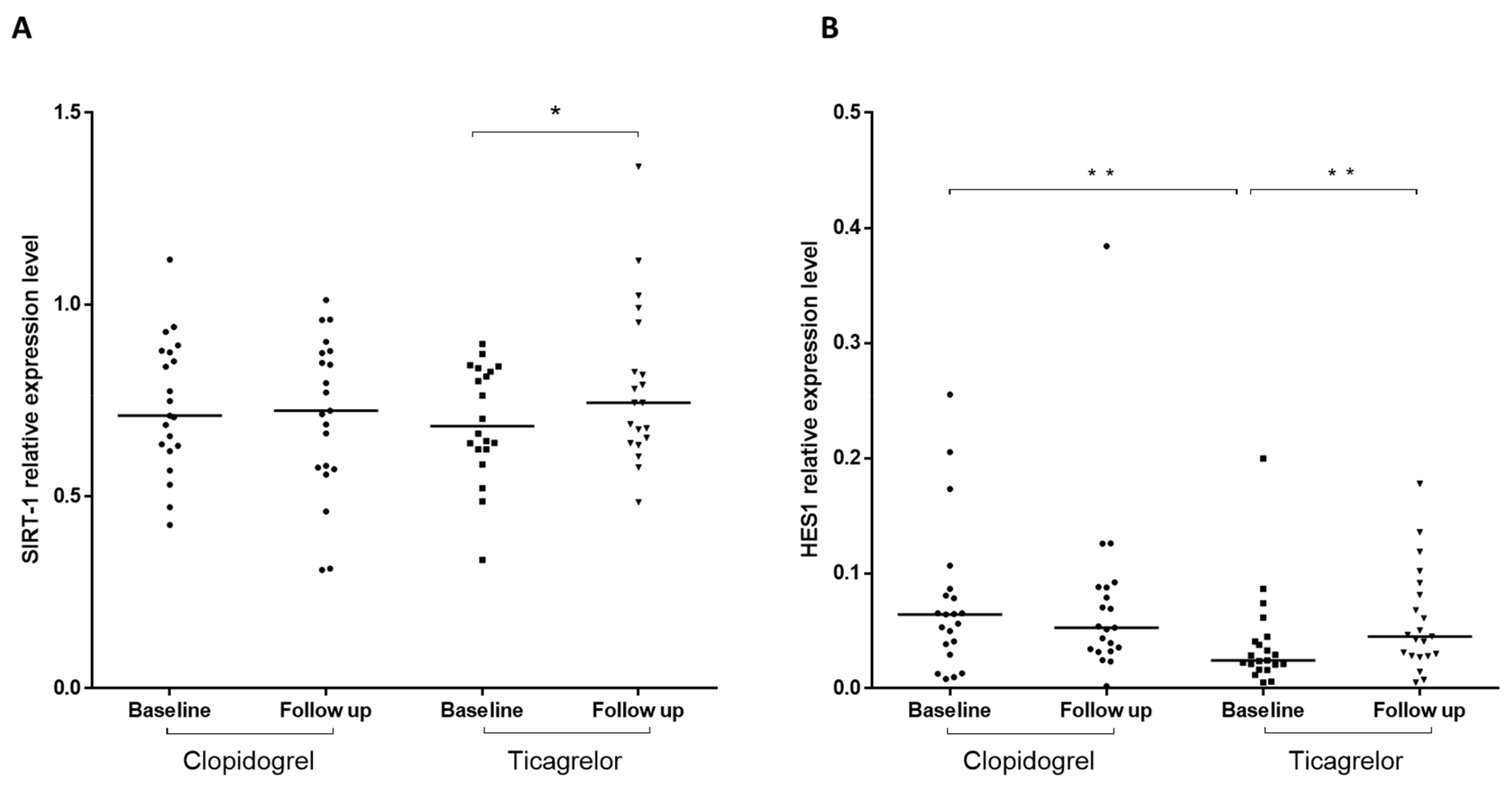
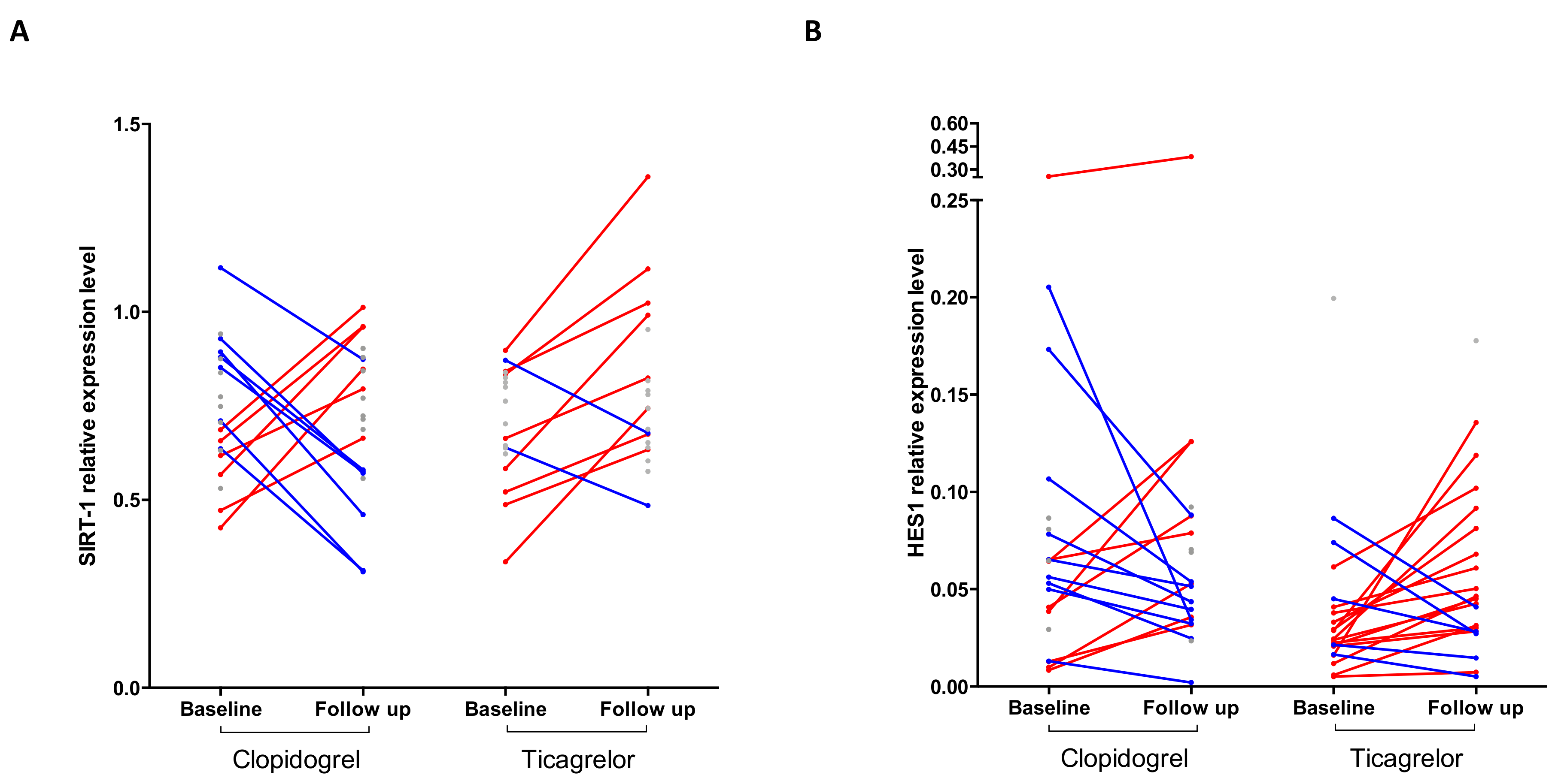
2.1. Ticagrelor, But Not Clopidogrel, Increases SIRT1 and HES1 mRNA Levels

We compared the mRNA expression levels of RORγt (Th17 cells transcription factor), FoxP3 (Tregs transcription factor), NLRP3, ICAM1, SIRT1, Notch ligands JAG1 and DLL4, and of HES1, Notch target gene, in RNA isolated from peripheral blood cells obtained from stable CAD/COPD patients following PCI before (T0) and after 1-month (T30) treatment with ticagrelor or clopidogrel. At T0, mRNA levels of RORγt, FoxP3, NLRP3, ICAM1, and JAG1 did not differ between the treatment groups (Figure 1A–E), whereas HES1 mRNA levels were lower in ticagrelor, compared to clopidogrel (Figure 2B). In both ticagrelor- and clopidogrel-treated groups, we found no significant differences between T0 and T30 in the levels of RORγt, FoxP3, NLRP3, ICAM1, and JAG1 mRNAs (Figure 1A–E). The results relative to DLL4 mRNA are not shown because the expression level of this ligand was too low to provide a reliable assessment of the expression of this gene.
SIRT1 and HES1 mRNAs were significantly increased at T30 compared to T0 following ticagrelor but not clopidogrel treatment (Figure 2A,B). Before–after analyses showed that unchanged levels of RORγt, FoxP3, NLRP3, ICAM1, and JAG1 mRNAs between T0 and T30 were not due to lack of response of these genes expression to each drug, but rather to a heterogeneous response characterized by similar number of patients showing no difference, increased or reduced levels of the specific mRNA in response to treatment (Supplementary Figure S2A–F). Instead, the response of SIRT1 to ticagrelor showed prevalently no changes (10/20 patients) or increased (8/20 patients) mRNA levels and reduced levels only in 2/20 patients. On the contrary, in the clopidogrel group the changes in SIRT1 mRNA were equally distributed between patients showing no changes (8/21), increased (6/21), or reduced levels (7/21) of mRNA (Figure 3A). Similarly, in the ticagrelor group HES1 mRNA increased in 15/21 patients and was unchanged in 1/21 patients or decreased in 5/21 patients. In the clopidogrel-treated group the levels of HES1 mRNA were increased in 8/21 patients, decreased in 9/21 patients, or unchanged in 4/21 patients (Figure 3B).
Severity of diseases, co-morbidities, and pharmacological treatment can determine differences in gene expression. Furthermore, drug interaction may modulate ticagrelor concentration and therefore effectiveness of the treatment [52]. To identify possible associations between disease severity, comorbidity or drug treatment, we performed correlation analyses between these variables and changes in SIRT1 or HES1 after a 1-month treatment. Interestingly, we found that being a man is associated with an increase in SIRT1 expression after treatment (R = 0.357; p = 0.022). We also found an inverse association between the variations of SIRT1 and treatment with inhaled corticosteroids (R = −0339; p = 0.003) or long-acting beta2 agonist (R = −0.311; p = 0.048) (Supplementary Figure S3). These data show that men respond better than women to ticagrelor with regard to SIRT1 expression and suggest that drugs for the management of COPD may affect the effectiveness of treatment with ticagrelor. On the contrary, we found no association between other co-morbidities, COPD severity, and treatment response. Due to the high homogeneity of patients with stable CAD and the relative small study population, it was not possible to stratify patients according to CAD severity.

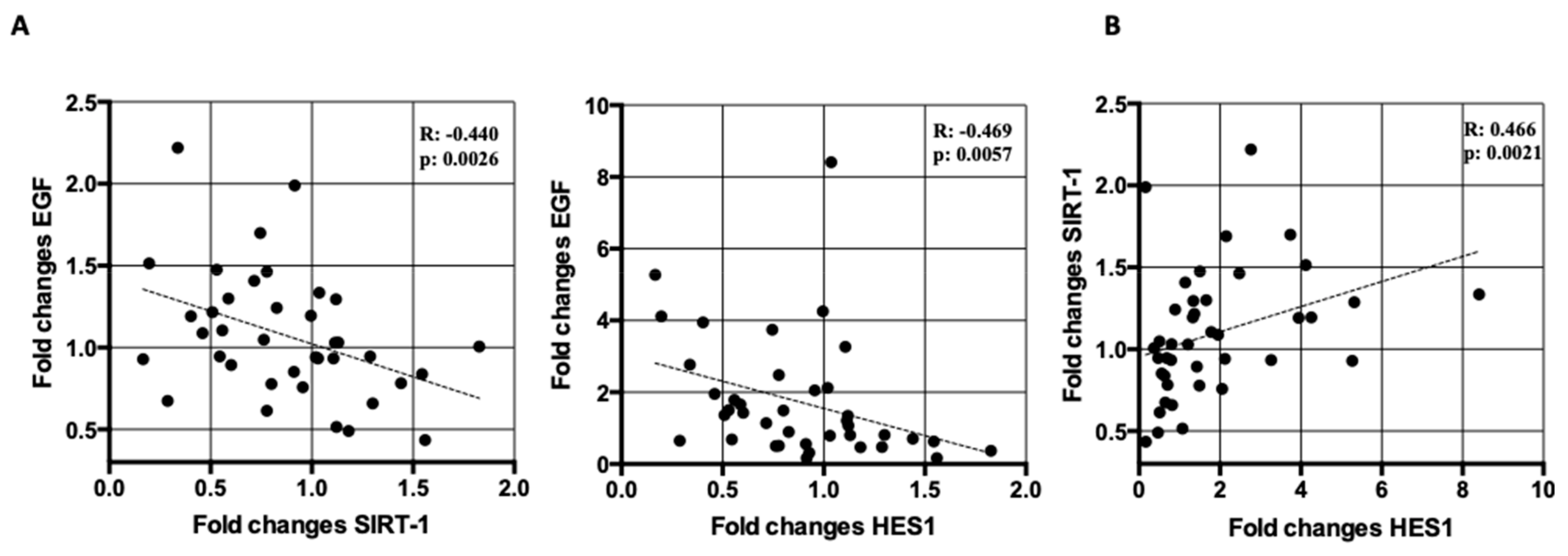
2.2. Correlation Analyses Between SIRT1 and HES1 mRNA Levels and Markers of Endothelial Dysfunction in Treated Patients

Endothelial dysfunction (ED) comprises alterations of the endothelial cell (EC) physiology (including increased expression of pro-inflammatory cell adhesion molecules (CAMs), such as intercellular adhesion molecule-1 (ICAM1) and vascular cell adhesion molecule-1 (VCAM1), impaired nitric oxide (NO) production and signaling, EC apoptosis, and increased vascular permeability) that can be reflected in changes in the serum of patients. We have developed an ex-vivo assay to determine the levels of apoptosis in the endothelium of patients. The assay involves the determination of the number of annexin V/propidium iodide positive apoptotic human umbilical vein endothelial cells (HUVECs) following 48 h of cultivation in the presence of medium containing 20% of patients’ serum. We have shown that the levels of apoptosis in HUVECs treated with serum from stable and acute CAD patients reflect the gravity of the disease [53,54]. Furthermore, we have reported that this assay can reliably detect a reduction in apoptosis in HUVECs treated with serum from stable CAD/COPD patients following treatment with ticagrelor [6] or from subjects with hypercholesterolemia and low-moderate cardiovascular risk, following eight weeks consumption of a red yeast rice, polymethoxyflavones, and antioxidants containing nutraceutical supplement [55]. In another study by our group, this approach was used, in parallel with measurements of changes in brachial wall shear stress and flow mediated dilation, to assess ED in patients that underwent surgical or transcatheter aortic valve replacement [56]. Of relevance, in stable CAD/COPD patients, following ticagrelor treatment we also found decreased serum levels of epidermal growth factor (EGF), increased NO generation in HUVECs treated with patient’s serum, measured by diaminofluorescein (DAF) assay, and attenuated reactive oxygen species (ROS) production in PBMC isolated from patients, assessed by flow cytometric analysis [2,6]. In order to determine whether the observed changes of SIRT1 and HES1 mRNA could be related to these markers of endothelial function, and whether the different effect of the drugs on SIRT1 and HES1 mRNA could be explained by differences in antiplatelet activity, we conducted association analyses between changes in SIRT1 and HES1 mRNA and changes in the levels of EGF, ROS, apoptosis, NO and platelets reactive units (PRU) after 1-month treatment (changes were defined as the ratio between values at T30 over values at T0).
Following treatment with ticagrelor or clopidogrel, we found correlations between changes in HES1 and SIRT1 mRNA levels and changes in the levels of EGF (HES1, R = −0.469, p < 0.01; SIRT1, R = −0.440, p < 0.01) (Figure 4A). In addition, changes in HES1 mRNA levels also correlated with changes in SIRT1 mRNA levels (R = 0.466, p < 0.01) (Figure 4B). On the contrary, we found no correlation among changes in HES1 or SIRT1 and ROS (HES1, R = −0.150, p = 0.36; SIRT1, R = −0.78, p = 0.65), apoptosis (HES1, R = 0.176, p = 0.27; SIRT1, R = 0.077, p = 0.63) and NO (HES1, R = 0.002, p = 0.99; SIRT1, R = −0.05, p = 0.76). Similarly, no correlation was found between changes in HES1 or SIRT1 mRNA and changes in the levels of PRU (HES1, R = −0.163, p = 0.31; SIRT1, R = −0.207, p = 0.20).

3. Discussion

Although concerns remain as to dosage equivalence, the PLATO (PLATelet inhibition and clinical Outcome) trial results showed that ticagrelor is superior to clopidogrel in preventing ischemic events in patients with acute coronary syndrome (ACS) [5]. To test the effect of ticagrelor over clopidogrel in patients with CAD, we selected a population of patients with stable CAD and chronic inflammation supported by the presence of a COPD diagnosis. COPD is a well-known chronic condition associated with inflammation and ED [57,58]. A similar approach has already been used in the study of Mangiacapra et al. who enrolled patients with stable CAD and type 2 diabetes to study the effect of ticagrelor and clopidogrel on endothelial function. In this case, diabetic patients were selected because they are a well-established subgroup of patients with ED, independently by cardiologic clinical presentation [7]. The choice to investigate the effects of ticagrelor on ED is in apparent contrast with a study showing that the anti-aggregatory effects of ticagrelor are less pronounced during acute inflammation [59], but is supported by data showing pleiotropic effects of ticagrelor beyond its potent antiplatelet effects [60,61]. Consistently, we found reduced levels of surrogate markers of endothelial dysfunction in patients with stable CAD and concomitant COPD following PCI and 1-month treatment with ticagrelor, but not with clopidogrel [6]. We now report that, in the same patients, treatment with ticagrelor, but not with clopidogrel, results in increased of levels of SIRT1 and HES1 mRNAs in peripheral blood cells.
Ticagrelor, unlike clopidogrel, has antiplatelet- and P2Y12-independent activities that seem to involve increased levels of adenosine/cAMP in plasma able to reduce both systemic inflammation and oxidative stress [9,10], although a single study has shown that ticagrelor is not able to increase adenosine circulating levels [62]. In order to gain more insights on the molecular pathways modulated by ticagrelor, we determined, in RNA isolated from peripheral blood cells of stable CAD patients following PCI, the effect of 1-month treatment with ticagrelor or clopidogrel on mRNA expression levels of inflammation- and oxidative stress-related genes, such as ICAM1, NLRP3, SIRT1, JAG1, DLL4, HES1, RORγt (Th17 marker), and FoxP3 (Tregs marker). Among the mRNAs analyzed, we found an increase of SIRT1 mRNA levels after treatment with ticagrelor but not clopidogrel. Specifically, we found that, although the number of patients with increased levels of SIRT1 were comparable in both treatment groups (6/21 in clopidogrel and 8/20 in ticagrelor), in the ticagrelor-treated group, only 2/20 patients showed a significant reduction of SIRT1 levels, which instead was detected in 7/21 patients treated with clopidogrel (Figure 3A). Our results, suggesting that ticagrelor may counteract SIRT1 reduction in RNA from peripheral blood of stable CAD/COPD patients, are consistent with previous reports of reduced activity of SIRT1 in serum of COPD patients [39] and inhibition of SIRT1 gene in peripheral monocytes in ACS and stable CAD patients [38]. SIRT1, one of the class III NAD+-deacetylases sirtuin family [63], has gained great attention for its impact on the regulation of aging-related processes [36]. SIRT1 blunts inflammatory response and oxidative stress mainly through the deacetylation of target proteins, such as Forkhead box O3 A (Foxo3a) [64], activator protein-1 (AP-1) [65], nuclear factor kappa B (NF-κB) [66], Notch1 [67,68], and NLRP3 [69]. Furthermore, SIRT1 interferes with Toll-like receptor (TLR) 2- mediated monocyte adhesion to the vascular endothelium, an early step in the pathogenesis of CAD [70]. Of interest, SIRT1 genetic overexpression significantly decreased oxidative stress in cigarette smoke-exposed rodent lungs [71].
We also found increased expression of HES1 in more than a half of ticagrelor-treated patients (15/21), compared to the clopidogrel group, in which the number of patients with increased or decreased HES1 mRNA levels were similar (8/21 and 9/21, up- and down-regulated, respectively) (Figure 3B). HES1, a transcription factor belonging to the Hairy and Enhancer of Split (HES) family, is required for differentiation of T and B cells [72] and plays a major role in the reduction of inflammation and neutrophil-mediated responses by controlling production of macrophage-derived chemokines [73,74]. In macrophages, HES1 functions as a feedback inhibitor of production of pro-inflammatory cytokines, such as IL (Interleukin)-6 and IL-12 [75,76]. HES1 is a target gene and essential transducer of the Notch pathway, implicated in the regulation of cell fate decisions, such as differentiation, proliferation and survival [77]. The Notch pathway is triggered by the interaction between the Notch receptors (Notch 1–4) and ligands (DLL1, 3, 4 and JAGGED-1, -2), on adjacent cells, leading to the release of the active form of Notch which translocates into the nucleus, where it promotes the transcription of its target genes [77]. Notch regulates heart and vascular functions during development and is involved in the repair of the damaged and/or stressed myocardium [29,78,79]. Dysregulation of the Notch pathway has been linked to atherosclerosis [80,81] and to COPD [82]. In COPD patients, reduced levels of NOTCH1 and HES1 have been shown in the endothelium [33] and in the airways epithelium [32].
Our data, analyzed in the context of the existing literature, suggest that increased levels of SIRT1 and HES1 in RNA of peripheral blood cells of stable CAD/COPD patients may be part of the protective action of ticagrelor. Of interest, we found a positive correlation between changes of SIRT1 and HES1 mRNA levels after the treatment with ticagrelor or clopidogrel (Figure 4B). Little is known about SIRT1 and HES1 interaction. SIRT1 physically interacts with HES1 leading to stimulation of its role as negative regulator of transcription [83], whereas in neural stem cells SIRT1 through inhibition of HES1 expression mediates response of the cell to glucose [84] and induces their differentiation [85]. The biological meaning of this association in the context of ticagrelor/clopidogrel treated stable CAD/COPD patients deserves further studies.
Correlation analysis showed that SIRT1 and HES1 changes were not associated to changes in platelet activity, indicating that the ticagrelor effect on these parameters is not directly related to the inhibition of P2Y12. On the contrary, changes in both mRNA SIRT1 and HES1 levels negatively correlated with changes in EGF levels (Figure 4A). High levels of EGF and its receptor EGFR have been detected in lung tissue specimens of COPD patients [86] and high airway immunoreactivity has been associated to mucin hypersecretion in COPD [87]. EGF is involved in endothelial dysfunction, neointimal hyperplasia, cardiac hypertrophy and remodeling [88] and, as shown by us, reduction in circulating levels of EGF could be involved in the ticagrelor-mediated improvement of endothelial function in stable CAD/COPD patients [2]. The cross-talk between EGF and Notch signaling is well characterized: EGF and Notch pathways can cooperate in either synergistic or antagonistic fashion in malignancies [89] and the EGF, via EGFR, acts as a negative regulator of HES1 expression in keratinocytes [90]. On the contrary, little is known on the cross-talk between EGF/SIRT1. In cancer, SIRT1 is upregulated during EGF-mediated epithelial-to-mesenchymal transition [91], while in vascular smooth muscle cells, resveratrol, a SIRT1 stimulator, interferes with EGF-induced ROS production [92]. The negative correlation found in this study may be suggestive of a cross-talk among EGF, SIRT1, and/or HES1 in peripheral blood of stable CAD/COPD patients, according to which ticagrelor-mediated EGF-reduction may lead to the induction of SIRT1/HES1 axis.
A limitation of this study is represented by the use of RNA isolated from whole peripheral blood, which contains different cell types (lymphocytes, monocytes, and granulocytes). Therefore, no conclusions can be drawn with respect to the molecular mechanism involved in SIRT1 and HES1 upregulation, which could be due to the effect of the drug either on the expression of these genes or on the size of specific populations of cells expressing these genes. The use of whole blood could have also reduced the sensitivity of our assay, thus allowing the detection, at follow up, of significant changes in the expression of SIRT1 and HES1 but of none of the other genes analyzed (RORγt, FoxP3, NLRP3, ICAM1, JAG1, and DLL4).
Another limitation is the lack of information on the deacetylase activity of SIRT1 in our samples that, as shown by Conti and collaborators, would have provided more specific information, compared to the mRNA, about the role of SIRT1 [39] in mediating the pleiotropic effects of ticagrelor in stable CAD/COPD patients. This limitation could explain the lack of association between changes in SIRT1 mRNA and neither the other inflammation markers investigated (NLRP3, ICAM1, RORγt, and FoxP3), known to be regulated by SIRT1 [64,66,67,69] nor the surrogate markers of endothelial function (apoptosis, ROS and NO levels), previously found by us to be reduced by ticagrelor treatment [6].
A third limitation of the study is represented by the relatively small number of patients and by the impossibility to conduct statistical power analysis due to lack of published data on the effects of ticagrelor/clopidogrel on the expression of the studied genes. Therefore, this should be considered a pilot study, providing information for future studies aimed to validate our findings, which could be relevant not only for dissecting the mechanism of action of ticagrelor but also for providing novel blood markers (SIRT1 and HES1 mRNA) able to predict whether the single patient will benefit of effects of ticagrelor beyond its antiplatelet activity.

4. Material and Methods

4.1. Study Design and Population/Randomization and Interventions

This is a sub-study of the clinical trial “The comparisoN between ticAgrelor and clopidogrel effect on endoTHelial platelet ANd iNflammation parameters in patiEnts with stable coronary artery disease and chronic obstructiVE pulmonaRy disease undergoing percutaneous coronary intervention (NATHAN-NEVER)” registered at www.clinicaltrials.gov (NCT02519608, September 2015). The protocol was approved by “Comitato Etico Unico della Provincia di Ferrara”. Written informed consent was given by all subjects in accordance with the Declaration of Helsinki. This study was an investigator-initiated, prospective, single-center, randomized, open-label phase II trial involving 46 consecutive patients with stable coronary artery disease requiring coronary artery angiography (CAA) and PCI, and concomitant COPD. The diagnosis of COPD was based on spirometry data according to international guidelines and in particular by the presence of not fully reversible airflow limitation defined as a ratio between forced expiratory volume at 1 s (FEV1) and forced vital capacity (FVC) ratio < 0.7 after the administration of a bronchodilator [93]. An independent blinded reviewer analyzed spirometry and documentation available to confirm COPD diagnosis and to adjudicate severity of airflow limitation based on post-bronchodilator FEV1 (from mild to very severe). Patients were randomly assigned to receive clopidogrel (n = 23) or ticagrelor (n = 23) on top of standard therapy with aspirin. The primary endpoint of the study was the 1-month rate of HUVECs apoptosis and the rate of apoptosis after 1 month was significantly lower in patients treated with ticagrelor (7.4 ± 1.3% vs. 9.3 ± 1.5%, p < 0.001), satisfying the pre-specified primary endpoint [6]. The study design, the characteristics of the study participants, including the severity of both diseases, comorbidities, and drug treatment, the outcomes of this trial are described in [6].

4.2. Blood Samples

A 21-gauge needle was used to collect blood samples from an antecubital vein of patients before PCI and drug administration, and at the 1-month visit. Details of the sampling procedures are reported in [6]. After the withdrawal, blood samples were stored frozen until RNA extraction.

4.3. RNA Isolation and cDNA Synthesis

RNA was isolated from blood samples using the QIAamp RNA Blood Mini Kit (Qiagen, Carlsbad, CA, USA). One volume of blood was mixed with 5 volumes of buffer EL (erythrocyte lysis) and incubated for 10 to 15 min on ice, vortexing briefly 2 times during incubation to selectively lyse red blood cells. White cells were then collected by centrifugation at 400× g for 10 min at 4 °C and supernatant was removed. White cells were lysed using highly denaturing conditions to inactivate the RNases. After homogenization using the QIAshredder spin column, the sample was applied to the QIAamp spin column. Total RNA binds to the QIAamp membrane and contaminants are washed away, leaving pure RNA to be eluted in 30–100 µL RNase-free water. RNA concentration and purity were determined by NanoDrop 2000 spectrophotometer (Thermo Fisher Scientific, Waltham, MA, USA). RNA was treated with the RNase-Free DNase Set (Qiagen, Carlsbad, CA, USA), in order to eliminate DNA contamination. The 100 ng of total RNA were reverse transcribed to cDNA using the SuperScript™ III First-Strand Synthesis SuperMix (Life Technologies, Carlsbad, CA, USA).

4.4. Droplet Digital PCR Reaction

For droplet digital (dd) PCR, 2 µL of cDNA (1 ng/µL) were used in each reaction. 20 µL of a solution containing cDNA, primers and QX200™ ddPCR™ EvaGreen Supermix (Bio-Rad, Hercules, CA, USA) were used for droplets preparation using a Bio-Rad QX200 droplet generator (Bio-Rad, Hercules, CA, USA). Emulsified samples were transferred to a 96-well plate, sealed with the PX1 PCR plate sealer (Bio-Rad, Hercules, CA, USA) and amplified using the SimpliAmp thermal cycler, (Applied Biosystems, Waltham, CA, USA). The parameters for ddPCR were: 95 °C enzyme activation step for 5 min followed by 40 cycles of a two-step cycling protocol (95 °C for 30 s and 60 °C for 1 min). The sequences of primers used are shown in Supplementary Table S1. At the end of thermal cycling, a QX200 droplet reader (Bio-Rad, Hercules, CA, USA) was used to quantify the number of generated droplets from each reaction mix. Expression data was extracted using QuantaSoft software (Bio-Rad, Hercules, CA, USA) and the absolute quantity of cDNA per sample (copies/µL) was normalized to the average number of copies of GUSB (β-glucuronidase) mRNA in each sample.

4.5. Statistical Analysis

Normal distribution of the variables was verified with the D’Agostino–Pearson normality test and with the Shapiro–Wilk test (alpha = 0.05). Variables were presented as scatter plot with median. Outliers were identified and removed. Normally distributed variables were checked for homoscedasticity by Levene test. Follow up toward baseline groups were compared employing repeated measures ANOVA to take into account for samples dependency. Student t test for independent measures was employed to test statistical significance in the mean differences of gene expression levels at baselines between treatments. p-values ≤ 0.05 were considered statistically significant. Correlations between continuous variables were tested by Spearman’s correlation coefficient correlation. Correlation of baseline categorical variables and gene expression was tested by point-biserial correlation. Statistical analysis was performed with GraphPad Prism version 7.0 (GraphPad software Inc., San Diego, CA, USA) and R (R Foundation for Statistical Computing, Vienna, Austria, 2019).

5. Conclusions

In conclusion, we showed that the effect of ticagrelor related to the mitigation of endothelial dysfunction in stable CAD/COPD patients may be mediated, at least in part, by its capacity to increase SIRT1 and HES1 mRNAs levels (Supplementary Figure S1). These findings are consistent with studies suggesting that the pleiotropic effects of ticagrelor on the vascular system may be ascribed to a reduction of systemic inflammation and oxidative stress.

Supplementary Materials

Supplementary materials can be found at https://www.mdpi.com/1422-0067/21/5/1576/s1.

Author Contributions

Conceptualization, G.A., P.R., and G.C.; Formal analysis, G.A., F.V.D.S., F.F., F.M., M.M., A.P. (Antonio Pannuti), and R.F.; Funding acquisition, G.C.; Investigation, G.A., L.M., V.M., and I.B.; Project administration, G.C.; Resources, R.P., L.S.C., A.P. (Anna Piredda), and A.S.; Supervision, P.R.; Validation, G.A.; Writing—original draft, G.A., R.F., and P.R.; Writing—review & editing, G.A., F.V.D.S., F.F., F.M., M.M., A.P. (Antonio Pannuti), and P.R. All authors have read and agreed to the published version of the manuscript.

Funding

This study was partially supported by a research grant [ESR-14–10659] from AstraZeneca, which had no role in the collection, analysis, interpretation of data, writing of the report, and the decision to submit the paper for publication.

Conflicts of Interest

Gianluca Campo has received honoraria for lectures from AstraZeneca, Menarini, Abbott Vascular, Boston Scientific. The other authors declare that there is no conflict of interest regarding the publication of this paper.

References

  1. Cannon, C.P.; Harrington, R.A.; James, S.; Ardissino, D.; Becker, R.C.; Emanuelsson, H.; Husted, S.; Katus, H.; Keltai, M.; Khurmi, N.S.; et al. Comparison of ticagrelor with clopidogrel in patients with a planned invasive strategy for acute coronary syndromes (PLATO): A randomised double-blind study. Lancet 2010, 375, 283–293. [Google Scholar] [CrossRef]
  2. Vieceli Dalla Sega, F.; Fortini, F.; Aquila, G.; Pavasini, R.; Biscaglia, S.; Bernucci, D.; Del Franco, A.; Tonet, E.; Rizzo, P.; Ferrari, R.; et al. Ticagrelor Improves Endothelial Function by Decreasing Circulating Epidermal Growth Factor (EGF). Front. Physiol. 2018, 9, 337. [Google Scholar] [CrossRef] [PubMed]
  3. Fromonot, J.; Dignat-Georges, F.; Rossi, P.; Mottola, G.; Kipson, N.; Ruf, J.; Bonello, L.; Guieu, R.; Paganelli, F. Ticagrelor Improves Peripheral Arterial Function in Acute Coronary Syndrome Patients: Relationship With Adenosine Plasma Level. J. Am. Coll Cardiol. 2016, 67, 1967–1968. [Google Scholar] [CrossRef] [PubMed]
  4. Cattaneo, M.; Schulz, R.; Nylander, S. Adenosine-mediated effects of ticagrelor: Evidence and potential clinical relevance. J. Am. Coll Cardiol. 2014, 63, 2503–2509. [Google Scholar] [CrossRef] [PubMed] [Green Version]
  5. Wallentin, L.; Becker, R.C.; Budaj, A.; Cannon, C.P.; Emanuelsson, H.; Held, C.; Horrow, J.; Husted, S.; James, S.; Katus, H.; et al. Ticagrelor versus clopidogrel in patients with acute coronary syndromes. N. Engl. J. Med. 2009, 361, 1045–1057. [Google Scholar] [CrossRef]
  6. Campo, G.; Vieceli Dalla Sega, F.; Pavasini, R.; Aquila, G.; Gallo, F.; Fortini, F.; Tonet, E.; Cimaglia, P.; Del Franco, A.; Pestelli, G.; et al. Biological effects of ticagrelor over clopidogrel in patients with stable coronary artery disease and chronic obstructive pulmonary disease. Thromb Haemost 2017, 117, 1208–1216. [Google Scholar] [CrossRef] [Green Version]
  7. Mangiacapra, F.; Panaioli, E.; Colaiori, I.; Ricottini, E.; Lauria Pantano, A.; Pozzilli, P.; Barbato, E.; Di Sciascio, G. Clopidogrel Versus Ticagrelor for Antiplatelet Maintenance in Diabetic Patients Treated With Percutaneous Coronary Intervention: Results of the CLOTILDIA Study (Clopidogrel High Dose Versus Ticagrelor for Antiplatelet Maintenance in Diabetic Patients). Circulation 2016, 134, 835–837. [Google Scholar] [CrossRef] [Green Version]
  8. Bonaca, M.P.; Bhatt, D.L.; Cohen, M.; Steg, P.G.; Storey, R.F.; Jensen, E.C.; Magnani, G.; Bansilal, S.; Fish, M.P.; Im, K.; et al. Long-term use of ticagrelor in patients with prior myocardial infarction. N. Engl. J. Med. 2015, 372, 1791–1800. [Google Scholar] [CrossRef] [Green Version]
  9. Bonello, L.; Laine, M.; Kipson, N.; Mancini, J.; Helal, O.; Fromonot, J.; Gariboldi, V.; Condo, J.; Thuny, F.; Frere, C.; et al. Ticagrelor increases adenosine plasma concentration in patients with an acute coronary syndrome. J. Am. Coll Cardiol. 2014, 63, 872–877. [Google Scholar] [CrossRef]
  10. Li, X.; Wang, Q.; Xue, Y.; Chen, J.; Lv, Q. Ticagrelor Compared with Clopidogrel Increased Adenosine and Cyclic Adenosine Monophosphate Plasma Concentration in Acute Coronary Syndrome Patients. Basic Clin. Pharmacol. Toxicol. 2017, 120, 610–614. [Google Scholar] [CrossRef] [Green Version]
  11. Ramkumar, V.; Hallam, D.M.; Nie, Z. Adenosine, oxidative stress and cytoprotection. Jpn. J. Pharmacol. 2001, 86, 265–274. [Google Scholar] [CrossRef] [PubMed] [Green Version]
  12. Cekic, C.; Linden, J. Purinergic regulation of the immune system. Nat. Rev. Immunol. 2016, 16, 177–192. [Google Scholar] [CrossRef] [PubMed]
  13. Vieceli Dalla Sega, F.; Fortini, F.; Aquila, G.; Campo, G.; Vaccarezza, M.; Rizzo, P. Notch Signaling Regulates Immune Responses in Atherosclerosis. Front. Immunol. 2019, 10, 1130. [Google Scholar] [CrossRef] [PubMed] [Green Version]
  14. Takahashi, H.K.; Iwagaki, H.; Hamano, R.; Wake, H.; Kanke, T.; Liu, K.; Yoshino, T.; Tanaka, N.; Nishibori, M. Effects of adenosine on adhesion molecule expression and cytokine production in human PBMC depend on the receptor subtype activated. Br. J. Pharmacol. 2007, 150, 816–822. [Google Scholar] [CrossRef] [PubMed] [Green Version]
  15. Yan, Y.; Jiang, W.; Liu, L.; Wang, X.; Ding, C.; Tian, Z.; Zhou, R. Dopamine controls systemic inflammation through inhibition of NLRP3 inflammasome. Cell 2015, 160, 62–73. [Google Scholar] [CrossRef] [PubMed] [Green Version]
  16. Sokolowska, M.; Chen, L.Y.; Liu, Y.; Martinez-Anton, A.; Qi, H.Y.; Logun, C.; Alsaaty, S.; Park, Y.H.; Kastner, D.L.; Chae, J.J.; et al. Prostaglandin E2 Inhibits NLRP3 Inflammasome Activation through EP4 Receptor and Intracellular Cyclic AMP in Human Macrophages. J. Immunol. 2015, 194, 5472–5487. [Google Scholar] [CrossRef] [Green Version]
  17. Zhou, W.; Chen, C.; Chen, Z.; Liu, L.; Jiang, J.; Wu, Z.; Zhao, M.; Chen, Y. NLRP3: A Novel Mediator in Cardiovascular Disease. J. Immunol. Res. 2018, 2018, 5702103. [Google Scholar] [CrossRef]
  18. Zou, X.L.; Chen, Z.G.; Zhang, T.T.; Feng, D.Y.; Li, H.T.; Yang, H.L. Th17/Treg homeostasis, but not Th1/Th2 homeostasis, is implicated in exacerbation of human bronchial asthma. Ther. Clin. Risk Manag. 2018, 14, 1627–1636. [Google Scholar] [CrossRef] [Green Version]
  19. Wang, L.; Wan, H.; Tang, W.; Ni, Y.; Hou, X.; Pan, L.; Song, Y.; Shi, G. Critical roles of adenosine A2A receptor in regulating the balance of Treg/Th17 cells in allergic asthma. Clin. Respir. J. 2018, 12, 149–157. [Google Scholar] [CrossRef]
  20. Francois, V.; Shehade, H.; Acolty, V.; Preyat, N.; Delree, P.; Moser, M.; Oldenhove, G. Intestinal immunopathology is associated with decreased CD73-generated adenosine during lethal infection. Mucosal Immunol. 2015, 8, 773–784. [Google Scholar] [CrossRef] [Green Version]
  21. Zarek, P.E.; Huang, C.T.; Lutz, E.R.; Kowalski, J.; Horton, M.R.; Linden, J.; Drake, C.G.; Powell, J.D. A2A receptor signaling promotes peripheral tolerance by inducing T-cell anergy and the generation of adaptive regulatory T cells. Blood 2008, 111, 251–259. [Google Scholar] [CrossRef] [PubMed]
  22. Potekhina, A.V.; Pylaeva, E.; Provatorov, S.; Ruleva, N.; Masenko, V.; Noeva, E.; Krasnikova, T.; Arefieva, T. Treg/Th17 balance in stable CAD patients with different stages of coronary atherosclerosis. Atherosclerosis 2015, 238, 17–21. [Google Scholar] [CrossRef] [PubMed]
  23. Cheng, X.; Yu, X.; Ding, Y.J.; Fu, Q.Q.; Xie, J.J.; Tang, T.T.; Yao, R.; Chen, Y.; Liao, Y.H. The Th17/Treg imbalance in patients with acute coronary syndrome. Clin. Immunol. 2008, 127, 89–97. [Google Scholar] [CrossRef] [PubMed]
  24. Li, X.N.; Pan, X.; Qiu, D. Imbalances of Th17 and Treg cells and their respective cytokines in COPD patients by disease stage. Int. J. Clin. Exp. Med. 2014, 7, 5324–5329. [Google Scholar] [PubMed]
  25. Wang, H.; Ying, H.; Wang, S.; Gu, X.; Weng, Y.; Peng, W.; Xia, D.; Yu, W. Imbalance of peripheral blood Th17 and Treg responses in patients with chronic obstructive pulmonary disease. Clin. Respir. J. 2015, 9, 330–341. [Google Scholar] [CrossRef] [PubMed]
  26. Sorrentino, C.; Hossain, F.; Rodriguez, P.C.; Sierra, R.A.; Pannuti, A.; Osborne, B.A.; Minter, L.M.; Miele, L.; Morello, S. Adenosine A2A Receptor Stimulation Inhibits TCR-Induced Notch1 Activation in CD8+T-Cells. Front. Immunol. 2019, 10, 162. [Google Scholar] [CrossRef]
  27. Bergstrom, I.; Backteman, K.; Lundberg, A.; Ernerudh, J.; Jonasson, L. Persistent accumulation of interferon-gamma-producing CD8+CD56+ T cells in blood from patients with coronary artery disease. Atherosclerosis 2012, 224, 515–520. [Google Scholar] [CrossRef] [Green Version]
  28. Kemeny, D.M.; Vyas, B.; Vukmanovic-Stejic, M.; Thomas, M.J.; Noble, A.; Loh, L.C.; O’Connor, B.J. CD8(+) T cell subsets and chronic obstructive pulmonary disease. Am. J. Respir. Crit. Care Med. 1999, 160, S33–S37. [Google Scholar] [CrossRef]
  29. Ferrari, R.; Rizzo, P. The Notch pathway: A novel target for myocardial remodelling therapy? Eur. Heart J. 2014, 35, 2140–2145. [Google Scholar] [CrossRef] [Green Version]
  30. Zhao, L.L.; Wang, C.; Hu, S.N.; Li, L.L.; Wang, Y.; Hou, J.B.; Yu, B.; Zhu, Y.C.; Jia, H.B.; Zhe, C.Y.; et al. P1788The relationship between the jagged1 levels of human blood mononuclear cell surface and plaque vulnerability. Eur. Heart J. 2017, 38. [Google Scholar] [CrossRef] [Green Version]
  31. Aquila, G.; Fortini, C.; Pannuti, A.; Delbue, S.; Pannella, M.; Morelli, M.B.; Caliceti, C.; Castriota, F.; de Mattei, M.; Ongaro, A.; et al. Distinct gene expression profiles associated with Notch ligands Delta-like 4 and Jagged1 in plaque material from peripheral artery disease patients: A pilot study. J. Transl. Med. 2017, 15, 98. [Google Scholar] [CrossRef] [PubMed]
  32. Tilley, A.E.; Harvey, B.G.; Heguy, A.; Hackett, N.R.; Wang, R.; O’Connor, T.P.; Crystal, R.G. Down-regulation of the notch pathway in human airway epithelium in association with smoking and chronic obstructive pulmonary disease. Am. J. Respir. Crit. Care Med. 2009, 179, 457–466. [Google Scholar] [CrossRef] [PubMed] [Green Version]
  33. Zong, D.; Li, J.; Cai, S.; He, S.; Liu, Q.; Jiang, J.; Chen, S.; Long, Y.; Chen, Y.; Chen, P.; et al. Notch1 regulates endothelial apoptosis via the ERK pathway in chronic obstructive pulmonary disease. Am. J. Physiol. Cell Physiol. 2018, 315, C330–C340. [Google Scholar] [CrossRef] [PubMed]
  34. Fortini, F.; Vieceli Dalla Sega, F.; Caliceti, C.; Lambertini, E.; Pannuti, A.; Peiffer, D.S.; Balla, C.; Rizzo, P. Estrogen-mediated protection against coronary heart disease: The role of the Notch pathway. J. Steroid. Biochem. Mol. Biol. 2019, 189, 87–100. [Google Scholar] [CrossRef] [PubMed]
  35. Vieceli Dalla Sega, F.; Aquila, G.; Fortini, F.; Vaccarezza, M.; Secchiero, P.; Rizzo, P.; Campo, G. Context-dependent function of ROS in the vascular endothelium: The role of the Notch pathway and shear stress. Biofactors 2017, 43, 475–485. [Google Scholar] [CrossRef]
  36. Salminen, A.; Kaarniranta, K.; Kauppinen, A. Crosstalk between Oxidative Stress and SIRT1: Impact on the Aging Process. Int. J. Mol. Sci. 2013, 14, 3834–3859. [Google Scholar] [CrossRef] [Green Version]
  37. Vachharajani, V.T.; Liu, T.; Wang, X.; Hoth, J.J.; Yoza, B.K.; McCall, C.E. Sirtuins Link Inflammation and Metabolism. J. Immunol. Res. 2016, 2016, 8167273. [Google Scholar] [CrossRef] [Green Version]
  38. Breitenstein, A.; Wyss, C.A.; Spescha, R.D.; Franzeck, F.C.; Hof, D.; Riwanto, M.; Hasun, M.; Akhmedov, A.; von Eckardstein, A.; Maier, W.; et al. Peripheral blood monocyte Sirt1 expression is reduced in patients with coronary artery disease. PLoS ONE 2013, 8, e53106. [Google Scholar] [CrossRef] [Green Version]
  39. Conti, V.; Corbi, G.; Manzo, V.; Malangone, P.; Vitale, C.; Maglio, A.; Cotugno, R.; Capaccio, D.; Marino, L.; Selleri, C.; et al. SIRT1 Activity in Peripheral Blood Mononuclear Cells Correlates with Altered Lung Function in Patients with Chronic Obstructive Pulmonary Disease. Oxidative Med. Cell. Longev. 2018, 2018, 8. [Google Scholar] [CrossRef]
  40. Bonello, L.; Frere, C.; Cointe, S.; Laine, M.; Mancini, J.; Thuny, F.; Kerbaul, F.; Lemesle, G.; Paganelli, F.; Guieu, R.; et al. Ticagrelor increases endothelial progenitor cell level compared to clopidogrel in acute coronary syndromes: A prospective randomized study. Int. J. Cardiol. 2015, 187, 502–507. [Google Scholar] [CrossRef]
  41. Torngren, K.; Ohman, J.; Salmi, H.; Larsson, J.; Erlinge, D. Ticagrelor improves peripheral arterial function in patients with a previous acute coronary syndrome. Cardiology 2013, 124, 252–258. [Google Scholar] [CrossRef] [PubMed]
  42. Alexopoulos, D.; Moulias, A.; Koutsogiannis, N.; Xanthopoulou, I.; Kakkavas, A.; Mavronasiou, E.; Davlouros, P.; Hahalis, G. Differential effect of ticagrelor versus prasugrel on coronary blood flow velocity in patients with non-ST-elevation acute coronary syndrome undergoing percutaneous coronary intervention: An exploratory study. Circ. Cardiovasc. Interv. 2013, 6, 277–283. [Google Scholar] [CrossRef] [PubMed] [Green Version]
  43. Storey, R.F.; James, S.K.; Siegbahn, A.; Varenhorst, C.; Held, C.; Ycas, J.; Husted, S.E.; Cannon, C.P.; Becker, R.C.; Steg, P.G.; et al. Lower mortality following pulmonary adverse events and sepsis with ticagrelor compared to clopidogrel in the PLATO study. Platelets 2014, 25, 517–525. [Google Scholar] [CrossRef]
  44. Armstrong, D.; Summers, C.; Ewart, L.; Nylander, S.; Sidaway, J.E.; van Giezen, J.J. Characterization of the adenosine pharmacology of ticagrelor reveals therapeutically relevant inhibition of equilibrative nucleoside transporter 1. J. Cardiovasc. Pharm. Ther. 2014, 19, 209–219. [Google Scholar] [CrossRef] [PubMed]
  45. Van Giezen, J.J.; Sidaway, J.; Glaves, P.; Kirk, I.; Bjorkman, J.A. Ticagrelor inhibits adenosine uptake in vitro and enhances adenosine-mediated hyperemia responses in a canine model. J. Cardiovasc. Pharm. Ther. 2012, 17, 164–172. [Google Scholar] [CrossRef] [PubMed]
  46. Wittfeldt, A.; Emanuelsson, H.; Brandrup-Wognsen, G.; van Giezen, J.J.; Jonasson, J.; Nylander, S.; Gan, L.M. Ticagrelor enhances adenosine-induced coronary vasodilatory responses in humans. J. Am. Coll Cardiol. 2013, 61, 723–727. [Google Scholar] [CrossRef] [PubMed] [Green Version]
  47. Aungraheeta, R.; Conibear, A.; Butler, M.; Kelly, E.; Nylander, S.; Mumford, A.; Mundell, S.J. Inverse agonism at the P2Y12 receptor and ENT1 transporter blockade contribute to platelet inhibition by ticagrelor. Blood 2016, 128, 2717–2728. [Google Scholar] [CrossRef]
  48. Thomas, M.; Judge, H.; Dockrell, D.; Sabroe, I.; Storey, R. 215 Ticagrelor Inhibits Release of Pro-inflammatory Cytokines TNF and IL-6 during Human Endotoxaemia. Heart 2014, 100, A117–A118. [Google Scholar] [CrossRef]
  49. Nylander, S.; Schulz, R. Effects of P2Y12 receptor antagonists beyond platelet inhibition--comparison of ticagrelor with thienopyridines. Br. J. Pharmacol. 2016, 173, 1163–1178. [Google Scholar] [CrossRef] [Green Version]
  50. Husted, S.; Storey, R.F.; Harrington, R.A.; Emanuelsson, H.; Cannon, C.P. Changes in inflammatory biomarkers in patients treated with ticagrelor or clopidogrel. Clin. Cardiol. 2010, 33, 206–212. [Google Scholar] [CrossRef]
  51. Ariotti, S.; Ortega-Paz, L.; van Leeuwen, M.; Brugaletta, S.; Leonardi, S.; Akkerhuis, K.M.; Rimoldi, S.F.; Janssens, G.; Gianni, U.; van den Berge, J.C.; et al. Effects of Ticagrelor, Prasugrel, or Clopidogrel on Endothelial Function and Other Vascular Biomarkers: A Randomized Crossover Study. JACC Cardiovasc. Interv. 2018, 11, 1576–1586. [Google Scholar] [CrossRef] [PubMed]
  52. Teng, R.; Butler, K. Effect of the CYP3A inhibitors, diltiazem and ketoconazole, on ticagrelor pharmacokinetics in healthy volunteers. J. Drug Assess. 2013, 2, 30–39. [Google Scholar] [CrossRef] [PubMed] [Green Version]
  53. Agnoletti, L.; Curello, S.; Bachetti, T.; Malacarne, F.; Gaia, G.; Comini, L.; Volterrani, M.; Bonetti, P.; Parrinello, G.; Cadei, M.; et al. Serum from patients with severe heart failure downregulates eNOS and is proapoptotic: Role of tumor necrosis factor-alpha. Circulation 1999, 100, 1983–1991. [Google Scholar] [CrossRef] [PubMed] [Green Version]
  54. Valgimigli, M.; Agnoletti, L.; Curello, S.; Comini, L.; Francolini, G.; Mastrorilli, F.; Merli, E.; Pirani, R.; Guardigli, G.; Grigolato, P.G.; et al. Serum from patients with acute coronary syndromes displays a proapoptotic effect on human endothelial cells: A possible link to pan-coronary syndromes. Circulation 2003, 107, 264–270. [Google Scholar] [CrossRef] [PubMed]
  55. Cimaglia, P.; Vieceli Dalla Sega, F.; Vitali, F.; Lodolini, V.; Bernucci, D.; Passarini, G.; Fortini, F.; Marracino, L.; Aquila, G.; Rizzo, P.; et al. Effectiveness of a Novel Nutraceutical Compound Containing Red Yeast Rice, Polymethoxyflavones and Antioxidants in the Modulation of Cholesterol Levels in Subjects With Hypercholesterolemia and Low-Moderate Cardiovascular Risk: The NIRVANA Study. Front. Physiol. 2019, 10, 217. [Google Scholar] [CrossRef] [PubMed]
  56. Moscarelli, M.; Devito, F.; Fattouch, K.; Lancellotti, P.; Ciccone, M.M.; Rizzo, P.; Gaudino, M.; Marchese, A.; Angelini, G.; Speziale, G. The effect of surgical versus transcatheter aortic valve replacement on endothelial function. An observational study. Int. J. Surg. 2019, 63, 1–7. [Google Scholar] [CrossRef] [PubMed] [Green Version]
  57. Fabbri, L.M.; Rabe, K.F. From COPD to chronic systemic inflammatory syndrome? Lancet 2007, 370, 797–799. [Google Scholar] [CrossRef]
  58. Szucs, B.; Szucs, C.; Petrekanits, M.; Varga, J.T. Molecular Characteristics and Treatment of Endothelial Dysfunction in Patients with COPD: A Review Article. Int J. Mol. Sci 2019, 20, 4329. [Google Scholar] [CrossRef] [Green Version]
  59. Imam, H.; Nguyen, T.H.; De Caterina, R.; Nooney, V.B.; Chong, C.R.; Horowitz, J.D.; Chirkov, Y.Y. Impaired adenylate cyclase signaling in acute myocardial ischemia: Impact on effectiveness of P2Y12 receptor antagonists. Thromb. Res. 2019, 181, 92–98. [Google Scholar] [CrossRef]
  60. Zheng, W.; Li, Y.; Tian, J.; Li, L.; Xie, L.; Mao, Q.; Tong, W.; Zhou, D.; Azzalini, L.; Zhao, X. Effects of Ticagrelor versus Clopidogrel in Patients with Coronary Bifurcation Lesions Undergoing Percutaneous Coronary Intervention. Biomed. Res. Int. 2019, 2019, 3170957. [Google Scholar] [CrossRef] [Green Version]
  61. Jeong, H.S.; Hong, S.J.; Cho, S.-A.; Kim, J.-H.; Cho, J.Y.; Lee, S.H.; Joo, H.J.; Park, J.H.; Yu, C.W.; Lim, D.-S. Comparison of Ticagrelor Versus Prasugrel for Inflammation, Vascular Function, and Circulating Endothelial Progenitor Cells in Diabetic Patients With Non–ST-Segment Elevation Acute Coronary Syndrome Requiring Coronary Stenting: A Prospective, Randomized, Crossover Trial. JACC: Cardiovasc. Interv. 2017, 10, 1646–1658. [Google Scholar]
  62. Van den Berg, T.N.; El Messaoudi, S.; Rongen, G.A.; van den Broek, P.H.; Bilos, A.; Donders, A.R.; Gomes, M.E.; Riksen, N.P. Ticagrelor Does Not Inhibit Adenosine Transport at Relevant Concentrations: A Randomized Cross-Over Study in Healthy Subjects In Vivo. PLoS ONE 2015, 10, e0137560. [Google Scholar] [CrossRef] [PubMed] [Green Version]
  63. McGuinness, D.; McGuinness, D.H.; McCaul, J.A.; Shiels, P.G. Sirtuins, bioageing, and cancer. J. Aging Res. 2011, 2011, 235754. [Google Scholar] [CrossRef] [PubMed] [Green Version]
  64. Beier, U.H.; Wang, L.; Bhatti, T.R.; Liu, Y.; Han, R.; Ge, G.; Hancock, W.W. Sirtuin-1 targeting promotes Foxp3+ T-regulatory cell function and prolongs allograft survival. Mol. Cell Biol. 2011, 31, 1022–1029. [Google Scholar] [CrossRef] [PubMed] [Green Version]
  65. Zhang, R.; Chen, H.Z.; Liu, J.J.; Jia, Y.Y.; Zhang, Z.Q.; Yang, R.F.; Zhang, Y.; Xu, J.; Wei, Y.S.; Liu, D.P.; et al. SIRT1 suppresses activator protein-1 transcriptional activity and cyclooxygenase-2 expression in macrophages. J. Biol. Chem. 2010, 285, 7097–7110. [Google Scholar] [CrossRef] [PubMed] [Green Version]
  66. Stein, S.; Schafer, N.; Breitenstein, A.; Besler, C.; Winnik, S.; Lohmann, C.; Heinrich, K.; Brokopp, C.E.; Handschin, C.; Landmesser, U.; et al. SIRT1 reduces endothelial activation without affecting vascular function in ApoE-/- mice. Aging (Albany NY) 2010, 2, 353–360. [Google Scholar] [CrossRef] [Green Version]
  67. Guarani, V.; Deflorian, G.; Franco, C.A.; Krüger, M.; Phng, L.K.; Bentley, K.; Toussaint, L.; Dequiedt, F.; Mostoslavsky, R.; Schmidt, M.H.H.; et al. Acetylation-dependent regulation of endothelial Notch signalling by the SIRT1 deacetylase. Nature 2011, 473, 234–238. [Google Scholar] [CrossRef]
  68. Bai, X.; He, T.; Liu, Y.; Zhang, J.; Li, X.; Shi, J.; Wang, K.; Han, F.; Zhang, W.; Zhang, Y.; et al. Acetylation-Dependent Regulation of Notch Signaling in Macrophages by SIRT1 Affects Sepsis Development. Front. Immunol. 2018, 9, 762. [Google Scholar] [CrossRef] [Green Version]
  69. Li, Y.; Yang, X.; He, Y.; Wang, W.; Zhang, J.; Zhang, W.; Jing, T.; Wang, B.; Lin, R. Negative regulation of NLRP3 inflammasome by SIRT1 in vascular endothelial cells. Immunobiology 2017, 222, 552–561. [Google Scholar] [CrossRef]
  70. Lee, S.J.; Baek, S.E.; Jang, M.A.; Kim, C.D. SIRT1 inhibits monocyte adhesion to the vascular endothelium by suppressing Mac-1 expression on monocytes. Exp. Mol. Med. 2019, 51, 47. [Google Scholar] [CrossRef] [Green Version]
  71. Yao, H.; Sundar, I.K.; Ahmad, T.; Lerner, C.; Gerloff, J.; Friedman, A.E.; Phipps, R.P.; Sime, P.J.; McBurney, M.W.; Guarente, L.; et al. SIRT1 protects against cigarette smoke-induced lung oxidative stress via a FOXO3-dependent mechanism. Am. J. Physiol. Lung Cell Mol. Physiol. 2014, 306, L816–L828. [Google Scholar] [CrossRef] [PubMed] [Green Version]
  72. Tanigaki, K.; Honjo, T. Regulation of lymphocyte development by Notch signaling. Nat. Immunol. 2007, 8, 451–456. [Google Scholar] [CrossRef] [PubMed]
  73. Shang, Y.; Smith, S.; Hu, X. Role of Notch signaling in regulating innate immunity and inflammation in health and disease. Protein Cell 2016, 7, 159–174. [Google Scholar] [CrossRef] [Green Version]
  74. Shang, Y.; Coppo, M.; He, T.; Ning, F.; Yu, L.; Kang, L.; Zhang, B.; Ju, C.; Qiao, Y.; Zhao, B.; et al. The transcriptional repressor Hes1 attenuates inflammation by regulating transcription elongation. Nature Immunol. 2016, 17, 930. [Google Scholar] [CrossRef] [PubMed] [Green Version]
  75. Zhang, X.; Li, X.; Ning, F.; Shang, Y.; Hu, X. TLE4 acts as a corepressor of Hes1 to inhibit inflammatory responses in macrophages. Protein Cell 2019, 10, 300–305. [Google Scholar] [CrossRef] [Green Version]
  76. Hu, X.; Chung, A.Y.; Wu, I.; Foldi, J.; Chen, J.; Ji, J.D.; Tateya, T.; Kang, Y.J.; Han, J.; Gessler, M.; et al. Integrated regulation of Toll-like receptor responses by Notch and interferon-gamma pathways. Immunity 2008, 29, 691–703. [Google Scholar] [CrossRef] [PubMed] [Green Version]
  77. Espinoza, I.; Miele, L. Notch inhibitors for cancer treatment. Pharmacol. Ther. 2013, 139, 95–110. [Google Scholar] [CrossRef] [Green Version]
  78. Rizzo, P.; Miele, L.; Ferrari, R. The Notch pathway: A crossroad between the life and death of the endothelium. Eur. Heart J. 2013, 34, 2504–2509. [Google Scholar] [CrossRef] [Green Version]
  79. Aquila, G.; Kostina, A.; Vieceli Dalla Sega, F.; Shlyakhto, E.; Kostareva, A.; Marracino, L.; Ferrari, R.; Rizzo, P.; Malaschicheva, A. The Notch pathway: A novel therapeutic target for cardiovascular diseases? Expert Opin. Ther. Targets 2019, 1–16. [Google Scholar] [CrossRef]
  80. Aquila, G.; Pannella, M.; Morelli, M.B.; Caliceti, C.; Fortini, C.; Rizzo, P.; Ferrari, R. The role of Notch pathway in cardiovascular diseases. Glob. Cardiol. Sci. Pract. 2013, 2013, 364–371. [Google Scholar] [CrossRef]
  81. Rizzo, P.; Ferrari, R. The Notch pathway: A new therapeutic target in atherosclerosis? Eur. Heart J. Suppl. 2015, 17, A74–A76. [Google Scholar] [CrossRef] [Green Version]
  82. Liu, X.; Qu, J.; Xue, W.; He, L.; Wang, J.; Xi, X.; Liu, X.; Yin, Y.; Qu, Y. Bioinformatics-based identification of potential microRNA biomarkers in frequent and non-frequent exacerbators of COPD. Int. J. Chron. Obstruct. Pulmon. Dis. 2018, 13, 1217–1228. [Google Scholar] [CrossRef] [PubMed] [Green Version]
  83. Takata, T.; Ishikawa, F. Human Sir2-related protein SIRT1 associates with the bHLH repressors HES1 and HEY2 and is involved in HES1- and HEY2-mediated transcriptional repression. Biochem. Biophys. Res. Commun. 2003, 301, 250–257. [Google Scholar] [CrossRef]
  84. Fusco, S.; Leone, L.; Barbati, S.A.; Samengo, D.; Piacentini, R.; Maulucci, G.; Toietta, G.; Spinelli, M.; McBurney, M.; Pani, G.; et al. A CREB-Sirt1-Hes1 Circuitry Mediates Neural Stem Cell Response to Glucose Availability. Cell Rep. 2016, 14, 1195–1205. [Google Scholar] [CrossRef] [PubMed] [Green Version]
  85. Hisahara, S.; Chiba, S.; Matsumoto, H.; Tanno, M.; Yagi, H.; Shimohama, S.; Sato, M.; Horio, Y. Histone deacetylase SIRT1 modulates neuronal differentiation by its nuclear translocation. Proc. Natl. Acad. Sci. USA 2008, 105, 15599–15604. [Google Scholar] [CrossRef] [Green Version]
  86. Anagnostis, A.; Neofytou, E.; Soulitzis, N.; Avgousti, M.; Stroumpos, C.; Drositis, I.; Siafakas, N.; Tzortzaki, E. Epidermal growth factor (EGF) and its receptors (ERBBs) in smokers with and without COPD: An index of epithelial repair imbalance? Eur. Respir. J. 2012, 40, P4585. [Google Scholar]
  87. Vallath, S.; Hynds, R.E.; Succony, L.; Janes, S.M.; Giangreco, A. Targeting EGFR signalling in chronic lung disease: Therapeutic challenges and opportunities. Eur. Respir. J. 2014, 44, 513–522. [Google Scholar] [CrossRef] [Green Version]
  88. Makki, N.; Thiel, K.W.; Miller Jr, F.J. The epidermal growth factor receptor and its ligands in cardiovascular disease. Int. J. Mol. Sci. 2013, 14, 20597–20613. [Google Scholar] [CrossRef] [Green Version]
  89. Pancewicz-Wojtkiewicz, J. Epidermal growth factor receptor and notch signaling in non-small-cell lung cancer. Cancer Med. 2016, 5, 3572–3578. [Google Scholar] [CrossRef]
  90. Kolev, V.; Mandinova, A.; Guinea-Viniegra, J.; Hu, B.; Lefort, K.; Lambertini, C.; Neel, V.; Dummer, R.; Wagner, E.F.; Dotto, G.P. EGFR signalling as a negative regulator of Notch1 gene transcription and function in proliferating keratinocytes and cancer. Nat. Cell Biol. 2008, 10, 902–911. [Google Scholar] [CrossRef] [Green Version]
  91. Byles, V.; Zhu, L.; Lovaas, J.D.; Chmilewski, L.K.; Wang, J.; Faller, D.V.; Dai, Y. SIRT1 induces EMT by cooperating with EMT transcription factors and enhances prostate cancer cell migration and metastasis. Oncogene 2012, 31, 4619–4629. [Google Scholar] [CrossRef] [PubMed] [Green Version]
  92. Schreiner, C.E.; Kumerz, M.; Gesslbauer, J.; Schachner, D.; Joa, H.; Erker, T.; Atanasov, A.G.; Heiss, E.H.; Dirsch, V.M. Resveratrol blocks Akt activation in angiotensin II- or EGF-stimulated vascular smooth muscle cells in a redox-independent manner. Cardiovasc Res. 2011, 90, 140–147. [Google Scholar] [CrossRef] [PubMed] [Green Version]
  93. From the Global Strategy for the Diagnosis, M.a.P.o.C. Global Initiative for Chronic Obstructive Lung Disease (GOLD), updated 2014. Available online: https://goldcopd.org/ (accessed on 24 March 2014).
Figure 1. Droplet digital (dd) PCR based analysis of the expression of inflammation- and oxidative stress-related genes in peripheral blood cells of stable coronary artery disease (CAD)/concomitant chronic obstructive pulmonary disease (COPD) patients following 1-month treatment with ticagrelor and clopidogrel. Scatter plots, with medians, of the expression levels of RORγt (A), FoxP3 (B), NLRP3 (C), ICAM1 (D), and JAG1 (E) are shown. The absolute quantity of cDNA (copies/µL) was normalized to the average number of copies of GUSB. Follow up vs. baseline gene expression values, ANOVA test. Comparison of gene expression levels at baseline, student t test.
Ijms 21 01576 g001
Figure 2. Droplet digital (dd) PCR based analysis of the levels of SIRT1 and HES1 mRNA in peripheral blood cells of stable CAD/COPD patients following 1-month treatment with ticagrelor or clopidogrel. Scatter plots, with medians, of the expression levels of SIRT1 (A) and HES1 (B) in peripheral blood cells of stable CAD/COPD patients following 1-month treatment with ticagrelor or clopidogrel. The absolute quantity of cDNA (copies/µL) was normalized to the average number of copies of GUSB. Follow up vs. baseline gene expression values, ANOVA test, * p < 0.05 and ** p < 0.01. Comparison of gene expression levels at baseline, student t test, ** p < 0.01.
Ijms 21 01576 g002
Figure 3. Before-after analysis of SIRT1 and HES1. Before-after plot of SIRT1 (A) and HES1 (B) gene expression in peripheral blood cells from patients before and after one month of treatment with clopidogrel or ticagrelor. For clarity, only changes in gene expression higher than 20% of the values at baseline are connected and color-coded (red for fold changes >1.2 and blue for <0.8).
Ijms 21 01576 g003
Figure 4. Correlation analyses. (A) Correlation between changes in SIRT1 and HES1 mRNA expression levels and changes in serum levels of Epidermal Growth Factor (EGF). Correlations were assessed by Spearman’s correlation test (HES1, R = −0.469, p < 0.01; SIRT1, R = −0.440, p < 0.01). (B) Correlation between changes in HES1 and SIRT1 mRNA expression levels. Correlations were assessed by Spearman’s correlation test (R = 0.466, p < 0.01). SIRT1 and HES1 changes were defined as ratio between mRNA levels after (T30) and before (T0) treatment with ticagrelor or clopidogrel. EGF changes were expressed as ratio between serum EGF concentration at T30 over serum EGF concentration at T0.
Ijms 21 01576 g004

