Article

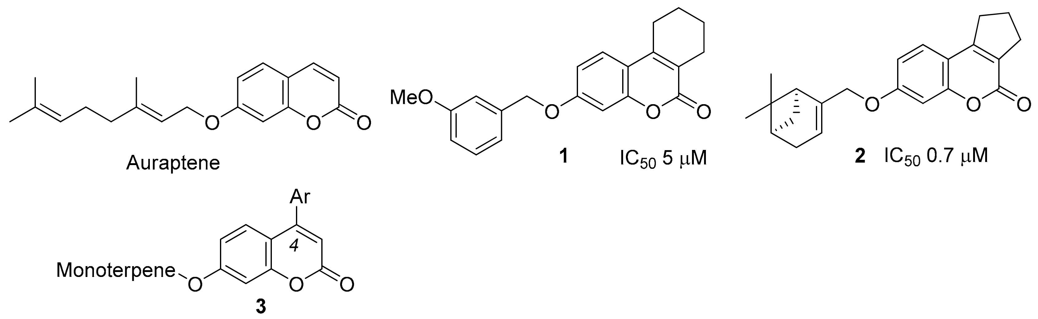
Promising New Inhibitors of Tyrosyl-DNA Phosphodiesterase I (Tdp 1) Combining 4-Arylcoumarin and Monoterpenoid Moieties as Components of Complex Antitumor Therapy

by
Tatyana M. Khomenko
1,
Alexandra L. Zakharenko
2,
Arina A. Chepanova
2,
Ekaterina S. Ilina
2,
Olga D. Zakharova
2,
Vasily I. Kaledin
3,
Valeriy P. Nikolin
3,
Nelly A. Popova
3,4,
Dina V. Korchagina
1,
Jóhannes Reynisson
5,
Raina Chand
6,
Daniel M. Ayine-Tora
6,
Jinal Patel
6,
Ivanhoe K. H. Leung
6,
Konstantin P. Volcho
1,4,*,
Nariman F. Salakhutdinov
1,4 and
Olga I. Lavrik
2,4,7
1
N.N. Vorozhtsov Novosibirsk Institute of Organic Chemistry, 9 acad. Lavrentjev ave., 630090 Novosibirsk, Russia
2
Novosibirsk Institute of Chemical Biology and Fundamental Medicine, 8, acad. Lavrentjev ave., 630090 Novosibirsk, Russia
3
Institute of Cytology and Genetics, 10, acad. Lavrentjev Ave., 630090 Novosibirsk, Russian
4
Novosibirsk State University, V. Zelman Institute for Medicine and Psychology and Department of Natural Sciences, 2, Pirogova str., 630090 Novosibirsk, Russia
5
School of Pharmacy and Bioengineering, Keele University, Hornbeam Building, Staffordshire ST5 5BG, UK
6
School of Chemical Sciences, The University of Auckland, Private Bag 92019, 1142 Auckland, New Zealand
7
Department of Physical and Chemical Biology and Biotechnology, Altai State University, 61, Lenina Ave., 656049 Barnaul, Russia
*
Author to whom correspondence should be addressed.
Int. J. Mol. Sci. 2020, 21(1), 126; https://doi.org/10.3390/ijms21010126
Submission received: 3 December 2019 / Revised: 19 December 2019 / Accepted: 20 December 2019 / Published: 23 December 2019

Abstract

:
Tyrosyl-DNA phosphodiesterase 1 (Tdp1) is an important DNA repair enzyme in humans, and a current and promising inhibition target for the development of new chemosensitizing agents due to its ability to remove DNA damage caused by topoisomerase 1 (Top1) poisons such as topotecan and irinotecan. Herein, we report our work on the synthesis and characterization of new Tdp1 inhibitors that combine the arylcoumarin (neoflavonoid) and monoterpenoid moieties. Our results showed that they are potent Tdp1 inhibitors with IC50 values in the submicromolar range. In vivo experiments with mice revealed that compound 3ba (IC50 0.62 µM) induced a significant increase in the antitumor effect of topotecan on the Krebs-2 ascites tumor model. Our results further strengthen the argument that Tdp1 is a druggable target with the potential to be developed into a clinically-potent adjunct therapy in conjunction with Top1 poisons.

1. Introduction

Natural and synthetic coumarins (2H-chromen-2-one) demonstrate diverse biological activities, and are often considered as a privileged scaffold [1,2,3,4,5,6]. In particular, a large number of coumarin derivatives with high antitumor activity have been found in recent years [7,8,9,10,11,12,13,14,15,16,17,18]. Natural derivatives of 7-hydroxycoumarin containing terpene fragments have also attracted the attention of the medicinal chemistry community [19,20,21]. The best-known compound of this structural type is auraptene (Figure 1), for which a variety of biological activities are known, including antitumor properties [22].
One of the current approaches to increase the efficacy of clinically-established antitumor therapy is the inhibition of DNA repair enzymes that counteract the effect of DNA-damaging chemotherapy agents [23,24,25]. One of these important enzymes is tyrosyl-DNA phosphodiesterase 1 (Tdp1) [26]. Tdp1 is involved in the repairing of damaged DNA, including the removal of lesions caused by topoisomerase 1 (Top1) inhibitors. Top1 inhibitors such as the camptothecin derivatives (CPTs), topotecan (tpc), and irinotecan are well-established antitumor agents [27] that are widely used [28]. Thus, Tdp1 reduces the impact of Top1 poisons, resulting in diminished DNA damage and reduced efficacy of this class of chemotherapeutic drugs.
Because Tdp1 repairs Top1/DNA cleavage complexes induced by CPTs, inhibitors of Tdp1 can enhance the sensitivity of cancer cells to CPT analogues [29]. Furthermore, increased Tdp1 expression counteracts the cytotoxicity of CPTs [30,31], and is frequently observed in cancers resistant to CPT therapy [31,32,33]. Convincing evidence exists from preclinical studies that the ratio of Tdp1/Top1 activity influences cellular sensitivity to Top1 inhibitors [34,35], and that the suppression of Tdp1 activity leads to an increase in the sensitivity of tumor cells to CPTs [32,36,37,38,39]. It is believed that targeted short-term treatment with a potent Tdp1 inhibitor will not lead to serious poisoning in normal cells. Indeed, it was shown that Tdp1-/- knockout mice were fertile and had a normal life expectancy, with no signs of premature aging [40]. Until now, no inhibitors of the Tdp1 enzyme have reached human clinical testing.
To date, many Tdp1 inhibitors have been identified. A major class of Tdp1 inhibitors comprises those based on natural products including usnic acid derivatives [41,42,43,44,45], coumarins [46], adamantanes [47,48,49], nucleoside analogs [50], dehydroabietylamine derivatives [51], chromenes [52], bile acids derivatives [53], and fungal products [54,55,56]. There are also early reports of Tdp1 inhibition based on diamidines [57], antibiotics [58,59], steroids [60], and other compounds [61]. Nevertheless, only a few Tdp1 inhibitors have been tested in cell- or cancer-models. Synergy with tpc has been demonstrated in vivo for just two Tdp1 inhibitors, both of which were derivatives of usnic acid, a natural product [43,44].
Previously, virtual screening of the InterBioScreen natural product library [62] and subsequent testing identified that 3-methoxybenzyl, a derivative of 7-hydroxycoumarin, annelated with the cyclohexane ring 1 (Figure 1) as a new structural type of Tdp1 inhibitors [46]. Further optimization of the inhibitor, including the replacement of the aromatic substituent in the phenolic group with bulky monoterpenoid substituents, allowed us to increase the potency by almost an order of magnitude, thereby reaching nanomolar activity [46]. Most importantly, the use of compound 2 in non-toxic doses significantly increased the cytotoxic activity of CPT in human cancer cells [46].
Based on molecular modeling, it was predicted that the attachment of an aromatic substituent at the fourth position of coumarin would be promising for enhanced binding. Note that 4-arylcoumarins are often considered a separate group of natural products, called neoflavones. Natural and synthetic neoflavones have low toxicities and exhibit a broad spectrum of biological activity, in particular against tumors [7]. The aim of this work was to synthesize neoflavone derivatives of structural type 3 (Figure 1) by varying both aromatic and monoterpene substituents to determine their inhibitory activity against Tdp1, and to study the synergistic effect with tpc, a clinically-important Top1 inhibitor, in in vivo experiments. As a result, it was discovered that arylcoumarins containing monoterpenoid substituents are indeed potent Tdp1 inhibitors and, most importantly, are able to enhance the antitumor activity of tpc in animal models.

2. Results and Discussion

2.1. Chemistry

The main approach to producing 4-arylcoumarins unsubstituted at the hydroxy group is acid-catalyzed Pechmann condensation between resorcinol 4 and ester of β-keto-carboxylic acids 5 [63] (Scheme 1). Using this approach, we synthesized 7-hydroxy-4-arylcoumarins 6a–d with yields of 63-81% by interaction of resorcinol 4 with esters 5a–d (Scheme 1). Ester 5a is commercially available, while compounds 5b–d were obtained by the reaction of substituted acetophenones 5b–d with diethyl carbonate in the presence of sodium hydride.
Monoterpenoid bromides 8a–d were obtained from the corresponding alcohols (geraniol, (−)-nopol, (−)- and (+)-myrtenols) by interaction with PBr3 according to the procedure [46] (Scheme 2). Geraniol and (−)-nopol were purchased from commercial sources, while (−)- and (+)-myrtenols were synthesized from (−)-myrtenal and (+)-α-pinene in accordance with the methods [46]. The choice of monoterpenoids was based on the results we obtained previously, i.e., when both the absolute configuration of the pinane cycle and the length of the bridge played important roles [46]. A further consideration was the desire to compare the activity data obtained with bulky bicyclic substituents with corresponding data for products with acyclic monoterpene fragments. Note that coumarin-containing derivatives of geraniol can be considered as analogues of the natural coumarin auraptene, having the same monoterpenoid fragment (Figure 1).
The target monoterpenoid-arylcoumarin hybrids 3 were synthesized by the reaction of 7-hydroxycoumarins 6a–d with monoterpenoid bromides 8a–d using DBU in DMF (Scheme 3). To compare and identify the importance of the monoterpenoid fragment, compounds 10a, c, d containing a benzyl substituent were also synthesized. The products were purified by recrystallization or column chromatography, and obtained with yields of 12–65%. In the case of the synthesis of nopol derivatives, a low conversion was observed, and double purification on SiO2 was required, for example, for compounds 3ab or 3cb with yields less than 20%).

2.2. Biology

A previously designed [64], real-time, hexadecameric oligonucleotide biosensor with 5(6)-carboxyfluorescein (FAM) at the 5′ end and fluorophore quencher BHQ1 (Black Hole Quencher-1) at the 3′-end was used to determine the inhibitory properties of the new compounds.
The results of the Tdp1 assay for the arylcoumarin derivatives are shown in Figure 2 and Supplementary Table S1. All arylcoumarin derivatives containing a geraniol residue (3aa–3da) showed high inhibitory activity, with IC50 values in the submicromolar range; compound 3ac with a bromine atom in the aromatic ring was slightly less active. Among the derivatives of nopol 3ab–3db, only the fluorine derivative 3bb showed a markedly lower activity; the remaining compounds were comparable in activity with the derivatives of geraniol. Since it was necessary to use column chromatography to obtain nopol-arylcoumarin hybrids, which complicates and increases the cost of the synthesis process, geraniol-containing inhibitors are more promising for further studies.
Almost all derivatives of (−)- and (+)-myrtenols (3ac–3dc and 3ad–3dd, respectively) showed similar inhibitory activity with IC50 values in the 0.4-1.0 µM range, except compound 3cc. Interestingly, compounds 10a, c, d containing a benzyl substituent instead of monoterpenoid fragments were significantly less active than most of their monoterpenoid-containing analogues, with an IC50 in the micromolar range.
An analysis of the cytotoxicity of the synthesized compounds was performed on cell lines of human breast adenocarcinoma MCF-7 and human cervical cancer HeLa. It turned out that cytotoxicity is absent or insignificant in the entire range of studied concentrations (up to 100 μM) for all the tested compounds, which makes it possible to use them as tumor sensitizers for currently-used antitumor drugs without introducing additional toxic burden (Figure 3).
Since most monoterpene-arylcoumarin hybrids showed comparable inhibitory activity against Tdp1 (~0.5 μM) and no or limited cytotoxicity, we selected a candidate for subsequent studies based on the following considerations. Derivatives of nopol were the most complex compounds to synthesize and purify. Therefore, they were excluded from further consideration. Since we previously obtained contradictory results in in vivo experiments with the myrtenol derivative 2 (unpublished data), in this work, we decided to focus on geraniol derivatives for the in vivo studies. Among the three derivatives of geraniol that showed similar inhibitory activity (Figure 2), we selected compound 3ba containing a fluorine atom in the para position of the aryl substituent, which can contribute to greater metabolic stability of the inhibitor [65]. In addition to the activity assay that we reported above, we wanted to confirm the interactions between compound 3ba and Tdp1 before progressing to in vivo studies. Using an intrinsic tryptophan fluorescence quenching assay that we previously applied to study the binding interactions of Tdp1 and its inhibitors [43,45,48,49,52], we evaluated the binding of compound 3ba to recombinant Tdp1. Clear quenching of the Tdp1 intrinsic fluorescence was observed upon the addition of 3ba (Supplementary Figure S1). Titration experiments were then performed to determine the dissociation constant (KD) of compound 3ba with Tdp1. A KD value of 63.0 ± 11 μM was obtained, indicating that it is a reasonable binder to the enzyme. This confirmed that the inhibition efficacy of compound 3ba was due to binding to the enzyme, and gave us confidence to progress with this compound towards in vivo studies.
A study of the influence of 3ba on the antitumor effect of tpc (topotecan) was performed using a murine Krebs-2 carcinoma model. An ascitic tumor model combines the advantages of in vitro and in vivo approaches in studying the cytotoxic effect of compounds, since ascitic cells grow in the context of the organism (in vivo), and the intraperitoneal administration of drug ensures its direct contact with tumor cells (in vitro). The experiments were performed using female C57BL/6 mice, which were injected intraperitoneally with 2 × 10⁵ ascitic cells on day zero. The mice were divided into six groups of ten animals each. Control group 1 did not receive treatment; group 2 received tpc in a single dose of 0.5 mg/kg of body weight intraperitoneally after 2 days; group 3 received tpc as described above, and 3ba at a dose of 80 mg/kg intraperitoneally; group 4 received tpc and 3ba 40 mg/kg; group 5 received tpc and 3ba 20 mg/kg; and finally, group 6 received 3ba 80 mg/kg only.
The combined use of 3ba at a maximum concentration of 80 mg/kg with tpc (group 3) led to a significant decrease in the weight of the ascitic tumor compared to the use of only tpc (Figure 4). The 3ba dose of 40 mg/kg also caused a decrease in ascites weight, although the difference between groups 2 and 4 was not significant. The dose of 3ba 20 mg/kg co-administered with tpc, as well as the use of 3ba (80 mg/kg) in the absence of tpc, did not affect tumor growth.
The number of tumor cells in ascites (Figure 5) in the control group (1750 million per mouse) and in group 2, which received only tpc (950 mln), was significantly different, i.e., by half, p = 0.005. The size of ascites in group 3 (tpc + 80 mg/kg 3ba) was very small; we managed to extract ascitic fluid to determine the number of cells in only one mouse; the number of tumor cells in this mouse was 250 mln. Differences in other groups are not significant.
We then examined the effect of 3ba in combination with tpc on the lifespan of mice. C57BL/6 mice were intraperitoneally inoculated with the ascites variant of Lewis carcinoma. Group 1: control without treatment; group 2: tpc 0.5 mg/kg intraperitoneally; group 3: tpc and 3ba intraperitoneally 120 mg/kg; and group 4: 3ba 120 mg/kg only. The results are given in Figure 6.
When using a combination of tpc and 3ba, a significant increase in lifespan was noted by 26% (p = 0.0065) compared with mice receiving only tpc, and by 42% compared with the control group (p = 0.0002). Monotherapy with tpc or 3ba at selected doses prolonged the life of experimental animals unreliably, i.e., by 13–19%, р > 0.05.

2.3. In Silico

2.3.1. Molecular Modeling

The 19 compounds were docked into the binding site of Tdp1 (PDB ID: 6DIE, resolution 1.78 Å) [66] with three water molecules (HOH 814, 821 and 1078). It has been shown that keeping these crystalline water molecules improves the prediction quality of the docking scaffold [45]. The modeling shows that all the ligands have a plausible binding mode and good scores with the four scoring functions used, i.e., Astex Statistical Potential (ASP) [67], improved Piecewise Linear Potential (ChemPLP) [68], ChemScore (CS) [69,70] and GoldScore (GS) [71]; the results are given in Table S2, Supplementary Information. Considering 3ba, one of the most active compounds, the coumarin moiety occupies the hydrophilic binding region, which contains amino acids such as threonine and glutamic acid, whilst the alkene side chain occupies the hydrophobic region formed by isoleucine, leucine, and phenylalanine. The carbonyl on the benzopyrone group forms hydrogen bonds with the amine side chain groups of Lys495 and Asn516. The predicted binding mode of 3ba is shown in Figure 7.

2.3.2. Chemical Space

The calculated molecular descriptors (MW (molecular weight), log P (water-octanol partition coefficient), HD (hydrogen bond donors), HA (hydrogen bond acceptors), PSA (polar surface area), and RB (rotatable bonds)) are given in Table S3. The log P values range from 4.4 and 6.3, lying between the drug-like and Known Drug Space (KDS), while the HD and PSA values are within the lead-like space (for the definition of lead-like, drug-like, and KDS regions, see [72] and Table S4). The molecular weight of the ligands is between 326.4 and 453.4 g·mol−1, falling in the drug-like chemical space. The main issue with these ligands is their relatively high lipophilicity, with Log P values reaching into the KDS.
The Known Drug Indexes (KDIs) of each ligand were calculated to gauge the balance of the molecular descriptor of the ligands (Table S5, Supplementary Information). This method is based on the statistical analysis of drugs in clinical use (KDS) and a weighted index for each of the six molecular descriptors used; both the summation (KDI2a) and multiplication (KDI2b) methods were used [73]. The KDI2a values range from 4.7 to 5.3, with a theoretical maximum of 6 and an average of 4.08 for known drugs. KDI2b ranges from 0.2 to 0.5, with a theoretical maximum of 1 and a KDS average of 0.18. This indicates that the majority of the ligands are well balanced. The most potent ligand 3ba has KDI2a of 4.90 and KDI2b of 0.27, while drugs with high bioavailability (>50%) have average KDI2a of 4.43 and KDI2b of 0.21, which shows that 3ba has a very good balance of physicochemical properties for bioavailability.

3. Materials and Methods

3.1. Chemistry Section

General Information. Reagents and solvents were purchased from commercial suppliers (Sigma-Aldrich, Acros) and used as received. GC-MS: Agilent 7890A gas chromatograph equipped with a quadrupole mass spectrometer Agilent 5975C as a detector; quartz column HP-5MS (copolymer 5%–diphenyl–95%–dimethylsiloxane) of length 30 m, internal diameter 0.25 mm and stationary phase film thickness 0.25 µm. Optical rotation: polAAr 3005 spectrometer. 1H and 13C NMR: Bruker DRX-500 apparatus at 500.13 MHz (1H) and 125.76 MHz (13C) and Bruker Avance—III 600 apparatus at 600.30 MHz (1H) and 150.95 MHz (13C), J in Hz; structure determinations by analyzing the 1H NMR spectra, including 1H–1H double resonance spectra and 1H–1H 2D homonuclear correlation, J-modulated 13C NMR spectra (JMOD), and 13C–1H 2D heteronuclear correlation with one-bond (C–H COSY, 1J(C,H) = 160 Hz, HSQC, 1J(C,H) = 145 Hz) and long-range spin-spin coupling constants (COLOC, 2,3J(C,H) = 10 Hz, HMBC, 2,3J(C,H) = 7 Hz). HR-MS: DFS Thermo Scientific spectrometer in a full scan mode (15–500 m/z, 70 eV electron impact ionization, direct sample administration).
Spectral and analytical investigations were carried out at the Multi-Access Chemical Research Center of Siberian Branch of Russian Academy of Sciences. All product yields are given for pure compounds purified by recrystallization from ethanol or isolated by column chromatography (SiO2; 60–200 μ; Macherey-Nagel). The purity of the target compounds was determined by GC-MS methods. All of the target compounds reported in this paper have purities of no less than 95%.

3.1.1. Synthesis of Compounds 5b–d

General procedure. To a stirred mixture of sodium hydride (3 mol equiv), washed with hexane (3 × 15 mL), and diethyl carbonate (4 mol equiv) in 50 mL of tetrahydrofuran (THF), the corresponding substituted acetophenone (1 mol equiv) was added dropwise over 30 min. The reaction mixture was refluxed for 4 h, and then poured into ice water, acidified with 5 mL of glacial acetic acid, and extracted with EtOAc (3 × 100 mL). The combined organic phase was washed with saturated sodium bicarbonate, brine, and water, and then dried over anhydrous Na2SO4 and evaporated in vacuo. The crude products were purified by silica gel column chromatography eluting with dichloromethane to afford 5b–d. The yields of 5b, 5c, and 5d were 80%, 89%, and 92%, respectively.

3.1.2. Synthesis of Compounds 6a–d

Syntheses were carried out from resorcinol 4 and appropriate β-keto esters (5a–d) in accordance with [46]. Conc. H2SO4 (2 mL, 37.6 mmol) was added dropwise to cooled (0–5 °C) solution of resorcinol 4 (2.3 g, 21 mmol) and appropriate β-keto esters (5a–d) (21 mmol) in dry ethanol (5 mL) with vigorously stirring. The reaction mixture was stirred at room temperature for 15 min, and then heated at 60 °C until it congealed. It was then left overnight at room temperature. Finally, it was poured into ice water (50 mL). The resulting solid was filtered off and crystallized from ethanol–water. The yields of 6a, 6b, 6c, and 6d were 81%, 79%, 73%, and 63% respectively.

3.1.3. Synthesis of Compounds 8a–d
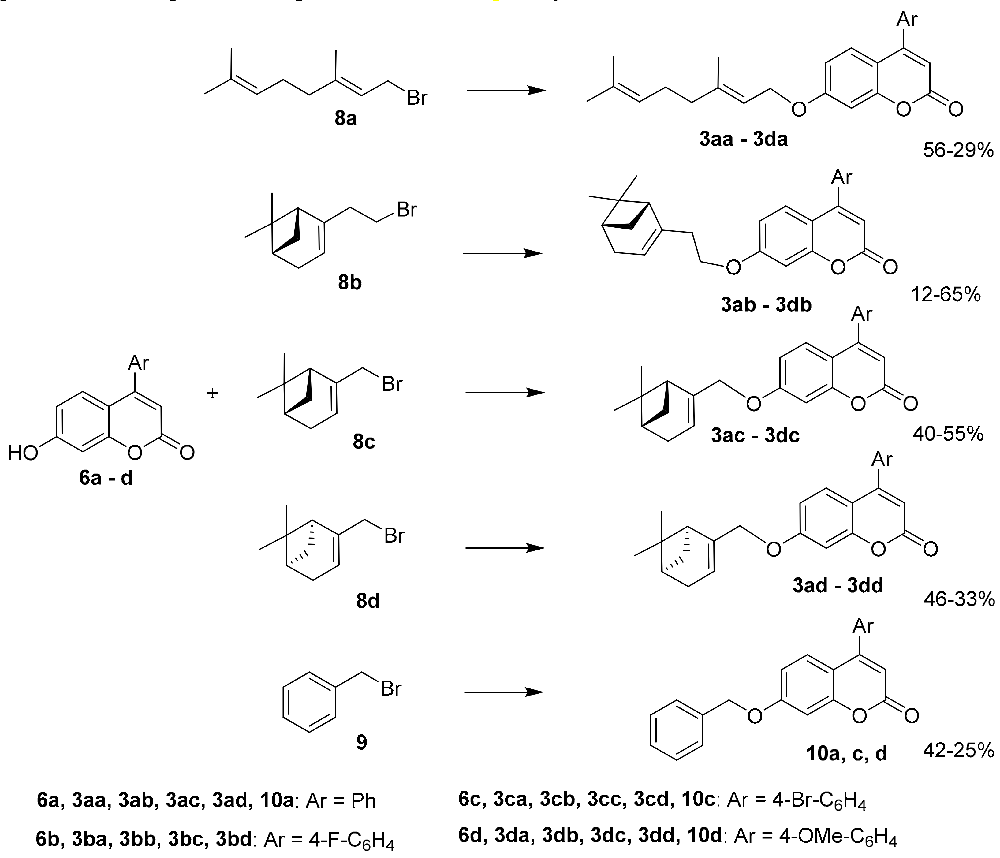
(+)-Myrtenal was synthesized according to the procedure [46] by the oxidation of (+)-α-pinene using t-BuOOH/SeO2 system with a 57% yield. (−)- The (+)-myrtenols were synthesized from the corresponding aldehydes via reduction to alcohols with NaBH4, as described above. NaBH4 (10.3 mmol) was added to a cooled (0–5 °C) solution of 10.3 mmol of the appropriate aldehyde in methanol (20 mL), and the reaction mixture was stirred for 3 h at room temperature. Then, 5% aqueous HCl was added to reach a pH of 4–5. The solvent was distilled off and the product was extracted using ether and dried with Na2SO4. The solvent was evaporated; the resulting alcohols (58% and 54% yields) were used in the synthesis without purification.
Bromides 8a–d were synthesized from geraniol, (−)-nopol, and (−)- and (+)-myrtenols via the aforementioned reaction with PBr3. PBr3 (8.9 mmol) was added to a cooled (0–5 °C) solution of the corresponding monoterpenoid alcohols (26.7 mmol) in dry ether (30 mL), and the reaction mixture was stirred for 2 h at room temperature. Saturated aqueous NaHCO3 was added, and the product was extracted with ether. The extracts were washed with brine, dried with Na2SO4, and evaporated. Compounds 8a, 8c, and 8d (with yields of 91%, 55%, and 60%, respectively) were sufficiently pure and used for the next step without purification. The compound 8b was purified by column chromatography on SiO2, eluent–hexane (yield 24%).

3.1.4. Synthesis of Compounds 3aa3da, 3ab3db, 3ac3dc, 3ad3dd, and 10a, c, d

Compounds 3aa–3da, 3ab–3db, 3ac–3dc, 3ad–3dd, and 10a, c, d were synthesized from coumarins 6a–d and the corresponding bromides 8a–d, 9 using DBU and DMF.
DBU (1.0 mmol) and corresponding bromide 8a–d, 9 (0.75 mmol) were added to compound 6a–d (0.5 mmol) in dry DMF (5 mL) at room temperature under stirring. The reaction mixture was stirred at room temperature for 15 min and then heated at 60 °C for 5 h. H2O (15 mL) was added and the product was extracted with ethyl acetate. The extracts were washed with brine, dried with Na2SO4, and evaporated. The products 3aa–3da, 3ab–3db, 3ac–3dc, 3ad–3dd, and 10a, c, d were isolated in the individual form a) by recrystallization from ethanol; or b) by column chromatography on silica gel using eluent–hexane, a solution containing from 25 to 100% ethyl acetate in hexane, and ethanol.
(E)-7-(3,7-Dimethylocta-2,6-dienyloxy)-4-phenyl-2H-chromen-2-one3aa. Yield 56%, method a. M.p. 54 °C. HRMS: 374.1879 [M]+; calcd. 374.1877 (C25H26O3)+. 1H NMR (CDCl3,δ ppm, J, Hz): 1.58 (br.s, 3H, CH3-24), 1.64 (m, 3H, all J < 1.5, CH3-23), 1.74 (m, 3H, all J < 1.5, CH3-25), 2.03-2.15 (m, 4H, 2H-19, 2H-20), 4.60 (d, 2H, J16,17 = 6.6, 2H-16), 5.06 (tm, 1H, J21,20 = 6.8, other J < 1.5, H-21), 5.45 (tm, 1H, J17,16 = 6.6, other J < 1.5, H-17), 6.18 (s, 1H, H-3), 6.77 (dd, 1H, J7,6 = 8.9, J7,9 = 2.5, H-7), 6.87 (d, 1H, J9,7 = 2.5, H-9), 7.34 (d, 1H, J6,7 = 8.9, H-6), 7.39-7.43 (m, 2H, H-11, H-15), 7.46-7.51 (m, 3H, H-12, H-13, H-14). 13C NMR (δ ppm, CDCl3): 155.85 (s, C-1), 161.12 (s, C-2), 111.61 (d, C-3), 155.70 (s, C-4), 112.28 (s, C-5), 127.75 (d, C-6), 112.81 (d, C-7), 161.99 (s, C-8), 101.75 (d, C-9), 135.52 (s, C-10), 128.23 (d, C-11, C-15), 128.66 (d, C-12, C-14), 129.40 (d, C-13), 65.38 (t, C-16), 118.34 (d, C-17), 142.19 (s, C-18), 39.37 (t, C-19), 26.11 (t, C-20), 123.49 (d, C-21), 131.78 (s, C-22), 25.50 (k, C-23), 17.56 (k, C-24), 16.63 (k, C-25).
(E)-7-(3,7-Dimethylocta-2,6-dienyloxy)-4-(4-fluorophenyl)-2H-chromen-2-one3ba. Yield 35%, method a. M.p. 72 °C. HRMS: 392.1778 [M]+; calcd. 392.1782 (C25H25FO3)+. 1H NMR (CDCl3,δ ppm, J, Hz): 1.58 (s, 3H, CH3-24), 1.64 (s, 3H, CH3-23), 1.74 (s, 3H, CH3-25), 2.02-2.16 (m, 4H, 2H-19, 2H-20), 4.60 (d, 2H, J16,17 = 6.5, 2H-16), 5.06 (tm, 1H, J21,20 = 6.8, other J < 2, H-21), 5.45 (tm, 1H, J17,16 = 6.5, other J < 2, H-17), 6.16 (s, 1H, H-3), 6.78 (dd, 1H, J7,6 = 8.9, J7,9 = 2.5, H-7), 6.87 (d, 1H, J9,7 = 2.5, H-9), 7.19 (dd, 2H, J12,11 = J14,15 = 8.7, J12(14),F = 8.7, H-12, H-14), 7.30 (d, 1H, J6,7 = 8.9, H-6), 7.41 (dd, 2H, J11,12 =J15,14 = 8.7, J11(15),F = 5.3, H-11, H-15). 13C NMR (δ ppm, CDCl3, JC,F, Hz): 155.86 (s, C-1), 160.98 (s, C-2), 111.77 (d, C-3), 154.66 (s, C-4), 112.17 (s, C-5), 127.51 (d, C-6), 112.95 (d, C-7), 162.12 (d, C-8), 101.84 (d, C-9), 131.52 (s, 4J = 3.4, C-10), 130.18 (d, 3J = 8.3, C-11, C-15), 115.89 (d, 2J = 21.4, C-12, C-14), 163.35 (s, 1J = 250.0, C-13), 65.43 (t, C-16), 118.29 (d, C-17), 142.30 (s, C-18), 39.39 (t, C-19), 26.13 (t, C-20), 123.49 (d, C-21), 131.82 (s, C-22), 25.51 (k, C-23), 17.57 (k, C-24), 16.64 (k, C-25).
(E)-4-(4-Bromophenyl)-7-(3,7-dimethylocta-2,6-dienyloxy)-2H-chromen-2-one3ca. Yield 40%, method a. M.p. 80 °C. HRMS: 452.0979 [M]+; calcd. 452.0982 (C25H25BrO3)+. 1H NMR (CDCl3,δ ppm, J, Hz): 1.58 (br.s, 3H, CH3-24), 1.64 (m, 3H, all J < 1.5, CH3-23), 1.74 (br.s, 3H, CH3-25), 2.03-2.14 (m, 4H, 2H-19, 2H-20), 4.59 (d, 2H, J16,17 = 6.6, 2H-16), 5.06 (tm, 1H, J21,20 = 6.7, other J ≤ 1.5, H-21), 5.44 (tm, 1H, J17,16 = 6.6, other J < 1.5, H-17), 6.16 (s, 1H, H-3), 6.78 (dd, 1H, J7,6 = 8.9, J7,9 = 2.5, H-7), 6.87 (d, 1H, J9,7 = 2.5, H-9), 7.28 (d, 1H, J6,7 = 8.9, H-6), 7.29 (br.d, 2H, J11,12 =J15,14 = 8.5, H-11, H-15), 7.63 (br.d, 2H, J12,11 = J14,15 = 8.5, H-12, H-14). 13C NMR (δ ppm, CDCl3): 155.82 (s, C-1), 160.92 (s, C-2), 111.65 (d, C-3), 154.52 (s, C-4), 111.83 (s, C-5), 127.43 (d, C-6), 112.99 (d, C-7), 162.13 (c, C-8), 101.78 (d, C-9), 134.31 (s, C-10), 129.84 (d, C-11, C-15), 131.98 (d, C-12, C-14), 123.87 (s, C-13), 65.39 (t, C-16), 118.16 (d, C-17), 142.38 (s, C-18), 39.37 (t, C-19), 26.08 (t, C-20), 123.45 (d, C-21), 131.84 (s, C-22), 25.54 (k, C-23), 17.59 (k, C-24), 16.65 (k, C-25).
(E)-7-(3,7-Dimethylocta-2,6-dienyloxy)-4-(4-methoxyphenyl)-2H-chromen-2-one3da. Yield 29%, method b. HRMS: 404.1980 [M]+; calcd. 404.1982 (C26H28O4)+. 1H NMR (CDCl3,δ ppm, J, Hz): 1.58 (s, 3H, CH3-24), 1.64 (m, 3H, all J < 2.0, CH3-23), 1.74 (m, 3H, all J < 1.5, CH3-25), 2.03-2.15 (m, 4H, 2H-19, 2H-20), 3.86 (s, 3H, CH3-26), 4.59 (d, 2H, J16,17 = 6.6, 2H-16), 5.06 (tm, 1H, J21,20 = 6.8, other J < 1.5, H-21), 5.45 (tm, 1H, J17,16 = 6.6, other J < 1.5, H-17), 6.16 (s, 1H, H-3), 6.78 (dd, 1H, J7,6 = 8.9, J7,9 = 2.5, H-7), 6.86 (d, 1H, J9,7 = 2.5, H-9), 7.00 (br.d, 2H, J12,11 = J14,15 = 8.7, H-12, H-14), 7.37 (br.d, 2H, J11,12 =J15,14 = 8.7, H-11, H-15), 7.41 (d, 1H, J6,7 = 8.9, H-6). 13C NMR (δ ppm, CDCl3): 155.85 (s, C-1), 161.34 (s, C-2), 111.08 (d, C-3), 155.40 (s, C-4), 112.39 (s, C-5), 127.78 (d, C-6), 112.74 (d, C-7), 161.87 (s, C-8), 101.70 (d, C-9), 127.74 (s, C-10), 129.73 (d, C-11, C-15), 114.12 (d, C-12, C-14), 160.60 (s, C-13), 65.34 (t, C-16), 118.31 (d, C-17), 142.21 (s, C-18), 39.38 (t, C-19), 26.10 (t, C-20), 123.48 (d, C-21), 131.80 (s, C-22), 25.52 (k, C-23), 17.57 (k, C-24), 16.63 (k, C-25), 55.28 (k, C-26).
7-(2-((1R,5S)-6,6-Dimethylbicyclo[3 1.1]hept-2-en-2-yl)ethoxy)-4-phenyl-2H-chromen-2-one3ab. Yield 12%, method b. HRMS: 386.1872 [M]+; calcd. 386.1877 (C26H26O3)+. [ α ] 589 22 = −20.0 (c = 0.53, CHCl3). 1H NMR (CDCl3,δ ppm, J, Hz): 0.81 (c, 3H, CH3-26), 1.16 (d, 1H, J24a,24s = 8.6, H-24a), 1.26 (s, 3H, CH3-25), 2.05-2.11 (m, 2H, H-21, H-23), 2.20 (dm, 1H, J20,20′ = 17.6, other J < 3.5, H-20), 2.26 (dm, 1H, J20′,20 = 17.6, other J < 3.5, H-20′), 2.37 (ddd, 1H, J24s,24a = 8.6, J24s,21 = J24s,23 = 5.6, H-24s), 2.40-2.52 (m, 2H, 2H-17), 3.98-4.06 (m, 2H, 2H-16), 5.32-5.36 (m, 1H, H-19), 6.18 (s, 1H, H-3), 6.74 (dd, 1H, J7,6 = 8.9, J7,9 = 2.5, H-7), 6.85 (d, 1H, J9,7 = 2.5, H-9), 7.34 (d, 1H, J6,7 = 8.9, H-6), 7.39-7.44 (m, 2H, H-11, H-15), 7.46-7.51 (m, 3H, H-12, H-13, H-14). 13C NMR (δ ppm, CDCl3): 155.91 (s, C-1), 161.11 (s, C-2), 111.63 (d, C-3), 155.71 (s, C-4), 112.30 (s, C-5), 127.80 (d, C-6), 112.63 (d, C-7), 162.07 (s, C-8), 101.50 (d, C-9), 135.54 (s, C-10), 128.25 (d, C-11, C-15), 128.68 (d, C-12, C-14), 129.42 (d, C-13), 66.90 (t, C-16), 36.07 (t, C-17), 143.89 (s, C-18), 118.99 (d, C-19), 31.27 (t, C-20), 40.64 (d, C-21), 37.97 (c, C-22), 45.79 (d, C-23), 31.54 (t, C-24), 26.18 (k, C-25), 21.08 (k, C-26).
7-(2-((1R,5S)-6,6-Dimethylbicyclo[3.1.1]hept-2-en-2-yl)ethoxy)-4-(4-fluorophenyl)-2H-chromen-2-one3bb. Yield 37%, method b. HRMS: 404.1774 [M]+; calcd. 404.1782 (C26H25FO3)+. [ α ] 589 22 = −17.4 (c = 0.78, CHCl3). 1H NMR (CDCl3,δ ppm, J, Hz): 0.81 (s, 3H, CH3-26), 1.16 (d, 1H, J24a,24s = 8.6, H-24a), 1.26 (s, 3H, CH3-25), 2.06-2.10 (m, 2H, H-21, H-23), 2.20 (dm, 1H, J20,20′ = 17.7, H-20), 2.27 (dm, 1H, J20′,20 = 17.7, H-20′), 2.36 (ddd, 1H, J24s,24a = 8.6, J24s,21 = J24s,23 = 5.6, H-24s), 2.40-2.51 (m, 2H, 2H-17), 4.02 (t, 2H, J16,17 = 7.0, 2H-16), 5.32-5.36 (m, 1H, H-19), 6.16 (s, 1H, H-3), 6.75 (dd, 1H, J7,6 = 8.9, J7,9 = 2.5, H-7), 6.84 (d, 1H, J9,7 = 2.5, H-9), 7.16-7.21 (m, 2H, J12,11 = J14,15 = 8.7, J12(14),F =8.7, H-12, H-14), 7.30 (d, 1H, J6,7 = 8.9, H-6), 7.38-7.43 (m, 2H, J11,12 = J15,14 = 8.7, J11(15),F = 5.3, H-11, H-15). 13C NMR (δ ppm, CDCl3, JC,F, Hz): 155.90 (s, C-1), 160.94 (s, C-2), 111.76 (d, C-3), 154.64 (s, C-4), 112.16 (s, C-5), 127.54 (d, C-6), 112.74 (d, C-7), 162.18 (s, C-8), 101.57 (d, C-9), 131.51 (s, 4J = 3.5, C-10), 130.18 (d, 3J = 8.3, C-11, C-15), 115.88 (d, 2J = 21.8, C-12, C-14), 66.93 (t, C-16), 36.05 (t, C-17), 143.86 (s, C-18), 119.02 (d, C-19), 31.27 (t, C-20), 40.64 (d, C-21), 37.97 (s, C-22), 45.79 (d, C-23), 31.54 (t, C-24), 26.17 (k, C-25), 21.08 (k, C-26).
4-(4-Bromophenyl)-7-(2-((1R,5S)-6,6-dimethylbicyclo[3.1.1]hept-2-en-2-yl)ethoxy)-2H-chromen-2-one3cb. Yield 15%, method b. [ α ] 589 26.6 = −17.9 (c = 0.58, CHCl3). HRMS: 464.0988 [M]+; calcd. 450.0982 (C26H25BrO3)+. 1H NMR (CDCl3,δ ppm, J, Hz): 0.80 (s, 3H, CH3-26), 1.15 (d, 1H, J24a,24s = 8.6, H-24a), 1.25 (s, 3H, CH3-25), 2.06-2.09 (m, 2H, H-21, H-23), 2.19 (dm, 1H, J20,20′ = 17.6, H-20), 2.26 (dm, 1H, J20′,20 = 17.6, H-20′), 2.36 (ddd, 1H, J24s,24a = 8.6, J24s,21 = J24s,23 = 5.6, H-24s), 2.40-2.51 (m, 2H, 2H-17), 3.98-4.04 (m, 2H, 2H-16), 5.33-5.36 (m, 1H, H-19), 6.16 (s, 1H, H-3), 6.75 (dd, 1H, J7,6 = 8.9, J7,9 = 2.5, H-7), 6.84 (d, 1H, J9,7 = 2.5, H-9), 7.28 (d, 1H, J6,7 = 8.9, H-6), 7.28-7.31 (m, 2H, J11,12 = J15,14 = 8.4, H-11, H-15), 7.61-7.64 (m, 2H, J12,11 = J14,15 = 8.4, H-12, H-14). 13C NMR (δ ppm, CDCl3): 155.86 (s, C-1), 160.87 (s, C-2), 111.65 (d, C-3), 154.49 (s, C-4), 111.81 (s, C-5), 127.44 (d, C-6), 112.79 (d, C-7), 162.19 (s, C-8), 101.50 (d, C-9), 134.30 (s, C-10), 129.84 (d, C-11, C-15), 131.97 (d, C-12, C-14), 123.86 (s, C-13), 66.87 (t, C-16), 36.01 (t, C-17), 143.78 (s, C-18), 119.03 (d, C-19), 31.23 (t, C-20), 40.54 (d, C-21), 37.95 (s, C-22), 45.65 (d, C-23), 31.51 (t, C-24), 26.14 (k, C-25), 21.08 (k, C-26).
7-(2-((1R,5S)-6,6-Dimethylbicyclo[3.1.1]hept-2-en-2-yl)ethoxy)-4-(4-methoxyphenyl)-2H-chromen-2-one3db. Yield 65%, method b. HRMS: 415.1906 [M-H]+; calcd. 415.1904 (C27H27O4)+. [ α ] 589 22 = −15.7 (c = 0.75, CHCl3). 1H NMR (CDCl3,δ ppm, J, Hz): 0.81 (s, 3H, CH3-27), 1.16 (d, 1H, J25a,25s = 8.6, H-25a), 1.26 (s, 3H, CH3-26), 2.05-2.11 (m, 2H, H-21, H-24), 2.19 (dm, 1H, 2J = 17.6, H-21), 2.26 (dm, 1H, 2J = 17.6, H-21′), 2.36 (ddd, 1H, J25s,25a = 8.6, J25s,22 = J25s,24 = 5.6, H-25s), 2.40-2.51 (m, 2H, 2H-18), 3.86 (s, 3H, CH3-16), 4.02 (t, 2H, J17,18 = 7.0, H-17), 5.32-5.36 (m, 1H, H-20), 6.15 (s, 1H, H-3), 6.75 (dd, 1H, J7,6 = 8.9, J7,9 = 2.5, H-7), 6.83 (d, 1H, J9,7 = 2.5, H-9), 7.01 (br.d, 2H, J12,11 = J14,15 = 8.7, H-12, H-14), 7.37 (br.d, 2H, J11,12 = J15,14 = 8.7, H-11, H-15), 7.40 (d, 1H, J6,7 = 8.9, H-6). 13C NMR (δ ppm, CDCl3): 155.93 (s, C-1), 161.28 (s, C-2), 111.12 (d, C-3), 155.38 (s, C-4), 112.43 (s, C-5), 127.81 (d, C-6), 112.54 (d, C-7), 161.97 (s, C-8), 101.49 (d, C-9), 127.79 (s, C-10), 129.74 (d, C-11, C-15), 114.16 (d, C-12, C-14), 160.64 (s, C-13), 55.30 (k, C-16), 66.88 (t, C-17), 36.07 (t, C-18), 143.90 (s, C-19), 118.97 (d, C-20), 31.27 (t, C-21), 40.64 (d, C-22), 37.97 (s, C-23), 45.79 (d, C-24), 31.54 (t, C-25), 26.17 (k, C-26), 21.08 (k, C-27).
7-(((1R,5S)-6,6-Dimethylbicyclo[3.1.1]hept-2-en-2-yl)methoxy)-4-phenyl-2H-chromen-2-one3ac. Yield 40%, method a. M.p. 106 °C. HRMS: 372.1717 [M]+; calcd. 372.1720 (C25H24O3)+. [ α ] 589 27.3 = −25.33 (c = 1.02, EtOH). 1H NMR (CDCl3,δ ppm, J, Hz): 0.82 (s, 3H, CH3-25), 1.18 (d, 1H, 2J = 8.7, H-23a), 1.28 (s, 3H, CH3-24), 2.08-2.14 (m, 1H, H-20), 2.20 (ddd, 1H, J22,20 = J22,23s = 5.6, J22,18 = 1.4, H-22), 2.26 (dm, 1H, 2J = 18.1, H-19), 2.33 (dm, 1H, 2J = 18.1, H-19′), 2.40 (ddd, 1H, 2J = 8.7, J23s,20= J23s,22 = 5.6, H-23s), 4.44 (dm, 1H, 2J = 12.4, other J ≤ 2.0, H-16), 4.47 (dm, 1H, 2J = 12.4, other J ≤ 2.0, H-16′), 5.61-5.64 (m, 1H, H-18), 6.19 (s, 1H, H-3), 6.77 (dd, 1H, J7,6 = 8.9, J7,9 = 2.5, H-7), 6.89 (d, 1H, J9,7 = 2.5, H-9), 7.33 (d, 1H, J6,7 = 8.9, H-6), 7.40-7.43 (m, 2H, H-11, H-15), 7.47-7.51 (m, 3H, H-12, H-13, H-14). 13C NMR (δ ppm, CDCl3): 155.81 (s, C-1), 161.24 (s, C-2), 111.65 (d, C-3), 155.76 (s, C-4), 112.32 (s, C-5), 127.68 (d, C-6), 112.94 (d, C-7), 162.14 (s, C-8), 102.00 (d, C-9), 135.55 (s, C-10), 128.27 (d, C-11, C-15), 128.68 (d, C-12, C-14), 129.42 (d, C-13), 71.12 (t, C-16), 142.92 (s, C-17), 121.34 (d, C-18), 31.18 (t, C-19), 40.71 (d, C-20), 38.00 (s, C-21), 43.11 (d, C-22), 31.40 (t, C-23), 26.02 (k, C-24), 20.97 (k, C-25).
7-(((1R,5S)-6,6-Dimethylbicyclo[3.1.1]hept-2-en-2-yl)methoxy)-4-(4-fluorophenyl)-2H-chromen-2-one3bc. Yield 53%, method b. [ α ] 589 22.0 = −15.6 (c = 0.68, CHCl3). HRMS: 390.1624 [M]+; calcd. 390.1626 (C25H23FO3)+. 1H NMR (CDCl3,δ ppm, J, Hz): 0.81 (s, 3H, CH3-25), 1.17 (d, 1H, J23a,23s = 8.7, H-23a), 1.28 (s, 3H, CH3-24), 2.11 (ddtd, 1H, J20,22 = J20,23s = 5.6, J20,19 = 2.9, J20,18 = 1.3, H-20), 2.20 (ddd, 1H, J22,20 = J22,23s = 5.6, J22,18 = 1.4, H-22), 2.26 (dm, 1H, J19,19, = 18.0, other J < 3.5, H-19), 2.33 (dm, 1H, J19′,19 = 18.0, other J < 3.5, H-19′), 2.40 (ddd, 1H, J23s,23a = 8.7, J23s,20= J23s,22 = 5.6, H-23s), 4.41-4.49 (m, 2H, 2H-16), 5.61-5.64 (m, 1H, H-18), 6.16 (s, 1H, H-3), 6.78 (dd, 1H, J7,6 = 8.9, J7,9 = 2.5, H-7), 6.88 (d, 1H, J9,7 = 2.5, H-9), 7.16-7.21 (m, 2H, J12,11 = J14,15 = 8.8, J12(14),F = 8.6, H-12, H-14), 7.29 (d, 1H, J6,7 = 8.9, H-6), 7.38-7.43 (m, 2H, J11,12 = J15,14 = 8.8, J11(15),F = 5.3, H-11, H-15). 13C NMR (δ ppm, CDCl3, JC,F, Hz): 155.80 (s, C-1), 161.02 (s, C-2), 111.78 (d, C-3), 154.66 (s, C-4), 112.18 (s, C-5), 127.42 (d, C-6), 113.03 (d, C-7), 162.23 (s, C-8), 102.07 (d, C-9), 131.52 (s, 4J = 3.5, C-10), 130.19 (d, 3J = 8.3, C-11, C-15), 115.88 (d, 2J = 21.8, C-12, C-14), 71.14 (t, C-16), 142.86 (s, C-17), 121.39 (d, C-18), 31.18 (t, C-19), 40.69 (d, C-20), 38.00 (s, C-21), 43.09 (d, C-22), 31.39 (t, C-23), 26.00 (k, C-24), 20.97 (k, C-25).
4-(4-Bromophenyl)-7-(((1R,5S)-6,6-dimethylbicyclo[3.1.1]hept-2-en-2-yl)methoxy)-2H-chromen-2-one3cc. Yield 36%, method a. M.p. 130 °C. HRMS: 450.0826 [M]+; calcd. 450.0825 (C25H23O3Br)+. [ α ] 589 26.4 = −13.9 (c = 0.52, CHCl3). 1H NMR (CDCl3,δ ppm, J, Hz): 0.80 (s, 3H, CH3-25), 1.16 (d, 1H, J23a,23s = 8.7, H-23a), 1.28 (s, 3H, CH3-24), 2.08-2.12 (m,1H, H-20), 2.19 (ddd, 1H, J22,20 = J22,23s = 5.6, J22,18 = 1.3, H-22), 2.25 (dm, 1H, J19,19, = 18.0, other J < 3.5, H-19), 2.33 (dm, 1H, J19,,19 = 18.0, other J < 3.5, H-19′), 2.40 (ddd, 1H, J23s,23a = 8.7, J23s,20= J23s,22 = 5.6, H-23s), 4.43-4.48 (m, 2H, 2H-16), 5.60-5.63 (m, 1H, H-18), 6.16 (s, 1H, H-3), 6.78 (dd, 1H, J7,6 = 8.9, J7,9 = 2.5, H-7), 6.88 (d, 1H, J9,7 = 2.5, H-9), 7.27 (d, 1H, J6,7 = 8.9, H-6), 7.30 (d, 2H, J11,12 = J15,14 = 8.5, H-11, H-15), 7.63 (d, 2H, J12,11 = J14,15 = 8.5, H-12, H-14). 13C NMR (δ ppm, CDCl3): 155.76 (s, C-1), 160.97 (s, C-2), 111.67 (d, C-3), 154.53 (s, C-4), 111.85 (s, C-5), 127.33 (d, C-6), 113.10 (d, C-7), 162.25 (s, C-8), 102.02 (d, C-9), 134.32 (s, C-10), 129.85 (d, C-11, C-15), 131.98 (d, C-12, C-14), 123.87 (s, C-13), 71.12 (t, C-16), 142.77 (s, C-17), 121.43 (d, C-18), 31.15 (t, C-19), 40.60 (d, C-20), 37.99 (s, C-21), 43.00 (d, C-22), 31.37 (t, C-23), 25.98 (k, C-24), 20.96 (k, C-25).
7-(((1R,5S)-6,6-Dimethylbicyclo[3.1.1]hept-2-en-2-yl)methoxy)-4-(4-methoxyphenyl)-2H-chromen-2-one3dc. Yield 55%, method b. M.p. 124 °C. [ α ] 589 30.6 = −17.3 (c = 0.82, CHCl3). HRMS: 402.1820 [M]+; calcd. 402.1827 (C26H26O4)+. 1H NMR (CDCl3,δ ppm, J, Hz): 0.81 (s, 3H, CH3-26), 1.17 (d, 1H, J24a,24s = 8.7, H-24a), 1.28 (s, 3H, CH3-25), 2.10 (ddtd, 1H, J21,23 = J21,24s = 5.6, J21,20 = 2.8, J21,19 = 1.3, H-21), 2.20 (ddd, 1H, J23,21 = J23,24s = 5.6, J23,19 = 1.4, H-23), 2.25 (dm, 1H, J20,20, = 18.0, other J < 3.0, H-20), 2.33 (dm, 1H, J20′,20 = 18.0, other J < 3.5, H-20′), 2.40 (ddd, 1H, J24s,24a = 8.7, J24s,21= J24s,23 = 5.6, H-24s), 3.85 (s, 3H, CH3-16), 4.41-4.49 (m, 2H, 2H-17), 5.60-5.63 (m, 1H, H-19), 6.14 (s, 1H, H-3), 6.77 (dd, 1H, J7,6 = 8.9, J7,9 = 2.5, H-7), 6.86 (d, 1H, J9,7 = 2.5, H-9), 7.00 (br.d, 2H, J12,11 = J14,15 = 8.8, H-12, H-14), 7.36 (br.d, 2H, J11,12 = J15,14 = 8.8, H-11, H-15), 7.39 (d, 1H, J6,7 = 8.9, H-6). 13C NMR (δ ppm, CDCl3): 155.80 (s, C-1), 161.30 (s, C-2), 111.11 (d, C-3), 155.36 (s, C-4), 112.41 (s, C-5), 127.67 (d, C-6), 112.79 (d, C-7), 161.99 (s, C-8), 101.96 (d, C-9), 127.77 (s, C-10), 129.72 (d, C-11, C-15), 114.13 (d, C-12, C-14), 160.62 (s, C-13), 55.27 (k, C-16), 71.06 (t, C-17), 142.91 (s, C-18), 121.26 (d, C-19), 31.15 (t, C-20), 40.67 (d, C-21), 37.97 (s, C-22), 43.07 (d, C-23), 31.37 (t, C-24), 25.99 (k, C-25), 20.94 (k, C-26).
7-(((1S,5R)-6,6-Dimethylbicyclo[3.1.1]hept-2-en-2-yl)methoxy)-4-phenyl-2H-chromen-2-one3ad. Yield 46%, method b. [ α ] 589 26.6 = +23.33 (c=1.02, EtOH). HRMS: 372.1718 [M]+; calcd. 372.1720 (C25H24O3)+. The 1H and 13C NMR spectra of 3ad correspond to the spectra of the enantiomer 3ac.
7-(((1S,5R)-6,6-Dimethylbicyclo[3.1.1]hept-2-en-2-yl)methoxy)-4-(4-fluorophenyl)-2H-chromen-2-one3bd. Yield 35%, method a. M.p. 129 °C. HRMS: 390.1628 [M]+; calcd. 390.1626 (C25H23FO3)+. [ α ] 589 22.0 = +22.3 (CHCl3, c = 0.53). The 1H and 13C NMR spectra of 3bd correspond to the spectra of the enantiomer 3bc.
4-(4-Bromophenyl)-7-(((1S,5R)-6,6-dimethylbicyclo[3.1.1]hept-2-en-2-yl)methoxy)-2H-chromen-2-one3cd. Yield 39%, method a. M.p. 138 °C. HRMS: 450.0825 [M]+; calcd. 450.0820 (C25H23BrO3)+. [ α ] 589 26.7 = +22.4 (c = 0.58, CHCl3). The 1H and 13C NMR spectra of 3cd correspond to the spectra of the enantiomer 3cc.
7-(((1S,5R)-6,6-Dimethylbicyclo[3.1.1]hept-2-en-2-yl)methoxy)-4-(4-methoxyphenyl)-2H-chromen-2-one3dd. Yield 33%, method a. M.p. 112 °C. HRMS: 402.1823 [M]+; calcd. 402.1827 (C26H26O4)+. [ α ] 589 22.0 = +20.8 (c = 0.72, CHCl3). The 1H and 13C NMR spectra of 3dd correspond to the spectra of the enantiomer 3dc.
7-(Benzyloxy)-4-phenyl-2H-chromen-2-one10a. Yield 42%, method b. M.p. 92 °C. HRMS: 328.1093 [M]+; calcd. 328.1094 (C22H16O3)+. 1H NMR (CDCl3,δ ppm, J, Hz): 5.13 (s, 2H, 2H-16), 6.19 (s, 1H, H-3), 6.85 (dd, 1H, J7,6 = 8.9, J7,9 = 2.5, H-7), 6.94 (d, 1H, J9,7 = 2.5, H-9), 7.31-7.35 (m, 1H, H-20), 7.35–7.44 (m, 7H, H-6, H-11, H-15, H-18, H-19, H-21, H-22), 7.46-7.52 (m, 3H, H-12, H-13, H-14). 13C NMR (δ ppm, CDCl3): 155.80 (s, C-1), 161.00 (s, C-2), 111.86 (d, C-3), 155.62 (s, C-4), 112.62 (s, C-5), 127.88 (d, C-6), 112.80 (d, C-7), 161.71 (s, C-8), 102.09 (d, C-9), 135.44 (s, C-10), 128.23 (d, C-11, C-15), 128.68 (d, C-12, C-14), 129.44 (d, C-13), 70.39 (t, C-16), 135.67 (s, C-17), 127.35 (d, C-18, C-22), 128.62 (d, C-19, C-21), 128.23 (d, C-20).
7-(Benzyloxy)-4-(4-bromophenyl)-2H-chromen-2-one10c. Yield 25%, method a. M.p. 128 °C. HRMS: 406.0204 [M]+; calcd. 406.0199 (C22H15BrO3)+. 1H NMR (CDCl3,δ ppm, J, Hz): 5.13 (s, 2H, 2H-16), 6.17 (s, 1H, H-3), 6.86 (dd, 1H, J7,6 = 8.9, J7,9 = 2.5, H-7), 6.94 (d, 1H, J9,7 = 2.5, H-9), 7.29 (dm, 2H, J11,12 = J15,14 = 8.5, H-11, H-15), 7.31 (d, 1H, J6,7 = 8.9, H-6), 7.32-7.35 (m, 1H, H-20), 7.37-7.43 (m, 4H, H-18, H-19, H-21, H-22), 7.63 (dm, 2H, J12,11 = J14,15 = 8.5, H-12, H-14). 13C NMR (δ ppm, CDCl3): 155.79 (s, C-1), 160.81 (s, C-2), 111.92 (d, C-3), 154.46 (s, C-4), 112.18 (s, C-5), 127.56 (d, C-6), 113.02 (d, C-7), 161.86 (s, C-8), 102.14 (d, C-9), 134.24 (s, C-10), 129.83 (d, C-11, C-15), 132.01 (d, C-12, C-14), 123.92 (s, C-13), 70.42 (t, C-16), 135.54 (s, C-17), 127.38 (d, C-18, C-22), 128.66 (d, C-19, C-21), 128.30 (d, C-20).
7-(Benzyloxy)-4-(4-methoxyphenyl)-2H-chromen-2-one10d. Yield 34%, method b. M.p. 143 °C. HRMS: 358.1198 [M]+; calcd. 358.1200 (C23H18O4)+. 1H NMR (CDCl3,δ ppm, J, Hz): 3.86 (s, 3H, CH3-16), 5.12 (s, 2H, 2H-17), 6.16 (s, 1H, H-3), 6.85 (dd, 1H, J7,6 = 8.9, J7,9 = 2.5, H-7), 6.93 (d, 1H, J9,7 = 2.5, H-9), 7.01 (dm, 2H, J12,11 = J14,15 = 8.7, H-12, H-14), 7.30-7.35 (m, 1H, H-21), 7.35–7.45 (m, 7H, H-6, H-11, H-15, H-19, H-20, H-22, H-23). 13C NMR (δ ppm, CDCl3): 155.82 (s, C-1), 161.18 (s, C-2), 111.34 (d, C-3), 155.30 (s, C-4), 112.75 (s, C-5), 127.90 (d, C-6), 112.72 (d, C-7), 161.61 (s, C-8), 102.07 (d, C-9), 127.68 (s, C-10), 129.72 (d, C-11, C-15), 114.16 (d, C-12, C-14), 160.65 (s, C-13), 55.29 (k, C-16), 70.37 (t, C-17), 135.70 (s, C-18), 127.36 (d, C-19, C-23), 128.61 (d, C-20, C-22), 128.22 (d, C-21).

3.2. Biology Section

Real-Time Detection of Tdp1 Activity. The Tdp1 activity measurements were carried out as described [64]. Briefly, Tdp1-biosensor with a final concentration of 50 nM was incubated in a volume of 200 μL containing buffer (50 mM Tris-HCl pH8.0, 50 mM NaCl, 7 mM β-mercaptoethanol) supplemented with purified 1.3 nM Tdp1. The reactions were incubated in a POLARstar OPTIMA fluorimeter, BMG LABTECH, GmbH, to measure fluorescence every 1 min (Ex485/Em520 nm). Tdp1 inhibition was calculated by comparing the rate of increase in fluorescence in the presence of the compound to that of DMSO control wells. IC50 values were determined using a 6-point concentration response curve. The data were imported into the MARS Data Analysis 2.0 program (BMG LABTECH), and the slope during the linear phase (here data from 0 to 7 min) was calculated.
Cell Culture Assays. Tumor cells from human mammary adenocarcinoma cell line MCF-7 and cervical cancer cell line HeLa (~2000 cells per well) were incubated for 24 h at 37 °C in IMDM medium (5% CO2), and then treated with the synthesized derivatives. After 72 h of cell incubation, the relative amount of alive cells was determined using standard colorimetric MTT test [74] or EZ4U Cell Proliferation and Cytotoxicity Assay (Biomedica, Austria), as per the manufacturer’s protocols.
Binding Assay. Synthetic DNA encoding human Tdp1 (residues 149-608) was cloned into pET-28a (+) (GenScript), which was then transformed into Escherichia coli BL21 (DE3) for recombinant protein production. Protein production was induced with 1 mM IPTG at 28 °C with overnight incubation. Purification of Tdp1 was performed using affinity and size exclusion chromatography. Intrinsic protein fluorescence was measured using PerkinElmer EnSpire Multimode Reader. The Tdp1 concentration was 10 µM, and the compound concentrations were 25 µM, 50 µM, 75 µM, 100 µM, 150 µM, and 250 µM. The buffer was composed of 20 mM Tris and 250 mM NaCl, pH 8. The excitation wavelength was 280 nm and the intrinsic fluorescence was measured at 350 nm. Compound control was performed using the buffer and compound only. The total volume per well was 30 µL. Dissociation constants (KD) were calculated using the following formula, that takes nonspecific binding into account.
I = I m a x   × L T K D + L T + N s L T
In this formula, I indicates changes in fluorescence intensity from the titration, Imax indicates the maximum fluorescence intensity change, [Lᴛ] is the titration ligand concentration, and Ns is the non-specific term. Non-linear curve fitting was conducted using SigmaPlot 13.0 (Systat Software, San Jose, CA, USA). Experiments were conducted in triplicate and the errors shown are standard derivations.
Lab animals. Thre-e to four-month-old male and female C57Bl/6 mice from the breeding colony of the Institute of Cytology and Genetics, SB RAS, were used in the study. The animals were kept on sawdust in plastic cages with 5–7 mice per cage, with free access to ground food (“Laboratorkorm”, Moskow, Russia) and tap water. All experiments were performed in accordance with protocols approved by the Animal Care and Use Committee of the Institute of Cytology and Genetics. Also, all experimental procedures were performed in accordance with the Directive 2010/63/EU for animal experiments.
Tumor models. The experimental tumor used was Lewis Lung Carcinoma (LLC) and Krebs-2. The animals were treated with tpc and the Tdp1 inhibitor 3ba two days after tumor transplantation. The tumor was transplanted into the muscles of the thigh by 0.2 million cells per mouse. Tpc (Sindan Pharma SRL, Romania) was administered intraperitoneally at a single dose of 0.5 mg/kg; Tdp1 inhibitor 3ba was administrated intraperitoneally at a single dose of 20, 40, or 80 mg/kg (for Krebs-2), or 120 mg/kg (for LLC) in 15% dimethyl sulfoxide (DMSO)–10% Tween-80 suspension in water (0.2 mL of suspension per mouse) simultaneously with tpc. Control mice were injected with a DMSO-Tween-80 mixture into the stomach.
The antitumor effect was assessed by the size and weight of the solid tumors at 18 days after transplantation. For estimations of daily gain in volume, the tumor nodules were periodically measured with a caliper.
Statistical analysis. The experimenter measuring and calculating the primary animal data (tumor size, lifespan) was blinded. After unblinding, the animal data were statistically processed using oneway ANOVA. Post-hoc testing was completed using Turkey’s Honestly Significant Difference (HSD). p < 0.05 was considered to be statistically significant. The statistical package STATISTICA version 12.5 was used for analysis. All results are expressed as mean ± SEM.

3.3. Modeling Section

Molecular modeling and chemical space. The compounds were docked against the crystal structure of Tdp1 (PDB ID: 6DIE, resolution 1.78 Å) [75], which was obtained from the Protein Data Bank (PDB) [76,77]. The Scigress version FJ 2.6 program [78] was used to prepare the crystal structure for docking, i.e., the hydrogen atoms were added, and the cocrystallized ligand benzene-1,2,4-tricarboxylic acid was removed, as well as crystallographic water molecules, except HOH 814, 821, and 1078. The Scigress software suite was also used to build the inhibitors, and the MM2 [77] force field was used to optimize the structures. The docking centre was defined as the position of a carbon on the ring of benzene-1, 2, 4-tricarboxylic acid (x = −6.052, y = −14.428, z = 33.998) with 10 Å radius. Fifty docking runs were allowed for each ligand with the default search efficiency (100%). The basic amino acids lysine and arginine were defined as protonated. Furthermore, aspartic and glutamic acids were assumed to be deprotonated. The GoldScore(GS) [71] and ChemScore (CS) [69,70], ChemPLP (Piecewise Linear Potential) [68], and ASP (AstexStatistical Potential) [67] scoring functions were implemented to validate the predicted binding modes and relative energies of the ligands using the GOLD v5.4.1 software suite (The Cambridge Crystallographic Data Centre, Cambridge, UK). The QikProp 3.2 [79] software package (Schrödinger, New York, USA) was used to calculate the molecular descriptors of the molecules; the reliability of this method has been established for the calculated descriptors [80].

4. Conclusions

Overall, we reported the synthesis and evaluation of novel Tdp1 inhibitors that combine the arylcoumarin and monoterpenoid moieties. Our results found that these compounds are good Tdp1 inhibitors with IC50 in the submicromolar or low submicromolar ranges. Compound 3ba showed a significant increase in the antitumor effect of tpc on Krebs-2 ascites in an in vivo tumor model. In addition, these compounds presented the good physicochemical properties required for oral bioavailability, making them good candidates for further development. Thus, this type of arylcoumarin-monoterpenoid hybrids represents an excellent starting point for the further development of adjuvant therapy against cancer in combination with Top 1 poisons.

Supplementary Materials

Supplementary materials can be found at https://www.mdpi.com/1422-0067/21/1/126/s1.

Author Contributions

Chemistry investigation, T.M.K., D.V.K. and K.P.V.; In vitro investigation, A.L.Z.; A.A.C., E.S.I., O.D.Z., J.P., I.K.H.L.; In vivo investigation, V.I.K., V.P.N., N.A.P., Modeling, J.R., R.C., D.M.A.-T.; Methodology, N.F.S. and O.I.L.; Project administration, K.P.V.; Supervision, K.P.V.; Writing—original draft, A.L.Z. and T.M.K.; Writing—review & editing, K.P.V., J.R., I.K.H.L., N.F.S., O.I.L. All authors have read and agreed to the published version of the manuscript.

Funding

This study was funded by the Russian Science Foundation grant N° 19-13-00040. A.A. Chepanova is grateful to Russian State funded budget project of ICBFM SB RAS No АААА-А17-117020210022-4 for financial support for Tdp1 purification.

Acknowledgments

Authors would like to acknowledge the Multi-Access Chemical Research Center SB RAS for spectral and analytical measurements.

Conflicts of Interest

The authors declare no conflict of interest. The funders had no role in the design of the study; in the collection, analyses, or interpretation of data; in the writing of the manuscript, or in the decision to publish the results.

Abbreviations

Tdp1Tyrosyl-DNA phosphodiesterase 1
Top1topoisomerase 1
CPTscamptothecin derivatives
tpctopotecan

References

  1. Stefanachi, A.; Leonetti, F.; Pisani, L.; Catto, M.; Carotti, A. Coumarin: A Natural, Privileged and Versatile Scaffold for Bioactive Compounds. Molecules 2018, 23, 250. [Google Scholar] [CrossRef] [PubMed] [Green Version]
  2. Schneider, P.; Schneider, G. Privileged Structures Revisited. Angew. Chem. Int. Ed. 2017, 56, 7971–7974. [Google Scholar] [CrossRef] [PubMed] [Green Version]
  3. Riveiro, M.E.; De Kimpe, N.; Moglioni, A.; Vázquez, R.; Monczor, F.; Shayo, C.; Davio, C. Coumarins: Old Compounds with Novel Promising Therapeutic Perspectives. Curr. Med. Chem. 2010, 17, 1325–1338. [Google Scholar] [CrossRef] [PubMed]
  4. Ibrar, A.; Shehzadi, S.A.; Saeed, F.; Khan, I. Developing hybrid molecule therapeutics for diverse enzyme inhibitory action: Active role of coumarin-based structural leads in drug discovery. Bioorg. Med. Chem. 2018, 26, 3731–3762. [Google Scholar] [CrossRef]
  5. Khomenko, T.M.; Zarubaev, V.V.; Orshanskaya, I.R.; Kadyrova, R.A.; Sannikova, V.A.; Korchagina, D.V.; Volcho, K.P.; Salakhutdinov, N.F. Anti-influenza activity of monoterpene-containing substituted coumarins. Bioorg. Med. Chem. Lett. 2017, 27, 2920–2925. [Google Scholar] [CrossRef]
  6. Singh, H.; Singh, J.V.; Bhagat, K.; Gulati, H.K.; Sanduja, M.; Kumar, N.; Kinarivala, N.; Sharma, S. Rational approaches, design strategies, structure activity relationship and mechanistic insights for therapeutic coumarin hybrids. Bioorg. Med. Chem. 2019, 27, 3477–3510. [Google Scholar] [CrossRef]
  7. Batran, R.Z.; Kassem, A.F.; Abbas, E.M.H.; Elseginy, S.A.; Mounier, M.M. Design, synthesis and molecular modeling of new 4-phenylcoumarin derivatives as tubulin polymerization inhibitors targeting MCF-7 breast cancer cells. Bioorg. Med. Chem. 2018, 26, 3474–3490. [Google Scholar] [CrossRef]
  8. Dhawan, S.; Kerru, N.; Awolade, P.; Singh-Pillay, A.; Saha, S.T.; Kaur, M.; Jonnalagadda, S.B.; Singh, P. Synthesis, computational studies and antiproliferative activities of coumarin-tagged 1,3,4-oxadiazole conjugates against MDA-MB-231 and MCF-7 human breast cancer cells. Bioorg. Med. Chem. 2018, 26, 5612–5623. [Google Scholar] [CrossRef]
  9. Yamaguchi, Y.; Nishizono, N.; Kobayashi, D.; Yoshimura, T.; Wada, K.; Oda, K. Evaluation of synthesized coumarin derivatives on aromatase inhibitory activity. Bioorg. Med. Chem. Lett. 2017, 27, 2645–2649. [Google Scholar] [CrossRef]
  10. Lingaraju, G.S.; Balaji, K.S.; Jayarama, S.; Anil, S.M.; Kiran, K.R.; Sadashiva, M.P. Synthesis of new coumarin tethered isoxazolines as potential anticancer agents. Bioorg. Med. Chem. Lett. 2018, 28, 3606–3612. [Google Scholar] [CrossRef]
  11. Wang, Y.; Liu, H.; Lu, P.; Mao, R.; Xue, X.; Fan, C.; She, J. Design, Synthesis, and In Vitro Evaluation of Novel 3,7-Disubstituted Coumarin Derivatives as Potent Anticancer Agents. Chem. Biol. Drug Des. 2015, 86, 637–647. [Google Scholar] [CrossRef] [PubMed]
  12. Bisi, A.; Cappadone, C.; Rampa, A.; Farruggia, G.; Sargenti, A.; Belluti, F.; Di Martino, R.M.C.; Malucelli, E.; Meluzzi, A.; Iotti, S.; et al. Coumarin derivatives as potential antitumor agents: Growth inhibition, apoptosis induction and multidrug resistance reverting activity. Eur. J. Med. Chem. 2017, 127, 577–585. [Google Scholar] [CrossRef] [PubMed]
  13. Ayati, A.; Bakhshaies, T.O.; Moghimi, S.; Esmaeili, R.; Majidzadeh-A, K.; Safavi, M.; Firoozpour, L.; Emami, S.; Foroumadi, A. Synthesis and biological evaluation of new coumarins bearing 2,4-diaminothiazole-5-carbonyl moiety. Eur. J. Med. Chem. 2018, 155, 483–491. [Google Scholar] [CrossRef] [PubMed]
  14. Xu, Z.; Zhao, S.J.; Deng, J.-L.; Wang, Q.; Lv, Z.-S.; Fan, Y.-L. Design, Synthesis, and Evaluation of Tetraethylene Glycol Tethered Isatin–Coumarin Hybrids as Novel Anticancer Agents. J. Heterocycl. Chem. 2009, 56, 400–405. [Google Scholar] [CrossRef]
  15. Nagarsenkar, A.; Guntuku, L.; Prajapti, S.K.; Guggilapu, S.D.; Sonar, R.; Vegi, G.M.N.; Babu, B.N. Umbelliferone–oxindole hybrids as novel apoptosis inducing agents. New J. Chem. 2017, 41, 12604–12610. [Google Scholar] [CrossRef]
  16. Rao, Y.J.; Goud, E.Y.; Hemasri, Y.; Jain, N.; Gabriella, S. Synthesis and Antiproliferative Activity of 6,7-Aryl/Hetaryl Coumarins. Russ. J. Gen. Chem. 2016, 86, 184–189. [Google Scholar]
  17. Zhang, L.; Xu, Z. Coumarin-containing hybrids and their anticancer activities. Eur. J. Med. Chem. 2019, 181, 111587. [Google Scholar] [CrossRef]
  18. Goud, N.S.; Pooladanda, V.; Mahammad, G.S.; Jakkula, P.; Gatreddi, S.; Qureshi, I.A.; Alvala, R.; Godugu, C.; Alvala, M. Synthesis and biological evaluation of morpholines linked coumarin–triazole hybrids as anticancer agents. Chem. Biol. Drug Des. 2019, 94, 1919–1929. [Google Scholar] [CrossRef]
  19. Fiorito, S.; Ianni, F.; Preziuso, F.; Epifano, F.; Scotti, L.; Bucciarelli, T.; Genovese, S. UHPLC-UV/Vis Quantitative Analysis of Hydroxylated and O-prenylated Coumarins in Pomegranate Seed Extracts. Molecules 2019, 24, 1963. [Google Scholar] [CrossRef] [Green Version]
  20. Ito, C.; Matsui, T.; Tokuda, H.; Tan, H.T.W.; Itoigawa, M. Cancer chemopreventive constituents from Melicope lunu-ankenda. Phytochem. Lett. 2017, 20, 172–176. [Google Scholar] [CrossRef]
  21. Sun, S.; Phrutivorapongkul, A.; Dibwe, D.F.; Balachandran, C.; Awale, S. Chemical Constituents of Thai Citrus hystrix and Their Antiausterity Activity against the PANC-1 Human Pancreatic Cancer Cell Line. J. Nat. Prod. 2018, 81, 1877–1883. [Google Scholar] [CrossRef] [PubMed]
  22. Genovese, S.; Epifano, F. Auraptene: A Natural Biologically Active Compound with Multiple Targets. Curr. Drug Targets 2011, 12, 381–386. [Google Scholar] [CrossRef] [PubMed]
  23. Jain, P.G.; Patel, B.D. Medicinal chemistry approaches of poly ADP-Ribose polymerase 1(PARP1) inhibitors as anticancer agents—A recent update. Eur. J. Med. Chem. 2019, 165, 198–215. [Google Scholar] [CrossRef] [PubMed]
  24. Laev, S.; Salakhutdinov, N.; Lavrik, O. Tyrosyl–DNA phosphodiesterase inhibitors: Progress and potential. Bioorg. Med. Chem. 2016, 24, 5017–5027. [Google Scholar] [CrossRef]
  25. Zakharenko, A.; Dyrkheeva, N.; Lavrik, O. Dual DNA topoisomerase 1 and tyrosyl-DNA phosphodiesterase 1 inhibition for improved anticancer activity. Med. Res. Rev. 2019, 39, 1427–1441. [Google Scholar] [CrossRef]
  26. Kawale, A.S.; Povirk, L.F. Tyrosyl–DNA phosphodiesterases: Rescuing the genome from the risks of relaxation. Nucleic Acids Res. 2018, 46, 520–537. [Google Scholar] [CrossRef] [Green Version]
  27. Ohmoto, A.; Yachida, S. Current status of poly(ADP-ribose) polymerase inhibitors and future directions. Onco Targets Ther. 2017, 26, 5195–5208. [Google Scholar] [CrossRef] [Green Version]
  28. Bailly, C. Irinotecan: 25 years of cancer treatment. Pharmacol. Res. 2019, 148, 104398. [Google Scholar] [CrossRef]
  29. Dean, R.A.; Fam, H.K.; An, J.; Choi, K.; Shimizu, Y.; Jones, S.J.; Boerkoel, C.F.; Interthal, H.; Pfeifer, T.A. Identification of a putative Tdp1 inhibitor (CD00509) by in vitro and cell-based assays. J. Biomol. Screen. 2014, 19, 1372–1382. [Google Scholar] [CrossRef] [Green Version]
  30. Barthelmes, H.U.; Habermeyer, M.; Christensen, M.O.; Mielke, C.; Interthal, H.; Pouliot, J.J.; Boege, F.; Marko, D. TDP1 overexpression in human cells counteracts DNA damage mediated by topoisomerases I and II. J. Biol. Chem. 2004, 279, 55618–55625. [Google Scholar] [CrossRef] [Green Version]
  31. Liu, C.; Zhou, S.; Begum, S.; Sidransky, D.; Westra, W.H.; Brock, M.; Califano, J.A. Increased expression and activity of repair genes TDP1 and XPF in non-small cell lung cancer. Lung Cancer 2007, 55, 303–311. [Google Scholar] [CrossRef] [PubMed] [Green Version]
  32. Fam, H.K.; Walton, C.; Mitra, S.A.; Chowdhury, M.; Osborne, N.; Choi, K.; Sun, G.; Wong, P.C.; O’Sullivan, M.J.; Turashvili, G.; et al. TDP1 and PARP1 deficiency are cytotoxic to rhabdomyosarcoma cells. Mol. Cancer Res. 2013, 11, 1179–1192. [Google Scholar] [CrossRef] [PubMed] [Green Version]
  33. Pappo, A.S.; Lyden, E.; Breitfeld, P.; Donaldson, S.S.; Wiener, E.; Parham, D.; Crews, K.R.; Houghton, P.; Meyer, W.H. Two consecutive phase II window trials of irinotecan alone or in combination with vincristine for the treatment of metastatic rhabdomyosarcoma: The Children’s Oncology Group. J. Clin. Oncol. 2007, 25, 362–369. [Google Scholar] [CrossRef] [PubMed]
  34. Meisenberg, C.; Ward, S.E.; Schmid, P.; El-Khamisy, S.F. TDP1/TOP1 Ratio as a Promising Indicator for the Response of Small Cell Lung Cancer to Topotecan. J. Cancer Sci. Ther. 2014, 6, 258–267. [Google Scholar] [CrossRef] [PubMed] [Green Version]
  35. Wang, W.; Rodriguez-Silva, M.; Acanda de la Rocha, A.M.; Wolf, A.L.; Lai, Y.; Liu, Y.; Reinhold, W.C.; Pommier, Y.; Chambers, J.W.; Tse-Dinh, Y.C. Tyrosyl-DNA Phosphodiesterase 1 and Topoisomerase I Activities as Predictive Indicators for Glioblastoma Susceptibility to Genotoxic Agents. Cancers 2019, 11, 1416. [Google Scholar] [CrossRef] [PubMed] [Green Version]
  36. Keil, A.; Frese-Schaper, M.; Steiner, S.K.; Korner, M.; Schmid, R.A.; Frese, S. The Topoisomerase I Inhibitor Irinotecan and the Tyrosyl-DNA Phosphodiesterase 1 Inhibitor Furamidine Synergistically Suppress Murine Lupus Nephritis. Arthritis Rheumatol. 2015, 67, 1858–1867. [Google Scholar] [CrossRef]
  37. He, X.; van Waardenburg, R.C.; Babaoglu, K.; Price, A.C.; Nitiss, K.C.; Nitiss, J.L.; Bjornsti, M.A.; White, S.W. Mutation of a conserved active site residue converts tyrosyl-DNA phosphodiesterase I into a DNA topoisomerase I-dependent poison. J. Mol. Biol. 2007, 372, 1070–1081. [Google Scholar] [CrossRef]
  38. Interthal, H.; Chen, H.J.; Kehl-Fie, T.E.; Zotzmann, J.; Leppard, J.B.; Champoux, J.J. SCAN1 mutant Tdp1 accumulates the enzyme—DNA intermediate and causes camptothecin hypersensitivity. EMBO J. 2005, 24, 2224–2233. [Google Scholar] [CrossRef] [Green Version]
  39. Murai, J.; Huang, S.Y.; Das, B.B.; Dexheimer, T.S.; Takeda, S.; Pommier, Y. Tyrosyl-DNA phosphodiesterase 1 (TDP1) repairs DNA damage induced by topoisomerases I and II and base alkylation in vertebrate cells. J. Biol. Chem. 2012, 287, 12848–12857. [Google Scholar] [CrossRef] [Green Version]
  40. Katyal, S.; El-Khamisy, S.F.; Russell, H.R.; Li, Y.; Ju, L.; Caldecott, K.W.; McKinnon, P.J. TDP1 facilitates chromosomal single-strand break repair in neurons and is neuroprotective in vivo. EMBO J. 2007, 26, 4720–4731. [Google Scholar] [CrossRef] [Green Version]
  41. Zakharenko, A.; Luzina, O.; Koval, O.; Nilov, D.; Gushchina, I.; Dyrkheeva, N.; Svedas, V.; Salakhutdinov, N.; Lavrik, O. Tyrosyl-DNA phosphodiesterase 1 inhibitors: Usnic acid enamines enhance the cytotoxic effect of camptothecin. J. Nat. Prod. 2016, 79, 2961–2967. [Google Scholar] [CrossRef] [PubMed]
  42. Zakharova, O.; Luzina, O.; Zakharenko, A.; Sokolov, D.; Filimonov, A.; Dyrkheeva, N.; Chepanova, A.; Ilina, E.; Ilyina, A.; Klabenkova, K.; et al. Synthesis and evaluation of aryliden- and hetarylidenfuranone derivatives of usnic acid as highly potent Tdp1 inhibitors. Bioorg. Med. Chem. 2018, 26, 4470–4480. [Google Scholar] [CrossRef] [PubMed]
  43. Zakharenko, A.L.; Luzina, O.A.; Sokolov, D.N.; Kaledin, V.I.; Nikolin, V.P.; Popova, N.A.; Patel, J.; Zakharova, O.D.; Chepanova, A.A.; Zafar, A.; et al. Novel tyrosyl-DNA phosphodiesterase 1 inhibitors enhance the therapeutic impact of topotecan on in vivo tumor models. Eur. J. Med. Chem. 2019, 161, 581–593. [Google Scholar] [CrossRef] [PubMed]
  44. Koldysheva, E.V.; Men’shchikova, A.P.; Lushnikova, E.L.; Popova, N.A.; Kaledin, V.I.; Nikolin, V.P.; Zakharenko, A.L.; Luzina, O.A.; Salakhutdinov, N.F.; Lavrik, O.I. Antimetastatic Activity of Combined Topotecan and Tyrosyl-DNA Phosphodiesterase-1 Inhibitor on Modeled Lewis Lung Carcinoma. Bull. Exp. Biol. Med. 2019, 166, 661–666. [Google Scholar] [CrossRef]
  45. Filimonov, A.S.; Chepanova, A.A.; Luzina, O.A.; Zakharenko, A.L.; Zakharova, O.D.; Ilina, E.S.; Dyrkheeva, N.S.; Kuprushkin, M.S.; Kolotaev, A.V.; Khachatryan, D.S.; et al. New Hydrazinothiazole Derivatives of Usnic Acid as Potent Tdp1 Inhibitors. Molecules 2019, 24, 3711. [Google Scholar] [CrossRef] [Green Version]
  46. Khomenko, T.; Zakharenko, A.; Odarchenko, T.; Arabshahi, H.J.; Sannikova, V.; Zakharova, O.; Korchagina, D.; Reynisson, J.; Volcho, K.; Salakhutdinov, N.; et al. New inhibitors of tyrosyl-DNA phosphodiesterase I (Tdp 1) combining 7-hydroxycoumarin and monoterpenoid moieties. Bioorg. Med. Chem. 2016, 24, 5573–5581. [Google Scholar] [CrossRef]
  47. Ponomarev, K.Y.; Suslov, E.V.; Zakharenko, A.L.; Zakharova, O.D.; Rogachev, A.D.; Korchagina, D.V.; Zafar, A.; Reynisson, J.; Nefedov, A.A.; Volcho, K.P.; et al. Aminoadamantanes containing monoterpene-derived fragments as potent tyrosyl-DNA phosphodiesterase 1 inhibitors. Bioorg. Chem. 2018, 76, 392–399. [Google Scholar] [CrossRef]
  48. Mozhaitsev, E.S.; Zakharenko, A.L.; Suslov, E.V.; Korchagina, D.V.; Zakharova, O.D.; Vasil’eva, I.A.; Chepanova, A.A.; Black, E.; Patel, J.; Chand, R.; et al. Novel Inhibitors of DNA Repair Enzyme TDP1 Combining Monoterpenoid and Adamantane Fragments. Anticancer Agents Med. Chem. 2019, 19, 463–472. [Google Scholar] [CrossRef]
  49. Chepanova, A.A.; Mozhaitsev, E.S.; Munkuev, A.A.; Suslov, E.V.; Korchagina, D.V.; Zakharova, O.D.; Zakharenko, A.L.; Patel, J.; Ayine-Tora, D.M.; Reynisson, J.; et al. The Development of Tyrosyl-DNA Phosphodiesterase 1 Inhibitors. Combination of Monoterpene and Adamantine Moieties via Amide or Thioamide Bridges. Appl. Sci. 2019, 9, 2767. [Google Scholar] [CrossRef] [Green Version]
  50. Komarova, A.O.; Drenichev, M.S.; Dyrkheeva, N.S.; Kulikova, I.V.; Oslovsky, V.E.; Zakharova, O.D.; Zakharenko, A.L.; Mikhailov, S.N.; Lavrik, O.I. Novel group of tyrosyl-DNA-phosphodiesterase 1 inhibitors based on disaccharide nucleosides as drug prototypes for anti-cancer therapy. J. Enzym. Inhib. Med. Chem. 2018, 33, 1415–1429. [Google Scholar] [CrossRef] [Green Version]
  51. Kovaleva, K.; Oleshko, O.; Mamontova, E.; Yarovaya, O.; Zakharova, O.; Zakharenko, A.; Kononova, A.; Dyrkheeva, N.; Cheresiz, S.; Pokrovsky, A.; et al. Dehydroabietylamine Ureas and Thioureas as Tyrosyl-DNA Phosphodiesterase 1 Inhibitors That Enhance the Antitumor Effect of Temozolomide on Glioblastoma Cells. J. Nat. Prod. 2019, 82, 2443–2450. [Google Scholar] [CrossRef] [PubMed]
  52. Li-Zhulanov, N.S.; Zakharenko, A.L.; Chepanova, A.A.; Patel, J.; Zafar, A.; Volcho, K.P.; Salakhutdinov, N.F.; Reynisson, J.; Leung, I.K.H.; Lavrik, O.I. A Novel Class of Tyrosyl-DNA Phosphodiesterase 1 Inhibitors That Contains the Octahydro-2H-chromen-4-ol Scaffold. Molecules 2018, 23, 2468. [Google Scholar] [CrossRef] [PubMed] [Green Version]
  53. Salomatina, O.V.; Popadyuk, I.I.; Zakharenko, A.L.; Zakharova, O.D.; Fadeev, D.S.; Komarova, N.I.; Reynisson, J.; Arabshahi, H.J.; Chand, R.; Volcho, K.P.; et al. Novel semisynthetic derivatives of bile acids as effective tyrosyl-DNA phosphodiesterase 1 inhibitors. Molecules 2018, 23, 679. [Google Scholar] [CrossRef] [PubMed] [Green Version]
  54. Zhang, Y.; Wang, Z.; Li, D.; Zang, W.; Zhu, H.; Wu, P.; Mei, Y.; Liang, Y. A polysaccharide from Antrodia cinnamomea mycelia exerts antitumor activity through blocking of TOP1/TDP1-mediated DNA repair pathway. Int. J. Biol. Macromol. 2018, 120, 1551–1560. [Google Scholar] [CrossRef] [PubMed]
  55. Tian, L.W.; Feng, Y.; Tran, T.D.; Shimizu, Y.; Pfeifer, T.; Vu, H.T.; Quinn, R.J. Achyrodimer F, a tyrosyl-DNA phosphodiesterase I inhibitor from an Australian fungus of the family Cortinariaceae. Bioorg. Med. Chem. Lett. 2017, 27, 4007–4010. [Google Scholar] [CrossRef] [PubMed]
  56. Takagi, M.; Ueda, J.Y.; Hwang, J.H.; Hashimoto, J.; Izumikawa, M.; Murakami, H.; Sekido, Y.; Shin-ya, K. Tyrosyl-DNA phosphodiesterase 1 inhibitor from an anamorphic fungus. J. Nat. Prod. 2012, 75, 764–767. [Google Scholar] [CrossRef]
  57. Antony, S.; Marchand, C.; Stephen, A.G.; Thibaut, L.; Agama, K.K.; Fisher, R.J.; Pommier, Y. Novel high-throughput electrochemiluminescent assay for identification of human tyrosyl-DNA phosphodiesterase (Tdp1) inhibitors and characterization of furamidine (NSC 305831) as an inhibitor of Tdp1. Nucleic Acids Res. 2007, 35, 4474–4484. [Google Scholar] [CrossRef] [Green Version]
  58. Huang, Y.; Qureshi, I.A.; Chen, H. Effects of phosphatidylinositol 4,5-bisphosphate and neomycin on phospholipase D: Kinetic studies. Mol. Cell. Biochem. 1999, 197, 195–201. [Google Scholar] [CrossRef]
  59. Liao, Z.; Thibaut, L.; Jobson, A.; Pommier, Y. Inhibition of human tyrosyl-DNA phosphodiesterase by aminoglycoside antibiotics and ribosome inhibitors. Mol. Pharmacol. 2006, 70, 366–372. [Google Scholar] [CrossRef] [Green Version]
  60. Dexheimer, T.S.; Gediya, L.K.; Stephen, A.G.; Weidlich, I.; Antony, S.; Marchand, C.; Interthal, H.; Nicklaus, M.; Fisher, R.J.; Njar, V.C.; et al. 4-Pregnen-21-ol-3,20-dione-21-(4-bromobenzenesulfonate) (NSC 88915) and related novel steroid derivatives as tyrosyl-DNA phosphodiesterase (Tdp1) inhibitors. J. Med. Chem. 2009, 52, 7122–7131. [Google Scholar] [CrossRef] [Green Version]
  61. Huang, S.N.; Pommier, Y.; Marchand, C. Tyrosyl-DNA Phosphodiesterase 1 (Tdp1) inhibitors. Expert Opin. Ther. Pat. 2011, 21, 1285–1292. [Google Scholar] [CrossRef] [PubMed] [Green Version]
  62. The Natural Compound InterBioScreen Collection. Available online: https://www.ibscreen.com/natural-compounds (accessed on 26 November 2019).
  63. Jung, J.-W.; Kim, N.-J.; Yun, H.; Han, Y.T. Recent Advances in Synthesis of 4-Arylcoumarins. Molecules 2018, 23, 2417. [Google Scholar] [CrossRef] [PubMed] [Green Version]
  64. Zakharenko, A.; Khomenko, T.; Zhukova, S.; Koval, O.; Zakharova, O.; Anarbaev, R.; Lebedeva, N.; Korchagina, D.; Komarova, N.; Vasil’ev, V.; et al. Synthesis and biological evaluation of novel tyrosyl-DNA phosphodiesterase 1 inhibitors with a benzopentathiepine moiety. Bioorg. Med. Chem. 2015, 23, 2044–2052. [Google Scholar] [CrossRef] [PubMed]
  65. Gillis, E.P.; Eastman, K.J.; Hill, M.D.; Donnelly, D.J.; Meanwell, N.A. Applications of Fluorine in Medicinal Chemistry. J. Med. Chem. 2015, 58, 8315–8359. [Google Scholar] [CrossRef] [PubMed]
  66. Lountos, G.T.; Zhao, X.Z.; Kiselev, E.; Tropea, J.E.; Needle, D.; Pommier, Y.; Burke, T.R.; Waugh, D.S. Identification of a ligand binding hot spot and structural motifs replicating aspects of tyrosyl-DNA phosphodiesterase I (TDP1) phosphoryl recognition by crystallographic fragment cocktail screening. Nucleic Acids Res. 2019, 47, 10134–10150. [Google Scholar] [CrossRef] [PubMed] [Green Version]
  67. Mooij, W.; Verdonk, M.L. General and targeted statistical potentials for protein–ligand interactions. Proteins 2005, 61, 272–287. [Google Scholar] [CrossRef]
  68. Korb, O.; Stutzle, T.; Exner, T.E. Empirical scoring functions for advanced protein−ligand docking with PLANTS. J. Chem. Inf. Model 2009, 49, 84–96. [Google Scholar] [CrossRef]
  69. Eldridge, M.D.; Murray, C.W.; Auton, T.R.; Paolini, G.V.; Mee, R.P. Empirical scoring functions: I. The development of a fast empirical scoring function to estimate the binding affinity of ligands in receptor complexes. J. Comput. Aided Mol. Des. 1997, 11, 425–445. [Google Scholar] [CrossRef]
  70. Verdonk, M.L.; Cole, J.C.; Hartshorn, M.J.; Murray, C.W.; Taylor, R.D. Improved protein–ligand docking using GOLD. Proteins 2003, 52, 609–623. [Google Scholar] [CrossRef]
  71. Jones, G.; Willett, P.; Glen, R.C.; Leach, A.R.; Taylor, R. Development and validation of a genetic algorithm for flexible docking. J. Mol. Biol. 1997, 267, 727–748. [Google Scholar] [CrossRef] [Green Version]
  72. Zhu, F.; Logan, G.; Reynisson, J. Wine Compounds as a Source for HTS Screening Collections. A Feasibility Study. Mol. Inf. 2012, 31, 847–855. [Google Scholar] [CrossRef] [PubMed]
  73. Eurtivong, C.; Reynisson, J. The Development of a Weighted Index to Optimise Compound Libraries for High Throughput Screening. Mol. Inf. 2018, 38, 100068. [Google Scholar] [CrossRef] [PubMed]
  74. Mosmann, T.J. Rapid colorimetric assay for cellular growth and survival: Application to proliferation and cytotoxicity assays. Immunol. Methods 1983, 65, 55–63. [Google Scholar] [CrossRef]
  75. Davies, D.R.; Interthal, H.; Champoux, J.J.; Hol, W.G.J. Insights into Substrate Binding and Catalytic Mechanism of Human Tyrosyl-DNA Phosphodiesterase (Tdp1) from Vanadate and Tungstate-inhibited Structures. J. Mol. Biol. 2002, 324, 917–932. [Google Scholar] [CrossRef]
  76. Berman, H.M.; Westbrook, J.; Feng, Z.; Gilliland, G.; Bhat, T.; Weissig, H.; Shindyalov, I.N.; Bourne, P.E. The protein data bank. Nucleic Acids Res. 2000, 28, 235–242. [Google Scholar] [CrossRef] [Green Version]
  77. Berman, H.; Henrick, K.; Nakamura, H. Announcing the worldwide protein data bank. Nat. Struct. Mol. Biol. 2003, 10, 980. [Google Scholar] [CrossRef]
  78. Scigress, version FJ 2.6 (EU 3.1.7); Fujitsu Limited: Tokyo, Japan, 2008–2016.
  79. QikProp, version 3.2; Schrödinger: New York, NY, USA, 2009.
  80. Ioakimidis, L.; Thoukydidis, L.; Naeem, S.; Mirza, A.; Reynisson, J. Benchmarking the Reliability of QikProp. Correlation between Experimental and Predicted Values. QSAR Comb. Sci. 2008, 27, 445–456. [Google Scholar] [CrossRef]
Figure 1. Examples of biologically-active coumarins: auraptene and compounds 13.
Ijms 21 00126 g001
Scheme 1. Synthesis of 7-hydroxy-4-arylcoumarins 6a–d.
Ijms 21 00126 sch001
Scheme 2. Synthesis of monoterpenoid bromides 8a–d.
Ijms 21 00126 sch002
Scheme 3. Synthesis of monoterpenoid-arylcoumarin hybrids.
Ijms 21 00126 sch003
Figure 2. The Tdp1 inhibitory activities of compounds 3aa3dd and 10a, c, d. Furamidine (Fur) was used as a positive control.
Ijms 21 00126 g002
Figure 3. The effect of compounds 3 on the survival of cells of the lines MCF-7 (a) and HeLa (b).
Ijms 21 00126 g003
Figure 4. Box plot of the 3ba influence on the antitumor effect of tpc against Krebs-2 carcinoma with intraperitoneal administration. P1–2 = 0.002; P1–3 = 0.00013; P2–3 = 0.04. The differences between group 2 and groups 4–6 are not significant.
Ijms 21 00126 g004
Figure 5. A box plot of the 3ba influence on the number of tumor cells in ascites.
Ijms 21 00126 g005
Figure 6. The influence of 3ba in combination with tpc on the lifespan of mice. The numbers above the boxes indicate the average lifespan in the group.
Ijms 21 00126 g006
Figure 7. The docked configuration of 3ba in the binding site of Tdp1 as predicted using the ChemPLP scoring function. (a) The protein surface is rendered. The ligand occupies the binding pocket. Blue depicts a hydrophilic region with a partial positive charge on the surface; brown depicts hydrophobic region with a partial negative charge and grey shows neutral areas. (b) Hydrogen bonds are shown as green lines between the ligand and residues Lys495 and Asn516. The water molecules also form hydrogen bonds with Ser514 and Lys459.
Ijms 21 00126 g007

