Article

Crystal Structures of CuCl2·2H2O (Eriochalcite) and NiCl2∙6H2O (Nickelbischofite) at Low Temperature: Full Refinement of Hydrogen Atoms Using Non-Spherical Atomic Scattering Factors

by
René T. Boeré
1,2
1
Department of Chemistry and Biochemistry, University of Lethbridge, Lethbridge, AB T1K 3M4, Canada
2
Canadian Centre for Research in Applied Fluorine Technologies, 4401 University Drive W., Lethbridge, AB T1K 3M4, Canada
Crystals 2023, 13(2), 293; https://doi.org/10.3390/cryst13020293
Submission received: 31 December 2022 / Revised: 3 February 2023 / Accepted: 5 February 2023 / Published: 9 February 2023
(This article belongs to the Special Issue Mineralogical Crystallography (3rd Edition))

Abstract

:
New structure determinations of CuCl2∙2H2O and NiCl2∙6H2O are reported from 100 K X-ray diffraction experiments using both Mo Kα and Cu Kα radiation. Combined density functional theory (ORCA) and non-spherical atomic scattering factor (NoSpherA2) computations enabled Hirshfeld atom refinements (HAR) using custom atom scattering factors based on accurately polarized atom electron densities. The water hydrogen atoms could be positionally refined resulting in distinctly longer O–H bond lengths than those reported from previous X-ray diffraction experiments, but in good agreement with legacy neutron diffraction studies. Anisotropic displacement factors were employed, for the first time in these compounds by any technique. The outcomes from using the different X-ray sources with this new HAR method are compared, and the precision of the H-atom refinements evaluated where possible.

1. Introduction

The simple salts of the transition metals and their hydrates are important in chemistry, technology, and mineralogy. They provided some of the earliest examples of “complex” ions with coordinated water ligands [1]. One of the best known is the intensely blue pentahydrate CuSO4·5H2O, the blue vitriol of copper, which is better formulated as [CuSO4(H2O)4]∙H2O to differentiate coordinated and co-crystallized water [2]. Similarly, the actual bonding in the common copper(II) chloride dihydrate (1, mineral form: eriochalcite [3]) and nickel(II) chloride hexahydrate (3, mineral form: nickelbischofite [4]), which are the focus of this paper, is not evident from their names. The relationships of the structures of the hydrates to those of the anhydrous salts CuCl2 (2) and NiCl2 (4) are important to their hygroscopic nature and their heats of hydration (Scheme 1).
Recently, there has been a flurry of interest in metal salt hydrates for thermochemical energy storage in conjunction with the implementation of green energy sources [5,6]. Both NiCl2 and CuCl2 have been considered for such applications [7]. Notably, the specific crystalline forms of anhydrate and hydrate have been invoked in the evaluation of materials for such applications [8]. These hydrates are generally well known, for example, 1 is correctly described in the comprehensive textbook Inorganic Chemistry by Housecraft and Sharpe [9]. However, 3 is incorrectly described therein as a salt of [Ni(OH2)6]2+, indicating there is an ongoing need for structural information on these foundational compounds.
Interest in the paramagnetic chlorides of Cu(II), especially 1 and 2, remains strong. The thermal behaviour of 1 has been monitored using Raman scattering spectroscopy, whereby the transition from the orthorhombic dihydrate to the monoclinic anhydrate could be related to the vibrational modes of the two phases [10]. The magnetic properties of 1 have recently been determined and contrasted to those of the deuteride CuCl2∙2D2O [11]. The unit cell volume in the deuterated system was found to be about 1.5% larger, implying slightly larger average separations between copper centres. The observed slightly weaker exchange interactions are consistent with the slightly smaller Tmax and Tc in CuCl2∙2D2O. A novel 2D paramagnetic NMR technique has been applied to powdered CuCl2∙2D2O, demonstrating that the paramagnetic shift anisotropy can act as a sensitive probe of distances in paramagnetic solids [12]. Raman spectroscopy has identified librational bands due to H2O at around 672 cm−1 in eriochalcite [13], in good agreement with previous work [10]. Key interest in the structures of 3 and 4 has been in relation to their antiferromagnetic structures at low temperatures [14,15,16]. Recent work has demonstrated that this antiferromagnetic character is preserved in nanoparticles of 4 [17]. Large single crystals of 3 are of great interest for optical UV filters, and the optical properties of such crystals have been carefully compared to those of aqueous solutions of Ni2+ salts [18].
Refinement with NoSpherA2. It is now recognized that improvements in crystal structure refinement models from X-ray diffraction are needed, since even routine datasets obtained on modern home-lab instruments are often of a quality that outpaces the assumptions of the independent atom method (IAM) employed as the default method in chemical and mineralogical structure determinations [19,20]. A characteristic feature of the IAM is the use of neutral atom scattering factors, which may not be appropriate in simple ionic crystal structures such as transition metal halides [21]. Another deficiency of the IAM is that H-nuclei must be placed too close to the atoms they are bonded to, whereas neutrons directly detect the nuclei and hence place them accurately in structures [22].
Fast and accurate density functional theory (DFT) methods now make it entirely feasible to compute accurate, custom, atom scattering factors that directly reflect the electron densities of atoms polarized correctly to the precise location of each atom in a structure. This approach remains a fully experimental structure determination, using the DFT to calculate custom atomic scattering factors, which are then used to refine the structure in standard applications. The method, known as Hirshfeld atom refinement (HAR) [19], has been implemented in the program NoSpherA2 [21], and incorporated into the popular structure determination package Olex2 v.1.5 [23]. HAR is undertaken with the ORCA [24] computational package in conjunction with X-ray structure refinement using olex2.refine [25], implemented with modern programming languages in an open-source, extensible environment. HAR is now becoming generally recognized as a reliable methodology to improve on IAM refinements [26], but whilst the concept that “hydrogen atoms can be located accurately and precisely by x-ray crystallography” has been endorsed [27], the uptake by chemical or mineralogical crystallographers has so far been minimal. Thus, only about 5 of the 168 citations of ref. [20] recorded in the Web of Science involve applications of the method, rather than focusing on theory or further method development. Encouragingly, some 15 of the citations of the NoSpherA2 method [21] deal primarily with applications, including some of our results [2,28,29]. Two previous applications of HAR to metal hydrates are especially relevant to this work [30,31].
Here, we report X-ray crystal structures using HAR on 1 and 3, enabling the refinement of anisotropic displacement ellipsoids for the first time. The precision of the placement of H-atoms is reviewed between structures determined with Cu Kα and Mo Kα sources, as well as with earlier neutron diffraction data.

2. Results and Discussion

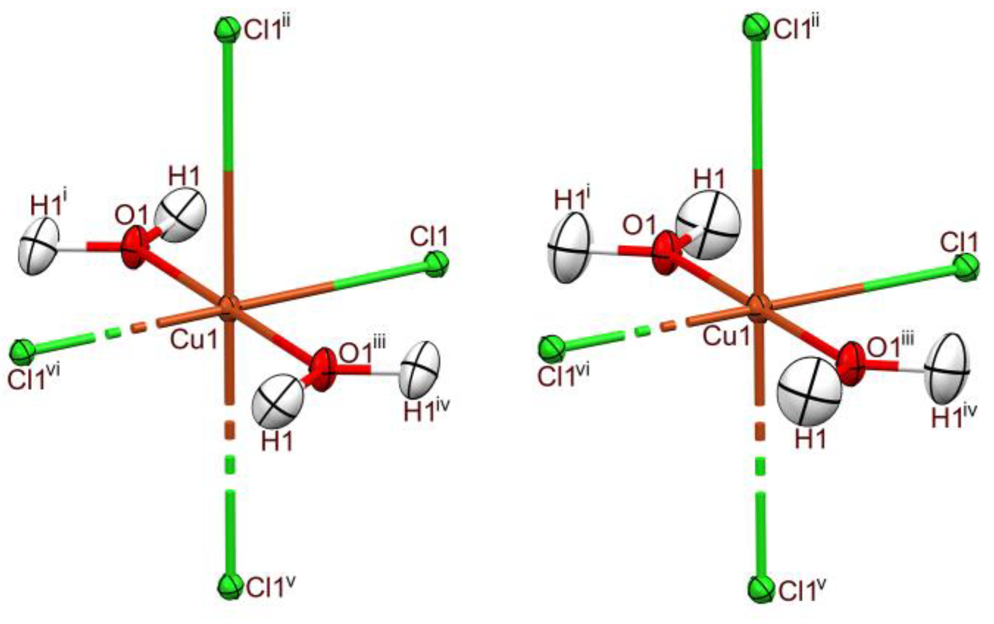
2.1. X-Ray Diffraction Structure of CuCl2∙2H2O

The local coordination environment at Cu is shown in Figure 1. CuCl2∙2H2O, 1, crystallizes in Pmna with the trans-disposed water molecules and the copper along a two-fold axis through the centre of the unit cell, and parallel to the a axis and the Cu, and four Cl ions on the mirror plane perpendicular to a. The primary structural data (Table 1) are in agreement with earlier X-ray [32,33,34,35] and neutron [36] diffraction data. The trans-disposed Cu–Cl1 and Cu–Cl1 vi bonds are short, 2.2870(3) Å, and along with the two water ligands form an orthogonal square planar geometry that is site-symmetry controlled. The longer, also trans-disposed Cu–Cl1 ii and Cu–Cl1 v bonds are at 2.9023(3) Å and are slightly offset from perpendicular, such that the Cl1–Cu1–Cl1 v angle is 91.10(1)°. The short–long pattern in bonds to Cl is expected for a “pseudo-Jahn-Teller” elongated d9 electron configuration at Cu(II) [37]. The Cu1–O1 distance is 1.9420(9) Å for the coordinated aqua ligand, in excellent agreement with 1.94 Å for bonds to equatorial water oxygen atoms found by Ohtaki in aqueous Cu(II) ions from X-ray scattering probability distribution curves [38]. The axial halides belong to CuCl2(OH2)2 moieties from the layers above and below the depicted molecule through perpendicular secondary bonding (see also Figure 2).
In the lattice structure of 1 (Figure 2), the layers of CuCl2(OH2)2 contain chains linked via bridging axial contacts; parts of two such chains are shown (the three left-side and three right--side Cu atoms labeled I in Figure 2a; in projection, these are the left and right I in Figure 2b). This layer is linked by Cl∙∙∙OH H-bonds to the next layers on either side (two pairs of bridged Cu atoms in these layers are labelled II in Figure 2a; in projection, the upper and lower II in Figure 2b). The “square planar” CuO2Cl2 moieties, involving the shorter Cu–Cl bonds in the type I and type II chains, are rotated by 77.9° from each other. The primary H-bonding contacts, ( C 1 1 ( 4 ) chains in Etter notation [39], labelled b in the figure) always involve a type I chloro ligand acceptor and a type II water O–H donor. Larger R 2 4 ( 8 ) rings are defined by four such H-bonds linking two type I and two type II centres (labelled a in the figure). For alternative representations of the lattice structure of 1, see [10].
The accurate placement of the H-atom position is of course essential for the description of the H-bond network (Table 2). The O1–H1 length determined as 0.941(14) Å from dataset 1a in our NoSpherA2 HAR is in good agreement with the 0.95 Å from the legacy RT neutron diffraction structure [36], but all our data exceed the precision of the very limited available neutron data. By contrast, the best (RT) X-ray diffraction structure reported before this work measured this value as 0.82 Å, i.e., about 14% shorter [35]. In the direct precursor IAM refinement of this dataset (olex2.refine), this O1–H1 length is even shorter at 0.783(17) Å. Hence, a distinct advantage of HAR is this accurate placement of H-atoms, which obviates the need to artificially adjust the bonds involved in H-bonding as was previously necessary [20]. The single water molecule in 1 is coordinated to the metal and the OH2 moiety is co-planar with equatorial CuCl2O2. This defines the O–H bond as Class 1 according to Chiari and Ferraris [40], those for which the envelope tip angle ε ≤ 30°. Chandler et al. using a metadata analysis (on neutron data), gave the range of such Class 1 coordinated water O–H bonds as 0.858–1.003 with a mean of 0.958 Å [41]. Our values from both datasets are very close to this mean.
Comparing structure models 1a and 1b shows that the s.u. of the derived parameters are ~40% higher in the latter. Thus, the Mo Kα data do give both an overall better structure (Table 1) and more precise H-atom determinations (Table 2). Looking ahead to 3a and 3b, the s.u. of the latter is 50% larger. Hence, the evidence is that Mo Kα is superior for XRD data modelled using HAR for metal hydrates.

2.2. Comparison to Structure of Anhydrous CuCl2

The relationship between the structures of 1 and anhydrous 2, the mineral known as tolbachite [42], is important for understanding the dynamics of the hydration/dehydration cycles involved in using the salt as a proposed thermal energy storage system [7,8]. Tolbachite, a distorted form of the cadmium iodide lattice type (Figure 3a), crystallizes in C2/c, and is centered only on a two-fold axis. It has four equatorial Cu–Cl bond distances, 2.263(6) Å, and two much longer apical Cu–Cl bond distances, 2.991(6) Å, as determined from an RT X-ray diffraction structure determination [39]. The trans-angles are exactly linear, but the cis angles are 92.4(2) and 93.6(2)°. The formation of the hydrate (Figure 3b) can be understood as the insertion of water into two of the four short Cu–Cl bonds, leading both to thermodynamically stronger Cu-O bonds and to a more robust, more elastic lattice, induced by the H-bonds from the water donors to the chloro acceptors that link chains in the offset {CuCl2} layers. Within layers, the equivalent Cl–Cu–Cl bridging bonds transform into the long–short alternation from the pseudo-Jahn-Teller distortions [37]. The mineral forms of both copper chlorides are extremely rare [42]. We are grateful for the access provided to the legacy crystal data for both X-ray and neutron diffraction by the Crystallographic Open Database (http://www.crystallography.net/cod/ accessed on 30 January 2023) [43].

2.3. X-ray Diffraction Structure of NiCl2∙6H2O

The local coordination environment at Ni is shown in Figure 4. NiCl2∙6H2O, 3, crystallizes in I2/m with Z′ = 0.25, a mirror bisecting the NiCl2 and O(2)H2 non-coordinated water as well as 1 ¯ symmetry at the Ni. The trans-disposed Cu–Cl and Cu–Cl i bonds are relatively short at 2.3949(3) Å (Table 3), and along with the four symmetry-equivalent water ligands O(1)H2 form a square planar coordination environment to give an overall close-to-ideal tetragonal coordination at this d8 Ni2+, affording a salt hydrate which is known to be paramagnetic [15,16]. The Ni–O distances are slightly longer than the 2.04 Å measured by Ohtaki from X-ray scattering experiments on aqueous solutions of Ni(II) ions [38]. The geometry of the Mo Kα structure determination is more precise than that obtained with copper.
There is a more complex H-bonding network in the lattice of 3 than found in 1 (Figure 5), as expected with the larger number of water molecules. It should be emphasized that the nominal count of six water molecules from the empirical formula consists of just these two kinds of water, one coordinated and one only H-bonded, in a 2:1 ratio. Consequently, there are just four unique H-bond types (Table 2).
The coordinated water molecule in 3, O(1)H2, belongs to Class 1′ according to Chiari and Ferraris, since the envelope tip angle ε in this structure is 44.1°, exceeding the limit of 30° [40]. In the metadata analysis of Chandler et al. [41], Class 1′ coordinated water O–H bonds range from 0.917 to 1.019 with a mean of 0.972 Å (neutron data). Notably, both O1-H1A and O1-H1B lengths, 0.965(15) and 0.953(14) Å from dataset 3a, fit comfortably within this range and are statistically indistinguishable from the mean value at the 99% confidence limit. They also fit the trend of longer bonds compared to structure 1a, fitting the pattern from Chandler’s analysis that Class 1′ > Class 1. However, very large datasets are essential to establish patterns with such small differences in the core data. By contrast, in the direct precursor IAM refinement of this X-ray dataset (olex2.refine), the four O–H lengths ranged from 0.808(11) to 0.851(13) Å, again showing the impact of the HAR.

2.4. Comparison to Structure of Anhydrous NiCl2

In the structure of anhydrous NiCl2, 4 (Figure 6a), each Ni atom is octahedrally coordinated by chloro ligands, but the latter each have three short bonds to Ni. These also interact with Cl atoms on neighbouring layers via contacts that exceed the sum of the v.d.W. radii by 0.18 Å [44]. In the hydrate (Figure 6b), all but two of the Ni–Cl bonds are replaced by thermodynamically stronger Ni–O bonds, and all the lattice-binding interactions are formed by the network of O–H∙∙∙O and O–H∙∙∙Cl hydrogen bonds (Table 2), producing a robust 3D lattice, in place of the layer structure, that is also much more elastic. These structural changes are relevant to use in proposed thermal energy storage systems [7,8].

3. Experimental Section

3.1. Sample Sources

The copper(II) chloride hydrate (CuCl2∙2H2O) crystals 1 were either sourced from Baker (Analyzed Reagent grade) and used as received, or synthesized by adding SO2Cl2 (13.63 mg, 0.101 mmol) in 1 mL CH2Cl2 to a suspension of CuCl (10.0 mg, 0.101 mmol) in 1 mL CH2Cl2 at 0 °C. The mixture was allowed to warm to room temperature and stirred for 30 min to complete the reaction. The solvent was evaporated, and the solid residues were dissolved in CH3CN. Small greenish-blue crystals of CuCl2·2H2O were grown by slow evaporation at room temperature and were identified by X-ray diffraction. Data obtained on these synthetic crystals were overall slightly superior to crystals taken from the bulk commercial salt.
The pale-green nickel(II) chloride hydrate crystals (NiCl2∙6H2O) 3 were obtained by recrystallization of commercial salt (BDH Reagents) by cooling warmed solutions in deionized H2O. Suitable, small, and well-formed crystals of both compounds were selected under the microscope and frozen in Paratone™ oil on 50 μm MiTeGen micromounts with the diffractometer Oxford Cryostream 800 device.

3.2. Crystallography

Data collection employed a Rigaku-Oxford Diffraction SuperNova dual microsource kappa diffractometer equipped with a Pilatus 200K HPC detector. Data collection, reduction, and correction were controlled using CrysAlisPro 1.171.42.63a (Rigaku Oxford Diffraction, 2022). Empirical absorption corrections were applied using spherical harmonics implemented in the SCALE3 ABSPACK scaling algorithm. The refinement employed NoSpherA2 [21], (NOn-SPHERical Atom-form-factors in Olex2) [23], an implementation of Hirshfeld atom refinement (HAR) that makes use of tailor-made aspherical atomic form factors calculated on-the-fly from a Hirshfeld-partitioned electron density (ED). The ED was calculated from a Gaussian basis set single determinant SCF wavefunction in DFT at the PBE/def2-TZVPP level of theory for a fragment of the crystal and a multiplicity of 2 for Cu d9 and 3 for Ni d8. The ORCA 5.0 software suite was employed on an i7-8700 CPU @ 3.20 GHz computer with 16.0 Gb RAM under Windows 10 [24]. NoSpherA2 was set for high accuracy partitioning. The crystal and structure refinement data are summarized in Table 4. Selected interatomic distances and angles for 1 are provided in Table 1 and for 3 in Table 3, and H-bonding data in Table 2. CheckCIF reports and data in CIF format are also available from the Supplementary Materials.
The following is a description of the operational strategy that was used in performing HAR with NoSpherA2 in this work. Of key importance is the integration of this method in the highly popular crystallographic graphical user interface (GUI) known as Olex2. The brief outline provided here should be used in conjunction with the theory provided in references [19] and [21]. Potential adopters of the method should certainly consult the excellent on-line documentation provided by the OlexSys organization (https://www.olexsys.org/ accessed on 30 January 2023) and many extremely useful videos on the Olex2 YouTube channel (https://www.youtube.com/channel/UCV6B2W8zlmXqkU2DbIviQow accessed on 30 January 2023). NoSpherA2 operates only in conjunction with the olex2.refine engine (i.e., not with ShelXL, though the input and most instructions from the ShelXL environment work seamlessly) [25]. However, only olex2.refine has the underlying software architecture able to input the custom form factors computed by NoSpherA2. In our group, we have learned that a full refinement to publishable standards should be undertaken with the IAM first using olex2.refine, which we archived in a separate folder. We used these archives to allow for comparisons in structure precision between the IAM and HAR models. We have discovered, particularly with organic compounds, an overall improvement in structure precision with NoSpherA2 that can approach 80% for high quality datasets [28,29]. Even for inorganics, there can be a measurable improvement where adequate markers are present (e.g., multiple S–O bonds [2]). A feature of implementing NoSpherA2 is that atom labels cannot be changed after the computational process starts, and so the IAM file also provides a “backup file”, from which the HAR must be restarted if significant changes to the model are later found to be necessary. Trying to make label or model changes to an active NoSpherA2 refinement model is not recommended.
A key innovation of NoSpherA2 in Olex2 is that the expanded structure required for a viable density functional theory (DFT) calculation can be arranged by symmetry and contact expansion in the GUI workspace. Very simply, whatever atoms are on the screen are computed. Thus, for 1, the full CuCl2(OH2)2 formula unit was used for the calculation (it would equally be possible to include the additional axial chlorides and compute [CuCl4(OH2)2]2-). Next, the appropriate charge and multiplicity for the computed entity must be assigned; therefore, for 2, the doublet state for the d9 electron configuration was employed. Similarly, for 3, [NiCl2(OH4)4]∙H2O was the computed entity (exactly as displayed in Figure 4a), and a multiplicity of 3 for the expected paramagnetic d8 electron configuration was selected. In practice, several combinations of the structure entity should be investigated to ensure that the DFT SCF calculation is able to model the atom electron densities sufficiently accurately to allow for optimal subsequent form factor calculations (such parallel models are best stored in separate working folders). In the work reported here, the ORCA 5.0 computational method was employed because we have found it to be reliable, and above all, very fast. The SCF calculations themselves took about 9 s in the case of 1a and a little over 4 s for 3a. The full combination of the final iterative sequence of ORCA DFT, NoSpherA2 form factor, and olex2.refine cycles were tracked with a stopwatch and amounted to about one minute for 1a and 1.5 min for 3a on the standard desktop PC described above. Larger molecules obviously took longer, and total computing times exceeding 20 min were encountered for some ~150 atom molecules studied in an earlier project, albeit using an older (slower) implementation of ORCA [29].

4. Conclusions

HAR using NoSpherA2 was undertaken for CuCl2∙2H2O, 1, and NiCl2∙6H2O, 3, from dual-wavelength single-crystal X-ray diffraction experiments. The obtained O–H lengths are indistinguishable from the legacy neutron diffraction structure determinations of these common metal salt hydrates. This report provides the first crystallographic models for 1 and 3, supposedly well-known structures, in which the hydrogen displacements ellipsoids have been fully refined. The very old and experimentally limited extant neutron refinements did not achieve this. Yet, notably, these neutron data have been used in extensive metadata analyses [41]. Therefore, there is an obvious need to revisit the structures of many of the crystalline hydrates of metal salts. Prospects for accurate H-atom placements in such salts by HAR with NoSpherA2 are extremely promising, at a modest experimental cost, and all who have access to good quality crystals, or have existing SC-XRD datasets on hand, are encouraged to apply this excellent, user-friendly method.

Supplementary Materials

The following supporting information can be downloaded at: https://www.mdpi.com/article/10.3390/cryst13020293/s1, Crystal Structure Reports for 1 and 3; structures in CIF format.

Funding

This research received no external funding.

Data Availability Statement

CSD 2233706; 2238551-2238553 contain the supplementary crystallographic data for this paper. These data can be obtained free of charge via www.ccdc.cam.ac.uk/data_request/cif, or by emailing [email protected], or by contacting The Cambridge Crystallographic Data Centre, 12 Union Road, Cambridge CB2 1EZ, UK; fax: +44-1223-336033 or from the Inorganic Crystal Structure Database, https://icsd.products.fiz-karlsruhe.de/ accessed on 28 January 2023.

Acknowledgments

M. A. Ibrahim prepared the synthetic sample of 1. The support of the University of Lethbridge for the experimental work and manuscript preparation is gratefully acknowledged. The university and the Faculty of Arts and Science for providing X-ray diffraction equipment is acknowledged. The anonymous referees of this article are thanked for their constructive advice that led to strengthening the content and arguments of the paper. Remaining errors are solely the author’s responsibility.

Conflicts of Interest

The author declares no conflict of interest.

References

  1. Kauffman, G.B. (Ed.) Coordination Chemistry: A Century of Progress; Oxford University Press: Oxford, UK, 1994. [Google Scholar]
  2. Ibrahim, M.A.; Boeré, R.T. The copper sulfate hydration cycle. Crystal structures of CuSO4 (Chalcocyanite), CuSO4∙H2O (Poitevinite), CuSO4∙3H2O (Bonattite) and CuSO4∙5H2O (Chalcanthite) at low temperature using non-spherical atomic scattering factors. New J. Chem. 2022, 46, 5479–5488. [Google Scholar] [CrossRef]
  3. Scacchi, A. Dell’eriocalco e del melanotallo, nuove specie di minerali. Rend. Dell’accademia Delle Sci. Fis. E Mat. Sez. Della Soc. R. Di Napoli 1870, 9, 86–89. [Google Scholar]
  4. Crook, W.W., III; Jambor, J.L. Nickelbischofite, a new nickel chloride hydrate. Can. Mineral. 1979, 17, 107–109. [Google Scholar]
  5. N’Tsoukpoe, K.E.; Schmidt, T.; Rammelberg, H.U.; Watts, B.A.; Ruck, W.K.L. A systematic multi-step screening of numerous salt hydrates for low temperature thermochemical energy storage. Appl. Energy 2014, 124, 1–16. [Google Scholar] [CrossRef]
  6. Glasser, L. Thermodynamics of Inorganic Hydration and of Humidity Control, with an Extensive Database of Salt Hydrate Pairs. J. Chem. Eng. Data 2014, 59, 526–530. [Google Scholar] [CrossRef]
  7. Donkers, P.A.J.; Sögütoglu, L.C.; Huinink, H.P.; Fischer, H.R.; Adan, O.C.G. A review of salt hydrates for seasonal heat storage in domestic applications. Appl. Energy 2017, 199, 45–68. [Google Scholar] [CrossRef]
  8. Kiyabu, S.; Girard, P.; Siegel, D.J. Discovery of Salt Hydrates for Thermal Energy Storage. J. Am. Chem. Soc. 2022, 144, 21617–21627. [Google Scholar] [CrossRef]
  9. Housecroft, C.E.; Sharpe, A.G. Inorganic Chemistry, 4th ed.; Pearson: Harlow, UK, 2012; pp. 761–766. [Google Scholar]
  10. Medeiros, F.E.O.; Araujo, B.S.; Ayala, A.P. Raman spectroscopy investigation of the thermal stability of the multiferroic CuCl2 and its hydrated form. Vib. Spectr. 2018, 99, 1–6. [Google Scholar] [CrossRef]
  11. DeFotis, G.C.; Hampton, A.S.; Van Dongen, M.J.; Komatsu, C.H.; Benday, N.S.; Davis, C.M.; Hays, K.; Wagner, M.J. Magnetism of CuCl2·2D2O and CuCl2·2H2O, and of CuBr2·6H2O. J. Magn. Magn. Mater. 2017, 434, 23–29. [Google Scholar] [CrossRef]
  12. Antonijevic, S.; Persson, E. Study of water dynamics and distances in paramagnetic solids by variable-temperature two-dimensional 2H NMR sectroscopy. J. Chem. Phys. 2007, 126, 014504. [Google Scholar] [CrossRef]
  13. Frost, R.L.; Williams, P.A.; Kloprogge, J.T.; Martens, W. Raman spectroscopy of the copper chloride minerals nantokite, eriochalcite and claringbullite—Implications for copper corrosion. Neues Jahrb. Mineral. Monats. 2003, 2003, 433–445. [Google Scholar] [CrossRef]
  14. Kleinberg, R. Crystal Structure of NiCl2·6H2O at Room Temperature and 4.2°K by Neutron Diffraction. J. Chem. Phys. 1969, 50, 4690–4696. [Google Scholar] [CrossRef]
  15. Kleinberg, R. Magnetic Structure of NiCl2·6H2O. J. Appl. Phys. 1967, 38, 1453–1454. [Google Scholar] [CrossRef]
  16. Spence, R.D.; Middents, P.; ElSaffar, Z.; Kleinberg, R. A Proton Resonance Study of the Magnetic Structure of Antiferromagnetic CoCl2·6H2O, CoBr2·6H20, NiCl2·6H2O, and NiBr2·6H2O. J. Appl. Phys. 1964, 35, 854–855. [Google Scholar] [CrossRef]
  17. Enyashin, A.N.; Ivanovskii, A.L. Magnetic properties of NiCl2 nanostructures. Comput. Mater. Sci. 2010, 49, 782–786. [Google Scholar] [CrossRef]
  18. Zajnullin, O.B.; Voloshin, A.E.; Komornikov, V.A.; Manomenova, V.L.; Rudneva, E.B.; Timakov, I.S.; Kovalev, S.I. Some Properties of NiCl2·6H2O Single Crystals. Phys. Sol. State 2019, 61, 2415–2417. [Google Scholar] [CrossRef]
  19. Capelli, S.C.; Burgi, H.B.; Dittrich, B.; Grabowsky, S.; Jayatilaka, D. Hirshfeld atom refinement. IUCrJ 2014, 1, 361–379. [Google Scholar] [CrossRef]
  20. Woińska, M.; Grabowsky, S.; Dominiak, P.M.; Woźniak, K.; Jayatilaka, D. Hydrogen atoms can be located accurately and precisely by x-ray crystallography. Sci. Adv. 2016, 2, e1600192. [Google Scholar] [CrossRef]
  21. Kleemiss, F.; Dolomanov, O.V.; Bodensteiner, M.; Peyerimhoff, N.; Midgley, L.; Bourhis, L.J.; Genoni, A.; Malaspina, L.A.; Jayatilaka, D.; Spencer, J.L.; et al. Accurate crystal structures and chemical properties from NoSpherA2. Chem. Sci. 2021, 12, 1675–1692. [Google Scholar] [CrossRef]
  22. Bacon, G.E. Neutron Diffraction, 3rd ed.; Clarendon Press: Oxford, UK, 1975. [Google Scholar]
  23. Dolomanov, O.V.; Bourhis, L.J.; Gildea, R.J.; Howard, J.A.K.; Puschmann, H. OLEX2: A complete structure solution, refinement and analysis program. J. Appl. Crystallogr. 2009, 42, 339–341. [Google Scholar] [CrossRef]
  24. Neese, F. Software update: The ORCA program system Version 5.0. WIREs Comput. Mol. Sci. 2022, 12, e1606. [Google Scholar] [CrossRef]
  25. Bourhis, L.J.; Dolomanov, O.V.; Gildea, R.J.; Howard, J.A.; Puschmann, H. The anatomy of a comprehensive constrained, restrained refinement program for the modern computing environment—Olex2 dissected. Acta Crystallogr. Sect. A 2015, 71, 59–75. [Google Scholar] [CrossRef] [PubMed] [Green Version]
  26. Cooper, R.I. Recent developments in the refinement and analysis of crystal structures. Struct. Bond. 2020, 185, 43–68. [Google Scholar] [CrossRef]
  27. Farrugia, L.J. Accurate H-atom parameters from X-ray diffraction data. IUCrJ 2014, 1, 265–266. [Google Scholar] [CrossRef]
  28. Hill, N.D.D.; Lilienthal, E.; Bender, C.O.; Boeré, R.T. Accurate crystal structures of C12H9CN, C12H8(CN)2, and C16H11CN valence isomers using nonspherical atomic scattering factors. J. Org. Chem. 2022, 87, 16213–16229. [Google Scholar] [CrossRef]
  29. Marszaukowski, F.; Boeré, R.T.; Wohnrath, K. Frustrated and realized hydrogen bonding in 4-hydroxy-3,5-ditertbutylphenylphosphine derivatives. Cryst. Growth Des. 2022, 22, 2512–2533. [Google Scholar] [CrossRef]
  30. Chocolatl Torres, M.; Bernès, S.; Kuri, U.S. Refinement of K[HgI3]·H2O using non-spherical atomic form factors. Acta Crystallogr. 2021, E77, 681–685. [Google Scholar] [CrossRef]
  31. Chrappová, J.; Pateda, Y.R.; Rakovský, E. Synthesis and Crystal Structure Analysis of NH4[Zn(cma)(H2O)2]·H2O Using IAM and HAR Approaches. J. Chem. Crystallogr. 2022. [Google Scholar] [CrossRef]
  32. Harker, D. The Crystal Structure of Cupric Chloride Dihydrate CuCl2·2H2O. Z. Kristallogr. 1936, 93, 136–145. [Google Scholar] [CrossRef]
  33. MacGillavry, C.H.; Bijvoet, J.M. Die Kristallstruktur der Cadmium- und Quecksilber-Diamin-Dihalogenide. Z. Kristallogr. 1936, 94, 231–245. [Google Scholar] [CrossRef]
  34. Engberg, A. An X-ray refinement of the crystal structure of copper(II) chloride dihydrate. Acta Chem. Scand. 1970, 24, 3510–3526. [Google Scholar] [CrossRef]
  35. Brownstein, S.; Han, N.F.; Gabe, E.; LePage, Y. A redetermination of the crystal structure of cupric chloride dihydrate. Z. Kristallogr. 1989, 189, 13–15. [Google Scholar] [CrossRef] [Green Version]
  36. Peterson, S.W.; Levy, H.A. Proton Positions in CuCl2·2H2O by Neutron Diffraction. J. Chem. Phys. 1957, 26, 220. [Google Scholar] [CrossRef]
  37. Halcrow, M.A. Jahn-Teller distortions in transition metal compounds, and their importance in functional molecular and inorganic materials. Chem. Soc. Rev. 2013, 42, 1784–1795. [Google Scholar] [CrossRef]
  38. Ohtaki, H.; Yamaguchi, T.; Maeda, M. X-ray diffraction studies of the structures of hydrated divalent transition-metal ions in aqueous solution. Bull. Chem. Soc. Jpn. 1976, 49, 701–708. [Google Scholar] [CrossRef]
  39. Etter, M.C. Encoding and decoding hydrogen-bond patterns of organic compounds. Acc. Chem. Res. 1990, 23, 120–126. [Google Scholar] [CrossRef]
  40. Chiari, G.; Ferraris, G. The water molecule in crystalline hydrates studied by neutron diffraction. Acta Crystallogr. Sect. B Struct. Crystallogr. Cryst. Chem. 1982, 38, 2331–2341. [Google Scholar] [CrossRef]
  41. Chandler, G.S.; Wajrak, M.; Khan, R.N. Neutron diffraction structures of water in crystalline hydrates of metal salts. Acta Crystallogr. B Struct. Sci. Cryst. Eng. Mater. 2015, 71, 275–284. [Google Scholar] [CrossRef]
  42. Burns, P.C.; Hawthorne, F.C. Tolbachite, CuCl2, the first example of Cu2+ octahedrally coordinated by Cl. Am. Mineral 1993, 78, 187–189. [Google Scholar]
  43. Grazulis, S.; Chateigner, D.; Downs, R.T.; Yokochi, A.T.; Quiros, M.; Lutterotti, L.; Manakova, E.; Butkus, J.; Moeck, P.; Le Bail, A. Crystallography Open Database—An open-access collection of crystal structures. J. Appl. Crystallogr. 2009, 42, 726–729. [Google Scholar] [CrossRef]
  44. Ferrari, A.; Braibanti, A.; Bigliardi, G. Refinement of the crystal structure of NiCl2 and of unit-cell parameters of some anhydrous chorides of divalent metals. Acta Crystallogr. 1963, 16, 846–847. [Google Scholar] [CrossRef]
Scheme 1. Local coordination environments of the hydrated and anhydrous chlorides.
Crystals 13 00293 sch001
Figure 1. Displacement ellipsoids plots (40% probability) of the asymmetric units in the crystal structures of 1 determined from single crystals at 100 K, with the atom numbering schemes: (left) 1a Mo Kα; (right) 1b Cu Kα. Expansions to show full coordination at one Cu(II) ion (symmetry operators: i x,-y,1-z; ii x,1-y,1-z; iii 1-x,1-y,1-z; iv 1-x,y,z; v x,1+y,z; vi x,1-y,1-z).
Crystals 13 00293 g001
Figure 2. The lattice structure of 1 (using Cu Kα data; H-atoms drawn as spheres of 0.15 Å radius and H-bonds drawn as dashed blue lines). (a) Projection along the ab bisector showing upper, middle, and lower layers. (b) Projection down the b axis. See text for further details.
Crystals 13 00293 g002
Figure 3. Comparative ”ball & stick” plots of the lattice structures of (a) 2 and (b) 1 drawn to scale. Projection down the b axis for 2 and down the a axis for 1, with each Cu centre six coordinates; unit cell boundaries for each are included. (The archival CIF COD9001506 is the source of 2 [42]).
Crystals 13 00293 g003
Figure 4. Displacement ellipsoids plots (50% probability) of the asymmetric units in the crystal structures of 3 determined from a single crystal at 100 K, with the atom numbering schemes: (left) 3a Mo Kα; (right) 3b Cu Kα. Expansions to show full coordination at one Ni(II) ion (symmetry operators: i 1-x, y,1-z; ii 1-x,1-y,1-z; iii x,1-y,z).
Crystals 13 00293 g004
Figure 5. The lattice structure of 3 (using Mo Kα data; H-atoms drawn as spheres of 0.15 Å radius and H-bonds drawn as dashed lines that are colour-coded to relative length: yellow < orange < red). Six uncoordinated O(2)H2 water molecules are circled for ease of identification. The two located furthest right show all H-bonds; two strong bonds as acceptors from coordinated O(1)H2, weaker bifurcated donation to two different O1, and a single intermediate donation to Cl1. Each coordinated water donates to Cl1 and to O2 and accepts from a different O2 water. See text for further details.
Crystals 13 00293 g005
Figure 6. Comparative “ball & stick” plots of the lattice structures of (a) 4 and (b) 3 drawn to scale. Projection parallel to the b axis for 4 and ~ perpendicular to the body diagonal axis for 3; unit cell boundaries for each are included. (The archival CIF COD2310380 was employed for 4 [44]).
Crystals 13 00293 g006
Table 1. Selected interatomic distances (Å) and angles (°) in structures 1a and 1b 1.
Atoms 2Mo Kα–1aCu Kα–1b
Cu1–Cl12.2870(3)2.2823(4)
Cu1–Cl1 ii2.9023(3)2.8973(4)
Cu1–O11.9420(9)1.9414(16)
O1–H10.941(14)0.95(2)
Cl1–Cu1–Cl1 vi180.0180.0
Cl1 ii –Cu1–Cl1v180.0180.0
O1–Cu1–O1 iii180.0180.0
Cl1–Cu1–O190.090.0
Cl1–Cu1–Cl1 v91.10(1)91.14(1)
Cl1 ii –Cu1–O190.090.0
Cu1–O1–H1123.7(10)126.5(16)
H1–O1–H1 i112.5(19)107(3)
1 Angles around Cu to O1 and Cl1 are symmetry controlled. 2 Sym codes: i x,-y,1-z; ii x,1-y,1-z; iii 1-x,1-y,1-z; v x,1+y,z; vi x,1-y,1-z.
Table 2. Hydrogen bonds and hydrogen displacement data for 1 and 3.
D-H∙∙∙Ad(D-H)/ÅD(H-A)/Åd(D-A)/ÅD-H-A/°Ueq
1
O1-H1∙∙∙Cl1 i a0.941(14)2.240(14)3.1686(7)169.2(14)0.030(4)
b0.95(2)2.22(2)3.1627(11)173(2)0.046(7)
c0.952.243.18172
3
O1-H1a∙∙∙O2 ii d0.965(15)1.783(14)2.7401(8)170.5(15)0.030(4)
e0.94(3)1.81(3)2.7395(16)168(3)0.029(7)
f0.94(2)1.80(2)2.74(1)174(2)0.044(4)
g0.961.772.73170.50.027(1)
O1-H1b∙∙∙Cl iii d0.953(14)2.288(15)3.2049(6)161.3(13)0.035(45)
e0.94(3)2.32(3)3.2013(12)155(2)0.029(7)
f0.94(2)2.30(2)3.21(1)164(2)0.043(4)
g0.972.263.19161.80.029(1)
O2-H2a∙∙∙Cl iv d0.957(19)2.209(19)3.1420(10)164.7(18)0.029(5)
e1.01(5)2.13(5)3.1368(18)175(4)0.035(11)
f1.07(3)2.11(3)3.17(2)165(1)0.015(5)
g0.97(1)2.16(1)3.109(7)168(1)0.022(3)
O2-H2b∙∙∙O1 v d0.91(2)2.259(18)3.0186(10)140.2(6)0.050(7)
e0.92(4)2.25(3)3.0176(18)140.3(9)0.029(10)
f0.96 (4)2.27(2)3.06(1)138.30.075(1)
g0.95(1)2.233.007138.40.025(3)
a Data in bold text are from structure 1a (Mo Kα data). b Data in normal text are from structure 1b (Cu Kα data). c Data in italics are from the 1957 single crystal neutron structure at RT and are obtained from the archived CIF COD1008760 [36]; neither s.u. values nor Ueq values are reported [36]. d Data in bold text are from structure 3a (Mo Kα data). e Data in normal text are from structure 3b (Cu Kα data). f Data in italics are from the 1969 single crystal neutron structure at RT and obtained from the archived CIF COD9012473 [14]. g Data in italic bold text are from the 1969 single crystal neutron structure at 4.2 K and obtained from the archived CIF COD90124734. Limited s.u. are available for this dataset due to restrictions imposed on the experiment by the cryostat; see the original article [14]. (Symmetry operators: i 1/2-x,+y,3/2-z; ii 3/2-x,1/2+y,1/2-z; iii 1/2+x,1/2+y,1/2+z; iv 1+x,+y,+z; v +x,1-y,+z).
Table 3. Selected interatomic distances (Å) and angles (°) in structures 3a and 3b 1.
Atoms 2Mo Kα–3aCu Kα–3b
Ni–Cl2.3949(3)2.3936(5)
Ni–O12.0681(5)2.0670(11)
O1–H1a0.965(15)0.94(3)
O1–H1b0.953(14)0.94(3)
O2–H2a0.957(19)1.01(5)
O2–H2b0.91(2)0.92(4)
Cl–Ni–Cl i180.0180.0
O1–Ni–O1 ii180.0180.0
O1 i–Ni–O1 iii180.0180.0
Cl–Ni–O189.137(17)89.17(3)
Cl–Ni–O1 ii90.863(17)90.83(3)
O1–Ni–O1 i93.19(3)93.14(6)
O1–Ni–O1 iii86.81(3)86.86(6)
Ni–O1–H1a115.2(8)115.0(17))
Ni–O1–H1b117.4(10)113.1(18)
H1a–O1–H1b106.9(13)114(2)
H2a–O2–H2b108(2)115(4)
1 Angles around Ni to O1 and Cl are symmetry controlled. 2 Sym codes: i 1-x, y,1-z; ii 1-x,1-y,1-z; iii x,1-y,z.
Table 4. Crystal and structure refinement data.
Parameter 1a1b3a3b
Empirical formula Cl2CuH4O2 Cl2CuH4O2 Cl2H12NiO6 Cl2H12NiO6
Formula weight 170.481 170.481 237.690 237.690
Temperature/K 100.01(10) 100.01(10) 99.98(10) 99.99(10)
Crystal system orthorhombic orthorhombic monoclinic monoclinic
Space group PmnaPmnaI2/mI2/m
a/Å 8.0553(3) 8.0405(4) 6.5628(4) 6.5579(2)
b/Å 3.7295(2) 3.7238(2) 7.0330(4) 7.0244(3)
c/Å 7.3674(3) 7.3585(4) 8.7326(6) 8.7291(3)
β/° 90 90 96.723(6) 96.702(4)
Volume/Å3 221.333(17) 220.32(2) 400.29(4) 399.36(3)
Z, Z’ 2, 0.252, 0.25 2, 0.25 2, 0.25
ρcalc/g/cm3 2.558 2.570 1.972 1.977
μ/mm−1 5.967 16.849 3.062 9.551
F(000) 166.0 166.0 244.0 244.0
Crystal size/mm3 0.21 × 0.04 × 0.02 0.16 × 0.07 × 0.06 0.18 × 0.1 × 0.03 0.23 × 0.09 × 0.04
Radiation Mo Kα (λ = 0.71073) Cu Kα (λ = 1.54184) Mo Kα (λ = 0.71073) Cu Kα (λ = 1.54184)
2Θ range data collect/° 7.5 to 68.94 16.32 to 149.94 7.36 to 71.44 16.04 to 160.34
Index ranges −13 ≤ h ≤ 13
−6 ≤ k ≤ 6
−12 ≤ l ≤ 12
−9 ≤ h ≤ 9
−4 ≤ k ≤ 4
−8 ≤ l ≤ 9
−10 ≤ h ≤ 9
−11 ≤ k ≤ 11
−14 ≤ l ≤ 13
−8 ≤ h ≤ 6
−8 ≤ k ≤ 8
−10 ≤ l ≤ 11
Reflections collected 20107 1200 4133 2052
Independent reflections 499 238 890 469
Rint, Rsigma0.0598, 0.01700.0186, 0.01080.0318, 0.02330.0364, 0.0227
Data/restraints/param499/6/25 238/0/26 890/0/56 469/24/56
Goodness-of-fit on F2 1.016 1.053 0.996 1.044
Final R indexes [I ≥ 2σ(I)] R1 = 0.0120, wR2 = 0.0257 R1 = 0.0133, wR2 = 0.0329 R1 = 0.0175, wR2 = 0.0396 R1 = 0.0276, wR2 = 0.0823
Final R indexes [all data] R1 = 0.0134, wR2 = 0.0264 R1 = 0.0133, wR2 = 0.0329 R1 = 0.0186, wR2 = 0.0400 R1 = 0.0295, wR2 = 0.0843
Largest diff. peak/hole/e Å−30.56/−0.42 0.26/−0.21 0.40/−0.50 0.46/−0.50
Accession codes (CCDC)2238551223370622385522238553
Disclaimer/Publisher’s Note: The statements, opinions and data contained in all publications are solely those of the individual author(s) and contributor(s) and not of MDPI and/or the editor(s). MDPI and/or the editor(s) disclaim responsibility for any injury to people or property resulting from any ideas, methods, instructions or products referred to in the content.

