Article

Quantum Confinement Effect and Photoenhancement of Photoluminescence of PbS and PbS/MnS Quantum Dots

by
Muhammad Safwan Zaini
1,
Josephine Ying Chyi Liew
1,2,
Shahrul Ainliah Alang Ahmad
2,3,
Abdul Rahman Mohmad
4 and
Mazliana Ahmad Kamarudin
1,*
1
Department of Physics, Faculty of Science, Universiti Putra Malaysia, UPM Serdang, Selangor Darul Ehsan 43400, Malaysia
2
Institute of Advanced Technology, Universiti Putra Malaysia, UPM Serdang, Selangor Darul Ehsan 43400, Malaysia
3
Department of Chemistry, Faculty of Science, Universiti Putra Malaysia, UPM Serdang, Selangor Darul Ehsan 43400, Malaysia
4
Institute of Microengineering and Nanoelectronics, Universiti Kebangsaan Malaysia, Bangi 43600, Malaysia
*
Author to whom correspondence should be addressed.
Appl. Sci. 2020, 10(18), 6282; https://doi.org/10.3390/app10186282
Submission received: 29 July 2020 / Revised: 12 August 2020 / Accepted: 15 August 2020 / Published: 9 September 2020
(This article belongs to the Section Materials Science and Engineering)

Abstract

:

Featured Application

Colloidal PbS quantum dots (QDs) that show a strong quantum confinement effect have potential towards photovoltaic application. Promising constant photoluminescence (PL) peak energies of PbS and PbS/MnS core shell QDs could be advantageous particularly for biological marker application.

Abstract

The quantum confinement effect and photoenhancement of photoluminescence (PL) of lead sulphide (PbS) quantum dots (QDs) and lead sulphide/manganese sulphide (PbS/MnS) core shell QDs capped with thiol ligands in aqueous solution were investigated. From PL results, the presence of MnS shells gives a strong confinement effect which translates to higher emission energy in PbS/MnS core shell QDs. Increasing MnS shell thickness from 0.3 to 1.5 monolayers (ML) causes a blueshift of PL peak energies as the charge carriers concentrated in the PbS core region. Enhancement of the PL intensity of colloidal PbS and PbS/MnS core shell QDs has been observed when the samples are illuminated above the band gap energy, under continuous irradiation for 40 min. Luminescence from PbS QDs and PbS/MnS core shell QDs can be strongly influenced by the interaction of water molecules and oxygen present in aqueous solution adsorbed on the QD surface. However, PbS/MnS core shell QDs with a shell thickness of 1.5 ML did not show a PL peak energy stability as it was redshifted after 25 min, probably due to wider size distribution of the QDs.

Graphical Abstract

1. Introduction

Colloidal quantum dots (QDs) have attracted great attention over the last decades because of their unique optical properties related to quantum confinement effects where their band gap can be tuned by adjusting their particle size [1,2]. This effect gives a significant impact on the energy band gap of the QDs. As the size of the particles reaches nanoscale, the movement of excitons is impeded. However, the total energy of excitons is significantly unaffected until the size of particles is smaller than the exciton Bohr radius. At this point, the excitation energy required to create electron–hole pairs starts to increase, and this inversely proportional phenomenon of excitation energy and QD size is known as quantum confinement. Furthermore, the effect of quantum confinement can be seen in the core shell QDs. It was reported that increasing thickness of the shell causes the photoluminescence (PL) peak to be shifted to a higher energy level [3]. Thus, properties of QDs such as photostability, bright luminescence, size dependent PL, narrow emission and broad absorption bands have caused colloidal QDs to receive extensive attention for the past decades [4].
Lead sulphide (PbS) QDs belong to the group of IV-VI semiconductors with a direct narrow band gap of 0.41 eV at room temperature [5]. Therefore, the band structure and electronic properties have distinguished PbS from II-VI (e.g., cadmium sulphide (CdS)) and III-V (e.g., indium arsenide (InAs)) semiconductors. PbS may offer unique opportunities to study the fundamental physics of quantum confinement due to the fact that it has a large exciton Bohr radius (~18 nm) which can achieve a strong quantum confinement effect with a small QD diameter (<10 nm) as compared to other groups of material [6,7]. This extreme confinement of charge carriers is predicted to enhance the quantization energy of nearly all material properties dependent on size [8]. Certainly, PbS QDs exhibit blueshifted PL onsets compared to the bulk crystals. In terms of technological perspective, PbS shows promising interest as it can provide size-tunable optical transitions at important near infrared (NIR) wavelengths. This may be advantageous to a variety of applications, including solar cells [9,10,11], dye imaging [12] and light emitting diodes [13] operating at NIR wavelengths.
From the examples above, practical applications of QDs are mainly related to the interaction of QDs with light, thus investigating the effects of laser light irradiation on the PL properties of QDs is one of the crucial tasks. Under ambient conditions, there are two possible effects on the influence of light on the QDs: (i) increase of luminescence intensity due to QD surface modification and (ii) degradation of PL spectra due to the photooxidation process. The combination of both effects is expected during prolonged light exposure and has been investigated experimentally [14,15,16,17,18]. It is clear that fluorescence enhancement is achieved through the use of appropriate surface passivation, which can eliminate surface defects that cause non-radiative relaxation [19]. One of the strategies is growing a wider bandgap semiconductor material towards the core QDs, so-called type-I core shell QDs. In such type of core shell QD, the exciton is confined in the core where the electron populates the energy in the conduction band while the hole is in the valence band [20,21].
It was reported that PL intensity is enhanced due to stabilization with surfactant molecules or surface ligand passivation [22,23,24]. The surfactant molecules such as trioctylphosphine oxide (TOPO) and hexadecylamine in organic solvents passivate the defects on the surface of the QDs resulting in enhancement. In addition, in terms of sample stability, attachment of capping ligands on core QDs results in unique properties, where the QDs will isolate from each other thus preventing them from agglomeration. In addition, in some cases, the enhancement of luminescence is related to the photooxidation of the surface of QDs [25,26,27]. The important part of this path is the photo-induced electron transfer from the QDs to the dissolved oxygen present in the solvent. If these electrons have been removed, the luminescence increases significantly.
Here we report the effects of laser exposure on the photoluminescence of PbS QDs and PbS/manganese sulphide (MnS) core shell QDs in aqueous solution with the emission in the near-infrared (NIR) region by illuminating the as-prepared samples. These QDs turned out to be stable in aqueous solution and the PL intensities were enhanced after continuous irradiation. The presence of a MnS shell on the PbS core QDs provides a strong quantum confinement effect with the variation of shell thicknesses.

2. Materials and Methods

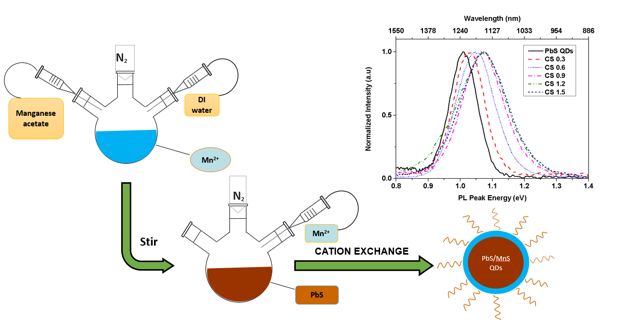
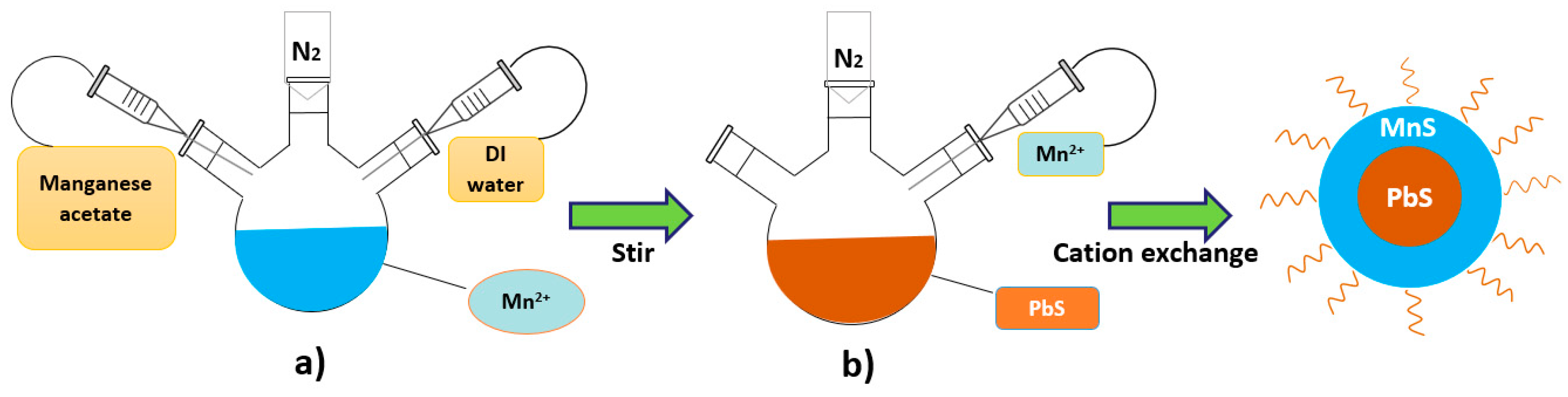
2.1. Sample Preparation

The sample preparation utilized lead (II) acetate, Pb(C2H3O2)2 (99.99%), to extract Pb2+ as a lead precursor and sodium sulfide, Na2S.9H2O (99.99%), to take S2− as a sulfide precursor. Thioglycerol, TGL (95%), and di-thioglycerol, DTG (95%), were used as capping ligands. In this experiment, the salts were purchased from R&M Chemicals (London, UK) and the ligands from Sigma Aldrich (Irvine, UK). All chemicals were used without any further purification. The synthesis was started with the preparation of PbS QDs in aqueous solution which followed the procedure as reported by Zaini et al. [28]. From this procedure, the typical size of the QDs obtained was approximately 6.00 ± 1.00 nm. The PbS solution was then aliquoted for the fabrication of PbS/MnS core shell QDs, where Mn2+ precursor was added into the PbS solution with different amounts of volume in order to vary the thickness. The cation exchange method was used, in which formation of the shell proceeds through cationic exchange between Pb2+ and Mn2+ ions. The schematic diagram of the preparation of PbS/MnS core shell QDs is shown in Figure 1. In this procedure, the molar ratios of Pb2+, S2− and Mn2+ precursors were fixed to 1, 0.3 and 0.2, respectively.
For the preparation of Mn2+ precursor, 0.086 g of manganese (II) acetate, Mn(CH3CO2).4H2O (98.5%, R&M Chemicals), in solid crystal was dissolved in 3.0 mL of deionized water. For the growth of the 0.3 monolayer (ML) thick MnS shell, 0.63 mL of Mn2+ precursor was injected into a three-neck flask containing PbS QDs that had been prepared earlier, and the solution was allowed to react for 30 min. Then, 7 mL of mixed solution was taken out and stored in the chemical refrigerator at a low controlled temperature of 4 °C to avoid oxidation and aggregation before proceeding to the characterization process. These steps were repeated by adding 0.51, 0.44, 0.30 and 0.25 mL of Mn2+ precursor into the same three-neck flask to grow the 0.6, 0.9, 1.2 and 1.5 ML shells, respectively. The PbS/MnS core shell QD samples were named as CS 0.3, CS 0.6, CS 0.9, CS 1.2 and CS 1.5. All of the steps were performed in ambient nitrogen (N2).

2.2. Sample Characterization

Customized photoluminescence (PL) spectroscopy was used in these experiments. A 532 nm green diode-pumped solid-state (DPSS) laser was used as an excitation source. The spectroscopy set up was equipped with a double monochromator (Horiba (iHR320)), lock-in amplifier, chopper and InGaAs detector. In detail, the emitted PL signal was collected by a lens and then focused onto the entrance slit of the monochromator. During the measurement, all of the samples were drop-casted on the glass substrate. The integration time was set at 1 s and the scanning range was fixed from 800 to 1600 nm. In this experiment, the PbS QD and PbS/MnS core shell QD (CS 0.3, CS 0.6, CS 0.9, CS 1.2 and CS 1.5) samples were exposed to the laser light for continuous irradiation time for 40 min with the power density of 5.66 W/cm−2.

3. Results

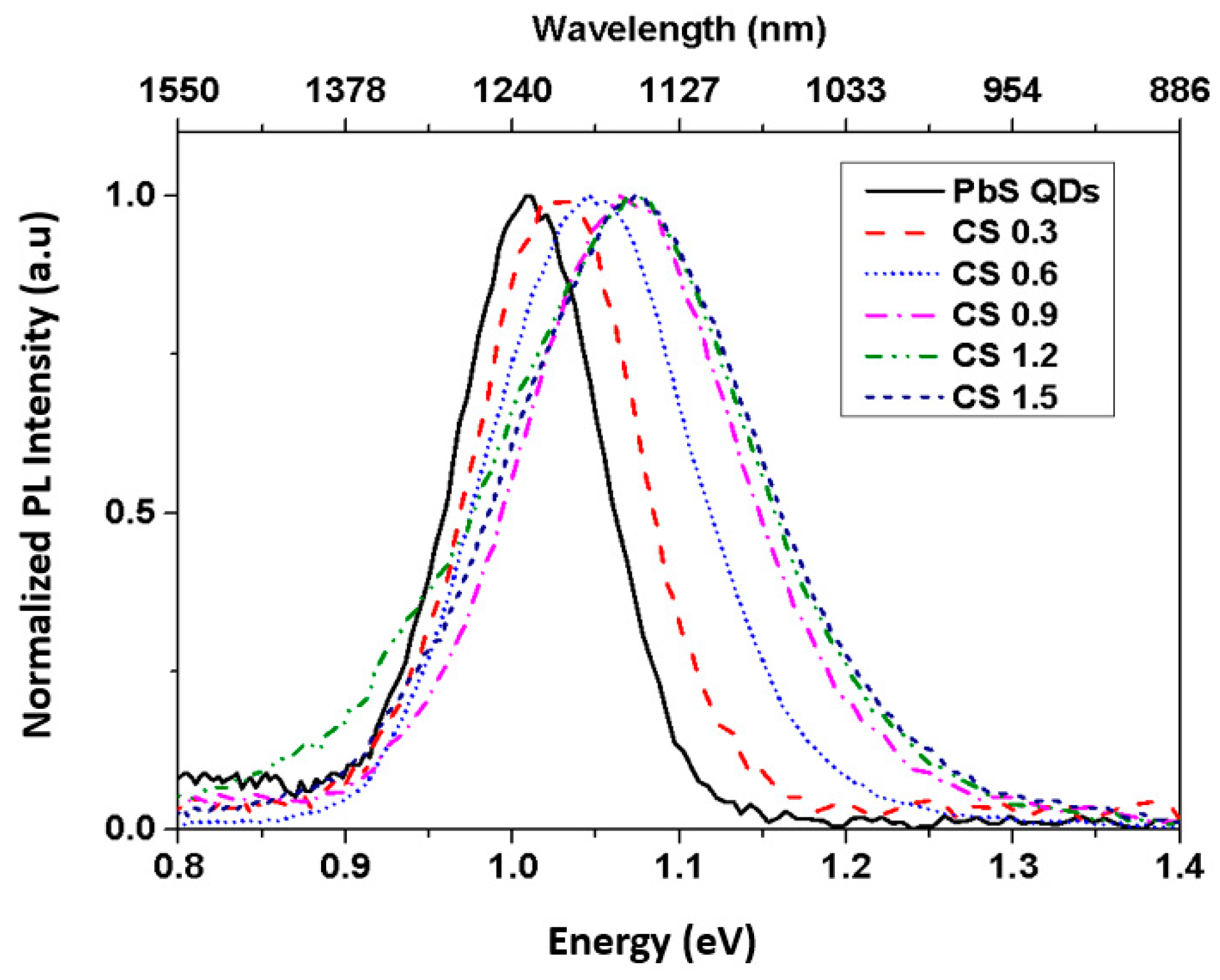
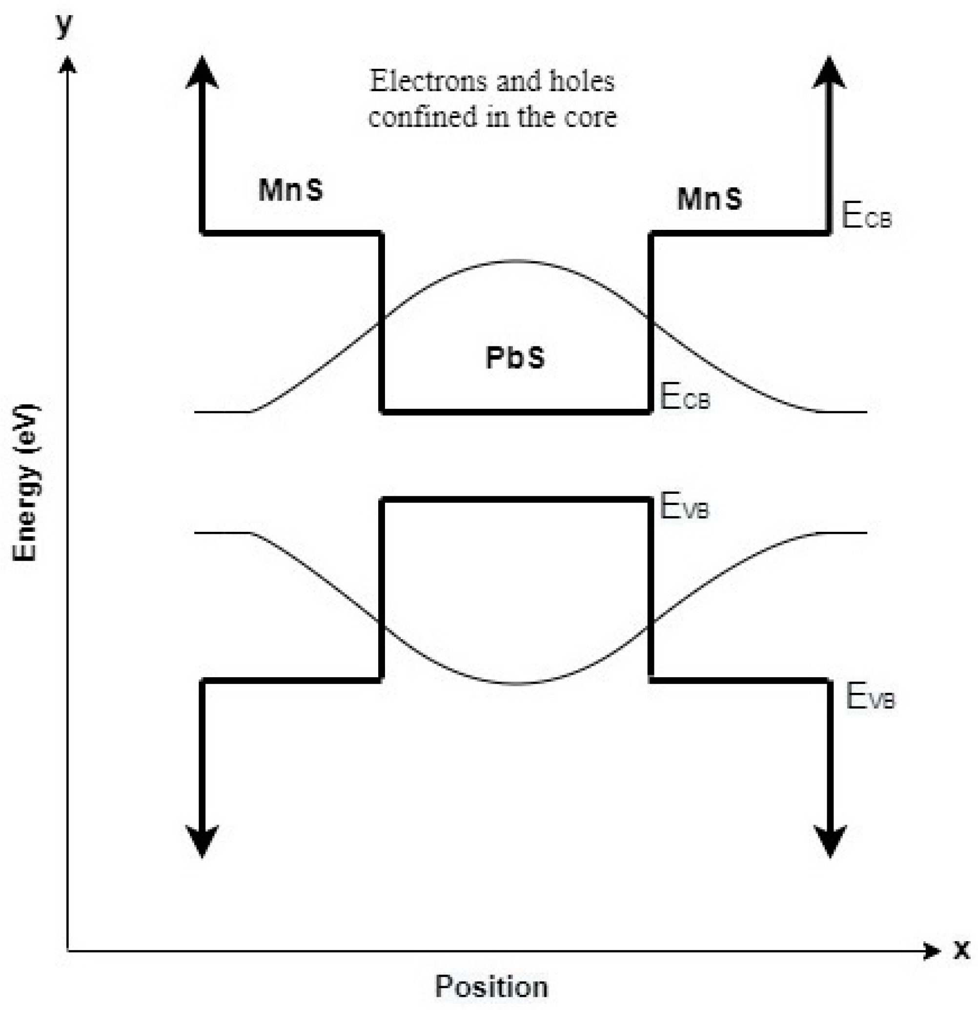
3.1. Quantum Confinement Effect in the PbS QDs and PbS/MnS Core Shell QDs

The effect of MnS shells on the PbS QDs as synthesized was investigated by observing the PL emissions of all samples at room temperature. Hence, different shell thicknesses of MnS shells showed different PL emissions compared to PbS QDs.
From Figure 2, it can be observed that the PL peak energy was blueshifted as the thickness of the MnS shell increased. The PL peak energies of the PbS QDs were centered at EPbS = 1.020 eV and gradually increased to 1.040, 1.051, 1.074, 1.080 and 1.082 eV for CS 0.3, CS 0.6, CS 0.9, CS 1.2 and CS 1.5, respectively. The significant blueshifts in PL emissions confirmed that MnS shells with different thicknesses were successfully grown, thus showing the effect of strong quantum confinement. In this case, overcoating the narrow band gap core material with the wider band gap shell material increased the confinement of charge carriers inside the core, causing the PL peak to shift to the higher energy level [29]. The blueshift of PL emissions after the presence of the shell was previously reported for type-I core shell QDs (CuInS2/Zn) [30]. In their case, the blueshift of a PL emission with the value of 130 nm was achieved due to the reduction of core size after cation exchange between the shell and core precursors. As in our PbS/MnS type-I core shell QDs, the charge carriers were concentrated in the PbS core and sandwiched between the large potential energy well of MnS, leading to fewer interactions of core-localized electron–hole pairs with surface traps than with bare QDs and thus increasing the PL peak energy. The band alignment of PbS/MnS core shell QDs is shown in Figure 3.
As the thickness of the MnS shell increased, the localization of charge carriers was increased, which translates to a strong carrier confinement and this is proven by a blueshift of PL peak energies. However, when optimum shell thickness is reached, the high energy excitons can tunnel through the core shell interface causing delocalization. This causes the confinement strength to be reduced as shown between CS 1.2 and CS 1.5 where their peak position shows a small shift of 0.2 meV.
Previous research also reported this effect on PbS/CdS type-I core shell QDs by using effective mass approximation with the single-band model [31]. It was stated that the addition of a CdS shell onto the PbS core QDs causes the radial probability of charge carriers to be shifted to the center of the core shell QD region, signaling stronger localization. This stronger carrier localization proves the stronger confinement which translates to higher transmission energy in PbS/CdS core shell QDs compared to their PbS core QD counterparts.

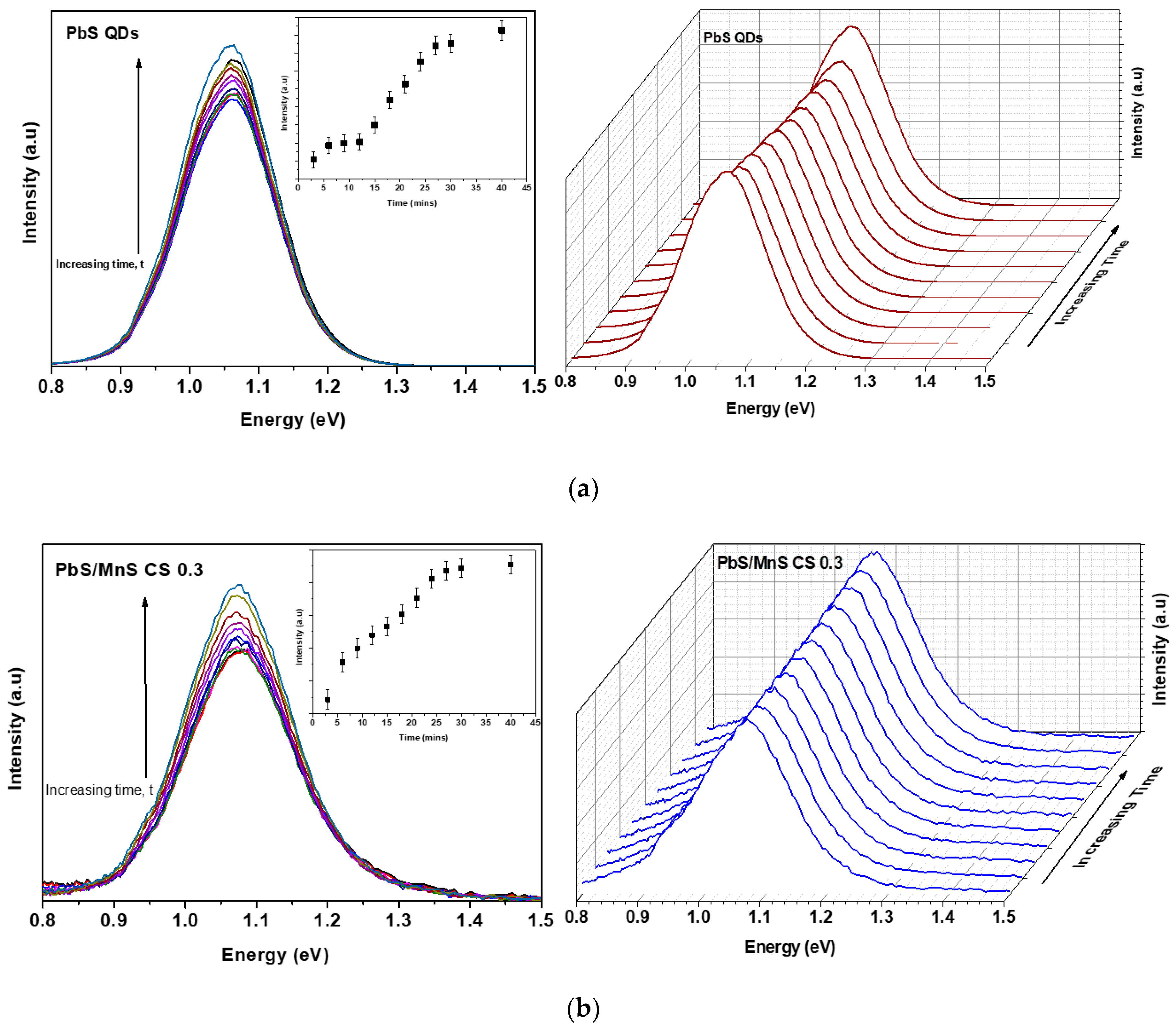
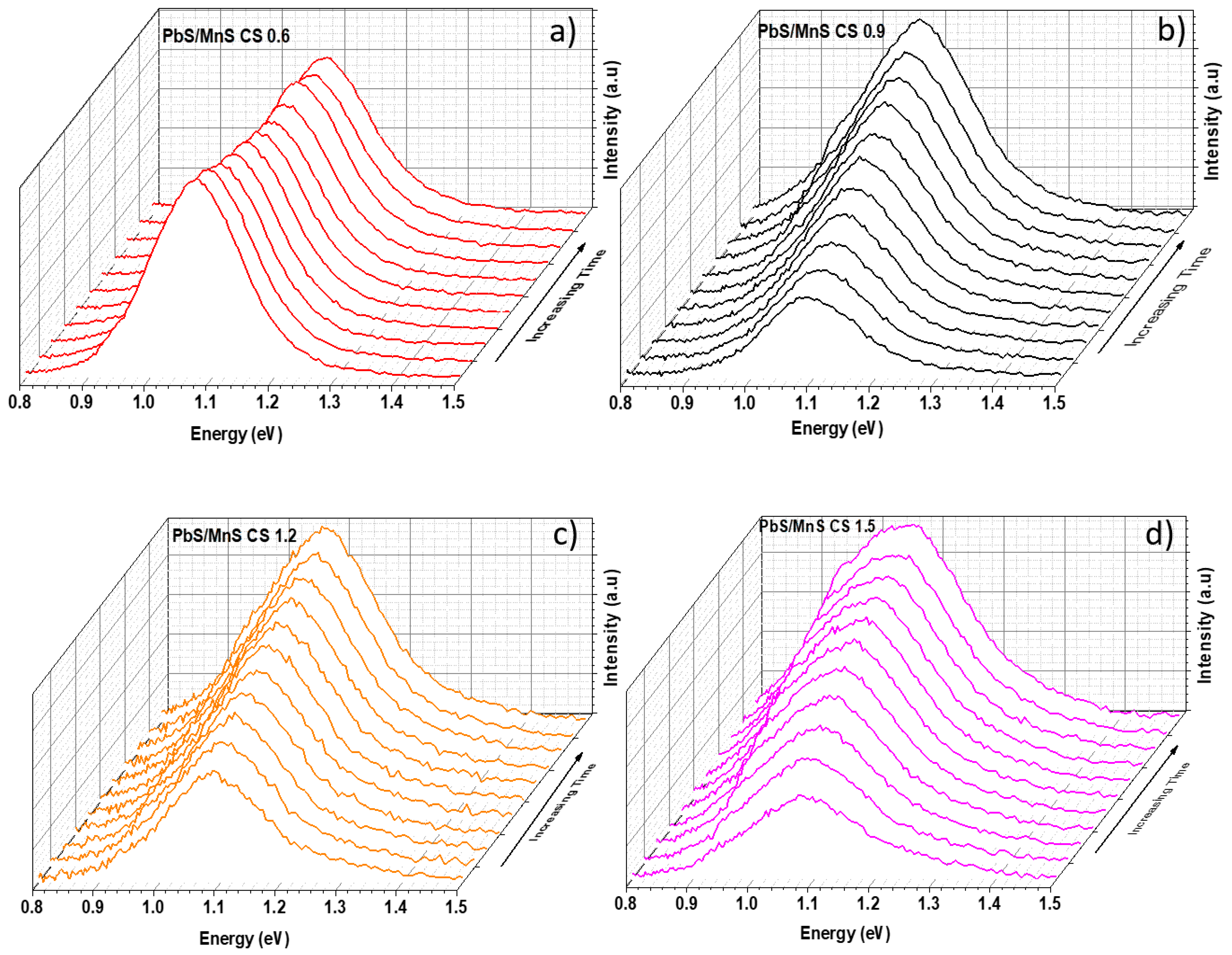
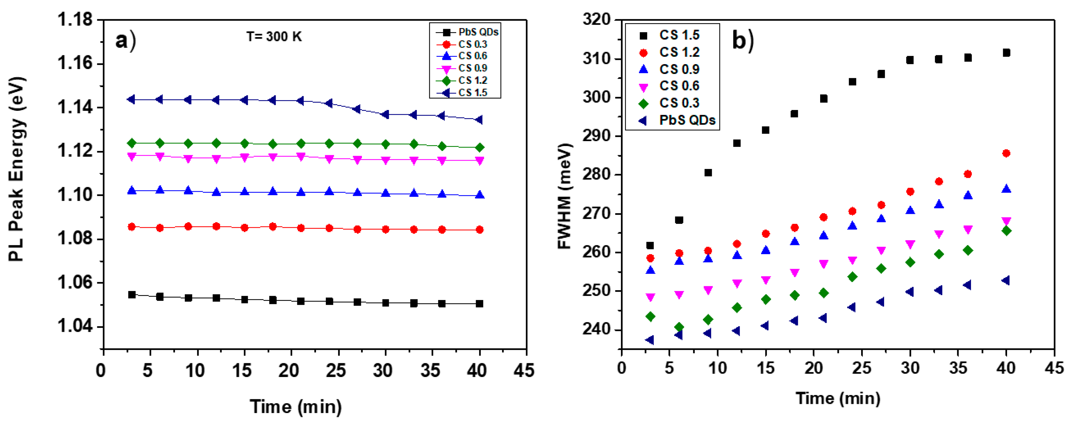
3.2. Photoenhancement of PbS QDs and PbS/MnS Core Shell QDs

Figure 4 and Figure 5 show the PL spectra of PbS QDs and PbS/MnS core shell QDs CS 0.3, CS 0.6, CS 0.9, CS 1.2 and CS 1.5. Generally, the PL peak intensities of the PbS QDs and PbS/MnS core shell QDs were continuously increased over a time of 40 min with no energy shifts observed in core shell samples except in CS 1.5.
It is well understood that the PL intensity enhancement depends on several factors, including the intensity of laser light, capping ligands and the presence of water [32]. Several experiments have been carried out with the aim of determining the atmospheric constituents involved in the activation procedure [33]. From our results, due to the fact that the PbS QDs and PbS/MnS core shell QDs were synthesized in an aqueous medium, it is likely because of the presence of oxygen in the aqueous solvent.
As discussed in the sample preparation section, the samples were synthesized in aqueous solution and the QDs were capped by the thiol ligands (TGL and DTG). Thiol ligands cause the PL intensity to be enhanced by smoothing the surface of the core QDs and removing surface defects. Previous research also reported the enhancement of PL intensity when water soluble QDs were exposed to continuous laser irradiation. This includes research from Wang et al., Kloepfer et al., and Ma et al., where they synthesized citrate-CdSe, 2-mercaptopropionic acid-CdSe and thioglycolic acid-CdTe, respectively [34,35,36]. However, their PL emission showed an inconsistent shift after continuous irradiation with a large energy shift of ~1.80–3.60 meV due to photocorrosion. Unlike in our samples that were capped by thiol ligands, PL peak energy of PbS showed a small shift (0.34 meV) and the behavior was improved with a MnS shell which showed a constant position that can be seen in Figure 6a.
In this work, the enhancement of PL intensity may be due to the adsorption of H2O molecules on the QD surface, and this pathway is the consequence of the stabilizing effect of H2O in coordination with the surface. In this case, at early illumination times, the water molecules adsorb on the surface of the QDs and passivate surface charge carrier traps thus increasing the exciton emission. The presence of oxygen in the aqueous solution speeds up PL activation as it causes a slow photocorrosion. This results in releasing Pb2+ based on the following reaction:
PbS + 2H2O + O2 →Pb2+ + SO42− + 4H+
where the presence of oxygen in water gives rises to the PL intensity [37]. While photoenhancement can be a simple phenomenon to improve the PL intensity of the QDs, this process is usually accompanied by a blueshift of the PL peak energy [33,38]. These effects are less pronounced in our samples, which could be advantageous particularly for biological marker applications where samples displayed promising constant positions of the PL peak energies after continuous irradiation with laser light.
Figure 6a shows a graph of PL peak energies of all samples with respect to time. It was noticed that the constant PL peak energies as the time increased were due to the passivation of capping ligands on the QDs. For PbS QDs, small shifts of 0.34 meV in the PL spectrum were detected after 40 min of continuous irradiation. Meanwhile, constant PL peak energies were obtained for PbS/MnS core shell QDs (CS 0.3, CS 0.6, CS 0.9, CS 1.2). However, the PL peak energy of PbS/MnS CS 1.5 was redshifted after 25 min. In this case, prolonged laser exposure caused the QDs to oxidize, which was due to the photooxidation process. Photooxidation of surface thiol groups (TGL and DTG) produces disulfide molecules which are water soluble and readily extracted from the surface of QDs and dissolved into the aqueous solution. This causes the hydrophobic surfaces of the QDs to be exposed to an aqueous environment, causing the QDs to stick together to form aggregates of QDs, hence increasing the QD size [39]. The increase in QD diameter due to MnS shell growth causes the PL peak energy to shift to a longer wavelength (redshift). In addition, this can be observed by the large broadening of the full width half maximum (FWHM) over time as shown in Figure 6b, indicating that either the size of PbS/MnS CS 1.5 was inconsistent or there was a wider size distribution after prolonged irradiation. In comparison with other samples, CS 1.5 showed a large linewidth shift with the value of ~50 meV after 40 min, almost twice the value of the other samples. In contrast, the FWHMs of CS 1.2, CS 0.9, CS 0.6, CS 0.3 and the PbS QDs showed a small linewidth shift with the values of 27, 21, 20, 22 and 15 meV, respectively. Furthermore, the redshifted PL peak energy of PbS/MnS CS 1.5 could be due to heat-induction by the laser light, also known as photoannealing. Laser light transmits heat and produces an increase in temperature of the sample, which induces the QDs to be ripened thus causing the redshift of PL peak energy.

4. Conclusions

In the present paper, we investigated the quantum confinement effect and photoenhancement of colloidal PbS QDs and PbS/MnS core shell QDs with various shell thicknesses after they were irradiated with continuous laser light for 40 min. At the beginning of the PL measurement, it was found that the PL peak energy was gradually increased with shell thickness due to the presence of MnS shells which confined charge carriers inside the core of PbS. As the thickness of the MnS shell increased from 0.3 to 1.5 ML, localization of charge carriers was also increased which translated to strong carrier confinement, and this was proven by a blueshift of PL peak energies up to 6 meV. After 40 min of continuous irradiation, the blueshifts of PL peak energies of 0.3 ML–1.5 ML PbS/MnS core shell samples were maintained as compared with PbS QDs. However, the enhancement of luminescence intensities was observed in PbS QDs and PbS/MnS core shell QDs. Oxygen present in the aqueous solvent accelerates PL activation as it induces a slow photocorrosion, thus increasing the PL intensity. Large broadening of the FWHM of PbS/MnS CS 1.5 with the value of ∼50 meV indicates wider size distribution. In comparison, the FWHMs of PbS/MnS CS 1.2, CS 0.9, CS 0.6, CS 0.3 and PbS QDs show a small linewidth shift with the values of 27, 21, 20, 22 and 15 meV, respectively. The effect of laser light on the photoluminescence of QDs can be considered as a useful standard for selection of appropriate nanocrystals for certain applications.

Author Contributions

M.S.Z. carried out the experiments and acquisition of data, analyzed and interpreted the data and prepared and revised the manuscript. J.Y.C.L. helped to draft and revise the manuscript. S.A.A.A. contributed in chemistry knowledge, participated in the design of the synthesis method and manuscript revision. A.R.M. contributed to the photoluminescence setup and manuscript revision. M.A.K. participated in designing the study, analyzing and interpreting the data and drafting a manuscript. All authors read and approved the final manuscript.

Funding

This work was supported by the Ministry of Higher Education Malaysia (FRGS/1/2018/STG07/UPM/02/14), (AKU254: HiCoE Phase 2), and Universiti Putra Malaysia (vot number: 9567500, 9664000).

Conflicts of Interest

The authors declare no conflict of interest.

References

  1. Manzoor, U.; Islam, M.; Tabassam, L.; Rahman, S.U. Quantum confinement effect in ZnO nanoparticles synthesized by co-precipitate method. Phys. E 2019, 41, 1669–1672. [Google Scholar] [CrossRef]
  2. Moreels, I.; Lambert, K.; Smeets, D.; Muynck, D.D.; Nollet, T.; Martins, J.C.; Vanhaecke, F.; Vantomme, A.; Delereu, C.; Allan, G.; et al. Size Dependent Optical Properties of Colloidal PbS Quantum Dots. ACS Nano 2009, 3, 3023–3030. [Google Scholar] [CrossRef] [PubMed] [Green Version]
  3. Binetti, E.; Striccoli, M.; Sibillano, T.; Giannini, C.; Brescia, R.; Falqui, A. Tuning light emission of PbS nanocrystals from infrared to visible range by cation exchange. Sci. Technol. Adv. Mater. 2015, 16, 055007. [Google Scholar] [CrossRef] [PubMed]
  4. Litvin, A.P.; Martynenko, I.V.; Purcell-Milton, F.; Baranov, A.V.; Fedorov, A.V.; Gunko, Y.K. Colloidal quantum dots for electronics. J. Mater. Chem. A 2017, 5, 13252. [Google Scholar] [CrossRef]
  5. Landry, M.L.; Morrell, T.E.; Karagounis, T.K.; Hsia, C.; Wang, C. Simple Syntheses of CdSe Quantum Dots. J. Chem. Educ. 2014, 91, 274–279. [Google Scholar] [CrossRef]
  6. Ahmad, W.; He, J.; Liu, Z.; Xu, K.; Chen, Z.; Yang, X.; Li, D.; Xia, Y.; Zhang, J.; Chen, C. Lead Selenide (PbSe) Colloidal Quantum Dot Solar Cells with >10% Efficiency. Adv. Mater. 2019, 31, 1900593. [Google Scholar] [CrossRef]
  7. Liu, H.; Zhong, H.; Zheng, F.; Xie, Y.; Li, D.; Wu, D.; Zhou, Z.; Sun, X.W.; Wang, K. Near-infrared lead chalcogenide quantum dots: Synthesis and applications in light emitting diodes. Chi. Phys. B 2019, 28, 12. [Google Scholar] [CrossRef] [Green Version]
  8. Wise, F.W. Lead Salt Quantum Dots: The Limit of Strong Quantum Confinement. Acc. Chem. Res. 2000, 33, 773–780. [Google Scholar] [CrossRef]
  9. Hu, L.; Zhang, Z.; Patterson, R.J.; Hu, Y.; Chen, W.; Chen, C.; Li, D.; Hu, C.; Ge, G.; Chen, Z.; et al. Achieving high-performance PbS quantum dot solar cells by improving hole extraction through Ag doping. Nano Energy 2018, 46, 212–219. [Google Scholar] [CrossRef]
  10. Sung, S.D.; Lim, I.; Kang, P.; Lee, C.; Lee, W.I. Design and development of highly efficient PbS quantum dot-sensitized solar cells working in an aqueous polysulfide electrolyte. Chem. Commun. 2013, 49, 6054. [Google Scholar] [CrossRef]
  11. Xu, J.; Wang, H.; Wang, Y.; Yang, S.; Ni, G.; Zou, B. Efficiency enhancement for solution-processed PbS quantum dots solar cells by inserting graphene oxide as hole-transporting and interface modifying layer. Org. Electron. 2018, 58, 270–275. [Google Scholar] [CrossRef]
  12. Jin, T.; Imamura, Y. Applications of Highly Bright PbS Quantum Dots to Non-Invasive Near-Infrared Fluorescence Imaging in the Second Optical Window. ECS J. Solid State Sci. Technol. 2015, 5, R3138. [Google Scholar] [CrossRef]
  13. Shulga, A.G.; Kahmann, S.; Dirin, D.N.; Graf, A.; Zaumseil, J.; Kovalenko, M.V.; Loi, M.A. Electroluminescence Generation in PbS Quantum Dot Light-Emitting Field-Effect Transistors with Solid-State Gating. ACS Nano 2018, 12, 12805–12813. [Google Scholar] [CrossRef] [Green Version]
  14. Chai, Q.; Zhou, H.; Lu, F. Enhanced infrared response of Si base p-n diode with assembled Ge QDs by thermal annealing. Appl. Surf. Sci. 2008, 254, 3376–3379. [Google Scholar]
  15. Uematsu, T.; Maenosono, S.; Yamaguchi, Y. Photoinduced Fluorescence Enhancement in Mono- and Multilayer Films of CdSe/ZnS Quantum Dots: Dependence on Intensity and Wavelength of Excitation Light. J. Phys. Chem. B 2005, 109, 8613–8618. [Google Scholar] [CrossRef]
  16. Jeong, S.; Achermann, M.; Nanda, J.; Ivanov, S.; Klimov, V.I.; Hollingsworth, J.A. Effect of Thiol-Thiolate Equilibrium on the Photophysical Properties of Aqueous CdSe/ZnS Nanocrystal Quantum Dots. J. Am. Chem. Soc. 2005, 127, 10126–10127. [Google Scholar] [CrossRef]
  17. Peng, H.; Zhang, L.; Soeller, C.; Travas-Sejdic, J. Preparation of water-soluble CdTe/CdS core/shell quantum dots with enhanced photostability. J. Lumin. 2007, 127, 721–726. [Google Scholar] [CrossRef]
  18. Zhibin, S.; Xi, Z.; Jing, Z.; Wansheng, L.; Weijie, G.; Cheng, L.; Tingzhu, W.; Yue, L.; Zhong, C. The Stability of Metal Halide Perovskite Nanocrystals—A Key Issue for the Application on Quantum-Dot-Based Micro Light-Emitting Diodes Display. Nanomaterials 2020, 10, 1375. [Google Scholar]
  19. Green, M. The nature of quantum dot capping ligands. J. Mater. Chem. 2010, 20, 5797–5809. [Google Scholar] [CrossRef]
  20. Jia, G. Excitons properties and quantum confinement in CdS/ZnS core/shell quantum dots. Opto. Adv. Mater. 2011, 5, 738–741. [Google Scholar]
  21. Vasudevan, D.; Gaddam, R.R.; Trinchi, A.; Cole, I. Core-shell quantum dots: Properties and applications. J. Alloys Compd. 2015, 636, 395–404. [Google Scholar] [CrossRef]
  22. Bao, H.; Gong, Y.; Li, Z.; Gao, M. Ehancement Effect of Illumination on the Photoluminescence of Water-Soluble CdTe Nanocrystals: Toward Highly Fluorescent CdTe/CdS Core-Shell Structure. Chem. Mater. 2004, 20, 3853–3859. [Google Scholar] [CrossRef]
  23. Shavel, A.; Gaponik, N.; Eychmuller, A. Efficient UV-Blue Photoluminescing Thiol-Stabilized Water-Soluble Alloyed ZnSe(S) Nanocrystals. J. Phys. Chem. B 2004, 108, 5905–5908. [Google Scholar] [CrossRef]
  24. Zhang, T.; Zhao, H.; Riabinina, D.; Chaker, M.; Ma, D. Concentration-Dependent Photoinduced Photoluminescence Enhancement in Colloidal PbS Quantum Dot Solution. J. Phys. Chem. C 2010, 114, 10153–10159. [Google Scholar] [CrossRef]
  25. Gaponik, N.; Talapin, D.V.; Rogach, A.L.; Hoppe, K.; Shevchenko, E.V.; Kornowski, A.; Eychmuller, A.; Weller, H. Thiol-Capping of CdTe Nanocrystals: An Alternative to Organometallic Synthetic Routes. J. Phys. Chem. B 2002, 106, 7177–7185. [Google Scholar] [CrossRef]
  26. Guo, J.; Yang, W.; Wang, C. Systematic Study of the Photoluminescence Dependence of Thiol-Capped CdTe Nanocrystals on the Reaction Conditions. J. Phys. Chem. B 2005, 109, 17467–17473. [Google Scholar] [CrossRef]
  27. Carolina-Carrion, C.; Cardenas, S.; Simonet, B.M.; Valcarcel, M. Quantum dots luminescence enhancement due to illumination with UV/Vis light. Chem. Commun. 2009, 35, 5214–5226. [Google Scholar] [CrossRef]
  28. Zaini, M.S.; Kamarudin, M.A.; Chyi, J.L.Y.; Ahmad, S.A.A.; Mohmad, A.R. Temperature and Power Dependence of Photoluminescence in PbS Quantum Dots Nanoparticles. Sains Malays. 2019, 48, 1281–1288. [Google Scholar] [CrossRef]
  29. Reiss, P.; Protiere, M.; Li, L. Core/Shell Semiconductor Nanocrystals. Small 2009, 5, 154–168. [Google Scholar] [CrossRef]
  30. Park, J.; Kim, S.W. CuInS2/ZnS core/shell quantum dots by cation exchange and their blue-shifted photoluminescence. J. Mater. Chem. 2011, 21, 3745–3750. [Google Scholar] [CrossRef]
  31. Shelawati, T.; Nurisya, M.S.; Tim, C.K.; Mazliana, A.K. Effects of step-potential on confinement strength of strain-induced type-I core-shell quantum dots. Superlattices Microstruct. 2019, 131, 95–103. [Google Scholar]
  32. Manna, L.; Scher, E.C.; Li, L.; Alivisatos, A.P. Epitaxial Growth and Photochemical Annealing of Graded CdS/ZnS Shells on Colloidal CdSe Nanorods. J. Am. Chem. Soc. 2002, 124, 7136–7145. [Google Scholar] [CrossRef] [PubMed]
  33. Jones, M.; Nedeljkovic, J.; Ellingson, R.J.; Nozik, A.J.; Rumbles, G. Photoenhancement of Luminescence in Colloidal CdSe Quantum Dot Solutions. J. Phys. Chem. B 2003, 107, 11346–11352. [Google Scholar] [CrossRef]
  34. Wang, Y.; Tang, Z.; Correa-duarte, M.A.; Liz-marza, L.M.; Kotov, N.A. Multicolor Luminescence Patterning by Photoactivation of Semiconductor Nanoparticle Films. J. Am. Chem. Soc. 2003, 125, 2830–2831. [Google Scholar] [CrossRef]
  35. Kloepfer, J.A.; Mielke, R.E.; Wong, M.S.; Nealson, K.H.; Stucky, G.; Nadeau, J.L. Quantum Dots as Strain-and Metabolism-Specific Microbiological Labels. Appl. Environ. Microb. 2003, 69, 4205–4213. [Google Scholar] [CrossRef] [PubMed] [Green Version]
  36. Ma, J.; Chen, J.; Guo, J.; Wang, C. Improvement of the photostability of thiol-capped CdTe quantum dots in aqueous solutions and living cells by surface treatment. Nanotechnology 2006, 17, 5875–5881. [Google Scholar] [CrossRef]
  37. Wang, Y.; Tang, Z.; Correa-Duarte, M.A.; Pastoriza-Santos, I.; Giersig, M.; Kotov, N.A.; Liz-Marzan, L.M. Mechanism of Strong Luminescence Photoactivation of Citrate-Stabilized Water-Soluble Nanoparticles with CdSe Cores. J. Phys. Chem. B 2004, 108, 15461–15469. [Google Scholar] [CrossRef]
  38. Zhelev, Z.; Jose, R.; Nagase, T.; Ohba, H.; Bakalova, R.; Ishikawa, M.; Baba, Y. Enhancement of the photoluminescence of CdSe quantum dots during long-term UV-irradiation: Privilege or fault in life science research. Mater. Today 2004, 75, 99–105. [Google Scholar] [CrossRef]
  39. Sato, K.; Kojima, S.; Hattori, S.; Chiba, T.; Ueda-Sarson, K.; Torimoto, T.; Tachibana, Y.; Kuwabata, S. Controlling surface reactions of CdS nanocrystals: Photoluminescence activation, photoetching and photostability under light irradiation. Nanotechnology 2007, 18, 465702. [Google Scholar] [CrossRef]
Figure 1. The schematic diagram of the preparation of PbS/MnS core shell quantum dots (QDs). (a) Preparation of Mn2+ precursor and (b) preparation of PbS/MnS core shell QDs.
Applsci 10 06282 g001
Figure 2. Room temperature photoluminescence (PL) spectra of the colloidal PbS QDs and PbS/MnS core shell QDs. The presence of the MnS shell causes PL peak energy to blueshift.
Applsci 10 06282 g002
Figure 3. Schematic representation of the band alignment of the PbS/MnS core shell QDs.
Applsci 10 06282 g003
Figure 4. PL spectra of PbS core QDs (a) showing enhancement of PL intensity, including the intensity trend over time (inset). PL spectra of PbS/MnS CS 0.3 (b) exposed to laser of power P = 100 mW and time exposure for 40 min at room temperature.
Applsci 10 06282 g004
Figure 5. PL spectra of PbS/MnS (a) CS 0.6, (b) CS 0.9, (c) CS 1.2 and (d) CS 1.5 exposed to laser of power p = 100 mW and time exposure up to 40 min at room temperature.
Applsci 10 06282 g005
Figure 6. (a) Graph of PL peak energies over time exposure to the laser, p = 100 mW. (b) Dependence of full width half maximum (FWHM) of the CS 1.5 PL spectrum on the irradiation time.
Applsci 10 06282 g006

