Glutathione Activates Type III Secretion System Through Vfr in Pseudomonas aeruginosa
 Yani Zhang1
  Chao Zhang1
  Xiao Du1
  Yun Zhou1
  Weina Kong1
  Gee W. Lau2
  Gukui Chen1
  Gurjeet Singh Kohli3,4
  Liang Yang3,5
  Tietao Wang1
  Haihua Liang1*
- 1Key Laboratory of Resource Biology and Biotechnology in Western China, College of Life Sciences, Northwest University, Ministry of Education, Xi'an, China
- 2Department of Pathobiology, University of Illinois at Urbana-Champaign, Champaign, IL, United States
- 3Singapore Centre for Environmental Life Sciences Engineering, Nanyang Technological University, Singapore, Singapore
- 4Alfred Wegener-Institut Helmholtz-Zentrum für Polar- und Meeresforschung, Bremerhaven, Germany
- 5School of Medicine, Southern University of Science and Technology, Shenzhen, China
Glutathione (GSH) is the most abundant antioxidant in all living organisms. Previously, we have shown that a deletion mutant in the glutathione synthetase gene (ΔgshB) decreases the expression of type III secretion system (T3SS) genes of Pseudomonas aeruginosa. However, the mechanism remains elusive. In this study, a comprehensive transcriptomic analysis of the GSH-deficient mutant ΔgshAΔgshB was used to elucidate the role of GSH in the pathogenesis of P. aeruginosa. The data show that the expression of genes in T3SS, type VI secretion system (T6SS) and some regulatory genes were impaired. ΔgshAΔgshB was attenuated in a mouse model of acute pneumonia, swimming and swarming motilities, and biofilm formation. Under T3SS inducing conditions, GSH enhanced the expression of T3SS in both wild-type PAO1 and ΔgshAΔgshB, but not in Δvfr. Genetic complementation of Δvfr restored the ability of GSH to induce the expression of T3SS genes. Site-directed mutagenesis based substitution of cysteine residues with alanine in Vfr protein abolished the induction of T3SS genes by GSH, confirming that GSH regulates T3SS genes through Vfr. Exposure to H2O2 decreased free thiol content on Vfr, indicating that the protein was sensitive to redox modification. Importantly, GSH restored the oxidized Vfr to reduced state. Collectively, these results suggest that GSH serves as an intracellular redox signal sensed by Vfr to upregulate T3SS expression in P. aeruginosa. Our work provides new insights into the role of GSH in P. aeruginosa pathogenesis.
Introduction
Pseudomonas aeruginosa is a Gram-negative opportunistic pathogen that cause acute and chronic infections (Gellatly and Hancock, 2013). Protein secretion systems are essential for bacteria to intimately interface with other bacteria and the host, particularly in the efficiency of infection (Filloux, 2011). Large multicomponent secretion apparatus, including both type III secretion system (T3SS) and type VI secretion system (T6SS), facilitate P. aeruginosa infection by directly injecting effector proteins into the cytoplasm of host cells to subvert the host immune response. Four known T3SS effectors that have been identified are ExoS, ExoT, ExoY, and ExoU. Once injected into the host cells, these effectors disrupt cytoskeletal organization and host cell membranes, resulting in cytotoxicity and death (Brutinel et al., 2008).
The ExsA of P. aeruginosa is the central regulator of the of T3SS gene expression, and is encoded by the last gene of the exsCEBA polycistronic mRNA (Yahr and Wolfgang, 2006). In response to varieties of environmental signals (e.g., low Ca2+) and direct contact with eukaryotic host cells, ExsA autoregulates its own transcription through direct activation of the exsC promoter (Brutinel et al., 2008). Under non-inducing conditions (e.g., high Ca2+), ExsE and ExsC form a complex, allowing the anti-activators ExsD to bind to ExsA and inhibit transcription activation. Under inducible or low Ca2+ conditions, ExsE is secreted, which in turn, free ExsC to form a complex with ExsD, freeing ExsA to activate the expression of T3SS genes (King et al., 2012). Additionally, T3SS expression is also influenced by ~30 extrinsic gene products, including the Vfr (Diaz et al., 2011). Vfr is a member of the 3′, 5′-cAMP receptor protein that serves as a global transcriptional regulator of P. aeruginosa (Fuchs et al., 2010). Prior genomic and proteomic studies demonstrate that more than 100 genes and 60 proteins in P. aeruginosa are positively or negatively regulated by Vfr (Suh et al., 2002; Wolfgang et al., 2003). Importantly, a recent study showed that Vfr directly binds to the promoter of exsA and activates its transcription (Marsden et al., 2016).
Glutathione (GSH) is the most abundant intracellular thiol and antioxidant that protects cells against free radicals and reactive oxygen intermediates in living organisms. GSH is synthesized from three amino acid precursors L-glutamate, L-cysteine, and glycine by a two-step process involving the enzymes γ-glutamate-cysteine ligase (GshA) and GSH synthetase (GshB) (Lushchak, 2012). GshA catalyzes the ligation of cysteine with glutamate to form γ-glutamylcysteine. GshB ligates γ-glutamylcysteine with glycine to generate glutathione. Previously, we have shown that P. aeruginosa mutant strain deficient in GshB expresses lower level of T3SS (Zhang and Wei, 2009). Additionally, GSH could alter P. aeruginosa's sensitivity to different antibiotics (Zhang and Duan, 2009). In this study, we discover that the GSH biosynthesis deficient mutant ΔgshAΔgshB expresses significantly lower level of T3SS. Further study supports a model in which the global regulator Vfr senses GSH and activates the expression of T3SS in P. aeruginosa.
Materials and Methods
Bacterial Strains and Growth Conditions
All bacterial strains, plasmids and primers used in this study are listed in Tables S1, S2. Bacteria were maintained in Luria Bertani (LB) broth or Pseudomonas Isolation Agar at 37°C. Antibiotics were added to the growth medium as needed: for E. coli: 15 μg/ml gentamicin (Gm), 50 μg/ml kanamycin (Kan), 100 μg/ml ampicillin (Amp), and 10 μg/ml tetracycline (Tet) in LB medium; For P. aeruginosa strain PAO1: 50 μg/ml Gm in LB medium or 150 μg/ml in Pseudomonas isolation agar (PIA), and 300 μg/ml trimethoprim (Tmp) and 500 μg/ml carbenicillin (Cb) in LB medium. Depletion of intracellular GSH in P. aeruginosa was performed with 1.5 mM diethylmaleate (DEM). Biofilm formation in flow chamber was performed in ABTG medium (1.51 mM (NH4)2SO4, 3.37 mM Na2HPO4, 2.2 mM KH2PO4, 5 mM NaCl, 1 mM MgCl2·6H2O, 100 μM CaCl2·2H2O, 1 μM FeCl3·6H2O, and supplemented with 0.4 g/l Glucose).
Construction of Plasmids and Deletion Mutants
The ΔgshB was as previously described (Zhang and Duan, 2009) (Table S1). For deletion of the gshA gene, a 1,948 bp fragment upstream of the start codon and a 1,966 bp fragment downstream of the stop codon flanking the gene were PCR amplified and digested with HindIII-XbaI and XbaI-EcoRI, respectively. The two fragments were then ligated into pEX18Tc plasmid, which was digested with EcoRI and HindIII, resulting in pEX18Tc-gshA, in which the whole gshA open reading frame (ORF) had been deleted. The resultant plasmid was electroporated into PAO1, followed by selection for single crossovers and double crossovers as described previously (Kong et al., 2015). Both ΔexsA and Δvfr were generated by using a similar strategy. Deletions of the gshA gene in ΔgshB mutant strain, and the vfr gene in ΔgshAΔgshB mutant strain were constructed using similar strategy. Successful deletions were verified by PCR.
To genetically complement the ΔgshB mutant, the promoter and coding regions of the gshB locus was PCR amplified from PAO1 genomic DNA using primers listed on Table S2, and integrated into ΔgshB genome at the attB site using the mini-CTX1 system (Hoang et al., 2000). The result was confirmed by PCR amplification. The complemented strain was named ΔgshB/gshB.
To genetically complement the Δvfr, a 916 kb fragment containing the vfr coding regions with its own promoter was PCR amplified from PAO1 genomic DNA using primers listed in Table S2, digested with BamHI and HindIII, and then ligated into a shuttle vector, pAK1900 (Poole et al., 1993), resulting in pAK-vfr. pAK-gshB was constructed using the similar strategy.
The plasmid pMS402 with a promoterless luxABCDE gene cluster was used to construct the lux reporter strains (Duan et al., 2003). For examples, the exoS promoter region were amplified by PCR and cloned into the pMS402, yielding pKD-exoS. Similar strategy was used to clone the promoters of exoY, exsA, exsC, and vfr in front of the promoterless lux, respectively. Apart from the plasmid-based lux reporter system, an integration plasmid CTX 6.1 derived from the plasmid mini-CTX-lux was used to construct the chromosomal fusion reporter (Becher and Schweizer, 2000). The pMS402 fragment containing the MCS kanamycin-resistance marker and the promoter-lux reporter cassette were ligated into CTX 6.1, yielding CTX-promoter-lux and electroporated into P. aeruginosa recipient strains. All constructs were sequenced to verify that no mutations had been incorporated.
H2O2 Susceptibility Measurements
For the agar diffusion assay, P. aeruginosa strains were cultured overnight in LB broth and adjusted to an optical density OD600 of 0.1, then, 20 μl of bacteria was suspended in 30 ml of LB soft agar (0.7% agar), mixed, and incubated on agar plates for 30 min. Then, 5 μl of 30% H2O2 was spotted onto each disk prepopulated on the agar plates containing the bacteria. After overnight incubation at 37°C, inhibition zones were measured. All experiments were performed in triplicate.
Growth Studies
P. aeruginosa strains were cultivated in LB medium overnight and then diluted 1:100 in fresh media and grown in 96-well plates with shaking at 37°C. Bacterial growth (OD600) was measured every 2 h for 24 h and each strain was assayed in triplicate.
Gene-Expression Assays
The level of gene expression was monitored by measuring luminescence production in black 96-well plates with transparent bottoms. Both luminescence (counts per second) and bacterial growth (OD595) were measured every 30 min for 24 h in a Victor2 Multilabel Counter (Perkin-Elmer) as previously described (Zhang et al., 2017).
Swimming, Swarming, and Twitching Motility Assays
Swimming and swarming motilities were assayed as described (Rashid and Kornberg, 2000). Swimming media consisting of 0.3% agar supplemented with 1% tryptone and 0.5% NaCl. Swarming media consisting of 0.4% agar supplemented with 0.8% nutrient broth and 0.5% glucose. Twitching medium consisting of 1% agar supplemented with 1% tryptone, 0.5% yeast extract and 1% NaCl. P. aeruginosa cultures were diluted to an OD600 of 0.1, and 2 μl of the diluted bacteria was center-spotted onto the surface of the agar plates. After the absorption of spotted bacterial inoculum, swimming plates were incubated at 30°C for 14–16 h, while swarming and twitching plates were incubated at 37°C for 14–16 h. Bacterial motility was imaged using a camera (LAS-3000, Tanon, China).
Biofilm Formation Assays
Biofilm formation in LB was performed as previously described (O'Toole and Kolter, 1998) with minor modifications. Briefly, 800 μl of the diluted bacterial suspension (OD600 = 0.1) was incubated in 24-well polystyrene plates. Three milliliters of the diluted bacteria were also incubated in borosilicate tubes to visualize biofilms. After 30 h of incubation at 37°C without shaking, planktonic bacterial cultures were gently removed. The plates and tubes were triple-washed with distilled water, and the adhered bacteria were stained with 0.1% crystal violet for 20 min. Then, the plates and tubes were washed with water to remove unbound dye and air dried. To quantify the biofilms on the 24-well polystyrene plate, 800 μl of 95% ethanol was added to each well to elute the crystal violet. The absorbance of the pooled eluent was measured at 590 nm by a microplate reader (Infinite M200 PRO, TECAN, Switzerland). Experiments were performed in triplicate, and results are shown as the mean ± s.d.
Development of biofilms in a flow chamber were performed as previously described (Chua et al., 2016). Various gfp-tagged P. aeruginosa strains were cultured overnight in 2 ml of LB medium in 37°C with shaking condition (200 rpm). The overnight cultures were diluted to 1:100 with ABTG medium and 500 μl of the diluted cultures were injected into the flow chambers and incubated for 1 h incubation in an inverted position without medium flow (Sternberg and Tolker-Nielsen, 2006). Then, the flow chambers were placed in upright position before the bacteria were fed with ABTG medium via Masterflex peristaltic pump (Cole-Parmer, United States) at the rate of 4 ml/h in 37°C to allow biofilm development. After 72 h, 300 μl of 3.34 μM SYTO9 (LIVE/DEAD™ BacLight™ Bacterial Viability Kit, ThermoFisher Scientific) was injected into each chamber to stain the bacteria for 15 min before subjected to confocal microscopy. The images of the SYTO9-stained biofilms were acquired using an LSM 780 confocal laser scanning microscope (Carl Zeiss, Germany) fitted with 20×/0.8 DICII objective lens and 488 nm argon multiline laser. Acquired images were further processed using the IMARIS Version 8 software (Bitplane AG, Zurich, Switzerland) to obtain the volume of biofilms.
Mouse Model of Acute Pneumonia Infection
All animal experiments were performed in strict accordance with the Regulations for the Administration of Affairs Concerning Experimental Animals approved by the State Council of People's Republic of China (11-14-1988). All animal procedures were approved by the Institutional Animal Care and Use Committee (IACUC) of the college of life sciences of Northwest University with a permit number: NW-02-2014. Acute lung infection was performed as described previously (Li et al., 2014). Briefly, stationary phase P. aeruginosa grown in LB were harvested, washed three times, and serially diluted in PBS and infected intranasally (1 × 10⁷ CFU) into the 6-week old male CD-1 mice. For bacterial burden determination, infected mice (n = 6) were euthanized at 16 h post-infection. Mouse lungs were aseptically removed and homogenized in 1 ml cold PBS with a Dounce homogenizer. Homogenates were serial diluted and plated on Pseudomonas isolation agar. Colony forming units (CFU) were determined after 24 h of incubation at 37°C. For histopathological analyses, lungs were inflated with 1 ml 10% neutral buffered formalin, harvested and immersed in the same solution for 24 h prior to paraffin and sectioning. Paraffin embedded sections (4- to 8-mm thickness) were stained with hematoxylin and eosin (H&E).
RNA-seq and Data Analyses
Identification of the GSH regulons in P. aeruginosa genome by RNA-seq was performed as previously described (Deng et al., 2014). Briefly, 10 ml of mid-log-phase of PAO1 and ΔgshAΔgshB strains were collected. An RNeasy minikit (Qiagen) was used for RNA purification with DNase I treatment. After removing rRNA by using the MICROBExpress kit (Ambion), mRNA was used to generate the cDNA library according to the TruSeq RNA sample prep kit protocol (Illumina), and sequenced using the HiSeq 2000 System. Bacterial RNA-seq reads were mapped to the P. aeruginosa genomes by using TopHat (version 2.0.0), with two mismatches allowed. Only the uniquely mapped reads were kept for subsequent analyses. The analysis of differentially expressed genes was performed using Cuffdiff software (version 2.0.0). The data have been uploaded to NCBI under BioProject (accession number PRJNA504392).
RNA Extraction and Quantitative Real-Time PCR
Overnight cultures of P. aeruginosa were diluted 1:100 with fresh LB broth and recultured until OD600 0.6. RNA was isolated from bacteria using the RNAprep pure cell/bacteria Kit (Tiangen Biotech, Beijing, China). cDNA was synthesized using the PrimeScript Reverse Transcriptase (TaKaRa) with random primers, then subjected to qRT-PCR using the SYBR Premix Ex Taq II (TaKaRa). The 30s ribosomal protein gene rpsL was used as an internal control. Data presented were averages of three independent cultures grown on different days.
Western Blot Assays
The production of ExoS was analyzed as previously described (Bleves et al., 2005). Overnight P. aeruginosa cultures were subcultured with 100-fod dilution in fresh LB containing 5 mM EGTA and 20 mM MgCl2 for 3 h. Bacterial supernatant and pellet were separated by centrifugation. Bacterial supernatant was treated with 15% trichloroacetic acid (TCA) at 4°C to precipitate proteins. Precipitated proteins were collected by centrifugation and washed with acetone. Then, protein samples from equivalent numbers of bacterial cells were loaded and separated on 12% SDS-PAGE gels. Proteins were transferred onto a polyvinylidene difluoride (PVDF) membrane and hybridized with a rabbit polyclonal antibody against ExoS generously provided by Professor Weihui Wu (Nankai University). The signal was detected by an ECL Plus kit (Amersham Biosciences). All Western blot assays were repeated a minimum of three times with independently derived proteins samples. Representative blots are shown.
Site-Directed Mutagenesis
Alanine (GCT) substitution of Cys20, Cys38, Cys97, Cys156, and Cys183 in Vfr was performed by using a site-directed mutagenesis kit (TransGen Biotech). All mutagenesis primers are listed in Table S2.
Protein Purification of Vfr
A P. aeruginosa vfr expression plasmid ptac-vfr (Table S1) was introduced into P. aeruginosa PA103 for Vfr purification as described, with modifications (Dasgupta et al., 2002). Briefly, cultures were grown to the density of OD600 0.6, and then isopropyl-β-D-thiogalactopyranoside (IPTG) was added to a final concentration of 1 mM to induce Vfr expression. Following an additional 7 h incubation at 37°C, bacteria were harvested by centrifugation at 10,000 × g for 4 min at 4°C. The cell pellet was resuspended in buffer A (80 mM KCl, 50 mM K3PO4, pH 7.2) with 10 mM PMSF at a ratio of 5 ml/g of cells (wet weight) and lysed by sonication in an ice bath. Lysates were centrifuged (11,000 rpm for 60 min at 4°C) to remove unbroken cells and insoluble materials. All subsequent steps were performed at 4°C. Soluble materials were filtered through a 0.45 μm filter and applied to a HiTrap HP column (GE Healthcare) equilibrated with buffer A. The column was washed with 10% buffer B (1M KCl, 50 mM K3PO4, pH 7.2) and eluted with a linear gradient from 10% to 100% buffer B over 40 ml using an ÄKTA Prime. Peak fractions were collected and enriched, and then load onto a Gel Filtration column (GE Healthcare) equilibrated with buffer A. Vfr-containing fractions were identified by SDS-PAGE, pooled and stored at −80°C until further use.
Detection Thiol Status of Vfr Protein With AMS
Cysteine thiols in protein were irreversibly modified with 4-acetamido-4′-maleiniDystil-bene-2,2′-disufonic acid (AMS), which adds 0.5 kDa mass to every modified cysteine (Si et al., 2015). The increase in protein mass was evident as delay of electrophoretic mobility of AMS-labeled proteins. Briefly, Vfr proteins were incubated with or without 1 mM DTT at room temperature for 30 min, and excess DTT was removed by ultrafiltration. GSH (15 mM) and H2O2 (500 mM) treated protein were analyzed with same strategy. The resulting proteins were incubated with 30 mM AMS for 30 min and then separated by non-reducing SDS-PAGE.
Circular Dichroism Spectroscopy Studies
CD measurements were performed by using a spectrometer (Chirascan V100, Applied Photophysics, England) equipped with a 1 mm path length quartz cell according to a method described previously (Kessenbrock and Groth, 2017). The CD spectra were recorded in the range from 190 to 260 nm. The helical content of protein was calculated based on change in molar ellipticity value.
Statistical Analysis
All experiments were performed in triplicate and independently repeated three times. Data were analyzed using an Unpaired Student's t-test and ANOVA, and expressed as the mean ± standard deviation. The differences between means were considered be significant at p < 0.05.
Results
ΔgshB and ΔgshAΔgshB Mutant Exhibits Motility and Biofilm Formation Defects
Our previous study revealed that GSH alters the sensitivity of P. aeruginosa to different antibiotics, as well as modulates the expression of T3SS genes (Zhang and Duan, 2009; Zhang and Wei, 2009). Reduction in the expression of T3SS may reduce the virulence of GSH-deficient P. aeruginosa mutant. gshA and gshB are responsible for GSH biosynthesis in P. aeruginosa. In other bacteria, such as E. coli and L. monocytogenes, ΔgshB strain lacks reduced GSH but instead accumulates the glutathione precursor compound γ-glutamylcysteine which partially functionally replace GSH (Fuchs and Warner, 1975; Gopal et al., 2005). Here, to investigate the role of GSH in P. aeruginosa, γ-glutamate-cysteine ligase mutant (ΔgshA) and double gshA gshB mutant (ΔgshAgshB) were constructed. ΔgshAgshB strain lacking both glutathione and γ-glutamylcysteine. When cultured in LB broth, ΔgshA, ΔgshB, and ΔgshAΔgshB mutant strains showed similar growth kinetics as PAO1 (Figure S1A). Because GSH is a major intracellular antioxidant protecting against environmental stresses, we assessed the susceptibility of these mutants to H2O2 in vitro. When compared to PAO1, ΔgshB and ΔgshAΔgshB were more susceptible to H2O2, while ΔgshAgshB mutant completely lacking GSH described greater susceptibility to H2O2 than ΔgshB mutant (Figure S1B; Table S4), confirming the protective function of GSH against oxidative stress and injury.
To determine the effects of GSH on P. aeruginosa pathogenesis, we compared the motility and biofilm of ΔgshB and ΔgshAΔgshB against the wild-type PAO1. Both ΔgshB and ΔgshAΔgshB were attenuated in swarming, swimming and twitching motility. The genetically complemented strain ΔgshB/gshB showed wild-type levels of motilities, suggesting that defects in motility were due to the lack of GSH (Figure 1A).
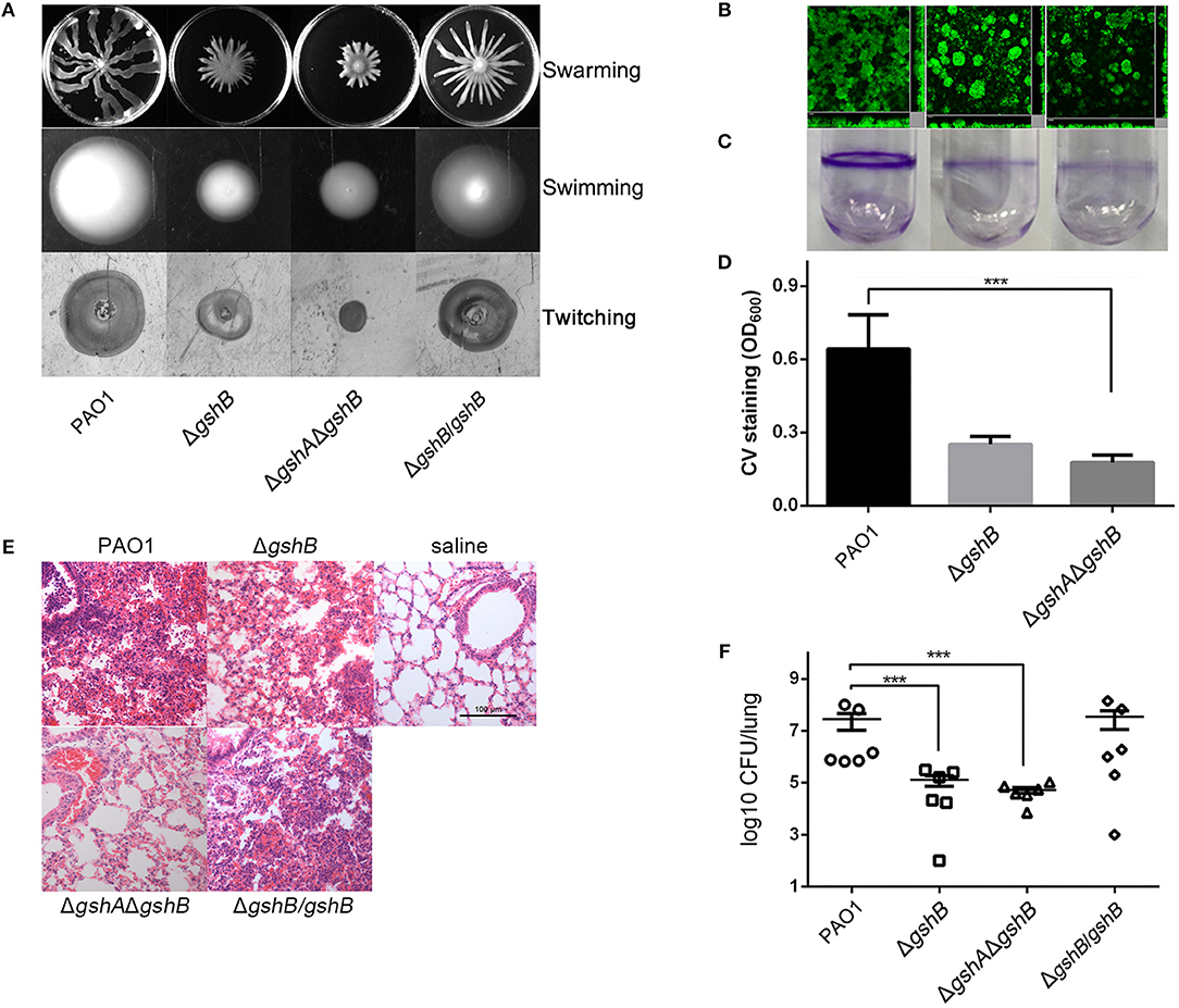
Figure 1. GSH is important in P. aeruginosa pathogenicity. (A) ΔgshB and ΔgshAΔgshB exhibited weaker swimming, swarming, and twitching motilities. The experiments were repeated at least three times with similar results. Images from one typical experiment are shown. (B) Biofilms of PAO1, ΔgshB and ΔgshAΔgshB grown in flow cells for 72 h and examined by confocal laser scanning microscopy. Experiments were performed in triplicate independently for three times. Representative images are shown. (C,D) Biofilm formation of PAO1, ΔgshB and ΔgshAΔgshB on borosilicate tube surface and quantified by the crystal violet (CV) staining methods. The results are averages based on three replicate tubes. (E) H&E stained mouse lungs 16 h post-infection by indicated P. aeruginosa strains. (F) ΔgshB and ΔgshAΔgshB are attenuated in acute pneumonia infection. Bacterial burden in CD-1 mouse lungs (n = 6) after 16 h of infection by various P. aeruginosa strains. Data represent average CFU/lung ± standard deviations. ***p < 0.001, when compared PAO1 to ΔgshB and ΔgshAΔgshB.
Biofilm formation is partially dependent on bacterial swarming motility (Shrout et al., 2006; Pamp and Tolker-Nielsen, 2007; Chow et al., 2011). We examined whether GSH deficiency impacted the ability of ΔgshB and ΔgshAΔgshB to produce biofilms. In the flow cell system, ΔgshB and ΔgshAΔgshB synthesized significantly lower amounts of biofilms than wild-type PAO1 (Figure 1B). Similarly, the crystal violet method revealed that biofilms formed by ΔgshB and ΔgshAΔgshB were between 2- and 3-fold lower than PAO1 (Figures 1C,D). These results suggest that the loss of GSH biosynthesis impairs both motility and biofilm formation of P. aeruginosa.
ΔgshB and ΔgshAΔgshB Are Attenuated in a Mouse Model of Acute Pneumonia
Defective motility and biofilm formation in ΔgshB and ΔgshAΔgshB suggest that GSH modulates P. aeruginosa pathogenicity. Therefore, we investigated whether both ΔgshB and ΔgshAΔgshB were attenuated in lung infection. As shown in Figure 1E, when compared to the saline vehicle control group, PAO1-infected lungs showed more severe inflammation with intense infiltration of proinflammatory cells and hemorrhaging involving small bronchi, bronchioles, and alveoli. In contrast, inflammatory cell recruitment was greatly reduced in mouse lungs infected with both ΔgshB and ΔgshAΔgshB. Additionally, the ΔgshAΔgshB mutant induced less severe pneumonia than the ΔgshB. Similar to the histopathological observation, the bacterial burden in mouse lungs infected with PAO1 were significantly higher than those infected by ΔgshB or ΔgshAΔgshB, with a median value of 2.8 × 10⁷ CFU/lung. Conversely, the average bacterial burden in mice infected with ΔgshB and ΔgshAΔgshB was approximately 100-fold and 200-fold lower, at 1.27 × 10⁵ and 5.24 × 10⁴ CFU/lung, respectively (Figure 1F). Genetic complementation of ΔgshB (ΔgshB/gshB) restored the lost virulence. Finally, ΔgshA was also attenuated in mouse model of acute pneumonia (Figure S1C). Collectively, these results indicate that GSH plays crucial role in conferring full virulence in P. aeruginosa.
Transcriptomic Analysis of the ΔgshAΔgshB
In attempt to have a comprehensive gauge of the function of GSH in P. aeruginosa, we compared the transcriptome of ΔgshAΔgshB to its parental wild-type PAO1 by RNA-seq analysis. A total of 676 genes with altered transcript levels were identified, with 294 genes showing an increase and 382 genes showing a decrease of least 2-fold (i.e., log2 = 2) (Table S3). The gshA/gshB deficiency altered the expression of numerous genes coding for proteins involved in regulatory functions, pathogenesis, translation, transporter, T3SS, T6SS, and metabolism (Figure 2A). To assess the reliability of the RNA-seq approach, we used the identical total RNA samples and determined by RT-qPCR the mRNA levels of one upregulated gene (PA5530) and five downregulated genes (chpA, hcp1, katA, nosR, ppyR). Expression levels of these genes closely resembled those obtained from the RNA-seq experiment (Figure 2B).
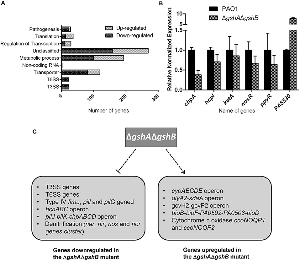
Figure 2. Comparison of the expression of selective genes between wild type PAO1 and ΔgshAΔgshB. (A) Functional categorization of differentially expressed genes according to their annotated function. Numbers of genes whose expression were altered in ΔgshAΔgshB compared to PAO1 were shown. (B) RT-PCR analysis of selective genes in PAO1 vs. ΔgshAΔgshB. All data shown were means ± SEM. RT-PCR assays were performed in triplicate. (C) Summary of major gene categories upregulated or repressed by GSH in P. aeruginosa.
Differentially expressed genes were classified into functional categories according to the PseudoCAP designations and information from other studies. Among the 676 genes, expression of those genes involved in motility, T3SS, T6SS, secreted factors (toxins, enzymes and alginate), and denitrification (nar, nir, nos, and nor) were repressed whereas expression of genes involved in quinol oxidase (cyo cluster), cytochrome c oxidases (ccoNOQP1 and ccoNOQP2), and amino acid biosynthesis were induced (Filiatrault et al., 2005) (Figure 2C). The nar, nir, and nos genes are required for anaerobic growth. In contrast, the cyo cluster, ccoNOQP1, and ccoNOQP2 are required for aerobic growth (Wu et al., 2005; Frangipani et al., 2008). Quinol oxidase operon and cytochrome c oxidases function as a low affinity terminal oxidase under high-oxygen, aerobic respiration conditions (Frangipani et al., 2008). These results suggest GSH modulates the expression of genes involved in respiratory pathways in P. aeruginosa. Importantly, the expression of 27 genes in T3SS secretion system, including exsA, exsD, exoY, exoS, pcrD, pcrG, pcrV, pscS, pscP, pscQ, and pscBCDEFGHIJ was reduced in ΔgshAΔgshB when compared to wild-type strain. Similarly, 28 genes involved in T6SS secretion were repressed in ΔgshAΔgshB (range from 2- to 6.5-fold), including both Hcp1 secretion island I (HSI-1) (PA1656-PA1690) and HSI-II (icmF1, ppkA, PA0086-PA0091) (Chen et al., 2015) (Table 1). Collectively, these results suggested that GSH regulates the expression of numerous virulence genes in P. aeruginosa. For the remainder of this study, we performed detailed analysis of the regulation of T3SS by GSH.
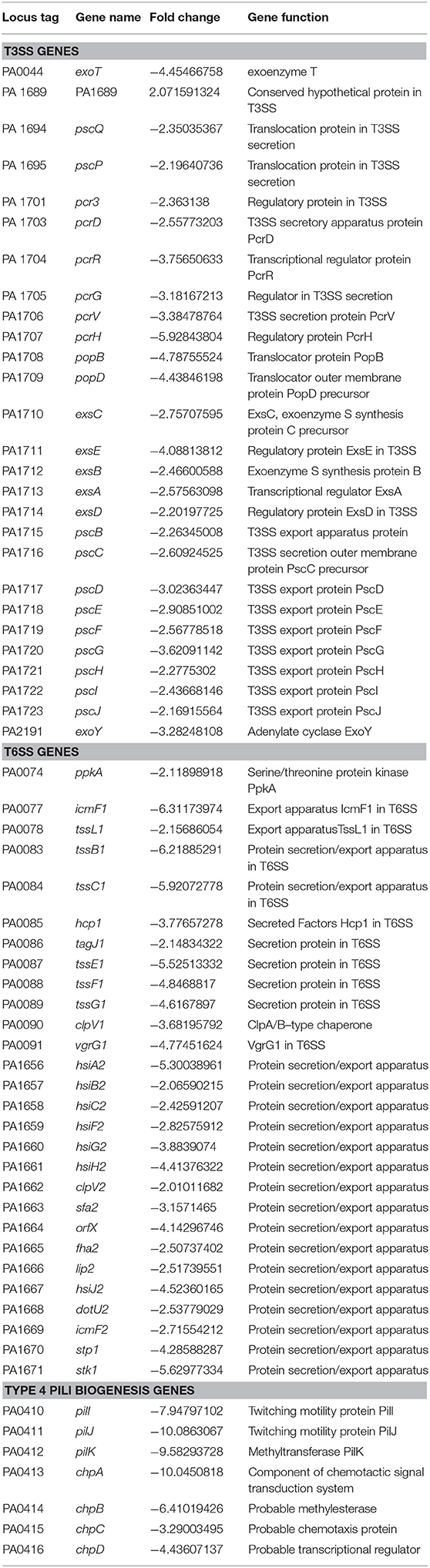
Table 1. Genes with altered expression in ΔgshAΔgshB when compared to parental wild-type PAO1.
GSH Upregulates T3SS Expression Through Vfr
We monitored the expression of T3SS genes, exoS and exoY, under inducing conditions (LB broth supplemented with 5 mM EGTA and 20 mM MgCl2). Under low Ca2+ conditions, the transcription of exoS (Figure 3A) and exoY (Figure S2) genes were significantly reduced in both ΔgshB and ΔgshAΔgshB compared to wild-type PAO1. Additionally, the expression of exoS in ΔgshAΔgshB was lower than that in ΔgshB (Figure 3A). The expression of exoS was partially restored in ΔgshB complemented with pAK-gshB (Figure 3B). To verify the involvement of GSH in T3SS expression, bacterial strains were treated with diethylmaleate (DEM) to deplete intracellular GSH. Significantly, the expression of exoS in DEM-treated PAO1 was reduced to levels comparable to that of untreated ΔgshAΔgshB mutant (Figure 3A). In contrast, addition of GSH significantly increased the expression of exoS by 2-fold in wild-type PAO1 (Figure 3A). The amount of secreted ExoS mirrored that of mRNA expression (Figure 3C). These results indicate that GSH plays an important role in regulating the expression of exoS.
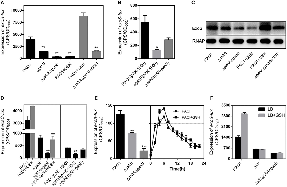
Figure 3. GSH upregulates the expression of T3SS genes through Vfr. All P. aeruginosa strains carrying plasmids, exoS-lux, PexsC-lux, and PexsA-lux were grown under T3SS inducing conditions (5 mM EGTA, 20 mM MgCl2) with or without GSH. The results shown are mean ± SEM. (A) The expression of exoS in the indicated lux reporter strains. (B) The expression of exoS in the genetically-complemented ΔgshB strain. (C) ExoS level in the indicated P. aeruginosa strains. Overnight bacterial cultures were subcultured in fresh media with or without 10 mM GSH for 3 h. Whole-cell extracts were resolved by SDS-PAGE and subjected to Western blotting. (D) The expression of PexsC in PAO1, ΔgshB, ΔgshAgshB and genetically-complemented ΔgshB strains in the presence or absence of 10 mM GSH. (E) The expression of PexsA in PAO1, ΔgshB and ΔgshAΔgshB. (F) The expression of exoS was induced by GSH in Δvfr and ΔvfrΔgshAΔgshB. *P < 0.05, **P < 0.01, ***P < 0.001, as determined by Student's t-test.
ExsA is the central regulator of T3SS gene expression. Previous studies have shown that EsxA binds to the promoters of T3SS genes and activates their transcription (Brutinel et al., 2008). exsA is co-transcribed with exsC, exsE, and exsB in the same operon under the control of the exsC promoter (PexsC) (Yahr and Frank, 1994). ExsA binds PexsC promoter and autoregulates its own expression. A recent study has identified a second promoter (PexsA) located immediately upstream of exsA (Marsden et al., 2016). Thus, we examined whether the induction of exoS by GSH was under the control of both PexsC and PexsA promoter. To facilitate the analysis, PexsC-lux and PexsA-lux plasmid was introduced into both ΔgshB and ΔgshAΔgshB. As expected, PexsC-lux and PexsA-lux reporter activity were drastically reduced when compared to PAO1 (Figures 3D,E). Genetic complementation of ΔgshB (ΔgshB/gshB) restored the expression of the exsCEBA operon. Importantly, the transcription of exsCEBA could be activated by GSH (Figure 3D). Because ExsA is an activator that feedback regulates its own operon, the loss of the exsCEBA transcription in the ΔgshB and ΔgshAΔgshB mutants was likely caused by reduced exsA expression. These results suggest that GSH is directly or indirectly regulating the expression of the exsCEBA operon.
An earlier study has shown that Vfr directly activates exsA transcription by binding to the promoter PexsA in P. aeruginosa (Marsden et al., 2016). Based on the aforementioned results, we speculated that the expression of T3SS modulated by GSH might be associated with Vfr. We generated a Δvfr mutant to examine this hypothesis. As expected, provision of GSH did not alter the expression of exoS gene in Δvfr (Figure 3F). Additionally, the ΔvfrΔgshAΔgshB triple mutant also failed to upregulate T3SS expression in the presence of exogenously provided GSH (Figure 3F). Importantly, the expression of exoS was significantly increased by GSH in Δvfr complemented strain (Figure 5A). Collectively, these results suggest that GSH regulate the expression of TSSS gene through Vfr.
In addition to Vfr and ExsA, the global transcriptional regulators RetS, LadS, and GacAS also regulate the expression of T3SS genes through the cAMP/Vfr pathway. RetS and LadS activities are mediated through the GacS/GacA/RsmZ. GacS activates the response regulator GacA by phosphotransfer. In turn, phosphorylated GacA positively regulates the expression of the two small RNAs, RsmY and RsmZ, which antagonize the activity of the regulator RNA-binding protein RsmA. Upregulation of rsmY and rsmZ and deletion of rsmA all result in the inhibition of the T3SS (Bordi et al., 2010). To clarify the relationship among GSH, GacS/GacA and T3SS, we examined the promoter activities of gacS, gacA, rsmY, and rsmZ in the ΔgshB. When compared to PAO1, a lower expression of gacA, rsmY, and rsmZ but not gacS was observed in ΔgshB and ΔgshAΔgshB (Figure 4A). In addition, the expression of gacA could be restored to wild-type level in complemented strain ΔgshB/gshB (Figure 4B). These results indicated that the GSH upregulates the expression of GacA in P. aeruginosa. Due to contradictory finding in the regulation of GacA and GacS, more studies are needed to determine the impact of GSH on GacA/GacS-mediated expression of T3SS.
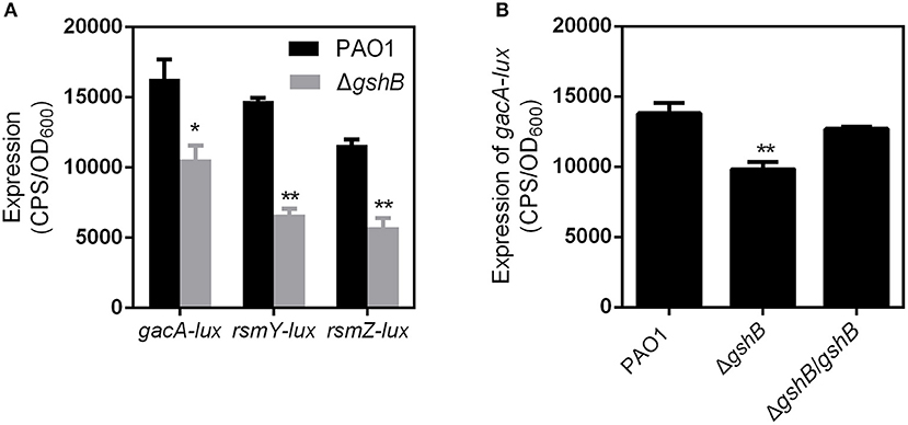
Figure 4. The expression of vfr, gacA, rsmY, and rsmZ genes in the indicated P. aeruginosa strains. (A) The expression of gacA, rsmY, and rsmZ genes in the PAO1 and ΔgshB strains. (B) The expression of gacA in the ΔgshB genetically-complemented strains. *P < 0.05, **P < 0.01 compared to PAO1 by Student's t-test.
Mutation of Cysteine Residues on Vfr Inhibits its Ability to Induce the Expression of T3SS Genes During Exposure to GSH
Recently studies have suggested GSH can function as a cofactor for several enzymes. In L. monocytogenes, GSH is a cofactor of PfrA and increases its activity at target genes (Reniere et al., 2015). Additionally, VirA, the main regulator of T6SS1 in Burkholderia pseudomallei, upregualtes virulence when VirA dimers are converted to monomers in the presence of GSH (Wong et al., 2015). The cysteine residues on both PrfA and VirA proteins were important for proper functioning and sensing of GSH. For Vfr, the five cysteine residues are located on positions 20, 38, 97, 156, and 183. To determine whether cysteine residues in the Vfr of P. aeruginosa were directly involved in the sensing of intracellular GSH, we performed cysteine-to-alanine substitution on each cysteine on pAK-vfr by site-directed mutagenesis. The plasmids containing mutated variants of vfr were introduced into Δvfr strain containing CTX-exoS-lux. Interestingly, all mutants have increased basal expression of exoS in the absence of exogenously supplied GSH, with VfrC20A and VfrC156A displayed the most significantly increase, followed by VfrC38A, VfrC97A, and VfrC183A (Figure 5A). Addition of GSH further enhanced exoS expression in all vfr mutants, suggesting all 5 cysteine residues were involved in GSH sensing (Figure 5A). Therefore, we generated a Vfr in which all five cysteine residues were mutated to alanine [Vfr(C/A)5]. Significantly, no increase in exoS expression was observed upon addition of GSH, indicating that intracellular GSH induced T3SS expression through Vfr (Figure 5A).
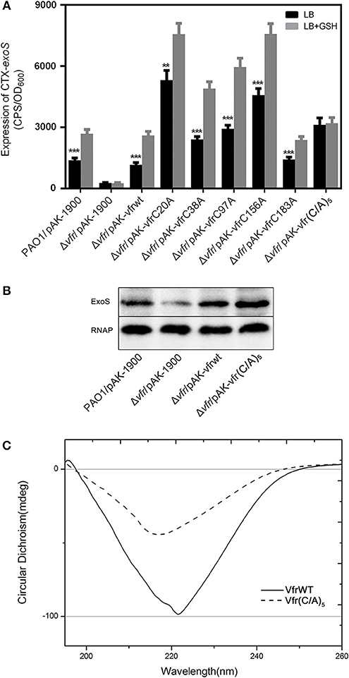
Figure 5. Cysteine residues in Vfr involved in signal transmission of glutathione. All P. aeruginosa strains carrying exoS-lux plasmids was grown under T3SS-inducing conditions (5 mM EGTA, 20 mM MgCl2) with or without 10 mM GSH. (A) Effect of cysteine-to-alanine mutation at positions 20, 38, 97, 156, and 183 of recombinant Vfr on the expression of exoS. (B) Secretion of ExoS in the indicated strains. **P < 0.01, ***P < 0.001 compared to PAO1 by Student's t-test. (C) Circular dichroism (CD) spectroscopy analysis of the secondary structure of Vfr and the Vfr (C/A)5.
Because we observed higher expression and secretion of exoS when cysteine residues of Vfr were substituted with alanine (Figures 5A,B), we assessed whether these pheotypes could be attributed by the conformation change of Vfr(C/A)5. Circular dichroism (CD) experiments were performed to understand the secondary and tertiary structure characteristics of wild type Vfr and Vfr(C/A)5. The far CD profile of wild-type Vfr exhibited a typical shape corresponding to a α-helical rich secondary structure revealing a broad negative minimum at 222 nm (Figure 5C). In the Vfr(C/A)5, the peak showed a decrease in negative ellipticities of the CD spectrum, which was a characteristic of secondary structure conformation change, indicating that cysteine residues were important for Vfr native conformation. Hence, the increased exoS expression in Vfr(C/A)5 was likely due to the conformation change.
Oxidized Vfr Is Reduced by GSH
Cysteine residues on proteins are the most sensitive amino acid to oxidative modification. Also, they are important for protein conformation, function, stress response, and signal transduction inside the cell (Kalinina et al., 2014). Thiol-mediated redox control in cell metabolism is attributed to the ability of the cysteine-thiol to reversibly change its redox state that modulate changes of protein structure, catalytic and regulatory activities (Meyer and Hell, 2005). We suspected that the five cysteine residues on Vfr might be involved in thiol-mediated redox regulation of Vfr function. The redox state of cysteine-thiol in wild-type Vfr protein was examined by detection of free thiol groups using the AMS covalent modification method. As shown in Figure 6A, the migration of non-DTT-treated, AMS-modified Vfr (lane 2) were same as DTT-treated, AMS-modified Vfr (lane 3) on non-reducing sodium dodecyl sulfate polyacrylamide gel electrophoresis (SDS-PAGE), indicating that all cysteine residues on the wild-type Vfr were in the thiol state. To determine whether cysteine-thiol could be oxidized, the Vfr protein was incubated with and without H2O2 and analyzed by AMS modification on a non-reducing SDS-PAGE. As shown in Figure 6B, H2O2-treated, AMS-modified Vfr (lane 4) migrated faster than its untreated counterpart (lane 2), suggesting that there was a lack of thiol group after H2O2 exposure, and one or more cysteine-thiols might have been oxidized.
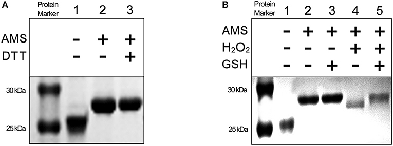
Figure 6. The redox state of cysteine thiol groups in Vfr under reducing and oxidizing conditions. Free thiol groups were covalently modified with AMS. Protein samples were resolved by using 15% non-reducing SDS-PAGE. (A) Vfr proteins (8 μM) treated with or without DTT (1 mM). (B) Vfr proteins (8 μM) treated with or without GSH (15 mM) and H2O2 (500 mM).
The cellular GSH is important for redox homeostasis and maintains the redox state of protein thiol groups (Meyer and Hell, 2005). We speculated that GSH might restore the oxidized form of Vfr to the reduced form. Following exposure to H2O2, oxidized Vfr proteins were treated with GSH. As expected, oxidized Vfr treated with GSH and modified with AMS (lane 5) had the same electrophoretic mobility as the untreated Vfr (lane 2) (Figure 6B). These results indicate that oxidized Vfr thiols can be restored to their reduced state in a reducing environment by GSH. Collectively, these data demonstrate that Vfr senses GSH-dependent redox environment to modulate the expression of T3SS genes.
Discussion
We have previously shown GSH is important for the expression of P. aeruginosa T3SS genes. In this study, we examined the mechanism underlying the regulation of T3SS by GSH. Our results clearly demonstrate that GSH is a critical signaling molecule that regulates T3SS gene expression through the activation of Vfr. Vfr-deficient mutant fails to activate T3SS gene expression even in the presence of GSH, suggesting that it is a critical factor mediating signal transduction of GSH-mediated gene modulation. Our finding is further validated by the finding that when all cysteine residues of Vfr were substituted with alanine, the expression of T3SS could not be induced by GSH in Δvfr [pAK-Vfr(C/A)5], suggesting these cysteine residues are directly involved in the sensing of GSH.
As a defensive measure, bacteria use redox sensitive transcription factors to sense and respond to oxidative stress (Lee et al., 2018). The thiol groups of cysteine residues in these proteins sense the redox state via reversible or irreversible modifications. Previous studies showed that several representative bacterial thiol-based redox-sensing proteins (VirA, OxyR, DksA, SpxA1, QsrA, OhrR/2-Cys, Spx, YodB, CrtJ, and CprK) regulate gene expression and play important roles during infection (Antelmann and Helmann, 2011; Wong et al., 2015). For example, the OxyR of P. aeruginosa senses cellular H2O2 level. High level of H2O2 cause cysteine residues in OxyR to form disulfide bonds and activate the transcription of antioxidant defense genes (Jo et al., 2015). However, when compared to OxyR, the VirA of B. pseudomallei works in opposite manner, where the oxidized dimers is reduced to its active monomeric form by GSH (Wong et al., 2015). GSH contributes to expression of L. monocytogenes virulence factors via allosteric binding to PrfA (Reniere et al., 2015). Similar to VirA, we propose that inactive, oxidized form of Vfr is converted to reduced active form in the presence of GSH. Therefore, Vfr in both ΔgshB and ΔgshAΔgshB is in the oxidized state and therefore, is unable to self-activates vfr transcription because of inability to synthesize GSH. Provision of GSH to ΔgshB and ΔgshAΔgshB restores Vfr activity through a reduction reaction that induces the expression of T3SS genes. Collectively, our results suggest that Vfr senses intracellular GSH levels to modulate the expression of ExsA and the downstream T3SS genes.
Our data show that the loss of gshB and gshA attenuates many virulence-associated phenotypes, including swimming, swarming and twitching motilities, biofilm formation, and virulence during acute pneumonia. RNA-seq analysis further corroborated that a number of genes, including ppyR, migA, PilG-pilK, chpA-chpD, associated with biofilm and motilities, were down regulated in ΔgshAΔgshB (Attila et al., 2008; Lau et al., 2009). These phenotypes are consistent with a recently published study (Wongsaroj et al., 2018). Finally, in the course of our studies, we have also discovered that the expression of a subset of known T6SS genes are decreased in ΔgshAΔgshB, suggesting that GSH also modulates the expression of T6SS system. However, the regulatory mechanism remains unclear.
In conclusion, the present work demonstrates that GSH represents a critical signaling molecule that activates the virulence of P. aeruginosa.
Author Contributions
YaZ and HL: conception and design of the study, analysis, and interpretation of data, drafting the manuscript. CZ, XD, YuZ, WK, GC, GK, LY, and TW: acquisition of data and analysis of data. GL: critically revised the manuscript. All the authors participated the idea discussion and reviewed the manuscript.
Funding
This work was supported by the National Nature Science Foundation of China (31622003, 31670080, 31670060) and Nature Science Foundation of Shaanxi Province, China (2017JM3037, 2016JZ007).
Conflict of Interest Statement
The authors declare that the research was conducted in the absence of any commercial or financial relationships that could be construed as a potential conflict of interest.
Supplementary Material
The Supplementary Material for this article can be found online at: https://www.frontiersin.org/articles/10.3389/fcimb.2019.00164/full#supplementary-material
References
Antelmann, H., and Helmann, J. D. (2011). Thiol-based redox switches and gene regulation. Antioxid. Redox Signal. 14, 1049–1063. doi: 10.1089/ars.2010.3400
Attila, C., Ueda, A., and Wood, T. K. (2008). PA2663 (PpyR) increases biofilm formation in Pseudomonas aeruginosa PAO1 through the psl operon and stimulates virulence and quorum-sensing phenotypes. Appl. Microbiol. Biotechnol. 78, 293–307. doi: 10.1007/s00253-007-1308-y
Becher, A., and Schweizer, H. P. (2000). Integration-proficient Pseudomonas aeruginosa vectors for isolation of single-copy chromosomal lacZ and lux gene fusions. Biotechniques 29, 948–950, 952. doi: 10.2144/00295bm04
Bleves, S., Soscia, C., Nogueira-Orlandi, P., Lazdunski, A., and Filloux, A. (2005). Quorum sensing negatively controls type III secretion regulon expression in Pseudomonas aeruginosa PAO1. J. Bacteriol. 187, 3898–3902. doi: 10.1128/JB.187.11.3898-3902.2005
Bordi, C., Lamy, M. C., Ventre, I., Termine, E., Hachani, A., and Fillet, S., et al. (2010). Regulatory RNAs and the HptB/RetS signalling pathways fine-tune Pseudomonas aeruginosa pathogenesis. Mol. Microbiol. 76, 1427–1443. doi: 10.1111/j.1365-2958.2010.07146.x
Brutinel, E. D., Vakulskas, C. A., Brady, K. M., and Yahr, T. L. (2008). Characterization of ExsA and of ExsA-dependent promoters required for expression of the Pseudomonas aeruginosa type III secretion system. Mol. Microbiol. 68, 657–671. doi: 10.1111/j.1365-2958.2008.06179.x
Chen, L., Zou, Y., She, P., and Wu, Y. (2015). Composition, function, and regulation of T6SS in Pseudomonas aeruginosa. Microbiol. Res. 172, 19–25. doi: 10.1016/j.micres.2015.01.004
Chow, S., Gu, K., Jiang, L., and Nassour, A. (2011). Salicylic acid affects swimming, twitching and swarming motility in Pseudomonas aeruginosa, resulting in decreased biofilm formation. J. Exp. Microbiol. Immunol. 15, 22–29. Available online at: https://www.microbiology.ubc.ca/sites/default/files/roles/drupal_ungrad/JEMI/15/JEMI15_22-29.pdf
Chua, S. L., Yam, J. K. H., Hao, P., Adav, S. S., Salido, M. M., and Liu, Y., et al. (2016). Selective labelling and eradication of antibiotic-tolerant bacterial populations in Pseudomonas aeruginosa biofilms. Nat. Commun. 7:10750. doi: 10.1038/ncomms10750
Dasgupta, N., Ferrell, E. P., Kanack, K. J., West, S. E., and Ramphal, R. (2002). fleQ, the gene encoding the major flagellar regulator of Pseudomonas aeruginosa, is sigma70 dependent and is downregulated by Vfr, a homolog of Escherichia coli cyclic AMP receptor protein. J. Bacteriol. 184, 5240–5250. doi: 10.1128/JB.184.19.5240-5250.2002
Deng, X., Liang, H., Ulanovskaya, O. A., Ji, Q., Zhou, T., and Sun, F., et al. (2014). Steady-state hydrogen peroxide induces glycolysis in Staphylococcus aureus and Pseudomonas aeruginosa. J. Bacteriol. 196, 2499–2513. doi: 10.1128/JB.01538-14
Diaz, M. R., King, J. M., and Yahr, T. L. (2011). Intrinsic and Extrinsic Regulation of type III secretion gene expression in Pseudomonas Aeruginosa. Front. Microbiol. 2:89. doi: 10.3389/fmicb.2011.00089
Duan, K., Dammel, C., Stein, J., Rabin, H., and Surette, M. G. (2003). Modulation of Pseudomonas aeruginosa gene expression by host microflora through interspecies communication. Mol. Microbiol. 50, 1477–1491. doi: 10.1046/j.1365-2958.2003.03803.x
Filiatrault, M. J., Wagner, V. E., Bushnell, D., Haidaris, C. G., Iglewski, B. H., and Passador, L. (2005). Effect of anaerobiosis and nitrate on gene expression in Pseudomonas aeruginosa. Infect. Immun. 73, 3764–3772. doi: 10.1128/IAI.73.6.3764-3772.2005
Filloux, A. (2011). Protein Secretion Systems in Pseudomonas aeruginosa: an essay on diversity, evolution, and function. Front. Microbiol. 2, 155. doi: 10.3389/fmicb.2011.00155
Frangipani, E., Slaveykova, V. I., Reimmann, C., and Haas, D. (2008). Adaptation of aerobically growing Pseudomonas aeruginosa to copper starvation. J. Bacteriol. 190, 6706–6717. doi: 10.1128/JB.00450-08
Fuchs, E. L., Brutinel, E. D., Jones, A. K., Fulcher, N. B., Urbanowski, M. L., and Yahr, T. L., et al. (2010). The Pseudomonas aeruginosa Vfr regulator controls global virulence factor expression through cyclic AMP-dependent and -independent mechanisms. J. Bacteriol. 192, 3553–3564. doi: 10.1128/JB.00363-10
Fuchs, J. A., and Warner, H. R. (1975). Isolation of an Escherichia coli mutant deficient in glutathione synthesis. J. Bacteriol. 124, 140–148.
Gellatly, S. L., and Hancock, R. E. (2013). Pseudomonas aeruginosa: new insights into pathogenesis and host defenses. Pathog. Dis. 67, 159–173. doi: 10.1111/2049-632X.12033
Gopal, S., Borovok, I., Ofer, A., Yanku, M., Cohen, G., and Goebel, W., et al. (2005). A multidomain fusion protein in Listeria monocytogenes catalyzes the two primary activities for glutathione biosynthesis. J. Bacteriol. 187, 3839–3847. doi: 10.1128/JB.187.11.3839-3847.2005
Hoang, T. T., Kutchma, A. J., Becher, A., and Schweizer, H. P. (2000). Integration-proficient plasmids for Pseudomonas aeruginosa: site-specific integration and use for engineering of reporter and expression strains. Plasmid 43, 59–72. doi: 10.1006/plas.1999.1441
Jo, I., Chung, I. Y., Bae, H. W., Kim, J. S., Song, S., and Cho, Y. H., et al. (2015). Structural details of the OxyR peroxide-sensing mechanism. Proc. Natl. Acad. Sci. USA. 112, 6443–6448. doi: 10.1073/pnas.1424495112
Kalinina, E. V., Chernov, N. N., and Novichkova, M. D. (2014). Role of glutathione, glutathione transferase, and glutaredoxin in regulation of redox-dependent processes. Biochem. Mosc. 79, 1562–1583. doi: 10.1134/S0006297914130082
Kessenbrock, M., and Groth, G. (2017). Circular dichroism and fluorescence spectroscopy to study protein structure and protein-protein interactions in ethylene signaling. Methods Mol. Biol. 1573, 141–159. doi: 10.1007/978-1-4939-6854-1_12
King, J. M., Brutinel, E. D., Marsden, A. E., Schubot, F. D., and Yahr, T. L. (2012). Orientation of Pseudomonas aeruginosa ExsA monomers bound to promoter DNA and base-specific contacts with the P(exoT) promoter. J. Bacteriol. 194, 2573–2585. doi: 10.1128/JB.00107-12
Kong, W., Zhao, J., Kang, H., Zhu, M., Zhou, T., and Deng, X., et al. (2015). ChIP-seq reveals the global regulator AlgR mediating cyclic di-GMP synthesis in Pseudomonas aeruginosa. Nucleic Acids Res. 43, 8268–8282. doi: 10.1093/nar/gkv747
Lau, P. C., Lindhout, T., Beveridge, T. J., Dutcher, J. R., and Lam, J. S. (2009). Differential lipopolysaccharide core capping leads to quantitative and correlated modifications of mechanical and structural properties in Pseudomonas aeruginosa biofilms. J. Bacteriol. 191, 6618–6631. doi: 10.1128/JB.00698-09
Lee, S. J., Kim, D. G., Lee, K. Y., Koo, J. S., and Lee, B. J. (2018). Regulatory mechanisms of thiol-based redox sensors: lessons learned from structural studies on prokaryotic redox sensors. Arch. Pharm. Res. 41, 583–593. doi: 10.1007/s12272-018-1036-0
Li, Y., Petrova, O. E., Su, S., Lau, G. W., Panmanee, W., and Na, R., et al. (2014). BdlA, DipA and induced dispersion contribute to acute virulence and chronic persistence of Pseudomonas aeruginosa. PLoS Pathog. 10:e1004168. doi: 10.1371/journal.ppat.1004168
Lushchak, V. I. (2012). Glutathione homeostasis and functions: potential targets for medical interventions. J. Amino Acids 2012:736837. doi: 10.1155/2012/736837
Marsden, A. E., Intile, P. J., Schulmeyer, K. H., Simmons-Patterson, E. R., Urbanowski, M. L., and Wolfgang, M. C., et al. (2016). Vfr directly activates exsA transcription to regulate expression of the Pseudomonas aeruginosa type III secretion system. J. Bacteriol. 198, 1442–1450. doi: 10.1128/JB.00049-16
Meyer, A. J., and Hell, R. (2005). Glutathione homeostasis and redox-regulation by sulfhydryl groups. Photosyn. Res. 86, 435–457. doi: 10.1007/s11120-005-8425-1
O'Toole, G. A., and Kolter, R. (1998). Initiation of biofilm formation in Pseudomonas fluorescens WCS365 proceeds via multiple, convergent signalling pathways: a genetic analysis. Mol. Microbiol. 28, 449–461.
Pamp, S. J., and Tolker-Nielsen, T. (2007). Multiple roles of biosurfactants in structural biofilm development by Pseudomonas aeruginosa. J. Bacteriol. 189, 2531–2539. doi: 10.1128/JB.01515-06
Poole, K., Neshat, S., Krebes, K., and Heinrichs, D. E. (1993). Cloning and nucleotide sequence analysis of the ferripyoverdine receptor gene fpvA of Pseudomonas aeruginosa. J. Bacteriol. 175, 4597–4604
Rashid, M. H., and Kornberg, A. (2000). Inorganic polyphosphate is needed for swimming, swarming, and twitching motilities of Pseudomonas aeruginosa. Proc. Natl. Acad. Sci. U. S. A. 97, 4885–4890. doi: 10.1073/pnas.060030097
Reniere, M. L., Whiteley, A. T., Hamilton, K. L., John, S. M., Lauer, P., and Brennan, R. G., et al. (2015). Glutathione activates virulence gene expression of an intracellular pathogen. Nature 517, 170–173. doi: 10.1038/nature14029
Shrout, J. D., Chopp, D. L., Just, C. L., Hentzer, M., Givskov, M., and Parsek, M. R. (2006). The impact of quorum sensing and swarming motility on Pseudomonas aeruginosa biofilm formation is nutritionally conditional. Mol. Microbiol. 62, 1264–1277. doi: 10.1111/j.1365-2958.2006.05421.x
Si, M., Xu, Y., Wang, T., Long, M., Ding, W., and Chen, C., et al. (2015). Functional characterization of a mycothiol peroxidase in Corynebacterium glutamicum that uses both mycoredoxin and thioredoxin reducing systems in the response to oxidative stress. Biochem. J. 469, 45–57. doi: 10.1042/BJ20141080
Sternberg, C., and Tolker-Nielsen, T. (2006). Growing and analyzing biofilms in flow cells. Curr. Protoc. Microbiol. 1B.2, 1–17. doi: 10.1002/9780471729259.mc01b02s00
Suh, S. J., Runyen-Janecky, L. J., Maleniak, T. C., Hager, P., MacGregor, C. H., and Zielinski-Mozny, N. A., et al. (2002). Effect of vfr mutation on global gene expression and catabolite repression control of Pseudomonas aeruginosa. Microbiology 148, 1561–1569. doi: 10.1099/00221287-148-5-1561
Wolfgang, M. C., Lee, V. T., Gilmore, M. E., and Lory, S. (2003). Coordinate regulation of bacterial virulence genes by a novel adenylate cyclase-dependent signaling pathway. Dev. Cell 4, 253–263. doi: 10.1016/S1534-5807(03)00019-4
Wong, J., Chen, Y., and Gan, Y. H. (2015). Host cytosolic glutathione sensing by a membrane histidine kinase activates the type VI secretion system in an intracellular bacterium. Cell Host Microbe 18, 38–48. doi: 10.1016/j.chom.2015.06.002
Wongsaroj, L., Saninjuk, K., Romsang, A., Duang-Nkern, J., Trinachartvanit, W., and Vattanaviboon, P., et al. (2018). Pseudomonas aeruginosa glutathione biosynthesis genes play multiple roles in stress protection, bacterial virulence and biofilm formation. PLoS ONE 13:e0205815. doi: 10.1371/journal.pone.0205815
Wu, M., Guina, T., Brittnacher, M., Nguyen, H., Eng, J., and Miller, S. I. (2005). The Pseudomonas aeruginosa proteome during anaerobic growth. J. Bacteriol. 187, 8185–8190. doi: 10.1128/JB.187.23.8185-8190.2005
Yahr, T. L., and Frank, D. W. (1994). Transcriptional organization of the trans-regulatory locus which controls exoenzyme S synthesis in Pseudomonas aeruginosa. J. Bacteriol. 176, 3832–3838.
Yahr, T. L., and Wolfgang, M. C. (2006). Transcriptional regulation of the Pseudomonas aeruginosa type III secretion system. Mol. Microbiol. 62, 631–640. doi: 10.1111/j.1365-2958.2006.05412.x
Zhang, Y., and Duan, K. (2009). Glutathione exhibits antibacterial activity and increases tetracycline efficacy against Pseudomonas aeruginosa. Sci. China. C. Life Sci. 52, 501–505. doi: 10.1007/s11427-009-0074-8
Zhang, Y., Qin, J., Tan, B., Kong, W., Chen, G., and Zhang, C., et al. (2017). The P-type ATPase PA1429 regulates quorum-sensing systems and bacterial virulence. Front. Microbiol. 8:2449. doi: 10.3389/fmicb.2017.02449
Keywords: glutathione, type III secretion system, Vfr, pathogenicity, Pseudomonas aeruginosa
Citation: Zhang Y, Zhang C, Du X, Zhou Y, Kong W, Lau GW, Chen G, Kohli GS, Yang L, Wang T and Liang H (2019) Glutathione Activates Type III Secretion System Through Vfr in Pseudomonas aeruginosa. Front. Cell. Infect. Microbiol. 9:164. doi: 10.3389/fcimb.2019.00164
Received: 15 January 2019; Accepted: 01 May 2019;
Published: 16 May 2019.
Edited by:
Michael J. Schurr, University of Colorado Denver, United StatesReviewed by:
Matthew L. Nilles, University of North Dakota, United StatesShouguang Jin, University of Florida, United States
Copyright © 2019 Zhang, Zhang, Du, Zhou, Kong, Lau, Chen, Kohli, Yang, Wang and Liang. This is an open-access article distributed under the terms of the Creative Commons Attribution License (CC BY). The use, distribution or reproduction in other forums is permitted, provided the original author(s) and the copyright owner(s) are credited and that the original publication in this journal is cited, in accordance with accepted academic practice. No use, distribution or reproduction is permitted which does not comply with these terms.
*Correspondence: Haihua Liang, lianghh@nwu.edu.cn