Bone Quantification Around Chitosan-Coated Titanium Dental Implants: A Preliminary Study by Micro-CT Analysis in Jaw of a Canine Model
 Nansi López-Valverde1
  Antonio López-Valverde2*
  Marta Paz Cortés3 Cinthia Rodríguez4
  Bruno Macedo De Sousa5 Juan Manuel Aragoneses3
- 1Department of Medicine and Medical Specialties, Faculty of Health Sciences, Universidad Alcalá de Henares, Alcalá de Henares, Spain
- 2Department of Surgery, University of Salamanca, Instituto de Investigación Biomédica de Sala-manca (IBSAL), Salamanca, Spain
- 3Faculty of Dentistry, Universidad Alfonso X El Sabio, Villanueva de la Cañada, Spain
- 4Department of Dentistry, Universidad Federico Henríquez y Carvajal, Santo Domingo, Dominican Republic
- 5Institute for Occlusion and Orofacial Pain Faculty of Medicine, University of Coimbra, Polo I‐Edifício Central Rua Larga, Coimbra, Portugal
Surface treatments of Ti in the dental implant industry are performed with the aim of in-creasing its bioactivity and osseointegration capacity. Chitosan (Cht) is a polysaccharide that has been proposed as a promising biomaterial in tissue engineering and bone regeneration, due to its ability to stimulate the recruitment and adhesion of osteogenic progenitor cells. The aim of our preliminary study was to evaluate, by micro-computed tomography (micro-CT), the osseointegration and bone formation around Cht-coated implants and to compare them with conventional surface-etched implants (SLA type). Four im-plants (8.5 mm length × 3.5 mm Ø) per hemiarch, were inserted into the jaws of five dogs, divided into two groups: chitosan-coated implant group (ChtG) and control group (CG). Twelve weeks after surgery, euthanasia was performed, and sectioned bone blocks were obtained and scanned by micro-CT and two bone parameters were measured: bone in contact with the implant surface (BCIS) and peri-implant bone area (PIBA). For BCIS and PIBA statistically significant values were obtained for the ChtG group with respect to CG (p = 0.005; p = 0.014 and p < 0.001 and p = 0.002, respectively). The results, despite the limitations, demonstrated the usefulness of chitosan coatings. However, studies with larger sample sizes and adequate experimental models would be necessary to confirm the results.
Introduction
Dental implant treatments have now become indispensable in clinical dental practice. The survival rate exceeds 90%, although studies on success rates are difficult to interpret, mainly due to a large number of variables, such as the surgical techniques used and the follow-up periods, in addition to the different criteria that have been proposed to define implant success (Simonis et al., 2010). Modern oral im-plantology uses different devices, in terms of size, shape, length, thickness and composition, from pure titanium (Ti) to titanium-aluminum-vanadium alloys (Ti-Al-V), due to their biocompatibility and high corrosion resistance (von Wilmowsky et al., 2014). However, the biological response of tissues can be improved by different surface treatments that provide both bioactivity and osseointegration capacity (Jemat et al., 2015). The first materials used in implantology provoked marked inflammatory reactions that led to the formation of fibrous tissue around the implant, with consequent failure; however, the latest generation materials, in addition to awakening metabolic activities, do not affect the normal biological metabolism, being considered as bioinductive materials (Li et al., 2019a; Hotchkiss et al., 2019), it is currently considered that certain changes on the surface of Ti play an active role in the control of the cellular response, resulting in reduced healing times and improved healing of the peri-implant area (Le Guéhennec et al., 2007a; Alfarsi et al., 2014). Current trends are directed not only towards achieving optimal osseointegrative surfaces, but also towards surfaces with antibacterial activity for prolonged periods of time, either by blocking microbial adhesion or by preventing late infections (Palla-Rubio et al., 2019).
Chitosan (Cht) is a polysaccharide derived from partially deacetylated chitin, formed by copolymers of glucosamine and N-acetylglucosamine. It possesses several amino groups attached to the main chain of the polysaccharide, which are readily available for chemical reaction and formation of salts with acids (Singla and Chawla, 2001). In recent years, it has been proposed as a promising biomaterial in certain dental and tissue engineering applications, in addition to being used as a cholesterol-lowering agent, hemostatic, drug carrier etc. (Ylitalo et al., 2002; Rojo and Deb, 2015; Ahsan et al., 2018; Hu et al., 2018; Ahn et al., 2021; Yu et al., 2022).
Some studies consider it as bactericidal and others as bacteriostatic, although its mechanism of action in both situations is not exactly known, since different factors have been proposed as contributing to its antibacterial action, among them, the amino groups of its structure and the origin of the chitin (Rabea et al., 2003; Matica et al., 2019). Likewise, its high biocompatibility, hydrophilicity and biodegradability, in addition to being non-toxic (Tajdini et al., 2010), are noteworthy. For all these reasons, its ability to increase cell adhesion and protein adsorption in Ti coatings has been highlighted, which would be beneficial for improving the osseointegration of dental implants (Bumgardner et al., 20032003; Bumgardner et al., 2003; López-Valverde et al., 2021). Muzzarelli et al. (Muzzarelli et al., 1994) demonstrated in a clinical trial on 10 patients, bone neoformation and mineralization of post-extraction sockets, due to the cationic nature and chelating ability of Cht; these results would highlight the potential of Cht coatings to support and facilitate osseointegration of orthopedic and craniofacial implants. Cht-based implants have been found to elicit minimal foreign body reaction, with little or no fibrous encapsulation and promote a rapid healing response (Kim et al., 2008). Most implant failures are due to poor early bone healing at the bone-implant interface (Sakka et al., 2012) and in this aspect, Cht has been proposed as a biomaterial with good bioactivity for osteogenesis (Hu et al., 2009).
However, as implant surface modifications have changed, investigations have become multifactorial in an attempt to develop detailed information on design optimization, resulting in difficulty in capturing the detailed bone response in a timely manner and with sufficient resolution by current conventional methods (Vandeweghe et al., 2013a).
The good film-forming ability of Cht allows its use in the coating of dental implants and the coated surfaces show good cell compatibility with osteoblastic and fibroblastic cells (Shukla et al., 2013a). Numerous studies of Ti coatings with Cht have been performed, with the attachment of Cht to the metal substrate being considered a challenge, either by electrophoretic deposition, layer-by-layer deposition, casting methods, spin coating and dip coating methods (Bumgardner et al., 20032003; Reddy Tiyyagura et al., 2016; Chen et al., 2017; Höhlinger et al., 2017). Dip coating of a substrate, used in our study, is a simple form of deposition, especially for small substrates, forming thin layer deposits, which can be further compacted, by heat treatment. Moreover, it is an economical way to deposit thin layers from chemical solutions, with relatively fair control over the layer thickness (Grosso, 2011). Dip coating is based on a steady flow condition, and the coating thickness is determined by the competition between viscous force, surface tension, gravity and substrate withdrawal rate (Scriven, 1988).
On the other hand, some researchers have pointed out the limitations of histomorphometry in providing quantitative and qualitative bone information, due to the dependence on slice position and possible interface damage during sample cutting and grinding procedures, in addition to relying on a small number of sections, which means a limited subset of the entire sample. All this, together with the sample preparation time, the destructive nature of the method and its cost, has led to the proposal of new evaluation techniques, such as micro computed tomography (micro-CT) analysis, which increase the performance of the evaluation, providing the same resolution capacity as conventional techniques and, above all, because they take advantage of the non-destructive nature of the specimens (Kampschulte et al., 2016; Becker et al., 2017; He et al., 2017).
In addition, micro-CT produces an improved resolution in the range approximately 1,000,000 times smaller than normal CT scanning, allowing a three-dimensional (3D) evaluation of the specimen in high resolution and providing 3D reconstructed images, to obtain a better understanding of the bone architecture, generated within the area of interest (AoI) (Szmukler-Moncler et al., 2004; Boyd et al., 2006; Peyrin et al., 2014).
Therefore, the aim of our study, was to evaluate the osseointegration and bone formation at crestal, mid and apical levels of Cht-coated Ti implants in the mandible of a canine model and to compare them with conventional implants with an etched surface (SLA type) without coating. The null hypothesis was that uncoated implants, with a conventional SLA-type etched surface, have the same osseointegration and bone formation capacity as implants coated with Cht.
Materials and Methods
Study Design
Forty implants (8.5 mm length x 3.5 mm Ø) were inserted in the jaws of 5 Foxhound dogs, four per hemiarch. They were randomly divided into two groups: a group of im-plants re-coated with chitosan (ChtG) and a group of control implants without coating (CG). Two bone parameters were measured around the implant, Bone in Contact to the Implant Surface (BCIS) and Peri-Implant Bone Area (PIBA) at three levels: crestal, middle and apical of each implant. The study protocol was approved on 24 July 2020 by the Ethics Committee of the Catholic University of Murcia (Spain) with code CE072004.
Implant Surface Topography
Bioner® Ti implants, grade 5 (TiAl6V4) (Sant Just Desvern, Barcelona, Spain) (CG), were etched by the proprietary Bioetch® method, which provides a homogeneous macro- and microtextured macropore surface in the 15–20 µm range (Höhlinger et al., 2017) (Figures 1A,B)

FIGURE 1. Implant surface topography. CG surface (A,B). ChtG coated implant surface (C).
Implant Surface Preparation
The chitosan coating (ChtG) was prepared according to the procedure described by Vakili et al. with slight modifications (Vakili and Asefnejad, 2020). 0.5% (w/v) chitosan was prepared in 0.5% (v/v) acidic solution by stirring the solution for 12 h on a magnetic stirrer. The film-forming solution was prepared following the procedure described by Zhang et al. with slight modifications (Zhang et al., 2019). Glycerol (0.4 g) was dispersed in 80 ml of acetic acid (1%, w/v) by stirring for at least 12 h (4°C). The prepared chitosan solution was added to the film-forming solution using a syringe pump (Infusomat® Space, Braun, Barcelona, Spain), at a rate of 50 ml/h, stirring by mechanical shaker at 800 rpm. The implants were coated with Cht by immersion in the prepared solution, coating the entire implant surface. The coated implants were then dried in a drying oven with rotary drum and air blowing at 25°C for the formation of a uniform film, with a relative humidity of 50%, to avoid cracking and deformation of the coating (Figure 1C). CG implants did not receive any surface coating. Both CG and ChtG implants were sterilized by gamma irradiation. This method of sterilization in ChtG was preferred so as not to give rise to sterilization biases with GC. Other methods, such as ethylene oxide, in addition to leaving residues detrimental to health, could damage the molecular structure of the coating and its susceptibility to degradation, although the effects of sterilization on the stability of the molecular structure and the mechanical properties of the coating itself are unclear. Certain in vitro studies have shown that the early stages of mineralization are essentially independent of the sterilization method (Ueno et al., 2012; Türker et al., 2014).
Surgical Protocol
The surgical procedures, supervised by a veterinary surgeon, were performed under general anesthesia, infusing Propofol® (Propovet, Abbott Laboratories Ltd., Queens-borough, Kent, United Kingdom), through a catheter installed in the cephalic vein. Anesthetic maintenance was performed by means of a volatile anesthetic (Isoflurane, IsoVet 1000 mg/g®, Piramal Critical Care B.V. Voorschoten, NL). In addition, a local anesthetic was administered to the surgical sites (articaine 40 mg, with 1% epinephrine, Ultracain®, Normon, Madrid, Spain). Three premolars and the first mandibular molar (P2, P3, P4 and M1) of each animal were extracted by odontosection (Figure 2). The placement of the implants in the empty sockets (Figure 2) was determined by the randomization program (http://www.randomization.com). The experimental animals were assigned to the two different implant surfaces: 20 implants with Cht from the test group (ChtG) and 20 uncoated implants from the commercial company Bioner (Bioetch®, Bioner Sistemas Implantológicos, Barcelona, Spain) (CG), randomly distributed among five dogs. Each dog received eight screw implants (8.5 mm length x 3.5 mm Ø in the premolar and molar area), four per hemiarch. Cover screws were placed to allow a submerged healing protocol (Figure 2). The implants were placed in the post-extraction sockets without friction of the implant with the alveolar walls (Figure 2), achieving primary stability of the implants in the apical area, so as not to damage the coating with the insertion forces, leaving the cylindrical part of the implant in contact with the blood clot and only the conical apical part in contact with the bone. Healing abutments were not placed to avoid bacterial contamination and contact with the antagonist teeth during chewing and to avoid disturbing the rest period of the implants. No grafting materials were used in the spaces between the alveolar walls and the placed implants (Figure 2). The flaps were closed with simple sutures (Silk 4-0®, Lorca Marín, Lorca, Spain). The animals were maintained on a soft diet from the time of surgery until the end of the study. Sacrifice was performed after 12 weeks, using pentothal natrium (Abbot Laboratories, Madrid, Spain) perfused through the carotid artery, after anesthesia of the animal. Sectioned bone blocks were obtained.
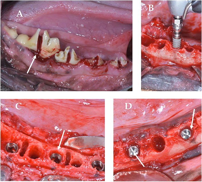
FIGURE 2. Surgery. (A), Odontosection; (B), Implant insertion; (C), Implant placed in subcrestal position with cover screws for submerged curing; (D), Lack of friction between the implant and the bone wall.
Micro-Computed Tomography Analysis
After euthanasia of animals (after 12 weeks of implants placement), the sections of the block were preserved and fixed in 10% neutral formalin. Image acquisitions were per-formed using a multimodal SPECT/CT Albira II ARS scanner (Bruker® Corporation, Karksruhe, Germany). The acquisition parameters were 45 kV, 0.2 mA, and 0.05 mm voxels. The acquisition slices were axial, 0.05 mm thick, and 800 to 1,000 images were obtained from each piece through a flat panel digital detector with 2,400 × 2,400 pixels and a FOV (field of view) of 70 mm × 70 mm. The implants were grouped according to the three axes (transverse, coronal, and sagittal). The sagital axis was used for BCIS and PIBA measurements. In all images the same color scale was used (0 min and 3 max) with the same parameters in FOV (%): 90 and zoom 0.6, with a hardness of 1. The areas of interest (AoIs) were manually fixed by three micro-CT cross-sections at crestal, mid and apical levels; the apical section avoided the conical area of the implant (Figure 3). The voxels in contact with the implant surface were excluded in the measurements because they were considered artifact zones, estimating a value of 0.5 mm higher than the implant diameter for the calculation of the BCIS AoI (4 mm Ø) and 2 mm for the calculation of the PIBA AoI (5.5 mm Ø) (Figure 4).
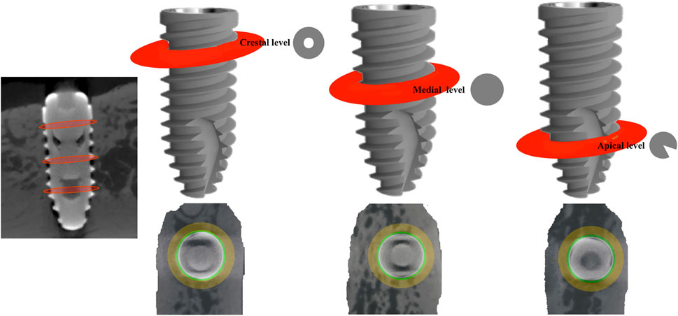
FIGURE 3. Areas of interest (AoI) delimited by three micro-CT slices at crestal, mid and apical level.
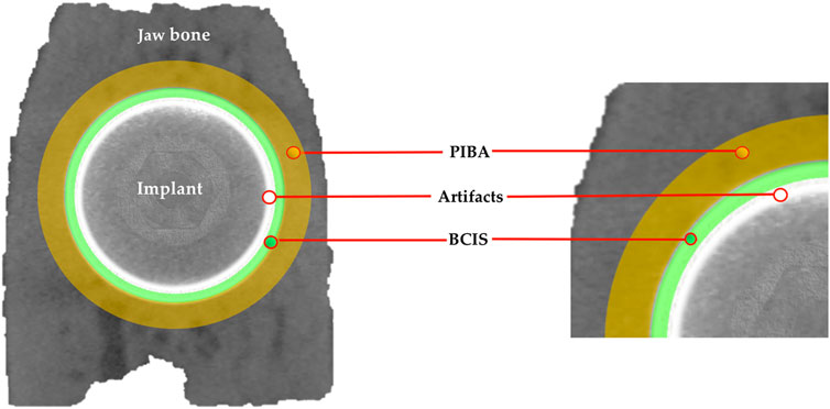
FIGURE 4. Artifact area in white color; bone in contact with the implant surface (BCIS) in green color and peri-implant bone area (PIBA) in yellow color.
The AMIDE tool allowed us to obtain the data in statistical form (Hounsfield Units), with maximums, minimums and deviations. AMIDE is a tool for visualizing, analyzing and registering volumetric medical image data sets (AMIDE, UCLA University, Los Angeles, CA, USA). It allows drawing two-dimensional and three-dimensional AoIs directly on the images and generating statistical data for these AoIs. 3D Slicer program (http://www.slicer.org) provided the 3D images of the bone-to-implant contact area (Figure 5).
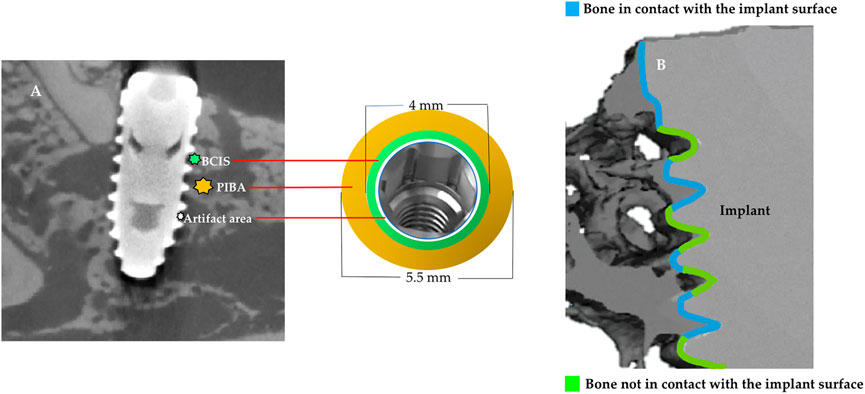
FIGURE 5. (A), Micro-CT image in sagittal plane and graph to represent the artifact area, bone in contact with the implant surface (BCIS) (4 mm Ø) and peri-implant bone area (PIBA) (5.5 mm Ø); (B), 3D image with the contact and non-contact surfaces of the bone with the implant.
Statistical Analysis
SPSS Statistics 26.0 (IBM, Chicago, IL, USA) was used as the statistical analysis program. Statistical analysis of the BCIS and PIBA variables in the crestal, mid and apical areas was performed for the experimental and control groups. The normality of the data generated by the microtomographic analyses was examined using the Shapiro-Wilk test. The mean and standard deviation of each group were proposed; the p-value and p for trend derived from the differences and changes in each group were presented, with a significance level of ≤0.05.
Results
All the implants used in the 5 dogs achieved osseointegration. Two parameters, Bone in Contact to the Implant Surface (BCIS) and Peri-Implant Bone Area (PIBA) were measured in crestal, mid and apical, resulting in a total of 120 sites for each measurement parameter, in the experimental group (ChtG) and in the control group (CG).
The mean value and standard deviation of the trends for each group are shown in Tables 1, 2. For BCIS the values in crestal, mid and apical, in the experimental group (ChtG), were 3,770.11 ± 245.60, 3,245.25 ± 1,477.08 and 4,196.82 ± 453.03, respectively and 3,829.29 ± 249.08, 3,958.75 ± 1,477.08 and 4,112.13 ± 112.6, respectively, in the control group (CG). Trend analysis in the experimental group showed a higher statistical significance (p = 0.005) with respect to the control group (p = 0.014). For PIBA the values in crestal, mid and apical, in the experimental group, were 3,613.00 ± 1,109.12, 3,905.75 ± 809.65 and 3,759.17 ± 944.73, respectively and 4,162.50 ± 618.02, 3,705.20 ± 1,045.86 and 3,832.71 ± 1,201.43, respectively, in the control group. Trend analysis in the experimental group showed considerable statistical significance (p < 0.001) versus the control group (p = 0.02). Figures 6, 7 represents the boxplots of the results.
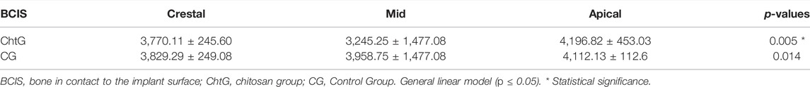
TABLE 1. Specific crestal, mid and apical trends in the experimental and control groups for BCIS (mean ± deviation).
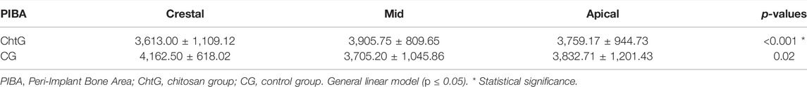
TABLE 2. Specific crestal, mid and apical trends in the experimental and control groups for PIBA (mean ± deviation).
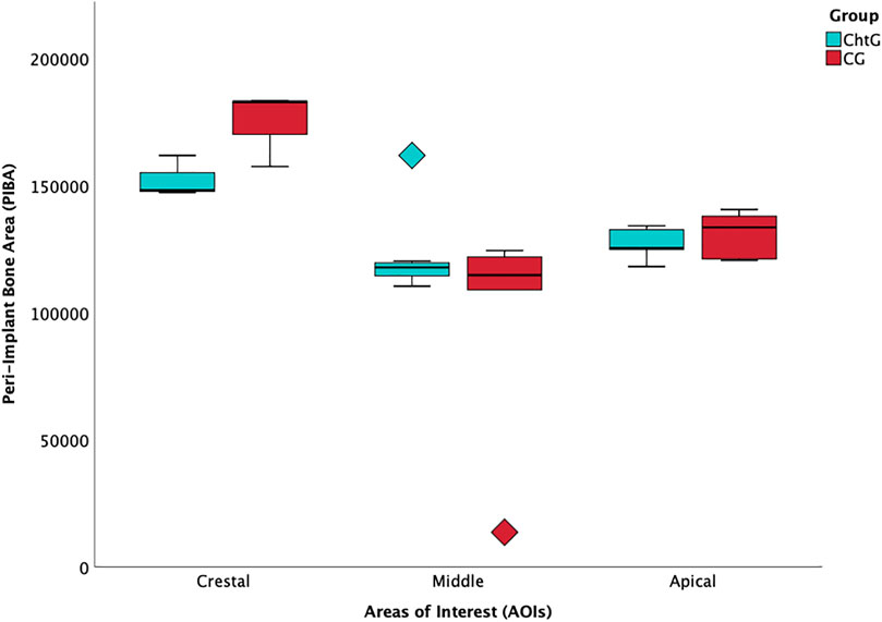
FIGURE 6. Boxplot of peri-implant bone area (PIBA) values at crestal, mid and apical levels.
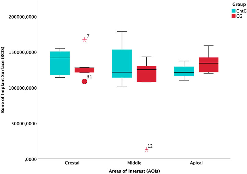
FIGURE 7. Boxplot of bone in contact with the implant surface (BCIS) values at crestal, mid and apical levels.
Discussion
Since Brånemark published his first study on the osseointegration of Ti implants in 1969, there have been numerous variations of their surfaces, all with the aim of achieving early and more durable osseointegration. The topography and roughness of the surfaces have been questioned in terms of osteoblastic cell differentiation and it has been shown that the attachment of this type of cells is greater on smooth surfaces, although rougher surfaces have been associated with greater cell differentiation (Bachle and Kohal, 2004; Esposito et al., 2005). Apart from surface characteristics, it is also known that osseointegration is affected by factors such as the biological compatibility of an implant (Annunziata and Guida, 2015). Therefore, to improve the bioactivity of implants, the surface can be modified by incorporating organic and inorganic phases either within or on the Ti oxide layer, using ions, inorganic molecules or organic molecules (Jiang et al., 2014; Anitua et al., 2015).
The evaluation of bone-implant contact provides evidence of an implant anchored in the bone and has traditionally been established as the most common method of evaluation, however, the concept of osseointegration has undergone variations and is now considered as “a reaction to a foreign body in which interfacial bone is formed as a defensive reaction to protect the implant from the tissues” (Albrektsson et al., 2017; Albrektsson and Wennerberg, 2019). The amount of bone in contact with the implant, as well as the frictional properties at the contact interface, are important parameters influencing bone-implant mechanics. However, the stability of implants in trabecular bone has been little studied and considering the reduced contact surface between trabecular bone and implant, it has been suggested that macroscopic phenomena such as trabeculae-implant mechanical fixation would dominate over the microscopic aspects such as friction (Huang et al., 2008; Wirth et al., 2010).
In the present study, a model was designed in the canine mandible, where 2 bone parameters were measured in the area surrounding the implant, Bone in Contact with the Implant Surface (BCIS) and Peri-implant Bone Area (PIBA), at crestal, mid and apical levels, both in the Cht-coated implant group (ChtG) and in the control group (CG), with conventional etched surface, and the results were analyzed by means of micro-CT.
Although no single animal species meets all the requirements of an ideal model, an understanding of the differences in bone architecture and remodeling between different species of experimental animals could help to select a suitable species. Most studies resort to modified and inadequate experimental models, both the experimental animal (rabbit rat...) and the implantation site, with extraoral surgical approaches (tibia, femur...), so that the results cannot be extrapolated to humans, among other reasons, because of the lack of cortical remodeling and the fact that the cessation of growth occurs much later in these species than in other mammals (Ferguson et al., 2018). On the other hand, in vitro cultures maintain tissues or organs without vascularization, limiting the supply of nutrients, oxygen and waste elimination and, therefore, the extrapolation of the results to the in vivo situation limits the model. All this, without taking into account the reduced lifespan of the cultured cells (Pizzoferrato et al., 1994). Our preclinical study used the mandible of a canine model, with greater similarity to the human in terms of bone architecture and remodeling. The dog, along with the pig, are considered valuable models for the study of tissues adjacent to dental implants, and large-breed dogs can support human dental implants (Pearce et al., 2007). To our knowledge, this is the first time that this experimental model has been used for the study of the effectiveness of Cht as a coating for dental implants.
Cht is a macromolecule that has achieved great attention in the biomedical industry, arousing great interest in bone regeneration (De Jonge et al., 2008; Ebhodaghe, 2021). It has the ability to stimulate the recruitment and adhesion of osteogenic progenitor cells, facilitating bone formation. In addition, it has been shown that no inflammatory or allergic reactions occur after topical application (Kim et al., 2003; Waibel et al., 2011; Azuma et al., 2015; Sukpaita et al., 2021).
Typically, toxic reagents, such as 3-isocyanatopropyltriethoxysilane and glutaraldehyde, are used to form Cht coatings for silanization and attachment to the Ti substrate but these techniques involve complex processing that hinders coating deposition and limits clinical applicability (Yuan et al., 2008). However, although efforts have been made to increase the bond strength of hydroxyapatite coatings on implant alloys, as they are brittle materials, it is unclear whether these high bond strengths would be necessary for the polymeric Cht material (Bumgardner et al., 2007).
The dip coating used in our study (Vakili and Asefnejad, 2020), in addition to resisting the forces used during implantation, because of the surgical technique employed, is an inexpensive way of depositing thin layers from chemical solutions, with relatively fair control over the thickness of the layer and offers the possibility of fine-tuning the amount of material that can be deposited and, therefore, the thickness of the final film. For these reasons, it is becoming increasingly popular not only in research and development laboratories, but also in industrial production. Grosso have proposed the immersion technique as a very suitable method to impregnate porosities, make nanocomposites or perform nanofusion. (Grosso, 2011); Brinker et al. (Brinker et al., 1992) pointed out wet methods, as suitable, since they are homogeneously organized in the final film, with adequate thickness control.
The amount of Cht can be adjusted by controlling the concentration of the Cht solution and certain authors have indicated that, as the amount of loaded Cht increases, its antibacterial properties increase; therefore, controlling the amount of loaded Cht would endow the Ti implant with better biological and anti-bacterial properties. Cht loading in 0.5% acidic solution, was considered adequate (Vakili and Asefnejad, 2020), although the optimization of the amount of loaded Cht, degradation rate and antibacterial effect still need to be further investigated (Li et al., 2019b). Most of the existing studies on the efficacy of Cht as a Ti coating, as we have noted above, are in vitro studies or in vivo studies on inadequate models, or that resort to complicated coating methods, or toxic products, which hinder or impair their clinical applicability (Bumgardner et al., 2007; Kung et al., 2011; Takanche et al., 2018a; Zhang et al., 2020).
Sukpaita et al. (Sukpaita et al., 2019) demonstrated the ability of Cht scaffolds to self-promote bone tissue and repair calvarial bone defects in mice. Tian et al. (Tian et al., 2014), in an in vitro study, indicated that Cht film loaded on a Ti surface would promote osteoblast proliferation and differentiation in a dose-dependent manner, which could represent a new approach in the treatment of Ti implants. Zhang et al. (Li et al., 2019b) showed that porous Ti with a Cht/Hydroxyapatite coating could promote osteoblast-like cell proliferation and differentiation and osseointegration in vivo. Bumgardner et al. (Bumgardner et al., 2007) evaluated the ability of Cht coatings on Ti to promote bone formation and osseointegration compared to calcium phosphate coatings and uncoated Ti, in a 12-weeks rabbit model, maintaining the hypothesis that it may not be important that the Cht coating persists long-term, once a good bone-implant interface has been established, in the same way that some investigators have speculated with calcium phosphate coatings (Yang et al., 2005).
Even heterotopic (extraskeletal) bone formation induced by Cht-collagen-coated Ti implants has been demonstrated in vivo (Kung et al., 2011). Overall, researchers conclude that Cht significantly accelerates the bone regeneration process and, therefore, in terms of its biocompatibility and osteoinductivity, it can be considered as a biomaterial of great relevance in human bone healing (Ge et al., 2004; Pang et al., 2005; Guzmán-Morales et al., 2009; Muzzarelli, 2009; Ezoddini-Ardakani et al., 2012; Mututuvari et al., 2013), which is consistent with the results obtained in our research.
The good film-forming ability of Cht allows its use in the coating of dental or orthopedic implants, and the coated surfaces have been shown to possess good cellular compatibility with fibroblast cells. Klokkevold et al. (Klokkevold et al., 1996) reported that chitosan films enhanced osteoprogenitor cell differentiation, facilitated bone formation, and inhibited fibroblast proliferation. Moreover, the activity of Cht against bacteria such as Escherichia coli, Streptococcus mutans, Staphylococcus aureus, Bacillus subtilis and Actinomyces naeslundii (Renoud et al., 2012; Lin et al., 2021) and to prevent oxidative damage caused by free radicals (Ngo et al., 2011) has also been demonstrated. Takanche et al. in an in vivo study on osteoporotic rat jaws demonstrated, by micro-CT, that Cht-coated Ti implants increased the volume and density of newly formed bone and implant osseointegration, as well as the upregulation of bone morphogenetic protein, by inhibiting osteoclastogenesis. All these and other demonstrated properties make this biopolymer a good biocompatible and bioactive osteoconductor and a useful coating for orthopedic and craniofacial implant devices.
However, in the future, it will be necessary to determine the bond strength of the coatings and changes in bond strength over time and to evaluate the degradation of the coating in lysozyme solutions in the oral environment, as degradation rates may be different in the presence of this enzyme and changes in degradation would be important for the development of the implant-tissue interface (Yuan et al., 2008). It will also be necessary to determine whether changes in initial cell growth, or differences in Cht degradation, result in less cell mineralization. In addition, it will need to be determined whether surface morphology, roughness, and coating chemistry could be related to cell and tissue responses.
In our study, two bone parameters in the vicinity of the im-plant (BCIS and PIBA) were measured by micro-CT. The best statistical significance was obtained for PIBA, in the experimental group (ChtG) (p < 0.001), despite the fact that all the inserted implants obtained optimal osseointegration. The maximum value was obtained for PIBA at the crestal level of M1 in ChG (6,347.05 ± 413.2) and the minimum for BCIS at the mid-level of P2 in CG (1765.03 ± 358.01).
Micro-CT currently allows observation of bone tissue in a three-dimensional manner, as well as quantitative analysis in several sections, which is not possible by histomorphometric analysis. It detects only mineralized tissue and is therefore suitable for analyzing the bony annulus and assessing bone formation around implants during healing periods; moreover, histological studies could not be performed in clinical trials (Vandeweghe et al., 2013b; Nakahara et al., 2019). Rebaudi et al. (Rebaudi et al., 2004), have proposed it as a suitable technique for the analysis of peri-implant bone tissues, proposing it as a non-destructive evaluation, which allows the analysis of the bone-implant interface. Lyu and Lee (Lyu and Lee, 2021), in a study on rabbit tibiae, reported that the measurement of bone in contact with the implant by micro-CT is feasible to evaluate implant osseointegration, obtaining results similar to histomorphometric ones, although they recognize that the method needs further optimization. Likewise, they recognize that, in most cases, they can only use one or two histological sections per implant for histomorphometric evaluation, which could lead to an over- or underestimation of the peri-implant bone.
Nevertheless, we are aware of our limitations, in terms of small sample size and difficulty in accurately detecting the bone in close contact with the implant surface, due to the thin layer of noise in the surrounding area and the microtomography settings. and the difficulty of accurately detecting the bone in close contact with the implant surface, due to the thin layer of noise in the surrounding area and the microtomography settings. These drawbacks should be taken into account when interpreting the results. In future ongoing studies, the samples will be studied by histomorphometric analysis to compare the results.
Conclusion
The results of this preliminary study demonstrated the usefulness of Cht coatings on Ti surfaces to improve the osseointegration of dental implants. In addition, within the limitations, the use of nondestructive micro-CT analysis, seems to be useful to evaluate bone healing in the surroundings of the implant surfaces.
Since the design of the present study allowed only a preliminary analysis, the data obtained could serve as a basis for the design of future studies.
Data Availability Statement
The original contributions presented in the study are included in the article/Supplementary Material, further inquiries can be directed to the corresponding author.
Ethics Statement
The animal study was reviewed and approved by Ethics Committee of the Catholic University of Murcia (Spain) with code CE072004.
Author Contributions
Conceptualization, NL-V and AL-V; methodology, NL-V; formal analysis, AL-V; investigation, NL-V and BMS; writing-original draft preparation AL-V; data curation, JMA, MPC, and CR; supervision, JMA and AL-V. All authors have read and agreed to the published version of the manuscript. All authors have read and agreed to the published version of the manuscript.
Conflict of Interest
The authors declare that the research was conducted in the absence of any commercial or financial relationships that could be construed as a potential conflict of interest.
Publisher’s Note
All claims expressed in this article are solely those of the authors and do not necessarily represent those of their affiliated organizations, or those of the publisher, the editors and the reviewers. Any product that may be evaluated in this article, or claim that may be made by its manufacturer, is not guaranteed or endorsed by the publisher.
References
Ahn, S.-I., Cho, S., and Choi, N.-J. (2021). Effectiveness of Chitosan as a Dietary Supplement in Lowering Cholesterol in Murine Models: A Meta-Analysis. Mar. Drugs 19, 26. doi:10.3390/md19010026
Ahsan, S. M., Thomas, M., Reddy, K. K., Sooraparaju, S. G., Asthana, A., and Bhatnagar, I. (2018). Chitosan as Biomaterial in Drug Delivery and Tissue Engineering. Int. J. Biol. Macromolecules 110, 97–109. doi:10.1016/j.ijbiomac.2017.08.140
Albrektsson, T., Chrcanovic, B., Jacobsson, M., and Wennerberg, A. (2017). Osseointegration of Implants-A Biological and Clinical Overview. JSM Dent Surg. 2, 1–6.
Albrektsson, T., and Wennerberg, A. (2019). On Osseointegration in Relation to Implant Surfaces. Clin. Implant Dent Relat. Res. 21 (Suppl. 1), 4–7. doi:10.1111/cid.12742
Alfarsi, M. A., Hamlet, S. M., and Ivanovski, S. (2014). Titanium Surface Hydrophilicity Modulates the Human Macrophage Inflammatory Cytokine Response. J. Biomed. Mater. Res. 102, 60–67. doi:10.1002/jbm.a.34666
Anitua, E., Prado, R., Orive, G., and Tejero, R. (2015). Effects of Calcium-Modified Titanium Implant Surfaces on Platelet Activation, Clot Formation, and Osseointegration. J. Biomed. Mater. Res. 103, 969–980. doi:10.1002/jbm.a.35240
Annunziata, M., and Guida, L. (2015). The Effect of Titanium Surface Modifications on Dental Implant Osseointegration. Front. Oral Biol. 17, 62–77. doi:10.1159/000381694
Azuma, K., Osaki, T., Minami, S., and Okamoto, Y. (2015). Anticancer and Anti-inflammatory Properties of Chitin and Chitosan Oligosacchrides. J. Funct. Biomater. 6, 33–49. doi:10.3390/jfb6010033
Bachle, M., and Kohal, R. J. (2004). A Systematic Review of the Influence of Different Titanium Surfaces on Proliferation, Differentiation and Protein Synthesis of Osteoblast-like MG63 Cells. Clin. Oral Implants Res. 15, 683–692. doi:10.1111/j.1600-0501.2004.01054.x
Becker, K., Klitzsch, I., Stauber, M., and Schwarz, F. (2017). Three-dimensional Assessment of Crestal Bone Levels at Titanium Implants with Different Abutment Microstructures and Insertion Depths Using Micro-computed Tomography. Clin. Oral Impl. Res. 28, 671–676. doi:10.1111/clr.12860
Boyd, S. K., Moser, S., Kuhn, M., Klinck, R. J., Krauze, P. L., Müller, R., et al. (2006). Evaluation of Three-Dimensional Image Registration Methodologies for In Vivo Micro-computed Tomography. Ann. Biomed. Eng. 34, 1587–1599. doi:10.1007/s10439-006-9168-7
Brinker, C. J., Hurd, A. J., Schunk, P. R., Frye, G. C., and Ashley, C. S. (1992). Review of Sol-Gel Thin Film Formation. J. Non-Crystalline Sol. 147–148, 424–436. doi:10.1016/S0022-3093(05)80653-2
Bumgardner, J. D., Chesnutt, B. M., Yuan, Y., Yang, Y., Appleford, M., Oh, S., et al. (2007). The Integration of Chitosan-Coated Titanium in Bone: an In Vivo Study in Rabbits. Implant Dent 16, 66–79. doi:10.1097/ID.0b013e3180312011
Bumgardner, J. D., Wiser, R., Elder, S. H., Jouett, R., Yang, Y., and Ong, J. L. (2003). Contact Angle, Protein Adsorption and Osteoblast Precursor Cell Attachment to Chitosan Coatings Bonded to Titanium. J. Biomater. Sci. Polym. Edition 14, 1401–1409. doi:10.1163/156856203322599734
Bumgardner, J. D., Wiser, R., Gerard, P. D., Bergin, P., Chestnutt, B., Marini, M., et al. (20032003). Chitosan: Potential Use as a Bioactive Coating for Orthopaedic and Craniofacial/dental Implants. J. Biomater. Sci. Polym. Edition 14 (5), 423–438. doi:10.1163/156856203766652048
Chen, R., Cai, X., Ma, K., Zhou, Y., Wang, Y., and Jiang, T. (2017). The Fabrication of Double-Layered Chitosan/gelatin/genipin Nanosphere Coating for Sequential and Controlled Release of Therapeutic Proteins. Biofabrication 9 (2), 025028. doi:10.1088/1758-5090/aa70c3
De Jonge, L. T., Leeuwenburgh, S. C. G., Wolke, J. G. A., and Jansen, J. A. (2008). Organic-inorganic Surface Modifications for Titanium Implant Surfaces. Pharm. Res. 25, 2357–2369. doi:10.1007/s11095-008-9617-0
Ebhodaghe, S. O. (2021). Natural Polymeric Scaffolds for Tissue Engineering Applications. J. Biomater. Sci. Polym. Ed. 32, 2144–2194. doi:10.1080/09205063.2021.1958185
Esposito, M., Coulthard, P., Thomsen, P., and Worthington, H. V. (2005). The Role of Implant Surface Modifications, Shape and Material on the success of Osseointegrated Dental Implants. A Cochrane Systematic Review. Eur. J. Prosthodont. Restor Dent 13, 15–31.
Ezoddini-Ardakani, F., Navabazam, A., Fatehi, F., Danesh-Ardekani, M., Khadem, S., and Rouhi, G. (2012). Histologic Evaluation of Chitosan as an Accelerator of Bone Regeneration in Microdrilled Rat Tibias. Dental Res. J. 9, 694–699.
Ferguson, J. C., Tangl, S., Barnewitz, D., Genzel, A., Heimel, P., Hruschka, V., et al. (2018). A Large Animal Model for Standardized Testing of Bone Regeneration Strategies. BMC Vet. Res. 14, 330. doi:10.1186/s12917-018-1648-0
Ge, Z., Baguenard, S., Lim, L. Y., Wee, A., and Khor, E. (2004). Hydroxyapatite–chitin Materials as Potential Tissue Engineered Bone Substitutes. Biomaterials 25, 1049–1058. doi:10.1016/s0142-9612(03)00612-4
Grosso, D. (2011). How to Exploit the Full Potential of the Dip-Coating Process to Better Control Film Formation. J. Mater. Chem. 21, 17033–17038. doi:10.1039/c1jm12837j
Guzmán-Morales, J., El-Gabalawy, H., Pham, M. H., Tran-Khanh, N., Marc, D., McKee, W. W., et al. (2009). Effect of Chitosan Particles and Dexamethasone on Human Bone Marrow Stromal Cell Osteogenesis and Angiogenic Factor Secretion. Bone 45, 617–626. doi:10.1016/j.bone.2009.06.014
He, T., Cao, C., Xu, Z., Li, G., Cao, H., Liu, X., et al. (2017). A Comparison of Micro-CT and Histomorphometry for Evaluation of Osseointegration of PEO-Coated Titanium Implants in a Rat Model. Sci. Rep. 7, 16270. doi:10.1038/s41598-017-16465-4
Höhlinger, M., Heise, S., Wagener, V., Boccaccini, A. R., and Virtanen, S. (2017). Developing Surface Pre-treatments for Electrophoretic Deposition of Biofunctional Chitosan-Bioactive Glass Coatings on a WE43 Magnesium alloy. Appl. Surf. Sci. 405, 441–448. doi:10.1016/j.apsusc.2017.02.049
Hotchkiss, K. M., Sowers, K. T., and Olivares-Navarrete, R. (2019). Novel In Vitro Comparative Model of Osteogenic and Inflammatory Cell Response to Dental Implants. Dental Mater. 35, 176–184. doi:10.1016/j.dental.2018.11.011
Hu, Y., Cai, K., Luo, Z., Zhang, R., Yang, L., Deng, L., et al. (2009). Surface Mediated In Situ Differentiation of Mesenchymal Stem Cells on Gene-Functionalized Titanium Films Fabricated by Layer-By-Layer Technique. Biomaterials 30, 3626–3635. doi:10.1016/j.biomaterials.2009.03.037
Hu, Z., Zhang, D.-Y., Lu, S.-T., Li, P.-W., and Li, S.-D. (2018). Chitosan-Based Composite Materials for Prospective Hemostatic Applications. Mar. Drugs 16, 273. doi:10.3390/md16080273
Huang, H.-L., Hsu, J.-T., Fuh, L.-J., Tu, M.-G., Ko, C.-C., and Shen, Y.-W. (2008). Bone Stress and Interfacial Sliding Analysis of Implant Designs on an Immediately Loaded Maxillary Implant: a Non-linear Finite Element Study. J. Dentistry 36, 409–417. doi:10.1016/j.jdent.2008.02.015
Jemat, A., Ghazali, M. J., Razali, M., and Otsuka, Y. (2015). Surface Modifications and Their Effects on Titanium Dental Implants. Biomed. Res. Int. 2015, 1, 11. doi:10.1155/2015/791725
Jiang, X., Wang, G., Li, J., Zhang, W., Xu, L., Pan, H., et al. (2014). Magnesium Ion Implantation on a Micro/nanostructured Titanium Surface Promotes its Bioactivity and Osteogenic Differentiation Function. Ijn 9, 2387–2398. doi:10.2147/IJN.S58357
Kampschulte, M., Langheinirch, A., Sender, J., Litzlbauer, H., Althöhn, U., Schwab, J., et al. (2016). Nano-Computed Tomography: Technique and Applications. Fortschr Röntgenstr 188, 146–154. doi:10.1055/s-0041-106541
Kim, I.-Y., Seo, S.-J., Moon, H.-S., Yoo, M.-K., Park, I.-Y., Kim, B.-C., et al. (2008). Chitosan and its Derivatives for Tissue Engineering Applications. Biotechnol. Adv. 26, 1–21. doi:10.1016/j.biotechadv.2007.07.009
Kim, S. E., Park, J. H., Cho, Y. W., Chung, H., Jeong, S. Y., Lee, E. B., et al. (2003). Porous Chitosan Scaffold Containing Microspheres Loaded with Transforming Growth Factor-Beta1: Implications for Cartilage Tissue Engineering. J. Controlled Release 91, 365–374. doi:10.1016/s0168-3659(03)00274-8
Klokkevold, P. R., Vandemark, L., Kenney, E. B., and Bernard, G. W. (1996). Osteogenesis Enhanced by Chitosan (Poly-N-Acetyl Glucosaminoglycan) In Vitro. J. Periodontol. 67, 170–175. doi:10.1902/jop.1996.67.11.1170
Kung, S., Devlin, H., Fu, E., Ho, K. Y., Liang, S. Y., and Hsieh, Y. D. (2011). The Osteoinductive Effect of Chitosan-Collagen Composites Around Pure Titanium Implant Surfaces in Rats. J. Periodontal Res. 46, 126–133. doi:10.1111/j.1600-0765.2010.01322.x
Le Guéhennec, L., Soueidan, A., Layrolle, P., and Amouriq, Y. (2007). Surface Treatments of Titanium Dental Implants for Rapid Osseointegration. Dental Mater. 23, 844–854. doi:10.1016/j.dental.2006.06.025
Li, B., Xia, X., Guo, M., Jiang, Y., Li, Y., Zhang, Z., et al. (2019). Biological and Antibacterial Properties of the Micro-nanostructured Hydroxyapatite/chitosan Coating on Titanium. Sci. Rep. 9 (1), 14052. doi:10.1038/s41598-019-49941-0
Li, X., Qi, M., Sun, X., Weir, M. D., Tay, F. R., Oates, T. W., et al. (2019). Surface Treatments on Titanium Implants via Nanostructured Ceria for Antibacterial and Anti-inflammatory Capabilities. Acta Biomater. 94, 627–643. doi:10.1016/j.actbio.2019.06.023
Lin, M. H., Wang, Y. H., Kuo, C. H., Ou, S. F., Huang, P. Z., Song, T. Y., et al. (2021). Hybrid ZnO/chitosan Antimicrobial Coatings with Enhanced Mechanical and Bioactive Properties for Titanium Implants. Carbohydr. Polym. 257, 117639. doi:10.1016/j.carbpol.2021.117639
López-Valverde, N., López-Valverde, A., and Ramírez, J. M. (2021). Systematic Review of Effectiveness of Chitosan as a Biofunctionalizer of Titanium Implants. Biology 10, 102. doi:10.3390/biology10020102
Lyu, H. Z., and Lee, J. H. (2021). Correlation between Two-Dimensional Micro-CT and Histomorphometry for Assessment of the Implant Osseointegration in Rabbit Tibia Model. Biomater. Res. 2511. doi:10.1186/s40824-021-00213-x
Matica, M. A., Aachmann, F. L., Tøndervik, A., Sletta, H., and Ostafe, V. (2019). Chitosan as a Wound Dressing Starting Material: Antimicrobial Properties and Mode of Action. Ijms 20, 5889. doi:10.3390/ijms20235889
Mututuvari, T. M., Harkins, A. L., and Tran, C. D. (2013). Facile Synthesis, Characterization, and Antimicrobial Activity of Cellulose–Chitosan–Hydroxyapatite Composite Material: A Potential Material for Bone Tissue Engineering. J. Bio-medical Mater. Res. A 101A, 3266–3277. doi:10.1002/jbm.a.34636
Muzzarelli, R. A. A. (2009). Chitins and Chitosans for the Repair of Wounded Skin, Nerve, Cartilage and Bone. Carbohydr. Polym. 76, 167–182. doi:10.1016/j.carbpol.2008.11.002
Muzzarelli, R. A. A., Mattioli-Belmonte, M., Tietz, C., Biagini, R., Ferioli, G., Brunelli, M. A., et al. (1994). Stimulatory Effect on Bone Formation Exerted by a Modified Chitosan. Biomaterials 15, 1075–1081. doi:10.1016/0142-9612(94)90093-0
Nakahara, K., Haga‐Tsujimura, M., Igarashi, K., Kobayashi, E., Schaller, B., Lang, N. P., et al. (2019). Single‐staged Implant Placement Using the Bone Ring Technique with and without Membrane Placement: Micro‐CT Analysis in a Preclinical In Vivo Study. Clin. Oral Impl Res. 31, 29-36. doi:10.1111/clr.13543
Ngo, D. H., Qian, Z. J., Ngo, D. N., Vo, T. S., Wijesekara, I., and Kim, S. K. (2011). Gallyl Chitooligosaccharides Inhibit Intracellular Free Radical-Mediated Oxidation. Food Chem. 128, 974e981. doi:10.1016/j.foodchem.2011.03.128
Palla-Rubio, B., Araújo-Gomes, N., Fernández-Gutiérrez, M., Rojo, L., Suay, J., Gurruchaga, M., et al. (2019). Synthesis and Characterization of Silica-Chitosan Hybrid Materials as Antibacterial Coatings for Titanium Implants. Carbohydr. Polym. 203, 331–341. doi:10.1016/j.carbpol.2018.09.064
Pang, E.-K., Paik, J.-W., Kim, S.-K., Jung, U.-W., Kim, C.-S., Cho, K.-S., et al. (2005). Effects of Chitosan on Human Periodontal Ligament Fibroblasts In Vitro and on Bone Formation in Rat Calvarial Defects. J. Periodontol. 76, 1526–1533. doi:10.1902/jop.2005.76.9.1526
Pearce, A., Richards, R. G., Richards, R., Milz, S., Schneider, E., and Pearce, S. G. (2007). Animal Models for Implant Biomaterial Research in Bone: a Review. Eur. Cell Mater 13, 1–10. doi:10.1016/0267-6605(94)90081-710.22203/ecm.v013a01
Peyrin, F., Dong, P., Pacureanu, A., and Langer, M. (2014). Micro- and Nano-CT for the Study of Bone Ultrastructure. Curr. Osteoporos. Rep. 12, 465–474. doi:10.1007/s11914-014-0233-0
Pizzoferrato, A., Ciapetti, G., Stea, S., Cenni, E., Arciola, C. R., Granchi, D., et al. (1994). Cell Culture Methods for Testing Biocompatibility. Clin. Mater. 15, 173–190. doi:10.1016/0267-6605(94)90081-7
Rabea, E. I., Badawy, M. E.-T., Stevens, C. V., Smagghe, G., and Steurbaut, W. (2003). Chitosan as Antimicrobial Agent: Applications and Mode of Action. Biomacromolecules 4, 1457–1465. doi:10.3390/ijms2023588910.1021/bm034130m
Rebaudi, A., Koller, B., Laib, A., and Trisi, P. (2004). Microcomputed Tomographic Analysis of the Peri-Implant Bone. Int. J. Periodontics Restorative Dent 24, 316–325.
Reddy Tiyyagura, H., Rudolf, R., Gorgieva, S., Fuchs-Godec, R., Boyapati, V. R., Mantravadi, K. M., et al. (2016). The Chitosan Coating and Processing Effect on the Physiological Corrosion Behaviour of Porous Magnesium Monoliths. Prog. Org. Coat. 99, 147–156. doi:10.1016/j.porgcoat.2016.05.019
Renoud, P., Toury, B., Benayoun, S., Attik, G., and Grosgogeat, B. (2012). Functionalization of Titanium with Chitosan via Silanation: Evaluation of Biological and Mechanical Performances. PLoS One 7 (7), e39367. doi:10.1371/journal.pone.0039367
Rojo, L., and Deb, S. (2015). Polymer Therapeutics in Relation to Dentistry. Front. Oral Biol. 17, 13–21. doi:10.1159/000381688
Sakka, S., Baroudi, K., and Nassani, M. Z. (2012). Factors Associated with Early and Late Failure of Dental Implants. J. Invest. Clin. Dent 3, 258–261. doi:10.1111/j.2041-1626.2012.00162.x
Scriven, L. E. (1988). Physics and Applications of DIP Coating and Spin Coating. MRS Proc. 121, 717. doi:10.1557/PROC-121-717
Shukla, S. K., Mishra, A. K., Arotiba, O. A., and Mamba, B. B. (2013). Chitosan-based Nanomaterials: a State-Of-The-Art Review. Int. J. Biol. Macromolecules 59, 46–58. doi:10.1016/j.ijbiomac.2013.04.043
Simonis, P., Dufour, T., and Tenenbaum, H. (2010). Long-term Implant Survival and success: a 10-16-year Follow-Up of Non-submerged Dental Implants. Clin. Oral Implants Res. 21, 772–777. doi:10.1111/j.1600-0501.2010.01912.x
Singla, A. K., and Chawla, M. (2001). Chitosan: Some Pharmaceutical and Biological Aspects-Aan Update. J. Pharm. Pharmacol. 53, 1047–1067. doi:10.1211/0022357011776441
Sukpaita, T., Chirachanchai, S., Pimkhaokham, A., and Ampornaramveth, R. S. (2021). Chitosan-Based Scaffold for Mineralized Tissues Regeneration. Mar. Drugs 19 (10), 551. doi:10.3390/md19100551
Sukpaita, T., Chirachanchai, S., Suwattanachai, P., Everts, V., Pimkhaokham, A., and Ampornaramveth, R. S. (2019). In Vivo Bone Regeneration Induced by a Scaffold of Chitosan/Dicarboxylic Acid Seeded with Human Periodontal Ligament Cells. Int. J. Mol. Sci. 20 (19), 4883. doi:10.3390/ijms20194883
Szmukler-Moncler, S., Testori, T., and Bernard, J. P. (2004). Etched Implants: a Comparative Surface Analysis of Four Implant Systems. J. Biomed. Mater. Res. 69B, 46–57. doi:10.1002/jbm.b.20021
Tajdini, F., Amini, M. A., Nafissi-Varcheh, N., and Faramarzi, M. A. (2010). Production, Physiochemical and Antimicrobial Properties of Fungal Chitosan from Rhizomucor Miehei and Mucor Racemosus. Int. J. Biol. Macromolecules 47, 180–183. doi:10.1016/j.ijbiomac.2010.05.002
Takanche, J. S., Kim, J. E., Kim, J. S., Lee, M. H., Jeon, J. G., Park, I. S., et al. (2018). Chitosan-gold Nanoparticles Mediated Gene Delivery of C-Myb Facilitates Osseointegration of Dental Implants in Ovariectomized Rat. Artif. Cell Nanomed Bio-technol 46 (Suppl. 3), S807–S817. doi:10.1080/21691401.2018.1513940
Tian, A., Zhai, J. J., Peng, Y., Zhang, L., Teng, M. H., Liao, J., et al. (2014). Osteoblast Response to Titanium Surfaces Coated with Strontium Ranelate-Loaded Chitosan Film. Int. J. Oral Maxillofac. Implants 29, 1446–1453. doi:10.11607/jomi.3806
Türker, N. S., Özer, A. Y., Kutlu, B., Nohutcu, R., Sungur, A., Bilgili, H., et al. (2014). The Effect of Gamma Radiation Sterilization on Dental Biomaterials. Tissue Eng. Regen. Med. 11, 341–349. doi:10.1007/s13770-014-0016-9
Ueno, T., Takeuchi, M., Hori, N., Iwasa, F., Minamikawa, H., Igarashi, Y., et al. (2012). Gamma ray Treatment Enhances Bioactivity and Osseointegration Capability of Titanium. J. Biomed. Mater. Res. 100B, 2279–2287. doi:10.1002/jbm.b.32799
Vakili, N., and Asefnejad, A. (2020). Titanium Coating: Introducing an Antibacterial and Bioactive Chitosan-Alginate Film on Titanium by Spin Coating. Biomed. Tech. (Berl) 65, 621–630. doi:10.1515/bmt-2018-0108
Vandeweghe, S., Coelho, P. G., Vanhove, C., Wennerberg, A., and Jimbo, R. (2013). Utilizing Micro-computed Tomography to Evaluate Bone Structure Surrounding Dental Implants: A Comparison with Histomorphometry. J. Biomed. Mater. Res. 101, 1259–1266. doi:10.1002/jbm.b.32938
Vandeweghe, S., Coelho, P. G., Vanhove, C., Wennerberg, A., and Jimbo, R. (2013). Utilizing Micro‐computed Tomography to Evaluate Bone Structure Surrounding Dental Implants: A Comparison with Histomorphometry. J. Biomed. Mater. Res. B, Appl. Biomater. 101, 1259–1266. doi:10.1002/jbm.b.32938
von Wilmowsky, C., Moest, T., Nkenke, E., Stelzle, F., and Schlegel, K. A. (2014). Implants in Bone: Part I. A Current Overview about Tissue Response, Surface Modifications and Future Perspectives. Oral Maxillofac. Surg. 18, 243–257. doi:10.1007/s10006-013-0398-1
Waibel, K. H., Haney, B., Moore, M., Whisman, B., and Gomez, R. (2011). Safety of Chitosan Bandages in Shellfish Allergic Patients. Mil. Med. 176, 1153–1156. doi:10.7205/milmed-d-11-00150
Wirth, A., Müller, R., Müller, R., and van Lenthe, G. (2010). Computational Analyses of Small Endosseous Implants in Osteoporotic Bone. eCM 20, 58–71. doi:10.22203/ecm.v020a06
Yang, Y., Kim, K. H., and Ong, J. L. (2005). A Review on Calcium Phosphate Coatings Produced Using Sputtering Process–An Alternative to Plasma Spraying. Biomaterials 26, 327–337. doi:10.1016/j.biomaterials.2004.02.029
Ylitalo, R., Lehtinen, S., Wuolijoki, E., Ylitalo, P., and Lehtimäki, T. (2002). Cholesterol-lowering Properties and Safety of Chitosan. Arzneimittelforschung 52, 1–7. doi:10.1055/s-0031-1299848
Yu, D., Feng, J., You, H., Zhou, S., Bai, Y., He, J., et al. (2022). The Microstructure, Antibacterial and Antitumor Activities of Chitosan Oligosaccharides and Derivatives. Mar. Drugs 20, 69. doi:10.3390/md20010069
Yuan, Y., Chesnutt, B. M., Wright, L., Haggard, W. O., and Bumgardner, J. D. (2008). Mechanical Property, Degradation Rate, and Bone Cell Growth of Chitosan Coated Titanium Influenced by Degree of Deacetylation of Chitosan. J. Biomed. Mater. Res. B Appl. Biomater. 86B, 245–252. doi:10.1002/jbm.b.31012
Zhang, C., Wang, Z., Li, Y., Yang, Y., Ju, X., and He, R. (2019). The Preparation and Physiochemical Characterization of Rapeseed Protein Hydrolysate-Chitosan Composite Films. Food Chem. 272, 694–701. doi:10.1016/j.foodchem.2018.08.097
Keywords: titanium dental implant, chitosan-coating, micro-computed tomography, canine model, osteointegration
Citation: López-Valverde N, López-Valverde A, Cortés MP, Rodríguez C, Macedo De Sousa B and Aragoneses JM (2022) Bone Quantification Around Chitosan-Coated Titanium Dental Implants: A Preliminary Study by Micro-CT Analysis in Jaw of a Canine Model. Front. Bioeng. Biotechnol. 10:858786. doi: 10.3389/fbioe.2022.858786
Received: 20 January 2022; Accepted: 17 March 2022;
Published: 07 April 2022.
Edited by:
Min Jiang, Nanjing Tech University, ChinaReviewed by:
Sara Ferraris, Politecnico di Torino, ItalyAtakan Tevlek, Middle East Technical University, Turkey
Copyright © 2022 López-Valverde, López-Valverde, Cortés, Rodríguez, Macedo De Sousa and Aragoneses. This is an open-access article distributed under the terms of the Creative Commons Attribution License (CC BY). The use, distribution or reproduction in other forums is permitted, provided the original author(s) and the copyright owner(s) are credited and that the original publication in this journal is cited, in accordance with accepted academic practice. No use, distribution or reproduction is permitted which does not comply with these terms.
*Correspondence: Antonio López-Valverde, alopezvalverde@usal.es