Introduction: The in-situ fenestration of stent-grafts represents a considerable breakthrough which has allowed for a greater number of patients with Aortic pathologies to be treated urgently with percutaneous treatments as a bailout technique [1],[2]. This technique has been in development for 12 years since Dr. McWilliams first proposed it in 2003 [3]. However, most of the research to date has focused on the feasibility of the technique but ignored the issue of the durability of fenestrated sent-graft [4],[5]. We hereby performed a series of in-vitro, in-situ fenestrations to study the influence of the three commercially available polyester stent-grafts in response to laser perforation and balloon dilatation.
Materials and methods: Three types of aortic stent-grafts, the Zenith TX2 (multifilament woven fabric), the Anaconda (multifilament woven fabric) and the Valiant (monofilament woven fabric) were used to perform an in-situ fenestration in vitro. The perforation was initially performed using a laser probe with 1.7 mm, 2.0 mm or 2.3 mm in diameter which was then dilated by an angioplasty balloon with 8 mm, 10 mm or 12 mm in diameter. The fenestrations were observed by gross observations, light microscopy and SEM. The MIXED procedure of the SAS program was used for statistical analysis after the area and the maximal length (in warp and weft of fabrics) of the fenestrated ostia was measured (Figure 1).
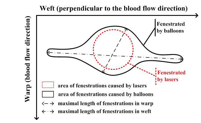
Figure 1. Schematic diagram illustrating the surface areas and the lengths of the fenestrations.
Results and Discussion: The in-situ fenestration procedure is feasible in all the devices. However, each device demonstrated different degrees of fraying and/or tearing (Figure 2). The monofilament twill weave structure (Medtronic Valiant) tore in weft while the multifilament plain weave structure (Anaconda Vascutek and Zenith TX2 Cook) showed more fraying. The area of the fenestration of Valiant was the largest (range from 32.4 mm2 to 72.1 mm2) among all devices. The one of Zenith TX2 (range from 8.0 mm2 to 34.6 mm2) was a little bigger than the one of Anaconda (range from 5.1 mm2 to 28.7 mm2). The size and directions of tearing were more predictable with the 8 mm diameter balloon whereas the results obtained with the 10 and 12 mm diameter balloons were more unpredictable. The fenestrations were free of melting of the yarns and blackening of the filaments.
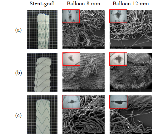
Figure 2. Fenestration with laser probe in 2.3 mm followed balloons.
(a) Zenith TX2; (b) Anaconda; (c) Valiant
Conclusions: The area and length of fenestration was more predictable with the 8 mm angioplasty balloon. The fabric of Valiant was easier to tear and caused an over enlarged fenestration compared other devices. Thus, the surgeon should make a prudent selection on the stent-grafts and the instruments of fenestration. The in-situ fenestration must currently be restricted to urgent and emergent cases using laser probe and an 8 mm diameter balloon.
This project was supported by the 111 Project (B07024) and Shanghai Construction of College Experimental Technique Team Project (101-07-0053014).
References:
[1] Manning BJ, Ivancev K, Harris PL. In situ fenestration in the aortic arch. J Vasc Surg. 2010;52:491-494.
[2] Redlinger RE, Ahanchi SS, Panneton JM. In situ laser fenestration during emergent thoracic endovascular aortic repair is an effective method for left subclavian artery revascularization. J Vasc Surg. 2013;58:1171-1177.
[3] Parodi JC, Palmaz JC, Barone HD. Transfemoral intraluminal graft implantation for abdominal aortic aneurysms. Ann Vasc Surg. 1991;5:491-499.
[4] Tse LW. First Clinical Case of Antegrade In Situ Fenestration of an Aortic Stent-Graft. J Endovasc Ther. 2012;19:721-722.
[5] Sonesson B, Resch T, Allers M, Malina M. Endovascular total aortic arch replacement by in situ stent graft fenestration technique. J Vasc Surg. 2009;49:1589-1591.