Multimodal AI for prediction of distant metastasis in carcinoma patients
 Isaac Olatunji
 Feng Cui
- Thomas H. Gosnell School of Life Science, Rochester Institute of Technology, Rochester, NY, United States
Metastasis of cancer is directly related to death in almost all cases, however a lot is yet to be understood about this process. Despite advancements in the available radiological investigation techniques, not all cases of Distant Metastasis (DM) are diagnosed at initial clinical presentation. Also, there are currently no standard biomarkers of metastasis. Early, accurate diagnosis of DM is however crucial for clinical decision making, and planning of appropriate management strategies. Previous works have achieved little success in attempts to predict DM from either clinical, genomic, radiology, or histopathology data. In this work we attempt a multimodal approach to predict the presence of DM in cancer patients by combining gene expression data, clinical data and histopathology images. We tested a novel combination of Random Forest (RF) algorithm with an optimization technique for gene selection, and investigated if gene expression pattern in the primary tissues of three cancer types (Bladder Carcinoma, Pancreatic Adenocarcinoma, and Head and Neck Squamous Carcinoma) with DM are similar or different. Gene expression biomarkers of DM identified by our proposed method outperformed Differentially Expressed Genes (DEGs) identified by the DESeq2 software package in the task of predicting presence or absence of DM. Genes involved in DM tend to be more cancer type specific rather than general across all cancers. Our results also indicate that multimodal data is more predictive of metastasis than either of the three unimodal data tested, and genomic data provides the highest contribution by a wide margin. The results re-emphasize the importance for availability of sufficient image data when a weakly supervised training technique is used. Code is made available at: https://github.com/rit-cui-lab/Multimodal-AI-for-Prediction-of-Distant-Metastasis-in-Carcinoma-Patients.
Introduction
Proper management planning for carcinoma patients requires accurate diagnosis, and prognosis prediction. A major prognostic factor, metastasis, however is not always diagnosed at initial patient presentation. Metastasis refers to the dissemination of cancer cells away from the initial site of origin to form colonies at distant organs. This single hallmark of malignancies is responsible for the highest proportion (approximately 90%) of cancer related mortalities (Fares et al., 2020). In HNSCC (Head and Neck Squamous Cell Carcinoma), about 10% of patients present with Distant Metastasis (DM) at diagnosis, while 20%–25% are detected during the disease course (Pisani et al., 2020). Pancreatic cancer is usually diagnosed at a late stage, and DM is quite common at presentation, however, multidetector computed tomography (MDCT) which is currently the optimal preoperative investigation has poor sensitivity to liver and peritoneal metastasis, the most common metastatic sites (Liu et al., 2018). Cases of metastatic non-muscle-invasive bladder cancer have also been reported (Xu et al., 2022). Early diagnosis of metastatic cancer is critical if patients will benefit from systemic therapies, alongside other appropriate management plans. The mechanisms behind metastasis, however, are very complex, and a lot is still not understood. Currently, there are no standard biomarkers of metastasis (Wang et al., 2018), and identification of biomarkers that are associated with metastasis will be useful in guiding clinical decisions, and as basis for development of new therapies.
Past studies have investigated the presence or absence of generalizable links between genes involved in the metastasis of different cancer types, either on the basis of mutation, or expression level. However, metastasis has proven to be a complex molecular and biochemical process. While similar mutation rate, and expression pattern have been reported in selected genes across groups of metastatic cancer, there are multiple claims that there are in fact no specific cancer metastatic genes. In a study carried out by Liu et al. (2017), mutation rates of TP53 was significantly different between primary and metastatic samples in seven cancer types, while PTEN mutation level was different in five cancer types. Copy number variations also differ significantly in all 15 cancer types examined. Nguyen et al. (2022) also implicated TP53, PTEN, CDKN2A, and MYC as significantly mutated genes in the metastasis of various subsets of cancer types.
In specific cancer types, designated differentially expressed signature genes are the basis of some of the past attempts to stratify cancer patients based on the risk of DM (van de Vijver et al., 2002). A recent study (Kaur et al., 2022) carried out in triple negative breast cancer samples using DESeq2 software identified a total of 1738 differentially expressed genes between metastatic and non-metastatic primary samples, 3 of which are part of the 70 prognostic signature genes in (van de Vijver et al., 2002). An analogous study in renal cell carcinoma noted some of the identified differentially expressed genes as predictive of metastasis-free survival, and overall survival (Ho et al., 2017). In melanoma, measurement of Breslow’s thickness of the primary tumor was correlated with the level of expression of specific genes, and the transition to metastatic tumor (Riker et al., 2008). While all of these reports suggest that genomic data may actually be predictive of metastasis, the inconsistent patterns of gene mutation and expression seen in different cancer samples makes it a challenge to precisely identify their importance in specific cases.
The wealth of morphological information contained in the tumor microenvironment is routinely exploited by pathologists for making definitive diagnosis of cancer and predicting patient prognosis. However, aside from being time consuming, this tedious process has also been associated with inter- and intra-observer variability that sometimes lead to unresolved diagnosis or worse, errors in diagnosis. With the use of Convolutional Neural Network (CNN), histopathology images have proven to be good predictors of malignancy status, important molecular biomarkers of various clinical and research relevance, as well as other cellular and extracellular processes (Mungenast et al., 2021). While many deep learning models have achieved relatively high metrics in detecting tumors within histopathology images of metastatic lymph node samples (Chuang et al., 2021; Wen et al., 2021; Huang et al., 2022), the difficult problem of predicting DM from primary samples remains a challenge, and most of the past attempts on this and similar tasks have struggled with relatively average model performance (Zhao, 2020; Brinker et al., 2021; Kiehl et al., 2021; Schiele et al., 2021).
Recent trends of use of multimodal data for prediction of diagnosis and outcomes in cancer patients have reported mostly improved metrics compared to use of singular modes of data (Mobadersany et al., 2018; Chen et al., 2022). In the case of metastasis prediction, past works have used singular or multimodal data combining clinical (Ali, 2020), genomic (Yuan et al., 2019), radiological (Liu et al., 2020), and histopathology (Zhao, 2020) data, however, to the best of our knowledge, at the time of this writing, this is one of the first works that combines transcriptomic data, clinical data, and histopathology images from primary tumor samples to predict DM.
In this work, we attempt to predict DM using gene expression data, clinical data, and histopathology images from primary tumors of three carcinoma types - Pancreatic Adenocarcinoma (PAAD), Bladder Carcinoma (BLCA), and Head and Neck Squamous cell Carcinoma (HNSC). The contributions of this research include:
- We identify genomic markers of DM in three different carcinoma types using a novel combination of the random forest algorithm with an optimized feature selection approach described in (Mori et al., 2021). Genes selected via this method performed better in prediction of DM than differentially expressed genes (DEGs) derived from DESeq2 analysis in our study, as well as in comparison to methods from other similar studies. These biomarkers could be further investigated for development of new diagnostics and therapies against DM.
- Using various machine learning techniques, we investigate and substantiate claims that genes involved in DM tend to be more cancer type specific rather than general across all cancer types, and that there are no specific cancer metastasis genes.
- We built separate models to predict DM from gene expression data, clinical data, or histopathology images, as well as multimodal combinations of the three data types. Models metrics show that multimodal data provides an edge for prediction of DM over genomic, clinical or histopathology data. However, the genomic data has the highest contribution by a wide margin.
- Unlike with genomic data in which features tend to be more cancer type specific, models built from histopathology image dataset of all three cancer types in total had better metrics than those built from a single cancer type image dataset, emphasizing the importance of sufficient data to build a robust model when a weakly supervised technique is used.
Methods
Dataset
Barcode of patients with DM in the TCGA-HNSC, TCGA-BLCA, and TCGA-PAAD projects were retrieved from Genomic Data Commons (GDC) Data Portal, and gene expression data, clinical data, and histopathology images of these patients were downloaded. The few number of available cases of DM is a common challenge in most studies on metastasis. BLCA, and PAAD were selected based on the number of cases of DM in these cancer types that are available on The Cancer Genome Atlas (TCGA), and the number of cases with available corresponding genomic, image, and clinical data. A few cancer types were dropped during this selection process due to the absence of some variables in their records. For example, Skin Cell Carcinoma (SKCM) was eliminated due to missing “number_of_lymph_nodes_positive_by_HE” variable which is present in BLCA, PAAD, and HNSC datasets. HNSC was added based on the interest of one of the authors. See Supplementary Material S1 for file ID and barcode of samples. BLCA (N = 80) records had 59 male, and 21 female patients. Average age in PAAD (N = 58; Male = 32; Female = 26), and HNSC(N = 24; Male = 21; Female = 3) records is 63, and 61 respectively. The race of patients in the BLCA cohort are (White = 66; Black or African American = 6; Asian = 5), and (White = 47; Black or African American = 3; Asian = 5) for PAAD, while HNSC had (White = 19; Black or African American = 4; Asian = 1). The T, and N staging, and Number of lymph nodes positive by hematoxylin and eosin (HE) staining in each cancer type is shown in Table 1.
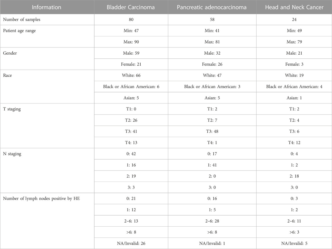
TABLE 1. Information about metastatic datasets.
Machine learning for identification of transcriptomic biomarkers and prediction of distant metastasis
We designed a study that utilized machine learning techniques to simultaneously identify biomarkers of DM in each of BLCA, PAAD, and HNSC, and investigate if genes involved in DM are similar across the three cancer types. Each cancer type dataset was pre-processed separately. Equal numbers of complementary gene expression data of samples without DM (BLCA = 80; PAAD = 58; HNSC = 24) were downloaded for each of the cancer types, and we ensured that there were no overlapping samples between the groups with DM, and those without DM. After extracting Transcript Per Million (TPM)) normalized values of protein-coding genes from the individual records, all samples in a group were merged to create a table, and NaN values were replaced with zero. Other initial preprocessing steps include log to base ten transformation of data to adjust for the wide and non-linear values, removal of genes with a value of zero in greater than 80% of the samples, and selection of only top 10000 genes with highest variance across all samples.
To identify important genes that are involved in DM, we utilized the Random Forest (RF) algorithm with an optimization technique described in (Mori Y. et al., 2021). RF is a supervised ML algorithm that creates multiple decision trees from bootstrapped samples data and randomly selected features to output a result, that is, based on a voting system. It works well with high dimensional data, and is a commonly utilized technique in gene expression data analysis. The optimization technique we employed consists of running the RF algorithm over (N = 1000) iterations to classify samples as DM positive or DM negative, and selecting the top (K = 100) most important genes at each of the N instances. This is followed by ranking the important genes overall based on how many times each gene appears (i.e., frequency) in K over the N iterations. The algorithm was used to classify samples in each of the groups based on the presence or absence of DM (Figure 1). We selected the top 50 overall highest ranked genes for BLCA, PAAD, and HNSC. Also, the three cancer types datasets were combined to derive the “ALL3” group, and the above steps were repeated to select the top 50 highest ranked genes in this group as well (Figure 1).
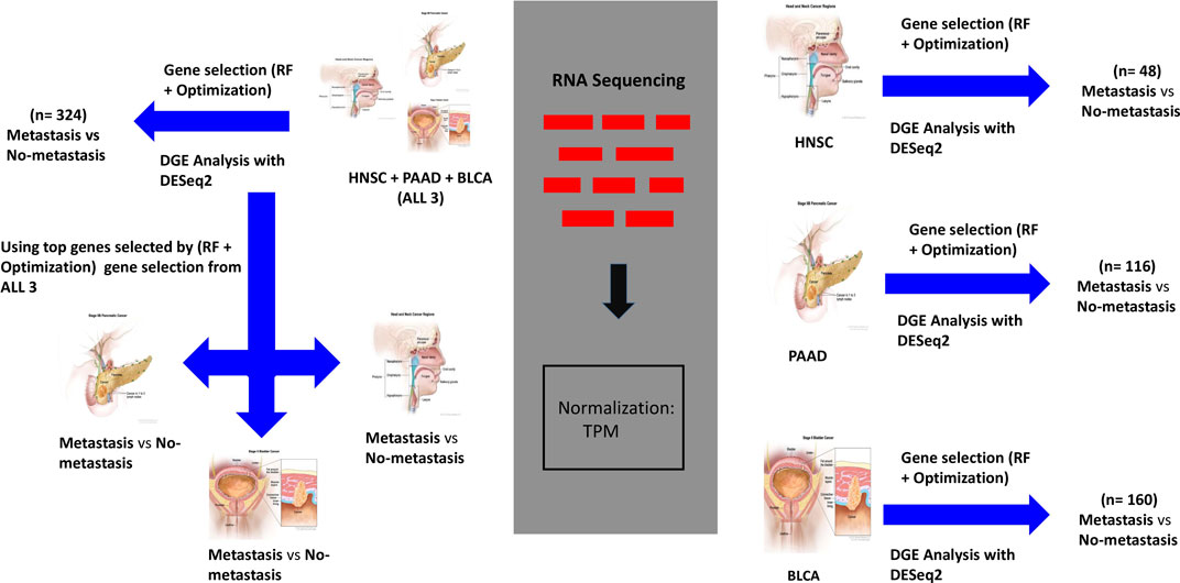
FIGURE 1. Workflow for analysis of genomic data. On the right, gene expression datasets of HNSC, PAAD, and BLCA are separately passed through the (RF + Optimization) gene selection algorithm, and a parallel DGE Analysis. Separate ML models are trained on highest ranked selected genes, and DEGs to predict DM samples. On the top left, the same processes carried out on ALL3 (combined dataset of HNSC, PAAD, and BLCA) dataset. On the lower left, genes selected from ALL3 dataset are used as features to train new models on each of the cancer types (i.e., other groups- HNSC, PAAD, BLCA) dataset. Cancer images source: National Cancer Institute.
To assess the strength of the selected biomarkers for prediction of DM, and to investigate if genes involved in DM are similar across the three cancer types, first, we used only the selected genes in each of the groups as features to train multiple ML models for the task of DM prediction. Support Vector Machine (SVM), K-Nearest Neighbor (KNN) and RF models were trained on datasets from each of the four (BLCA, PAAD, HNSC ALL3) groups (Figure 1), and the metrics were evaluated. Next, we looked for overlaps between different combinations of the selected genes in the four groups. Furthermore, we trained new ML (SVM, RF, and KNN) models on BLCA, PAAD, and HNSC datasets, but with genes selected from the combined dataset of the three cancer types (i.e., ALL3 group) (Figure 1). Lastly, we created a list of union of selected genes from each of the three cancer types (i.e., BLCA + PAAD + HNSC), and these were used as variables to predict presence or absence of DM in each of the three cancer dataset.
Differential Gene Expression with DESeq2
To further assess the strength of genes selected by the proposed method for predicting DM, under the same study design, we carried out an analysis to identify Differentially Expressed Genes (DEGs) between samples with DM and those without DM in each of the four groups. Instead of the TPM normalized values, raw counts of unstranded RNASeq were extracted from the TCGA RNASeq records. A sample information table was also generated. Differential Gene Expression (DGE) analysis was performed in R using the DESeq2 Bioconductor package. DESeq2 detects DEGs by normalizing raw count values of genes in the experimental groups, fitting negative binomial generalized linear models for each gene, and detecting significance by Wald test (Love et al., 2014). Threshold was set at p-adjusted value of 0.05, after initial set threshold of log fold-change of 1 and p-adjusted value of 0.05 yielded only five, one and seven DEGs in BLCA, PAAD and HNSC respectively. Thereafter, top DEGs were used as features to classify DM samples in each of the study groups (Figure 1). DEGs from this analysis and metrics of models trained on them are compared to those of genes selected via the (RF + Optimization) method.
Convolutional neural network for histopathology images analysis and prediction of distant metastasis
We downloaded diagnostic pathology Whole Slide Histopathology Images (WSIs) of patients with DM in the TCGA projects of BLCA, HNSC, and PAAD from GDC Data Portal. The number of samples in each group reduced slightly after collating a list of only patients with available histopathology images, and RNASeq data, and clinical data. Number of samples with DM in BLCA, PAAD, and HNSC groups are 50, 44, and 18 respectively. Again, we downloaded random complementary WSIs (BLCA = 55; PAAD = 51; HNSA = 22) of patients without DM for each of the cancer types, and ensured no overlap between samples (Supplementary Material S1). Total number of samples in each group was split in an 80:20 ratio for models training and testing.
WSIs preprocessing
One WSI was preprocessed per patient. Due to the typical large size of WSIs which hovers around 100000 pixels in both horizontal and vertical axis, we isolated representative regions within each WSI for analysis. First, a maximum of 500 random non-overlapping patches of size 512 * 512 pixels were extracted from each image at 20 × magnification. These were reduced to a maximum of 200 patches after checking for a tissue area of at least 80% in each patch. To ensure selection of tumor representative regions we imitated the concept of high cellularity described in (Riasatian, 2021) which assumes that high grade tumors contain more cellular areas than normal tissues. Hence, we used a pretrained U-Net nuclei segmentation model which was trained on breast cancer histopathology images to rank the patches based on cellular content. The top 60 highest ranked patches were selected for each patient. Further random manual inspection led to the exclusion of a few more patches before Macenko normalization. At the end of initial preprocessing steps, BLCA, PAAD, HNSC, and ALL3 groups had a total of 4936, 4348, 1856, and 11154 training patches respectively, of which 25% were for validation.
CNN training and multimodal fusion of genomic and imaging data
A DenseNet121 model, pretrained on Imagenet data, and KimiaNet (Riasatian, 2021) were chosen for this study. This allows us to evaluate the effect of keeping weights of lower layers of a model fine-tuned on domain histopathology images (i.e., KimiaNet) on performance. DenseNet is a CNN architecture that attempts to solve the problem of varnishing gradient associated with deep neural networks by cumulatively concatenating features output of a layer within the architecture to input of subsequent upper layers, (Huang et al., 2016; 2017). In total, DenseNet121 contains 1 7 × 7 convolution, 58 3 × 3 convolution, 61 1 × 1 convolution, 4 average pooling, and 1 fully connected layers. KimiaNet is a DenseNet121 architecture Imagenet-pretrained CNN model fine-tuned on about 250,000 histopathology images. We replaced the classification layers in the models with 1 GlobalAveragePooling2D, 3 Dropout (0.1, 0.2, and 0.05), and 3 Dense (512, 32, and 1) layers, to train a new classifier head on top of the base convolutional layers, using a weakly supervised technique. The last hidden layer of the new classifier heads has 32 nodes which is subsequently used as features extractor. The modified DenseNet121 was separately trained on data from each of the four study groups (HNSC, BLCA, PAAD, ALL3) for a binary classification task of DM prediction (Figure 2). Loss function was binary cross-entropy, and optimizer, Adam with a learning rate of 0.0001. Training epoch was set at 40.
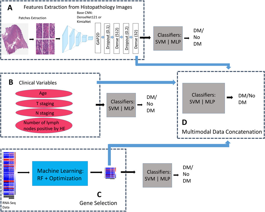
FIGURE 2. Binary classifiers trained on the same patients’ records but with different data types for comparisons between unimodal, and multimodal data models. (A) Additional Dense layers trained on DenseNet121, and KimiaNet CNN for features extraction from histopathology images. Classifiers are trained on image features to predict presence or absence of DM. (B) Classifiers are trained on only clinical variables to predict presence or absence of DM. (C) Classifiers are trained on genes selected by the (RF + Optimization) algorithm to predict presence or absence of DM (D) Image features are fused with selected genes, and clinical features to train multimodal models for prediction of DM.
For prediction on the test sets, patient level features were derived by passing all patches for each patient through the trained feature extractor models, and averaging all patch features from a slide. We trained traditional machine learning algorithm- SVM and a single layer MLP on the patient level features to classify samples as DM positive or DM negative (Figure 2). Considering this approach we have taken, HNSC group was dropped here based on small sample size.
Multimodal concatenation of patient level features derived from the CNN models to log transformed values of TPM normalized RNASeq data of selected DM associated genes from above was carried out. For BLCA, and PAAD groups, the 50 genes selected using the (RF + Optimization) technique were combined with 32 features derived from histopathology images of each patient to train classifiers, and predict presence or absence of DM (Figure 2). 150 genes, consisting of 50 each selected from BLCA, PAAD, and HNSC were combined with 32 image features to predict DM in the ALL3 group (Figure 2).
Likely due to the effect of a larger training dataset given the weakly supervised training technique that was employed, it was observed from early results that image only models trained on data from the ALL3 group generally performed better in their predictions on the test sets than other groups. Similarly, multimodal models of genomic and histopathology image were generally better than unimodal model of either data type in the ALL3 dataset, while metrics of multimodal models are generally lower than either genomic or image only models in other individual cancer dataset (i.e., BLCA, or PAAD). Therefore further experiments were carried out on the ALL3 dataset. We trained fully connected layers on KimiaNet, a DenseNet121 architecture model finetuned on about 250,000 histopathology images to extract features from the images in the ALL3 dataset. Metrics of multimodal models built from these features, and genomic data were compared to those from DenseNet121.
Four variables—Age, T stage, N stage, and Number of lymph nodes positive by HE—were selected from the clinical data, and concatenated with genomic, and histopathology features to predict presence or absence of DM in the ALL3 dataset (Figure 2). As part of the clinical data preprocessing, invalid/NaN values in the age variable were replaced by the rounded mean age of the cancer type while invalid/NaN T staging, N staging and number of positive lymph node values were replaced with 0. Min-max normalization was applied to age, and number of positive lymph node variables.
Results
ML identified gene expression biomarkers of distant metastasis
In each cancer type, samples with DM were combined with the same size of samples without DM. Also, datasets from the three cancer types were combined to derive the “ALL3” group. Following pre-processing, the optimized RF algorithm, as described in the methods section was used for binary classification of the samples (i.e., DM vs. non-DM), and simultaneous gene selection (top 50) in each of the groups (Supplementary Material S2). While RF is a popular ML based gene selection technique (Díaz-Uriarte and Alvarez de Andrés, 2006; Wenric and Ruhollah, 2018; Nivedhitha et al., 2020), to the best of our knowledge, at the time of this writing, this is the first time RF is combined with this optimization method for the task of gene selection. The top 5 genes associated with metastasis in BLCA are FKBP6, ASIC5, MAPK8IP1, F11R, and PABPC5, while CTSV, BIRC5, SERPINA7, CST2, KLHL3 are the highest ranked genes in PAAD. In HNSC, TM4SF1, C19orf18, EXPH5, FKBP2, PTPRZ1 are the highest ranked genes, and ALL3 group had CHRNG, CPT1B, CGREF1, GPR31, SPTBN5. Past publications have confirmed the activities of some of these genes in the metastasis of various cancers, either as oncogene or as tumor suppressors (Table 2). There are others whose roles in cancer metastasis are yet to be explored. A functional annotation search for the selected 50 genes on pantherdb.org revealed very similar classification patterns in all of the three cancer types. Approximately 50%–60% of the genes had no known category, and the most common functional classification of those with known categories in all the cancers are protein binding, and catalytic activity (Figure 3).
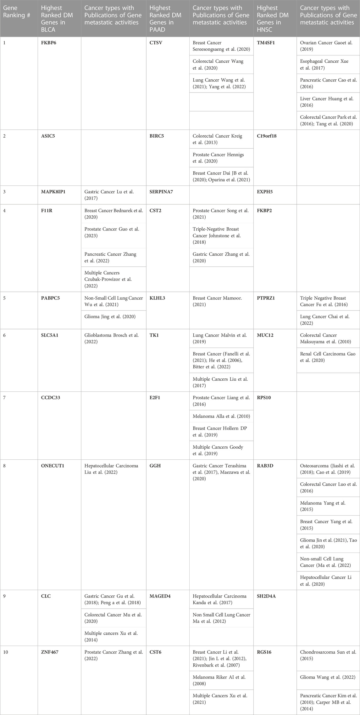
TABLE 2. Top 10 genes selected by the ML (RF + Optimization) technique in BLCA, PAAD, HNSC, and various types of malignancies in which their involvement in metastasis have been reported in past literatures.
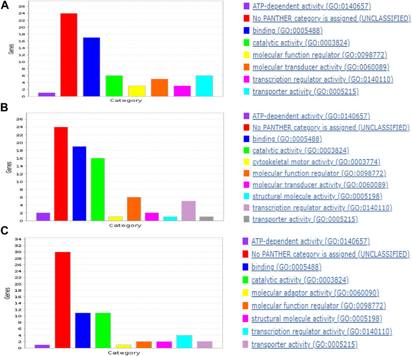
FIGURE 3. Functional classification of selected genes in (A) BLCA, (B) PAAD, and (C) HNSC.
Dataset in each group was randomly split into train and test set in a 75:25 ratio, and SVM with linear kernel, KNN, and RF models were trained on the highest ranked 30, 25, 20, 15, 10, 5, 3, 2, and 1 gene from list of selected top 50 genes. Outcomes of single instance predictions, and five-fold cross-validation on the test sets were recorded (Supplementary Material S3).
To test the strength of genes selected based on the (RF + Optimization) method as predictors of DM, a DGE analysis was carried out with the DESeq2 software package to identify DEGs between the DM and non-DM samples. With a threshold adjusted p-value of 0.05, the number of DEGs in BLCA, PAAD, HNSC, and ALL3 are 229, 2142, 1100, and 658 respectively. Of these 5, 39, 19, and 13 overlap with the 50 genes selected using the ML method in each of the respective groups. Presence or absence of DM was predicted in the four groups using DEGs with the lowest 30, 25, 20, 15, 10, 5, 3, 2, and 1 adjusted p-value as variables. In almost all cases, genes selected using the ML (RF + optimization) techniques had higher evaluation metrics (Accuracy, F1-score, AUROC) than those selected via DESeq2 DGE analysis in the task of DM prediction (Figure 4). Highest mean AUROC score achieved over a five-fold cross validation was 0.87, 0.92, 0.97, 0.79 in BLCA, PAAD, HNSC, and ALL3 groups respectively, compared to 0.65, 0.80, 0.92, 0.62 with DESeq2 DGE analysis, when models were trained on top 15 selected genes (Figure 4; Supplementary Material S3).
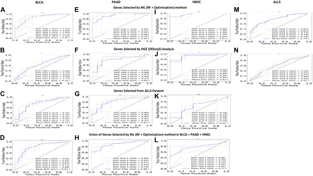
FIGURE 4. (A, E, I, M) Five-fold cross validation ROC curves, and mean ROC curve when 15 highest ranked genes selected by (RF + Optimization) method from each of the groups are used to predict presence or absence of DM. These are higher than other predictions within the same study group. (B, F, J, N) Five-fold cross validation ROC curves, and mean ROC curve when 15 highest ranked DEGs (p adjusted value = 0.05) are used to predict presence or absence of DM. (C, G, K) Five-fold cross validation ROC curves, and mean ROC curve when 15 highest ranked genes selected by (RF + Optimization) method from ALL3 group are used to predict presence or absence of DM in other (BLCA, PAAD, HNSC) study groups. (D, H, L) Five-fold cross validation ROC curves, and mean ROC curve when a union of the 15 highest ranked genes selected by (RF + Optimization) method from each of BLCA, PAAD, and HNSC groups (total = 45) are used to predict presence or absence of DM in each cancer type.
Expression profile of genes associated with DM in primary tumors differ across cancer types
To investigate if primary tumors of different cancer types share similar gene expression profiles in metastasis, first, we looked for overlap between various combinations of the selected 50 genes in each group. There was no overlap between the different combinations of BLCA, PAAD or HNC. However, in the ALL3 group, 7 genes (CGREF1, SPTBN5, TAS1R3, FAM241B, EL5, CSPG5, and MAPK8IP1), and 6 genes (CFAP45, CST2, BIRC5, MAGED4, CST6, and FAIM2) overlap with those selected in BLCA, and PAAD respectively (Figure 5; Table 3). Also, it was observed that there was at least one member of the ZNF and FKBP gene family present within the list of selected genes in BLCA, and HNSC. Jen and Wang (2016) extensively reviewed multiple studies on the role of ZNF (Zinc Finger) gene family proteins in cancer progression, and metastasis, acting both as oncogenes, and tumor suppressors. Various studies (Fong et al., 2003; Sun et al., 2021) have also implicated members of the FKBP family in cancer metastasis.
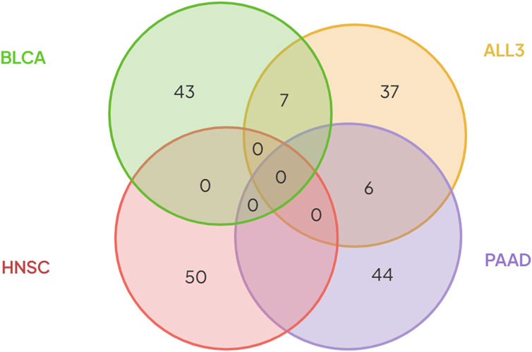
FIGURE 5. There was no overlap between genes selected using the (RF + Optimization) method in either of the three cancer types- BLCA, PAAD, and HNSC. Six genes selected in PAAD, and seven genes in BLCA were also present in the list of genes selected from the ALL3 group.
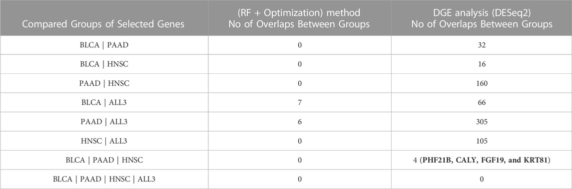
TABLE 3. Number of overlapping genes between combinations of the different groups of genes selected by (RF + Optimization) method, and DGE analysis by DESeq2.
Furthermore, we trained new ML (SVM, RF, and KNN) models on BLCA, PAAD, and HNSC datasets, but with genes selected from the combined dataset of the three cancer types (i.e., ALL3 group) (Figure 1). These models performed lesser than in cases of predictions of DM using genes selected in individual cancer types. Highest mean AUROC score of 0.76, 0.82 and 0.64 was achieved in BLCA, PAAD, and HNSC datasets respectively against 0.87, 0.92, and 0.97 of models trained on selected genes derived from individual cancer types (Figure 4; Supplementary Material S3). To further confirm this specificity of genes in each cancer type, a union list of selected top 15 genes in the three cancer types - BLCA, PAAD, and HNSC was created. Results show a drop in mean AUROC scores from 0.87, 0.92, and 0.97 in BLCA, PAAD, and HNSC datasets respectively when 15 genes generated from each cancer type were used as variables to 0.83, 0.88, and 0.77 when the total of 45 genes in the list of unified genes were used as variables to predict DM (Figure 4; Supplementary Material S3).
Histopathology images and clinical and genomic data for multimodal prediction of DM
DenseNet121 was trained on histopathology images from BLCA, PAAD, and ALL3 groups, after the HNSC group was dropped due to small sample size. One WSI was preprocessed per patient as described in the methods section. For each group, classification models—SVM, and MLP were trained on patient level features extracted by the trained CNN models, and prediction was carried out on the test set features (Figure 2). The image features were also combined with genomic features from the same patient to build multimodal (Image + Genomic) models.
Early results showed that metrics of multimodal (Image + Genomic) models built from the ALL3 dataset generally improved on image, or genomic unimodal models by 1–3 percent margin, with genomic data contributing more to the multimodal metrics by a large margin. Following a five-fold Monte Carlo cross-validation, a multimodal (Image + Genomic) SVM classifier produced a mean AUROC score of 0.77, while the corresponding image and genomic unimodal models produced a score of 0.58, and 0.74 respectively. The mean AUROC scores from MLP classifiers are (Image + Genomic = 0.81; Image = 0.59; Genomic = 0.80). In either BLCA or PAAD dataset, results of multimodal (Image + Genomic) models mostly improved on those of the image only models, however they were generally below that of genomic only models. The highest mean AUROC derived from a BLCA dataset multimodal (Image + Genomic) model is 0.62, and the corresponding values from image only, and genomic only model are 0.57, and 0.80 respectively. Similar pattern was seen in the PAAD dataset [Image + Genomic = 0.85; Image = 0.51; Genomic = 0.90) (Figure 6; Supplementary Material S4)]. These outcomes emphasize the importance of a large dataset for building a more robust image model when a weakly supervised training technique is employed.
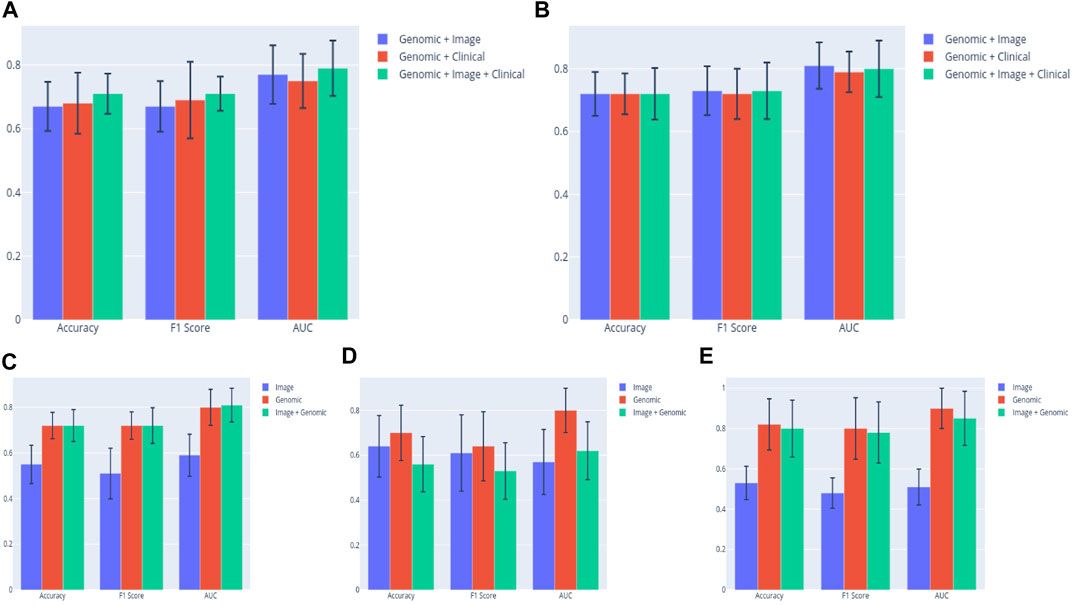
FIGURE 6. (A) SVM classifier clearly shows superiority of the multimodal (Clinical + Genomic + Image) model in the ALL3 dataset. (B) The performance of the (Clinical + Genomic + Image) model appears to be on par with the bimodal models when a MLP classifier is used. Comparing prediction metrics of the best performing bimodal (Image + Genomic) model in each group to those of unimodal models. Results from the bimodal model from the (C) ALL3 dataset are clearly better than those from unimodal (D) BLCA dataset, and (E) PAAD dataset.
Based on these early results, we carried out further experiments with the ALL3 dataset. To evaluate the effect of using a CNN model with lower layers pre-finetuned on histopathology domain images as features extractor, fully connected layers were trained on KimiaNet with the ALL3 dataset histopathology images, and features derived from the model were combined with genomic data to predict DM in the test set. There was generally no additional advantage observed in the results of KimiaNet over those from DenseNet121. Highest mean AUROC from the MLP multimodal model (KimiaNet Image + genomic data) is 0.77. Unimodal image and genomic models from the same dataset produced AUROC scores of 0.55, and 0.80 respectively. The same pattern was seen with the SVM classifier (Image + Genomic = 0.73; Image = 0.56; Genomic = 0.74) (Supplementary Material S4).
Lastly, four clinical variables—Age, T stage, N stage, and Number of lymph nodes positive by HE were concatenated with genomic, and histopathology features to train SVM, and MLP classifiers. This led to an improvement of the mean AUROC score of the Image + Genomic SVM model from 0.77 to a score of 0.79. Mean accuracy, and F1 scores also improved from 0.67 and 0.67 to 0.71 and 0.71 respectively. MLP classifier however produced a mean AUROC score of 0.80 against the score of 0.81 achieved with Genomic + Histopathology data (Figure 6). Combining clinical with genomic data improved the metrics of the clinical only models, however there was no marked improvement from metrics of the genomic only models (Figure 6; Tables 4, 5).
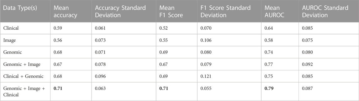
TABLE 4. Accuracy, F1, and AUROC scores derived from combinations of clinical, genomic and image data in the ALL3 dataset with a SVM classifier.
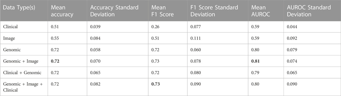
TABLE 5. Accuracy, F1, and AUROC scores derived from combinations of clinical, genomic and image data in the ALL3 dataset with a MLP classifier.
Discussion
Given that there was barely any overlap between genes selected from the different cancer types considered and that prediction of metastasis in each of the cancer types with a union of genes selected from the three cancer types or the ALL3 group produced inferior results, we have been able to substantiate the claim that metastatic genes are more cancer type specific, rather than general, across carcinomas. As only three carcinoma types are considered in this study, studies including a larger number of carcinoma types will be needed to further solidify our findings. As shown on (Table 3), some of the selected genes have however been identified to be involved in the metastasis of multiple cancers. These further confirm the theory that even though metastatic genes tend to be specific to each carcinoma type, this specificity is based more on group of genes rather than individual genes, and that there is no single metastatic gene as have been reported in (Nguyen et al., 2022). The AUROC scores, and other metrics reported from our study, and other similar studies call for future works and development of diagnostics and therapeutics against DM to perhaps be more focused on the carcinoma type, rather than a general one size fits all approach. The ZNF and FBKP family of genes which is present in the list of selected genes in the BLCA, and HNSC datasets have been severally associated with cancer progression, and metastasis in multiple cancers (Fong et al., 2003; Jen and Wang, 2016; Sun et al., 2021), and should be investigated more for their roles in cancer metastasis.
Furthermore, with our novel combination of the RF algorithm and the described optimization technique, we have identified separate gene expression biomarkers of DM in the individual cancer types. Metrics of models built with these genes (AUC: BLCA = 0.87; PAAD = 0.92; HNSC = 0.97) outperform those built from genes selected by DESeq2 DGE analysis (AUC: BLCA = 0.65; PAAD = 0.82; HNSC = 0.92). While it is important to note that the datasets in our study are focused specifically on DM, similar studies on related tasks have been reported. Using just RF algorithm for gene selection in a breast cancer study (Yao et al., 2022), achieved an AUC of 0.52 in a metastasis and recurrence prediction task. (Wu et al., 2017), and (Qiao et al., 2020) achieved AUC scores of 0.71 and 0.84 respectively when genes derived from DESeq2 DGE analysis, and Boruta algorithm in the latter were combined with clinical data for prediction of lymph node metastasis in HNSC. In predicting metastasis status in pancreatic cancer samples (Xue et al., 2021), achieved a highest AUC score of 0.72 using DEG identified by the edgeR package. A 51 gene signature produced an AUC score of 0.82 in prediction of lymph node metastasis in bladder cancer as reported by (Seiler et al., 2016).
The reported AUC scores from our study were derived from the highest ranked 15 genes out of the initially selected 50. Perhaps a protein-protein interaction analysis including the initial 50 genes could lead to selection of fewer genes with higher relevance than the ranking we have employed here, which may in turn lead to improved prediction metrics. The biomarkers discovered in each cancer type using the strict ML approach we have employed should be further investigated for their potential as diagnostic indicators, and as basis for development of new therapies against cancer metastasis.
Our results indicate that multimodal data (Genomic + Clinical + Histopathology) provides a predictive advantage over either of the unimodal data types in the task of DM detection, however the contribution of only gene expression data is highest by a wide margin. Similar studies have acknowledged the superiority of multimodal data for prediction of medical diagnosis and various outcomes in cancer patients (Mobadersany et al., 2018; Keihl et al., 2021; Chen et al., 2022), however some studies have shown that this is not always the case (Brinker et al., 2021; Vale-Silva and Rohr, 2021). Past multimodal studies on prediction of metastasis from histopathology images have mostly combined clinical data with histopathology images. This combination used in prediction of nodal metastasis led to an improved AUC score of 74% in a study by (Keihl et al., 2021). In contrast, there was a 0.2% decrease from the only image AUC score of 61.8% reported by (Brinker et al., 2021) when clinical, and cellular data were included. Results from our study also suggest the importance of a larger dataset of images when a weakly supervised technique is employed as noted with the superior metrics of the multimodal models including histopathology images in the ALL3 dataset compared to BLCA, and PAAD dataset. A recent study by (Yao et al., 2022) reported a higher accuracy in model built from only genomic data compared to that built on combined genomic, and histopathology data for prediction of metastasis and recurrence in breast cancer. However, an AUC score of 0.75 was achieved when gene expression, histopathology images and clinical data were combined.
The highest mean AUC score of 0.79 derived from the SVM classifier in our study was from multimodal combination of gene expression, clinical, histopathology data, while the score of 0.81 derived from the MLP classifier was obtained from a genomic + histopathology image model. Overall, our results indicate that combining clinical, genomic, and histopathology image data increases the prediction metrics for DM, however genomic data alone is a strong contributor.
Data availability statement
The original contributions presented in the study are included in the article/Supplementary Material, further inquiries can be directed to the corresponding author.
Author contributions
IO: Conceptualization, Methodology, Software, Data Analysis, Writing; FC: Conceptualization, Data Analysis, Writing. All authors listed have made a substantial, direct, and intellectual contribution to the work and approved it for publication. All authors contributed to the article and approved the submitted version.
Acknowledgments
We acknowledge the resources and assistance provided by RIT (Rochester Institute of Technology) Research Computing which has made this work possible.
Conflict of interest
The authors declare that the research was conducted in the absence of any commercial or financial relationships that could be construed as a potential conflict of interest.
Publisher’s note
All claims expressed in this article are solely those of the authors and do not necessarily represent those of their affiliated organizations, or those of the publisher, the editors and the reviewers. Any product that may be evaluated in this article, or claim that may be made by its manufacturer, is not guaranteed or endorsed by the publisher.
Supplementary material
The Supplementary Material for this article can be found online at: https://www.frontiersin.org/articles/10.3389/fbinf.2023.1131021/full#supplementary-material
References
Ali, B., Mubarik, F., Zahid, N., and Sattar, A. K. (2020). Clinicopathologic features predictive of distant metastasis in patients diagnosed with invasive breast cancer. JCO Glob. Oncol. 6, 1346–1351. doi:10.1200/GO.20.00257
Alla, V., Engelmann, D., An, N., Pahnke, J., Schmidt, A., Kunz, M., et al. (2010). E2F1 in melanoma progression and metastasis. JNCI J. Natl. Cancer Inst. 102 (2), 127–133. doi:10.1093/jnci/djp458
Bednarek, R., Selmi, A., Wojkowska, D., Karolczak, K., Popielarski, M., Stasiak, M., et al. (2020). Functional inhibition of F11 receptor (F11R/junctional adhesion molecule-A/JAM-A) activity by a F11R-derived peptide in breast cancer and its microenvironment. Breast Cancer Res. Treat. 179 (2), 325–335. doi:10.1007/s10549-019-05471-x
Bitter, E. E., Morris, R. M., Mortimer, T., Barlow, K., Schekall, A., Townsend, M. H., et al. (2022). “The potential effects of thymidine kinase 1 on breast cancer invasion,” in Proceedings of the American association for cancer research annual meeting 2022 (Philadelphia (PA): AACR; Cancer Res).
Brinker, T. J., Kiehl, L., Schmitt, M., Jutzi, T. B., Krieghoff-Henning, E. I., Krahl, D., et al. (2021). Deep learning approach to predict sentinel lymph node status directly from routine histology of primary melanoma tumours. Eur. J. Cancer 154, 227–234. doi:10.1016/j.ejca.2021.05.026
Brosch, P. K., Korsa, T., and Taban, D. (2022). Glucose and inositol transporters, SLC5A1 and SLC5A3, in glioblastoma cell migration. Cancers 14 (23), 5794. doi:10.3390/cancers14235794
Cao, J., Yang, J., Ramachandran, V., Arumugam, T., Deng, D., Li, Z., et al. (2016). TM4SF1 regulates pancreatic cancer migration and invasion in vitro and in vivo. Cell. Physiol. Biochem. 39, 740–750. doi:10.1159/000445664
Cao, K., Fang, Y., Wang, H., Jiang, Z., Guo, L., and Hu, Y. (2019). <p>The lncRNA HOXA11-AS regulates Rab3D expression by sponging miR-125a-5p promoting metastasis of osteosarcoma</p>. Cancer Manag. Res. 11, 4505–4518. doi:10.2147/CMAR.S196025
Carper, M. .B., Denvir, J., Boskovic, G., Primerano, D. A., and Claudio, P. P. (2014). RGS16, a novel p53 and pRb cross-talk candidate inhibits migration and invasion of pancreatic cancer cells. Genes. Cancer 5 (11-12), 420–435. doi:10.18632/genesandcancer.43
Chai, R. C., Liu, X., Pang, B., Liu, Y., Li, J., Li, Y., et al. (2022). Recurrent PTPRZ1-MET fusion and a high occurrence rate of MET exon 14 skipping in brain metastases. Cancer Sci. 113, 796–801. doi:10.1111/cas.15211
Chen, R. J., Lu, M. Y., Wang, Y., Williamson, D. F. K., Rodig, S. J., Lindeman, N. I., et al. (2022). Pathomic fusion: An integrated framework for fusing histopathology and genomic features for cancer diagnosis and prognosis. IEEE Trans. Med. Imaging 41 (4), 757–770. doi:10.1109/tmi.2020.3021387
Chuang, W. Y., Chen, C. C., Yu, W. H., Yeh, C. J., Chang, S. H., Ueng, S. H., et al. (2021). Identification of nodal micrometastasis in colorectal cancer using deep learning on annotation-free whole-slide images. Mod. Pathol. 34 (10), 1901–1911. doi:10.1038/s41379-021-00838-2
Czubak-Prowizor, K., Babinska, A., and Swiatkowska, M. (2022). The F11 receptor (F11r)/junctional adhesion molecule-A (JAM-A) (F11R/JAM-A) in cancer progression. Mol. Cell. Biochem. 477, 79–98. doi:10.1007/s11010-021-04259-2
Dai, J. B., Zhu, B., Lin, W. J., Gao, H. Y., Dai, H., Zheng, L., et al. (2020). Identification of prognostic significance of BIRC5 in breast cancer using integrative bioinformatics analysis. Biosci. Rep. 40 (2), BSR20193678. doi:10.1042/BSR20193678
Díaz-Uriarte, R., and Alvarez de Andrés, S. (2006). Gene selection and classification of microarray data using random forest. BMC Bioinforma. 7, 3. doi:10.1186/1471-2105-7-3
Fanelli, G. N., Scarpitta, R., Cinacchi, P., Fuochi, B., Szumera-Ciećkiewicz, A., De Ieso, K., et al. (2021). Immunohistochemistry for thymidine kinase-1 (TK1): A potential tool for the prognostic stratification of breast cancer patients. J. Clin. Med. 10, 5416. doi:10.3390/jcm10225416
Fares, J., Fares, M. Y., Khachfe, H. H., Salhab, H. A., and Fares, Y. (2020). Molecular principles of metastasis: A hallmark of cancer revisited. Sig Transduct. Target Ther. 5, 28. doi:10.1038/s41392-020-0134-x
Fong, S., Mounkes, L., Liu, Y., Maibaum, M., Alonzo, E., Desprez, P. Y., et al. (2003). Functional identification of distinct sets of antitumor activities mediated by the FKBP gene family. Proc. Natl. Acad. Sci. U. S. A. 100 (24), 14253–14258. doi:10.1073/pnas.2332307100
Fu, F., Xiao, X., Zhang, T., Zou, Q., Chen, Z., Pei, L., et al. (2016). Expression of receptor protein tyrosine phosphatase ζ is a risk factor for triple negative breast cancer relapse. Biomed. Rep. 4, 167–172. doi:10.3892/br.2016.570
Gao, C., Yao, H., Liu, H., Feng, Y., and Yang, Z. (2019). TM4SF1 is a potential target for anti-invasion and metastasis in ovarian cancer. BMC Cancer 19, 237. doi:10.1186/s12885-019-5417-7
Gao, S. L., Yin, R., Zhang, L. F., Wang, S. M., Chen, J. S., Wu, X. Y., et al. (2020). The oncogenic role of MUC12 in RCC progression depends on c-Jun/TGF-β signalling. J. Cell. Mol. Med. 24 (15), 8789–8802. doi:10.1111/jcmm.15515
Goody, D., Gupta, S. K., Engelmann, D., Spitschak, A., Marquardt, S., Mikkat, S., et al. (2019). Drug repositioning inferred from E2F1-coregulator interactions studies for the prevention and treatment of metastatic cancers. Theranostics 9 (5), 1490–1509. doi:10.7150/thno.29546
Gu, Z., Li, Y., Yang, X., Yu, M., Chen, Z., Zhao, C., et al. (2018). Overexpression of CLC-3 is regulated by XRCC5 and is a poor prognostic biomarker for gastric cancer. J. Hematol. Oncol. 11, 115. doi:10.1186/s13045-018-0660-y
Guo, X., Gu, Y., Guo, C., Pei, L., and Hao, C. (2023). LINC01146/F11R facilitates growth and metastasis of prostate cancer under the regulation of TGF-β. J. Steroid Biochem. Mol. Biol. 225, 106193. doi:10.1016/j.jsbmb.2022.106193
He, Q., Fornander, T., Johansson, H., Johansson, U., Hu, G. Z., Rutqvist, L. E., et al. (2006). Thymidine kinase 1 in serum predicts increased risk of distant or loco-regional recurrence following surgery in patients with early breast cancer. Anticancer Res. 26 (6C), 4753–4759.
Hennigs, J. K., Minner, S., Tennstedt, P., Löser, R., Huland, H., Klose, H., et al. (2020). Subcellular compartmentalization of survivin is associated with biological aggressiveness and prognosis in prostate cancer. Sci. Rep. 10, 3250. doi:10.1038/s41598-020-60064-9
Ho, T. H., Serie, D., Parasramka, M., Cheville, J., Bot, B., Tan, W., et al. (2017). Differential gene expression profiling of matched primary renal cell carcinoma and metastases reveals upregulation of extracellular matrix genes. Ann. Oncol. 28, 604–610. doi:10.1093/annonc/mdw652
Hollern, D. P., Swiatnicki, M. R., Rennhack, J. P., Misek, S. A., Matson, B. C., McAuliff, A., et al. (2019). E2F1 drives breast cancer metastasis by regulating the target gene FGF13 and altering cell migration. Sci. Rep. 9 (1), 10718. doi:10.1038/s41598-019-47218-0
Huang, G., Liu, Z., and Weinberger, K. Q. (2017). IEEE conference on computer vision and pattern recognition. USA: CVPR. doi:10.48550/arXiv.1608.06993
Huang, S. C., Chen, C. C., Lan, J., Hsieh, T. Y., Chuang, H. C., Chien, M. Y., et al. (2022). Deep neural network trained on gigapixel images improves lymph node metastasis detection in clinical settings. Nat. Commun. 13, 3347. doi:10.1038/s41467-022-30746-1
Huang, Y.-K., Fan, X.-G., and Qiu, F. (2016). TM4SF1 promotes proliferation, invasion, and metastasis in human liver cancer cells. Int. J. Mol. Sci. 17, 661. doi:10.3390/ijms17050661
Jen, J., and Wang, Y. C. (2016). Zinc finger proteins in cancer progression. J. Biomed. Sci. 23 (1), 53. doi:10.1186/s12929-016-0269-9
Jiashi, W., Chuang, Q., Zhenjun, Z., Guangbin, W., Bin, L., and Ming, H. (2018). MicroRNA-506-3p inhibits osteosarcoma cell proliferation and metastasis by suppressing RAB3D expression. Aging (Albany NY) 10, 1294–1305. doi:10.18632/aging.101468
Jin, H., Miao, H., Nie, Y. W., and Lin, Y. Y. (2021). Investigating resistin like beta (RETNLB) as a tumor promoter for oral squamous cell carcinoma. Head. Face Med. 17 (1), 20. doi:10.1186/s13005-021-00272-4
Jin, L., Zhang, Y., Li, H., Yao, L., Fu, D., Yao, X., et al. (2012). Differential secretome analysis reveals CST6 as a suppressor of breast cancer bone metastasis. Cell. Res. 22, 1356–1373. doi:10.1038/cr.2012.90
Jing, F., Ruan, X., Liu, X., Yang, C., Wang, D., Zheng, J., et al. (2020). The PABPC5/HCG15/znf331 feedback loop regulates vasculogenic mimicry of glioma via STAU1-mediated mRNA decay. Mol. Ther. Oncolytics 17, 216–231. doi:10.1016/j.omto.2020.03.017
Johnstone, C. N., Pattison, A. D., Gorringe, K. L., Harrison, P. F., Powell, D. R., Lock, P., et al. (2018). Functional and genomic characterisation of a xenograft model system for the study of metastasis in triple-negative breast cancer. Dis. Model. Mech. 11 (5), dmm032250. doi:10.1242/dmm.032250
Kanda, M., Murotani, K., Sugimoto, H., Miwa, T., Umeda, S., Suenaga, M., et al. (2017). An integrated multigene expression panel to predict long-term survival after curative hepatectomy in patients with hepatocellular carcinoma. Oncotarget 8, 71070–71079. doi:10.18632/oncotarget.20369
Kaur, J., Chandrashekar, D. S., Varga, Z., Sobottka, B., Janssen, E., Kowalski, J., et al. (2022). Distinct gene expression profiles of matched primary and metastatic triple-negative breast cancers. Cancers (Basel) 14 (10), 2447. doi:10.3390/cancers14102447
Kim, J. H., Lee, J. Y., Lee, K. T., Lee, K. H., and Jang, K. T. (2010). RGS16 and FosB underexpressed in pancreatic cancer with lymph node metastasis promote tumor progression. Tumour Biol. J. Int. Soc. Oncodevelopmental Biol. Med. 31 (5), 541–548. doi:10.1007/s13277-010-0067-z
Krieg, A., Werner, T. A., Verde, P. E., Stoecklein, N. H., and Knoefel, W. T. (2013). Prognostic and clinicopathological significance of survivin in colorectal cancer: A meta-analysis. PLOS ONE 8 (6), e65338. doi:10.1371/journal.pone.0065338
Kiehl, L., Kuntz, S., Höhn, J., Jutzi, T., Krieghoff-Henning, E., Kather, J. N., et al. (2021). Deep learning can predict lymph node status directly from histology in colorectal cancer. Eur. J. Cancer 157, 464–473. doi:10.1016/j.ejca.2021.08.039
Li, S., Liu, S., and Bai, Y. (2020). Filopodia associated promotes hepatocellular carcinoma metastasis by altering the metabolic status of cancer cells through RAB3D. Hepatology 73, 2361–2379. doi:10.1002/hep.31641
Li, X., Liang, Y., Lian, C., Peng, F., Xiao, Y., He, Y., et al. (2021). CST6 protein and peptides inhibit breast cancer bone metastasis by suppressing CTSB activity and osteoclastogenesis. Theranostics 11 (20), 9821–9832. doi:10.7150/thno.62187
Liang, Y., Lu, J., Mo, R., He, H., Xie, J., JiangZhong, F. W., et al. (2016). E2F1 promotes tumor cell invasion and migration through regulating CD147 in prostate cancer. Int. J. Oncol. 48, 1650–1658. doi:10.3892/ijo.2016.3364
Liu, G., Zhan, X., Dong, C., and Liu, L. (2017). Genomics alterations of metastatic and primary tissues across 15 cancer types. Sci. Rep. 7 (1), 13262. doi:10.1038/s41598-017-13650-3
Liu, X., Fu, Y., Chen, Y., Wu, J., Gao, W., Jiang, K., et al. (2018). Predictors of distant metastasis on exploration in patients with potentially resectable pancreatic cancer. BMC Gastroenterol. 18, 168. doi:10.1186/s12876-018-0891-y
Liu, Y., Higashitsuji, H., Itoh, K., Yamaguchi, K., Umemura, A., Itoh, Y., et al. (2022). Onecut1 partially contributes to the liver progenitor cell transition and acquisition of metastatic potential in hepatocellular carcinoma. bioRxiv. doi:10.1101/2022.09.20.508738
Liu, Z., Meng, X., Zhang, H., Li, Z., Liu, J., Sun, K., et al. (2020). Predicting distant metastasis and chemotherapy benefit in locally advanced rectal cancer. Nat. Commun. 11, 4308. doi:10.1038/s41467-020-18162-9
Love, M. I., Huber, W., and Anders, S. (2014). Moderated estimation of fold change and dispersion for RNA-seq data with DESeq2. Genome Biol. 15, 550. doi:10.1186/s13059-014-0550-8
Lu, Y., Wei, G., Liu, L., Mo, Y., Chen, Q., Xu, L., et al. (2017). Direct targeting of MAPK8IP1 by miR-10a-5p is a major mechanism for gastric cancer metastasis. Oncol. Lett. 13 (3), 1131–1136. doi:10.3892/ol.2016.5544
Luo, Y., Ye, G. Y., Qin, S. L., Mu, Y. F., Zhang, L., Qi, Y., et al. (2016). High expression of Rab3D predicts poor prognosis and associates with tumor progression in colorectal cancer. Int. J. Biochem. Cell. Biol. 75, 53–62. doi:10.1016/j.biocel.2016.03.017
Ma, J., Li, Q., and Li, Y. (2022). CircRNA PRH1-PRR4 stimulates RAB3D to regulate the malignant progression of NSCLC by sponging miR-877-5p. Thorac. Cancer 13, 690–701. doi:10.1111/1759-7714.14264
Ma, Q. Y., Pang, L. W., Chen, Z. M., Zhu, Y. J., Chen, G., and Chen, J. (2012). The significance of MAGED4 expression in non-small cell lung cancer as analyzed by real-time fluorescence quantitative PCR. Oncol. Lett. 4 (4), 733–738. doi:10.3892/ol.2012.786
Maezawa, Y., Sakamaki, K., Oue, N., Kimura, Y., Hashimoto, I., Hara, K., et al. (2020). High gamma-glutamyl hydrolase and low folylpolyglutamate synthetase expression as prognostic biomarkers in patients with locally advanced gastric cancer who were administrated postoperative adjuvant chemotherapy with S-1. J. Cancer Res. Clin. Oncol. 146, 75–86. doi:10.1007/s00432-019-03087-8
Malvi, P., Janostiak, R., Nagarajan, A., Cai, G., and Wajapeyee, N. (2019). Loss of thymidine kinase 1 inhibits lung cancer growth and metastatic attributes by reducing GDF15 expression. PLOS Genet. 15 (10), e1008439. doi:10.1371/journal.pgen.1008439
Mamoor, S. (2021). KLHL3 is a differentially expressed gene in human metastatic breast cancer, in the brain and in the lymph nodes. Preprint. doi:10.31219/osf.io/nghx4
Matsuyama, T., Ishikawa, T., Mogushi, K., Yoshida, T., Iida, S., Uetake, H., et al. (2010). MUC12 mRNA expression is an independent marker of prognosis in stage II and stage III colorectal cancer. Int. J. Cancer 127, 2292–2299. doi:10.1002/ijc.25256
Mobadersany, P., Yousefi, S., Amgad, M., Gutman, D. A., Barnholtz-Sloan, J. S., Velázquez Vega, J. .E., et al. (2018). Predicting cancer outcomes from histology and genomics using convolutional networks. Proc. Natl. Acad. Sci. U. S. A. 115 (13), E2970–E2979. doi:10.1073/pnas.1717139115
Mori, Y., Yokota, H., Hoshino, I., Iwatate, Y., Wakamatsu, K., Uno, T., et al. (2021). Deep learning-based gene selection in comprehensive gene analysis in pancreatic cancer. Sci. Rep. 11, 16521. doi:10.1038/s41598-021-95969-6
Mu, H., Mu, L., and Gao, J. (2020). Suppression of CLC-3 reduces the proliferation, invasion and migration of colorectal cancer through Wnt/β-catenin signaling pathway. Biochem. Biophysical Res. Commun. 533 (4), 1240–1246. doi:10.1016/j.bbrc.2020.09.125
Mungenast, F., Fernando, A., Nica, R., Boghiu, B., Lungu, B., Batra, J., et al. (2021). Next-generation digital histopathology of the tumor microenvironment. Genes. (Basel) 12 (4), 538. doi:10.3390/genes12040538
Nguyen, B., Fong, C., Luthra, A., Smith, S. A., DiNatale, R. G., Nandakumar, S., et al. (2022). Genomic characterization of metastatic patterns from prospective clinical sequencing of 25,000 patients. Cell. 185 (3), 563–575. doi:10.1016/j.cell.2022.01.003
Nivedhitha, M., Vincent, D. P. M., and S Kathiravan, C. C. Y. (2020). Machine learning based computational gene selection models: A survey, performance evaluation, open issues, and future research directions. Front. Genet. 11, 603808. doi:10.3389/fgene.2020.603808
Oparina, N., Erlandsson, M. C., Fäldt Beding, A., Parris, T., Helou, K., Karlsson, P., et al. (2021). Prognostic significance of BIRC5/survivin in breast cancer: Results from three independent cohorts. Cancers 13, 2209. doi:10.3390/cancers13092209
Park, Y. R., Lee, S. T., Kim, S. L., Liu, Y. C., Lee, M. R., Shin, J. H., et al. (2016). MicroRNA-9 suppresses cell migration and invasion through downregulation of TM4SF1 in colorectal cancer. Int. J. Oncol. 48, 2135–2413. doi:10.3892/ijo.2016.3430
Peng, J., Chen, W., Chen, J., Yuan, Y., Zhang, J., and He, Y. (2018). Overexpression of chloride channel-3 predicts unfavorable prognosis and promotes cellular invasion in gastric cancer. Cancer Manag. Res. 10, 1163–1175. doi:10.2147/CMAR.S159790
Pisani, P., Airoldi, M., Allais, A., Aluffi Valletti, P., Battista, M., Benazzo, M., et al. (2020). Metastatic disease in head & neck oncology. Acta otorhinolaryngol. Ital. organo uff. della Soc. ital. otorinolaringol. chir. cerv. facc. 40 (1), S1–S86. doi:10.14639/0392-100X-suppl.1-40-2020
Qiao, B., Zhao, M., Wu, J., Wu, H., Zhao, Y., Meng, F., et al. (2020). A novel RNA-seq-based model for preoperative prediction of lymph node metastasis in oral squamous cell carcinoma. BioMed Res. Int. 2020, 1–13. doi:10.1155/2020/4252580
Riasatian, A., Babaie, M., Maleki, D., and Kalra, S. (2021). Fine-Tuning and training of densenet for histopathology image representation using TCGA diagnostic slides. Med. Image Anal. 70, 102032. doi:10.1016/j.media.2021.102032
Riker, A. I., Enkemann, S. A., Fodstad, O., Liu, S., Ren, S., Morris, C., et al. (2008). The gene expression profiles of primary and metastatic melanoma yields a transition point of tumor progression and metastasis. BMC Med. Genomics 1, 13. doi:10.1186/1755-8794-1-13
Rivenbark, A. G., Livasy, C. A., Boyd, C. E., Keppler, D., and Coleman, W. B. (2007). Methylation-dependent silencing of CST6 in primary human breast tumors and metastatic lesions. Exp. Mol. Pathology 83 (2), 188–197. doi:10.1016/j.yexmp.2007.03.008
Schiele, S., Arndt, T. T., Martin, B., Miller, S., Bauer, S., Banner, B. M., et al. (2021). Deep learning prediction of metastasis in locally advanced colon cancer using binary histologic tumor images. Cancers (Basel) 13 (9), 2074. doi:10.3390/cancers13092074
Seiler, R., Lam, L. L., Erho, N., Takhar, M., Mitra, A. P., Buerki, C., et al. (2016). Prediction of lymph node metastasis in patients with bladder cancer using whole transcriptome gene expression signatures. J. Urol. 196 (4), 1036–1041. doi:10.1016/j.juro.2016.04.061
Sereesongsaeng, N., McDowell, S. H., Burrows, J. F., Scott, C. J., and Burden, R. E. (2020). Cathepsin V suppresses GATA3 protein expression in luminal A breast cancer. Breast Cancer Res. 22 (1), 139. doi:10.1186/s13058-020-01376-6
Song, F., Zhang, Y., Pan, Z., Yi, Y., Zheng, X., Wei, H., et al. (2021). Identification of novel key genes associated with the metastasis of prostate cancer based on bioinformatics prediction and validation. Cancer Cell. Int. 21, 559. doi:10.1186/s12935-021-02258-3
Sun, X., Charbonneau, C., Wei, L., Chen, Q., and Terek, R. M. (2015). miR-181a targets RGS16 to promote chondrosarcoma growth, angiogenesis, and metastasis. Mol. Cancer Res. 13 (9), 1347–1357. doi:10.1158/1541-7786.MCR-14-0697
Sun, Z., Qin, X., Fang, J., Tang, Y., and Fan, Y. (2021). Multi-omics analysis of the expression and prognosis for FKBP gene family in renal cancer. Front. Oncol. 11, 697534. doi:10.3389/fonc.2021.697534
Tang, Q., Chen, J., Di, Z., Yuan, W., Zhou, Z., Liu, Z., et al. (2020). TM4SF1 promotes EMT and cancer stemness via the Wnt/β-catenin/SOX2 pathway in colorectal cancer. J. Exp. Clin. Cancer Res. 39, 232. doi:10.1186/s13046-020-01690-z
Tao, J., Mingfa, L., Yan, L., Yuanzhi, L., Zhennan, X., Haoqi, H., et al. (2020). Lcn2-derived circular RNA (hsa_circ_0088732) inhibits cell apoptosis and promotes EMT in glioma via the miR-661/rab3d Axis. Front. Oncol. 10, 170. doi:10.3389/fonc.2020.00170
Terashima, M., Ichikawa, W., Ochiai, A., Kitada, K., Kurahashi, I., Sakuramoto, S., et al. (2017). TOP2A, GGH, and PECAM1 are associated with hematogenous, lymph node, and peritoneal recurrence in stage II/III gastric cancer patients enrolled in the ACTS-GC study. Oncotarget. 8 (34), 57574–57582. doi:10.18632/oncotarget.15895
Vale-Silva, L. A., and Rohr, K. (2021). Long-term cancer survival prediction using multimodal deep learning. Sci. Rep. 11, 13505. doi:10.1038/s41598-021-92799-4
van de Vijver, M. J., He, Y. D., van’t Veer, L. J., Dai, H., Hart, A. A., Voskuil, D. W., et al. (2002). A gene-expression signature as a predictor of survival in breast cancer. N. Engl. J. Med. 347, 1999–2009. doi:10.1056/NEJMoa021967
Wang, C., Xue, H., Zhao, R., Sun, Z., Gao, X., Qi, Y., et al. (2022). RGS16 regulated by let-7c-5p promotes glioma progression by activating PI3K-AKT pathway. Front. Med. 17, 143–155. doi:10.1007/s11684-022-0929-y
Wang, C. H., Wang, L. K., Wu, C. C., Chen, M., Shyu, R. Y., and Tsai, F. M. (2020). Cathepsin V mediates the tazarotene-induced gene 1-induced reduction in invasion in colorectal cancer cells. Cell. Biochem. biophysics 78, 483–494. doi:10.1007/s12013-020-00940-3
Wang, H., Peng, R., Wang, J., Qin, Z., and Xue, L. (2018). Circulating microRNAs as potential cancer biomarkers: The advantage and disadvantage. Clin. Epigenet 10, 59. doi:10.1186/s13148-018-0492-1
Wang, X., Chen, Y., Gao, Y., Zhang, H., Guan, Z., Dong, Z., et al. (2021). Predicting gastric cancer outcome from resected lymph node histopathology images using deep learning. Nat. Commun. 12, 1637. doi:10.1038/s41467-021-21674-7
Wen, W., Gao, Y., Yifeng, L., Zhang, N., Xiaodan, H., Lifei, Z., et al. (2021). Secreted cathepsin V promoted metastasis of lung cancer by modulating adhesion molecules. Available at: https://ssrn.com/abstract=3974544.
Wenric, S., and Ruhollah, S. (2018). Using supervised learning methods for gene selection in RNA-seq case-control studies. Front. Genet. 9, 297. doi:10.3389/fgene.2018.00297
Wu, S., Law, A., and Whipple, M. E. (2017). A bayesian network model of head and neck squamous cell carcinoma incorporating gene expression profiles. Stud. Health Technol. Inf. 245, 634–638.
Wu, Y., H Ni, D. Y., Niu, Y., Chen, K., Xu, J., Wang, F., et al. (2021). Driver and novel genes correlated with metastasis of non-small cell lung cancer: A comprehensive analysis. Pathology - Res. Pract. 224, 153551. doi:10.1016/j.prp.2021.153551
Xu, B., Jin, X., Min, L, Lulu, D., Hui, W., Guixian, L., et al. (2014). Chloride channel-3 promotes tumor metastasis by regulating membrane ruffling and is associated with poor survival. Oncotarget 6, 2434–2450. doi:10.18632/oncotarget.2966
Xu, D., Ding, S., Cao, M., Yu, X., Wang, H., Qiu, D., et al. (2021). A pan-cancer analysis of cystatin E/M reveals its dual functional effects and positive regulation of epithelial cell in human tumors. Front. Genet. 12, 733211. doi:10.3389/fgene.2021.733211
Xu, T., Gu, W., Wang, X., Xia, L., He, Y., Dong, F., et al. (2022). Distant metastasis without regional progression in non-muscle invasive bladder cancer: Case report and pooled analysis of literature. World J. Surg. Oncol. 20 (1), 226. doi:10.1186/s12957-022-02664-5
Xue, K., Zheng, H., Qian, X., Chen, Z., Gu, Y., Hu, Z., et al. (2021). Identification of key mRNAs as prediction models for early metastasis of pancreatic cancer based on LASSO. Front. Bioeng. Biotechnol. 9, 701039. doi:10.3389/fbioe.2021.701039
Xue, L., Yu, X., Jiang, X., Deng, X., Mao, L., Guo, L., et al. (2017). TM4SF1 promotes the self-renewal of esophageal cancer stem-like cells and is regulated by miR-141. Oncotarget 8, 19274–19284. doi:10.18632/oncotarget.13866
Yang, J., Wei, L., Xin'an, L., Yan, F., Lin, L., and Yongzhang, L. (2015). High expression of small GTPase Rab3D promotes cancer progression and metastasis. Oncotarget 6, 11125–11138. doi:10.18632/oncotarget.3575
Yang, L., Zeng, Q., Deng, Y., Qiu, Y., Yao, W., and Liao, Y. (2022). Glycosylated cathepsin V serves as a prognostic marker in lung cancer. Front. Oncol. 12, 876245. doi:10.3389/fonc.2022.876245
Yao, Y., Lv, Y., Tong, L., Liang, Y., Xi, S., Ji, B., et al. (2022). Icsda: A multi-modal deep learning model to predict breast cancer recurrence and metastasis risk by integrating pathological, clinical and gene expression data. Brief. Bioinform 23 (6), bbac448. doi:10.1093/bib/bbac448
Yuan, L., Guo, F., Wang, L., and Zou, Q. (2019). Prediction of tumor metastasis from sequencing data in the era of genome sequencing. Briefings Funct. Genomics 18 (6), 412–418. doi:10.1093/bfgp/elz010
Zhang, H., Zhang, R., Yao, J., Hu, X., Pu, Y., He, S., et al. (2022). Effect of F11R gene knockdown on malignant biological behaviors of pancreatic cancer cells. J. Oncol. 2022, 1–8. doi:10.1155/2022/3379027
Zhang, W. P., Wang, Y., Tan, D., and Xing, C. G. (2020). Cystatin 2 leads to a worse prognosis in patients with gastric cancer. J. Biol. Regul. Homeost. Agents 34 (6), 2059–2067. doi:10.23812/20-293-A
Zhao, Y. (2020). “Predicting lymph node metastasis using histopathological images based on multiple instance learning with deep graph convolution,” in 2020 IEEE/CVF Conference on Computer Vision and Pattern Recognition (CVPR), Seattle, WA, USA, 13-19 June 2020 (IEEE), 4836–4845. doi:10.1109/CVPR42600.2020.00489
Keywords: metastasis, cancer, multimodal, gene expression, histopathology, deep learning, machine learning
Citation: Olatunji I and Cui F (2023) Multimodal AI for prediction of distant metastasis in carcinoma patients. Front. Bioinform. 3:1131021. doi: 10.3389/fbinf.2023.1131021
Received: 24 December 2022; Accepted: 24 April 2023;
Published: 09 May 2023.
Edited by:
Li Xiao, University of Science and Technology of China, ChinaReviewed by:
Yi Han, University of Texas Southwestern Medical Center, United StatesBing Song, University of Texas Southwestern Medical Center, United States
Copyright © 2023 Olatunji and Cui. This is an open-access article distributed under the terms of the Creative Commons Attribution License (CC BY). The use, distribution or reproduction in other forums is permitted, provided the original author(s) and the copyright owner(s) are credited and that the original publication in this journal is cited, in accordance with accepted academic practice. No use, distribution or reproduction is permitted which does not comply with these terms.
*Correspondence: Feng Cui, fxcsbi@rit.edu