einstein (São Paulo). 23/May/2023;21:eRC0256.
Bullous lesions following phototherapy in a newborn
Marina Moura Toscano
![]() , Flavia Fernandes Cintra
, Flavia Fernandes Cintra
![]() , Ludmila Oliveira Resende
, Ludmila Oliveira Resende
![]() , Paula Casteleti
, Paula Casteleti
![]() , Lucas Hirano Arruda Moraes
, Lucas Hirano Arruda Moraes
![]() , Maria Cecilia da Matta Rivitti-Machado
, Maria Cecilia da Matta Rivitti-Machado
![]() , Marcello Menta Simonsen Nico
, Marcello Menta Simonsen Nico
![]() , Juliana Zoboli Del Bigio
, Juliana Zoboli Del Bigio
![]() , Werther Brunow de Carvalho
, Werther Brunow de Carvalho
![]()
DOI: 10.31744/einstein_journal/2023RC0256
ABSTRACT
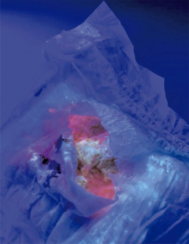
A male infant presented with progressive jaundice immediately after birth. Fecal acholia and choluria associated with extensive bullous skin lesions in his trunk, abdomen, and upper and lower limbs developed during phototherapy. Several diagnostic hypotheses were presented, including neonatal porphyria, hemochromatosis, Alagille syndrome, and neonatal lupus. A 24-hour urine sample for the dosage of urinary porphyrins was collected, showing high results (1823.6µg in 100mL). At 50 days of life, fluorescence spectroscopy using a Wood’s lamp revealed simultaneous bright red fluorescence of urine-stained diapers and sample blood. A definitive diagnosis of congenital erythropoietic porphyria was made following identification of a mutation of the uroporphyrinogen synthetases III gene on genetic testing. The patient was subsequently maintained in a low light environment since then, resulting in improvement of the lesions. Congenital erythropoietic porphyria is a disease of the group of porphyrias that presents shortly after birth with blistering occurring in regions exposed to the sun or other ultraviolet light. Atrophic scars, mutilated fingers, and bright red fluorescence of the urine and teeth may also be observed. There is no specific treatment, and prophylaxis comprising a total avoidance of sunlight is generally recommended. A high degree of suspicion is required for diagnosis. An early diagnosis can lead to less damage. Here, we present the case of a newborn with congenital erythropoietic porphyria diagnosed after presenting with bullous lesions secondary to phototherapy.
101