Label Free Imaging Through a 125μm Multimode Optical Fiber
- Abstract number
- 612
- Event
- European Microscopy Congress 2020
- DOI
- 10.22443/rms.emc2020.612
- Corresponding Email
- [email protected]
- Session
- PST.3 - New Instrumentation
- Authors
- Angel Cifuentes (1), Johanna Trägårdh (1), Tomáš Pikalek (1), Mojmír Šerý (1), Denis Akimov (2), Tobias Meyer (2, 3), Jürgen Popp (2, 3), Tomáš Čižmár (1, 2, 4)
- Affiliations
-
1. Institute of Scientific Instruments of the CAS
2. Leibniz Institute of Photonic Technology
3. Institute of Physical Chemistry, Friedrich Schiller University of Jena and Abbe Center of Photonics
4. Institute of Applied Optics, Friedrich Schiller University Jena
- Keywords
Microendoscopy MMF SLM wavefront-shaping
- Abstract text
In recent years, great advances have been made in developing minimal footprint micro-endoscopes using multimode optical fibers (MMF) [1]. By employing wavefront shaping methods the seemingly random speckle pattern resulting from the transport of coherent light through an MMF can be formed into a diffraction limited spot. This enables the implementation of multiple laser scanning techniques. In this work we show that this approach can be employed to realize label-free non-linear microcopy techniques such as coherent anti-Stokes Raman scattering (CARS) and second harmonic generation (SHG). Backscattered light makes epi-detection possible even though these processes emit light in the direction of beam propagation [2,3].
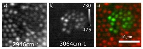
Figure 1. Epi CARS imaging of polymethyl methacrylate (PMMA) and polystyrene (PS) beads through a GRIN MMF using 1-2 ps laser pulses showing chemical selectivity. a) PMMA beads, Raman shift=2946 cm-1 b) PS beads, Raman shift=3064 cm-1, c) Composite image.
Using a spatial light modulator (SLM) and phase shifting interferometry we measure the transmission matrix (TM) of a 125μm graded index (GRIN) fiber with a 0.29 numerical aperture. This allows us to determine the light pattern needed at the fiber input in order to create a focused spot in which 1-2 ps long pump (660-675 nm) and Stokes (831 nm) beams overlap in time. This spot can be scanned over the sample at a distance of 50μm from the fiber facet to obtain an image. The chemical selectivity of CARS is demonstrated by imaging 2μm polystyrene beads (PS) and 2.5μm PMMA beads (Figure 1), with a pixel integration time as low as 1 ms for epi-detection.
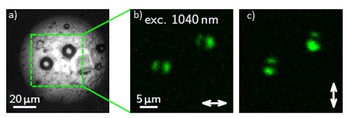
Additionally, we have used the same wave-front shaping technique to focus a 1040 nm long pulse and obtain SHG images (Figure 2). Our technique gives us the possibility of controlling the spot’s polarization, allowing polarization sensitive SHG techniques to be used. This could permit, for example, determining the orientation of collagen fibrils in tissue [4]. We show the possibility of utilizing this minimal footprint endoscope for this purpose.
Figure 2. a) White light transmission image of potato starch grains. b,c)SHG imaging of the same grains through a GRIN MMF using a ~140 fs 1040 nm laser. , b) Horizontal linearly polarized beam, c) Vertical linearly polarized beam.
- References
[1] Turtaev, S. et al, Light: Science & Applications 7.10 (2018).
[2] Evans, C. L. et al, PNAS, 102.46 (2005).
[3] Légaré, F., Pfeffer, C., and Olsen, B. R. Biophys. J 93.4 (2007).
[4] Stoller, P. et al, Biophys. J 82.6 (2002).