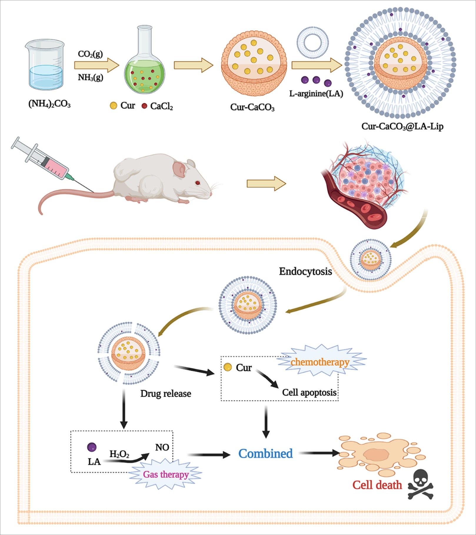
As shown in Fig. 1, CaCl2 was dissolved in ethanol and placed in a closed vessel along with (NH4)2CO3. (NH4)2CO3 gradually decomposed volatile NH3 and CO2 diffused into the ethanol and dissolved to form CO32− and NH4+. Under the alkaline condition formed by NH4+, CO32− and Ca2+ dissolved in ethanol reacted to form amorphous calcium carbonate cores, and due to the low water content of the reaction system (< 2%), the calcium carbonate cores did not undergo crystalline transformation during the growth process and CaCO3 nanoparticles were finally obtained. The gas diffusion reaction using organic solvent as a medium was shown to be a simple and feasible method for CaCO3 preparation 20.
TGA of Cur-CaCO3@LA-Lip
The TGA results of CaCO3 and CaCO3@Lip are shown in Fig. 7. The figure shows that the mass loss of CaCO3 and CaCO3@Lip decreases at a rate of about 5% and 7%, respectively, when the temperature is below 150°C. This difference is not significant, mainly because the mass loss at this stage is primarily the loss of water molecules from the surface and pore channels of CaCO3 and CaCO3@Lip carriers, and no degradation of macromolecules occurs. When the temperature rises to 150–400°C, the mass loss of CaCO3 and CaCO3@Lip starts to accelerate, and the increase in loss is due to the gradual decomposition of the CaCO3 matrix. When the temperature is 400°C, the residual masses of CaCO3 and CaCO3@Lip were 67.14% and 56.3%, respectively; this is mainly because the Lip in CaCO3@Lip also undergoes thermal decomposition, resulting in a higher final weight loss than CaCO3. In conclusion, CaCO3 and CaCO3@Lip carriers have good thermal stability.
In Vitro Release Studies
The in vitro release curve (Fig. 8) shows that the main release phase of Cur solution occurs within 8 hours, reaching 67.47% after 8 hours and 74.74% after 24 hours. The cumulative release of Cur-CaCO3@LA-Lip within 8 h only got 55.44%. In comparison, the cumulative release rate after 24 h reached 86.89%, indicating that Cur-CaCO3@LA-Lip exhibits good anti-surge release and sustained-release properties and can significantly improve the aqueous solubility of Cur, which helps to enhance the bioavailability of Cur.
Assessment of NO generation in cells
According to the results of the confocal fluorescence microscopy images in Fig. 9, cells treated with Cur-CaCO3@LA-Lip showed the strongest fluorescence signal in the tested preparations, indicating that a large amount of NO was produced in the cells. The weak signal from cells treated with LA only did not differ from the signal from untreated cells, which can be attributed to the endogenous NO already present in the cells 26. The above results suggest that Cur-CaCO3@LA-Lip in the internal tumor microenvironment is expected to exert combined anti-tumor effects through NO treatment with chemotherapy.
In vitro antitumor activity evaluation
To determine the cytotoxicity of Cur-CaCO3@LA-Lip, in this experiment, different concentrations of Cur, Cur-CaCO3, and Cur-CaCO3@LA-Lip were applied to the tumor cells for different times, and their inhibitory effects on cell proliferation were determined. The results are shown in Fig. 10; the cell proliferation inhibitory ability of Cur-CaCO3@LA-Lip was enhanced with the increase of Cur concentration and incubation time within 48 hours. The Cur-CaCO3@LA-Lip group showed stronger cytotoxicity than the free Cur group with increasing Cur concentration and incubation time. The corresponding IC50 for 24 h were 121.07 ± 34.70 µg/mL (Cur) and 62.07 ± 10.45 µg/mL (Cur-CaCO3@LA-Lip), and the corresponding IC50 for 48 h were 63.01 ± 4.36 µg/mL (Cur) and 42.35 ± 7.32 µg/mL (Cur-CaCO3@LA-Lip). The IC50 values of Cur-CaCO3@LA-Lip and free Cur groups were significantly different (p < 0.05) at both 24 h and 48 h of incubation, which can be explained by the superior cellular uptake efficiency provided by the nanocarriers. The cytotoxicity of the Cur-CaCO3@LA-Lip group was much better than that of the Cur-CaCO3 group due to the combination of chemotherapy and NO. Chemotherapy is the primary drug therapy in the clinical treatment of breast cancer, and gas therapy is an essential tool for adjuvant tumor treatment. Still, both are not ideal as a single treatment modality applied to breast cancer treatment. Since the mechanism of the cytotoxic effect of chemotherapeutic drugs and the means of NO production by gas therapy leading to cell death are very different, the construction of a nano-delivery system to co-deliver NO donors and chemotherapeutic drugs to tumor tissues is expected to be of significant research and application value to achieve efficient and low-toxic tumor combination therapy by complementing both chemotherapy and gas therapy 27.
Cellular uptake
The cellular uptake behavior of CaCO3 and Cur-CaCO3@Lip underlies their intracellular fate and is closely related to their biological function 28. The uptake effects were analyzed using inverted fluorescence microscopy. Figure 11A shows the fluorescence inverted microscopy of the free Cur, CaCO3, and Cur-CaCO3@Lip groups with 4T1 cells incubated for 2 h and 4 h, respectively, and Fig. 11B shows the mean fluorescence intensity (MFI) of Cur quantified by Image J. The results show that the intracellular fluorescence intensity of Cur-CaCO3@Lip at 2 h and 4 h was significantly higher than that of free Cur and CaCO3 groups, indicating that lipid wrapping can dramatically enhance the cellular uptake of nanoparticles, which may be due to the similar bilayer of lipid and cell membrane and good cellular biocompatibility, which can significantly improve the efficiency of nanoparticle endocytosis 29. The pattern of data obtained from the 4 h incubation was the same as that of the 2 h incubation. Still, the qualitative observation of fluorescence by inverted microscopy showed that the green fluorescence of Cur was stronger compared to the 2 h incubation, indicating that the uptake of Cur is time-dependent, with more uptake and eventually saturation with increasing time.
Effectiveness and analysis of in vivo anti-tumor therapy
To test whether Cur-CaCO3@LA-Lip could be used to achieve an effective combination therapy, its anti-tumor properties were evaluated in a 4T1 mouse model of breast cancer (Fig. 12A). The body weight of the mice was monitored during treatment, and the results are shown in Fig. 12B. As can be seen from the figure, the body weights of the mice in the different treatment groups did not change significantly. They were all within a reasonable range without significant weight loss, indicating the low side effects of Cur-CaCO3@LA-Lip NPs. Figures 12C, 12D, and 12E show the mean tumor weight results, photographs of tumors obtained from each group, and curves of changes in tumor volume in mice during treatment, respectively. Compared with the PBS group, the tumor growth of mice in the Cur group was slightly inhibited, mainly because Cur is not a chemotherapeutic agent such as Adriamycin, which has no significant anti-tumor effect. The tumor size of mice in the Cur-CaCO3 group was significantly reduced, mainly due to the anti-tumor effect of the CaCO3 carrier, which enhances the passive transport of Cur. Compared to the Cur-CaCO3 group, the tumor volume in the Cur-CaCO3@LA-Lip group was significantly suppressed due to the relatively high level of NO produced by LA, which can act as a cytotoxic and apoptosis-inducing agent for tumor treatment, thus enhancing the efficacy of Cur chemotherapy. Representative micrographs of H&E staining of tumor tissue from each group of mice are shown in Fig. 13F. The red color in the section is the cytoplasm, and the dark purple color is the nucleus 30,31. Compared to the control group, tumor necrosis was observed in the Cur, Cur-CaCO3, and Cur-CaCO3@LA-Lip groups, especially in the Cur-CaCO3@LA-Lip group. The primary manifestation of tumor necrosis was the loss of nuclei, and a small amount of inflammatory cell infiltration was seen at the edge of the necrotic tumor tissue 32. The Cur-CaCO3@LA-Lip group had the largest area of tumor necrosis after treatment and the best anti-tumor effect.
Preliminary biosafety evaluation
The hemolysis rates of the carrier materials CaCO3 and CaCO3@Lip were investigated by in vitro erythrocyte hemolysis assay, and the results are shown in Fig. 13A. The hemolysis rates of CaCO3 and CaCO3@Lip increased slightly with an increasing mass concentration in the experimentally set mass concentration range. Still, they were both below 5%, indicating that they have good biosafety for intravenous administration 33. To further confirm the in vivo safety of the Cur-CaCO3@LA-Lip formulation, we performed an H&E staining analysis on the mice's major organs (heart, liver, spleen, lung, and kidney), as shown in Fig. 13B. There were no significant pathological changes in the major organs of the mice in the Cur-CaCO3@LA-Lip treatment group, indicating that Cur-CaCO3@LA-Lip has good biocompatibility and does not cause substantial damage to the mice. The above results demonstrate that Cur-CaCO3@LA-Lip can achieve highly effective and low-toxicity therapeutic effects, reflecting the advantages and potential of LA combined with Cur for treating breast cancer.