Skin can repair itself after injury, but deep skin wounds generally heal with scar formation and do not regenerate HFs. HF regeneration is a sign of optimal wound healing[40, 41]. It was thought that HF morphogenesis mainly occurred during embryonic development, and in adult mammals, HFs destroyed by deep injury were irreparable. In 2007, Ito et al. observed functional HF regeneration in large skin wounds (> 1 cm2) in adult mice[42], which opened up new explorations into HF regeneration in wound healing.
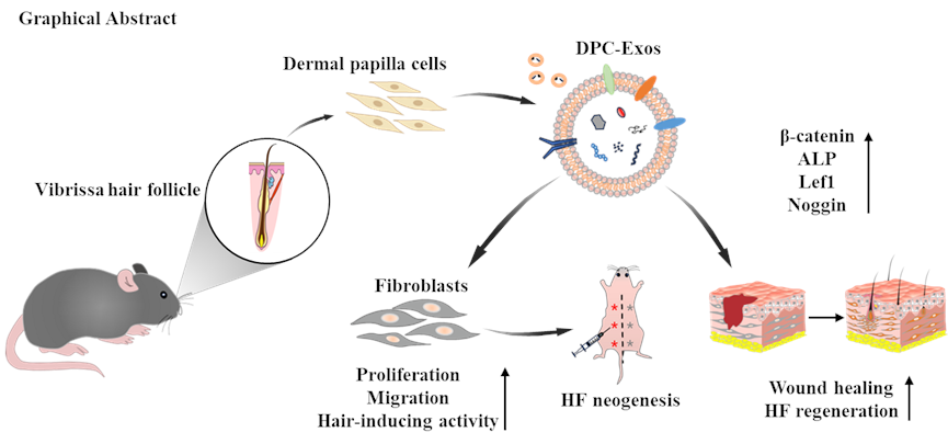
Wound healing is a complex biological process, involving a variety of cells, and fibroblasts are the primary cell population that determines wound healing outcomes. Li et al. found that exosomes derived from adipose stem cells can promote fibroblast proliferation and facilitate wound healing by activating the Wnt/β-catenin signaling pathway[15]. In this study, we found that similar to other stem cell-derived exosomes, DPC-Exos could improve the proliferation and migration of fibroblasts, which was undoubtedly helpful in promoting wound healing. Given the hair inductivity of DPCs and homology between DPCs and fibroblasts, multiple attempts have been made to promote the transformation of fibroblasts into DPCs, such as biomimetic hydrogel encapsulation culture[13], small molecule compounds inducing[12], and suspension culture with FGF2、PDGF and BIO[43]. These methods promote the hair-inducing ability of fibroblasts but are not suitable for wound healing. DPC-Exos, as a natural dermal signaling delivery system, could induce the proliferation and differentiation of hair follicle stem cells and hair matrix cells, and augment the hair-inductive capacity of dermal papilla spheres[44, 45]. By co-incubating fibroblasts with DPC-Exos, we found that the expression level of molecules associated with the hair-inducing capacity of DPCs (β-catenin, ALP, Lef1, and Noggin) was elevated in fibroblasts. Moreover, the hair reconstitution assay further confirmed the hair-inducing activity of fibroblasts after DCP-Exos treatment. These results suggest that DPC-Exos promotes the pro-regenerative phenotype of fibroblasts, and lays the foundation for DPC-Exos to promote HF regeneration in wound healing.
In the wound healing experiments, by tracking the area and hair growth of the wounds, we found that DPC-Exos could accelerate wound re-epithelialization. This is consistent with the promoting effect of other mesenchymal stem cell-derived exosomes on wound healing. In addition, there was significantly more HF neogenesis and less collagen deposition in the DPC-Exos treatment group. We hypothesize that DPC-Exos promoted HF regeneration by altering fibroblast phenotypes, and then we detected the expression level of molecules associated with hair-inducing capacity. The gene and protein expression levels of β-catenin, ALP, Lef1, and Noggin were elevated in the DPC-Exos treatment group. Additionally, the results of tissue immunofluorescence staining showed that the fluorescence intensity of β-catenin was significantly enhanced in the dermal layer and new hair follicles. Lef1 as a marker for newly formed hair follicles, exhibited higher fluorescence intensity in the DPC-Exos treatment group. These results supported our hypothesis that DPC-Exos could promote HF regeneration during wound healing by enhancing the hair-inducing activity of fibroblasts.
The Wnt/β-catenin signaling pathway plays an important role in HF regeneration and wound healing[46–48]. Accumulating evidence has demonstrated that activation of the Wnt/β-catenin signaling pathway could promote wound healing[49, 50]. During HF morphogenesis, the dermis provides the ‘first dermal signal’ that initiates the formation of epidermal placodes, and sustained β-catenin activity in the dermis leads to larger HF placodes and accelerates differentiation of HFs[49, 51]. In the study, we found that DPC-Exos enhanced the expression level of Wnt/β-catenin pathway transcription factors β-catenin and Lef1 in fibroblasts, based on this, we speculated that the effect of DPC-Exos on fibroblasts was related to the activation of Wnt/β-catenin signaling pathway. To verify this hypothesis, we used the Wnt inhibitor XAV939 to block the Wnt pathway in fibroblasts. The results of in vitro experiments showed that the Wnt inhibitor suppressed the proliferation and migration of fibroblasts, and this inhibitory effect could be partially alleviated by DPC-Exos. In addition, the Wnt inhibitor significantly reduced the protein expression level of β-catenin, ALP, Lef1, and Noggin in fibroblasts, and the expression level of these molecules could be partially restored by DPC-Exos. These results demonstrated that DPC-Exos could enhance the hair-inducing activity of fibroblasts by activating the Wnt/β-catenin signaling pathway.
For the role of the Wnt/β-catenin signaling pathway in wound healing, some studies showed that activation of the Wnt/β-catenin pathway in fibroblasts led to excessive collagen deposition and scarring[52, 53]. These findings increase the thinking about the regulation of the Wnt/β-catenin signaling pathway. It’s well known that beyond producing and maintaining extracellular matrix, fibroblasts also are critical to HF regeneration. Phan et al. pointed out that Lef1+ papillary fibroblasts can regenerate new HFs by interacting with the adjacent epidermal cells[11]. Mascharak et al. found that inhibiting the YAP signaling in fibroblasts could promote regenerative healing by activating the Wnt signaling pathway[40]. The Wnt/β-catenin signaling pathway is involved in multiple stages of wound healing, and inhibiting its activity would influence cell viability and wound healing. We found that inhibiting the Wnt pathway also significantly hindered wound re-epithelialization and HF regeneration, and this inhibition could be partially alleviated by DPC-Exos. Although some wounds were re-epithelialized in the XAV939 treatment group, there was no HF regeneration at the wound site. In addition, the expression level of β-catenin, ALP, Lef1, and Noggin was decreased after XAV939 treatment, and DPC-Exos partially restored the expression of these molecules. Meanwhile, compared to the DPC-Exos treated group, blocking the Wnt pathway weakened the effect of DPC-Exos on HF regeneration. These results indicated that DPC-Exos could promote HF regeneration by activating the Wnt/β-catenin signaling pathway.
Fibroblasts are highly dynamic cells and their heterogeneity and plasticity provide the possibility to change wound healing outcomes. Many studies have attenuated fibrotic repair by hindering the transformation of fibroblasts into myofibroblasts and disrupting YAP mechanotransduction in fibroblasts[54, 55]. However, these regulatory methods may have an impact on the activity of fibroblasts and thus influence the wound healing process. Some studies point out that regeneration and fibrosis are not completely excluded in cells during wound healing, and cells with pro-regeneration properties are also present in scars. But without intervention, the pro-fibrosis program predominates and leads to scarring. In addition, failing to regenerate skin appendages in wound healing is related to the lack of dermal regeneration signals rather than the lack of intrinsic regenerative ability of cells[40, 56]. Here, we introduced DPC-Exos, carrying regenerative dermal signaling, into wound healing, and found that DPC-Exos also worked to promote HF regeneration in case the integrity of the skin is compromised.
In this study, the results of our experiments confirmed that DPC-Exos could promote HF regeneration during wound healing. However, this is not enough for complete regenerative healing, which also requires the restoration of skin structure and strength. The contents of DPC-Exos, which are important for their function, still need to be explored. In addition, the regulatory targets of DPC-Exos in the Wnt/β-catenin signaling pathway have not been well elucidated. All of these require more in-depth research to figure out.