Construction and Expression of DspB-MagR
The magR gene were amplified using a pair of primers containing restriction site of endonuclease XhoI and then inserted at the downstream of dspB gene to construct the plasmid pET(DspB-MagR) (Fig. 1, Panel A), with a sequence encoding hexahistidine tag at the C-terminus of the recombinant protein DspB-MagR to facilitate the affinity purification.
Expression of DspB-MagR was induced by supplying IPTG to the culture containing E. coli host BL21 harboring the vector pET(DspB-MagR). Bacterial growth was significantly reduced upon the addition of IPTG (Fig. 1, Panel B), implying the involvement of bacterial metabolism in synthesizing the heterogeneric DspB-MagR retarded the bacterial growth. Expression of recombinant DspB-MagR was confirmed with SDS-PAGE electrophoresis (Fig. 1, Panel C), which showed that there was overexpression of protein with an apparent molecular weight between 50 and 70 kDa. This protein was assigned to be DspB-MagR with a theoretic molecular weight of 60 kDa.
Fast overexpression of heterogeneric protein sometimes leads to formation of inclusion body containing proteins of interests that are not correctly folded. In the present study, IPTG-induced DspB-MagR expression was attempted at two temperatures (that is, 16 oC and 37 oC) to avoid the formation of DspB-MagR inclusion body. It was observed that DspB-MagR expression level at 16 oC was lower than that at 37 oC. However, the soluble version of DspB-MagR at 16 oC was significantly improved (Fig. 1, Panel D). It might be due to that bacterial protein synthesis is slow at low temperature, which provides translated DspB-MagR with more time to fold properly.
Purification and Immobilization of DspB-MagR
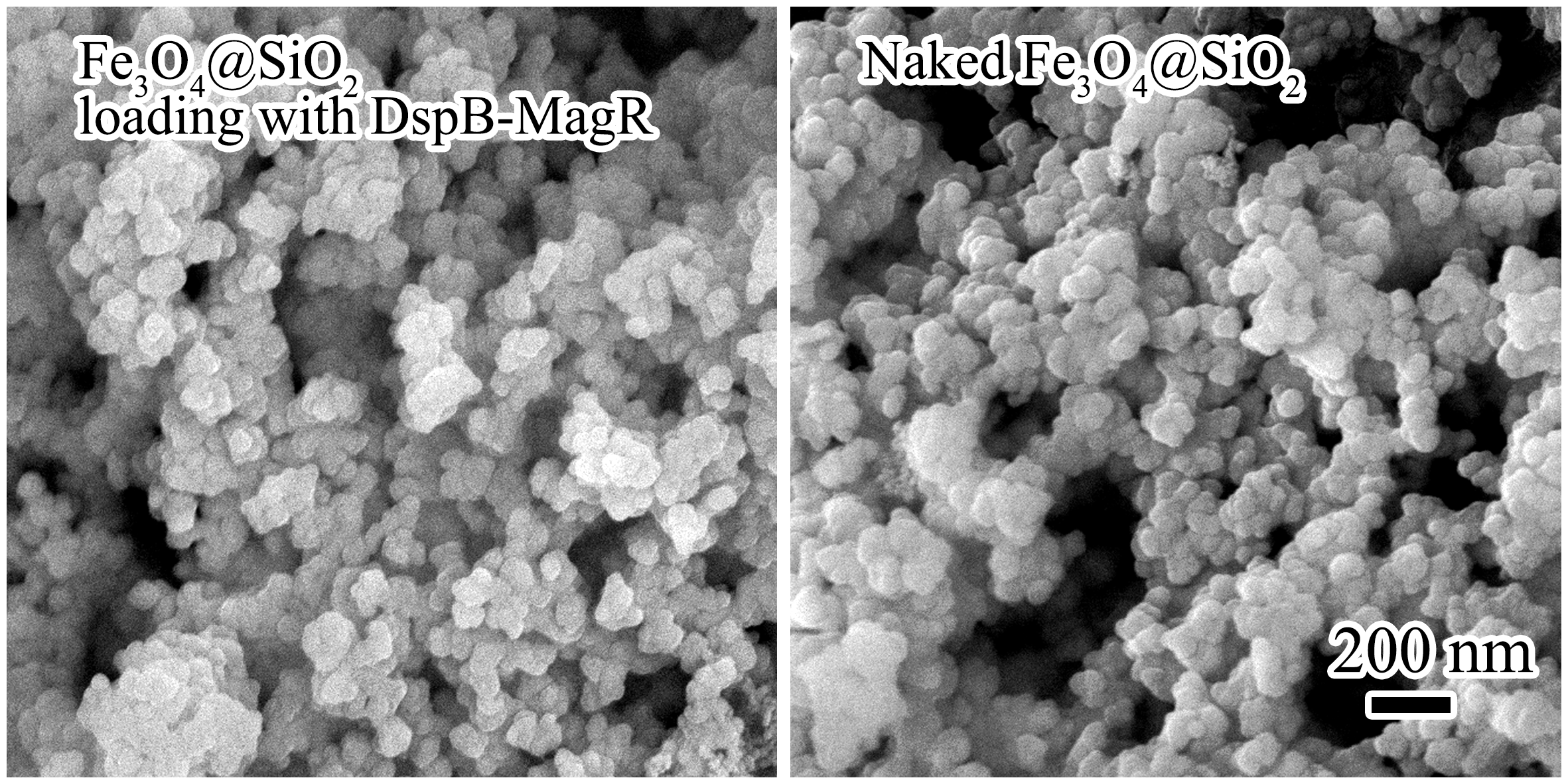
Expressed DspB-MagR was purified by using Ni-NTA affinity chromatography and immobilized on Fe3O4@SiO2 nanoparticles (Fig. 2, Panel A). The homogeneity of DspB-MagR was over 95% after purification and immobilization as determined by densitometric scanning of Coomassie blue stained SDS-polyacrylamide gels. SEM revealed no significant morphological changes of Fe3O4@SiO2 nanoparticles before and after loading with DspB-MagR (Figure S1, see Supporting Information). FTIR spectrometry of Fe3O4@SiO2 nanoparticles with or without DspB-MagR loading showed that there were characteristic bands of Si-O-Si band at 1095 cm− 1 and Fe-O band at 596 cm− 1, 576 cm− 1 (Fig. 2, Panel B). There was a shift of absorbance peak from 1634 cm− 1 (before protein loading) to 1652 cm− 1 (after protein loading), which was attributed to influence of the amine of polypeptide and proved the immobilization of DspB-MagR on Fe3O4@SiO2 nanoparticles. Immobilization of DspB-MagR on Fe3O4@SiO2 nanoparticles was also verified by TGA measurement (Fig. 2, Panel C). There was a weight loss of 3% and 5% for both Fe3O4@SiO2 nanoparticles with or without DspB-MagR loading when raising temperature up to 150 oC, which was due to water evaporation. When raising temperature from 150 oC to 350 oC, a significant weight loss of 1.25% was observed for Fe3O4@SiO2 nanoparticles loading with DspB-MagR due to the thermal decomposition of protein. Therefore, the loading capacity of Fe3O4@SiO2 nanoparticles to DspB-MagR was estimated as ~ 1.25 mg DspB-MagR per 100 mg nanoparticles. Zeta potential of Fe3O4@SiO2 nanoparticles with or without DspB-MagR loading was measured at different pH (Fig. 2, Panel D). Zeta potential is the electrostatic potential that exists at the shear plane of a particle, which is related to both surface charge and the particle’s local environment. It was observed that after loading with DspB-MagR, Fe3O4@SiO2 nanoparticles exhibited higher zeta potential than that of naked Fe3O4@SiO2 nanoparticles. This could be attributed to the ionization of functional NH2 groups of DspB-MagR protein.
Bioactivity of Immobilized DspB-MagR
Enzymatic activity of DspB-MagR before and after loading on Fe3O4@SiO2 nanoparticles was investigated under various conditions. Generally, the activity of enzymes is affected by factors such as pH, temperature, functional time [18]. It was observed that DspB-MagR exhibited similar spectrum of sensitivity to pH before and after loading on Fe3O4@SiO2 nanoparticles (Fig. 3, Panel A). Both showed highest activity in pH 6. Therefore, immobilization on Fe3O4@SiO2 nanoparticles did not change the pH sensitivity of DspB-MagR.
Sensitivity of DspB-MagR to temperature changed after loading on Fe3O4@SiO2 (Fig. 3, Panel B). Before immobilization, DspB-MagR exhibited highest activity at 30 oC. While the immobilized DspB-MagR exhibited highest activity at 37 oC that is the regular body temperature of human, implying that immobilization of DspB-MagR on Fe3O4@SiO2 nanoparticles is suited to medical purposes.
Stability of DspB-MagR over time was investigated before and after loading on Fe3O4@SiO2 nanoparticles (Fig. 3, Panel C). There were two phases of activity changes for both enzymes over time after incubation at their optimal temperature (30 oC for unloaded DspB-MagR and 37 oC for immobilized DspB-MagR). In the first two to three hours of incubation, both enzymes suffered a quick decrease and increase of the activity. Although the activity of both enzymes kept decreasing after three hours of incubation, immobilized DspB-MagR showed higher activity of than that of unloaded ones at sampling time, implying that immobilization of DspB-MagR on Fe3O4@SiO2 nanoparticles is beneficial for retaining the activity of DspB-MagR over time.
Degradation of Biofilm by Immobilized DspB-MagR
Bioactivity of DspB-MagR before and after loading on Fe3O4@SiO2 was assayed for detaching bacterial biofilms (Fig. 4). When testing with the standard strain of Staphylococcus aureus (SA), naked Fe3O4@SiO2 nanoparticles did not exhibit any effects on removing biofilm. Unloaded DspB-MagR showed limited function and removed about 10% of biofilm. After loading on Fe3O4@SiO2 nanoparticles, activity of DspB-MagR on removing biofilm was significantly improved and detached more than 50% of biofilm.
Activity of DspB-MagR loading on Fe3O4@SiO2 nanoparticles was further examined on removing biofilms forming by bacteria samples from clinic source, that is, one mixed bacterial species (MB) and three isolated and characterized species, Staphylococcus sp. (SS), Bacillus Cereus (BC), and Pseudomonas Putida (PP). Although it was not significant, immobilized DspB-MagR showed activity in degrading biofilm forming by mixed bacterial species or Pseudomonas Putida. As to Staphylococcus sp. or Bacillus Cereus, immobilized DspB-MagR exhibited significant activity on detaching biofilm, over 40% (Bacillus Cereus) or 60% (Staphylococcus sp.) of biofilms were removed.