The microparticles coated with nano-forest like structure(NFMPs) were fabricated as illustrated in Fig. 1b. Briefly, plain glass microparticles (GMP) was employed as typical model particles and drop-casted onto the a substrate, producing a single layer. 80 nm-thick zinc oxide (ZnO) layer was then sputtered onto the particles surfaceas an additional seed layer for further tree-trunk-like nanospikes structure growth. Subsequently, the nanospikes on the surface of microparticles were acquired via a hydrothermal approach, through which the substrate attaching particles was immersed into aqueous solutions containing 25 mM Zn(NO3)2 and 25 mM hexamethylenetetramine at 90°C for 1 h47. The extracted nanospikes microparticles (SMP) was coated with a ~ 10 nm sputtered alumina layer to avert potential cytotoxicity induced by ZnO. Eventually, the SMP were isolated and collected from the flat substrate by conducting brief sonication. The morphology of GMP and SMP was explored through applying scanning electron microscopy (SEM). As shown in Fig. 2a, the diameter of glass microparticle is about 52 µm with a flat surface. Treated with hydrothermal process, randomly protruding ZnO nanospiky structures appeared on the microparticles surface. The SMP, incubated in pure PDMS at 120°C for 72 h (Figure S1), were paired with silicone oil reagents, methoxy-terminated polydimethylsiloxane (CH3O-PDMS-OCH3) so as to functionalize microparticles with PDMS. Modification was thus accomplished, the PDMS brushes were grafted on the entire spiky microparticles and formed a dendritic structure. The functional nano-forest like structures microparticles were then washed with toluene three times to remove redundant silicone oil, separated by centrifugation, and dried. A glass substrate with nanospiky and coated with an aluminum oxide layer was modified by PDMS in order to locate the immobilization of PDMS chains. Further investigation was conducted via X-ray photoelectron spectroscopy (XPS) application. As shown in Fig. 2b, the spectrum illustrated a lower ratio of Al peaks, and higher ratio of Si 2p and Si 2s peaks, revealing the PDMS chains were immobilized onto the particles surface successfully.
The wetting behaviors of the NFMPs were evaluated by means of calculating their static contact angles (CAs) and sliding angles (SAs) towards water. The NFMPs were drop-casted on a flat substrate and an uniform film was manufactured. Meanwhile, smooth glass microparticles (GMPs), PDMS brush-modified glass microparticles (PMGMPs), and nanospiky structures microparticles (SMPs) without PDMS modified were employed to produce comparable thin films to tackle wetting tests (Fig. 2c). The results of the CAs measurements were statistically displayed in Fig. 2d. The CAs of several microparticles films to water were 62.8° (GMPs), 116.8° (SMPs), 125.2° (PMGMPs) and 132.6° (NFMPs), respectively. The results of the SAs measurements were statistically shown in Fig. 2e. The PDMS brush-modified microparticles, including PMGMPs and NFMPs, were highly slippery to water with low sliding angles at 20.0° and 8.0°, respectively, while the unmodified particles film failed to display similar features.
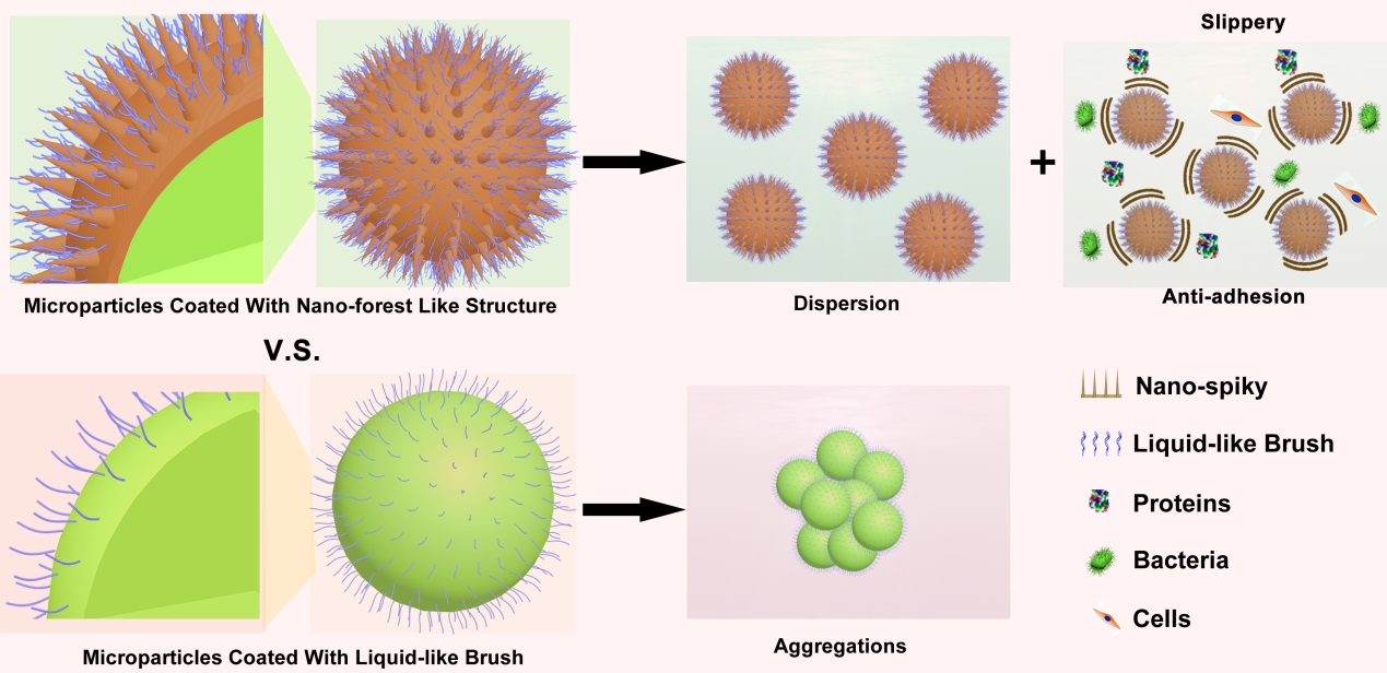
The superior dispersibility of particles is essential to be applied in clinical medical, which is affected by surface chemistry and surface morphology46. The dispersibility of hydrophobic NFMPs in water was further investigated. In the meantime, the GMPs, superficially nano-structural SMPs and chemical modification PDMS brush-grafted GMPs were taken as control groups. As shown in Fig. 2f and 2g, hydrophilic GMPs were dispersed well yet seen hydrophobic PMGMPs aggregation in water, on the contrary, hydrophobic nanospiky structures microparticles including SMPs and NFMPs still dispersed in water instead of producing aggregations.
Deliverable micron-sized biomaterials or microparticles are vulnerable to biofouling such as bacteria or cell adhesion, inflicting undesirable biofilms, and potentially infectious or evoking inflammation and fibrosis37. The adhesion of cells or bacteria was usually mediated by integrins or adhesin that was comprised of proteins and polysaccharide molecules48. Therefore, effective resistance to the adhesion of proteins and other biomolecules is crucial to prohibit biofouling. To evaluate the anti-biofouling performance of nano-forest like structure microparticles, their resistance to small molecules and proteins were initially explored. The NFMPs were incubated with representative fluorescent molecule fluorescein (FITC, MW = 389), representative proteins FITC conjugated bovine serum albumin (FITC-BSA, MW ~ 66 kDa) and fluorescent fibrinogen (Fg) in a static medium at 37°C for 24 hours. Meanwhile, non-modified by PDMS microparticles including GMPs and superficially nano-structural SMPs were conducted as the negative control groups. After incubation, all the microparticles including GMPs, SMPs, and NFMPs were washed once by PBS buffers (0.01 M, pH = 7.4), and the results were gained via applying a fluorescence microscope. Besides, the total relative fluorescence intensity was calculated by consulting Image J to determine the degree of adhesion by small molecules and proteins.
Representative fluorescence images revealing the adhesion of small molecules and proteins onto different particles surfaces were showcased in Fig. 3a and Figure S2.1. As expected, the fluorescence intensity of PDMS polymer functionalized particles was significantly diminished compared with unfunctionalized smooth or nanostructured particles, which hints that PDMS brush coating significantly inhibit the adhesion of small molecules and proteins. In further statistical experiment, the normalized fluorescence intensity of NFMPs group was evidently cut to 1/60 of the unmodified microparticles (Fig. 3b).
In addition, the adhesion of small molecules and proteins to PDMS brush-modified microparticles with different sizes (10 µm and 100 µm in diameter) were explored to study the anti-adhesion properties shown in the slippery ‘liquid-like’ coating surface. As mentioned in the protocol, the nanospikes were first produced on the particles surface, an aluminum oxide layer was then sputtered to protect the cells from ZnO cytotoxicity, which was functionalized by PDMS incubation. Representative SEM images demonstrated the nanospikes structures were evenly distributed to the surface of particles of different sizes (Fig. 3c). Two sizes of NFMPs were incubated with FITC, FITC-BSA and Fg at 37°C for 24h, respectively, and the corresponding GMPs and SMPs were applied as control tools. Follwing the completed incubation, PBS was washed once and observed via a fluorescence microscope. As shown in Fig. 3d, FigureS2.2 and Figure S2.3, all of the GMP and SMP groups showcased bright green fluorescence, indicating that small molecules and proteins stuck onto these particles. The NFMPs groups, either 10 µm and 100 µm in diameter, were almost dark on the contrary, revealing a highly effective withstand to small molecules and proteins after grafted with ‘liquid-like’ coating, regardless of particles size. Furthermore, Nano-forest like structures planar substrate (NFPS) was fabricated by utilizing the same approach, and its anti-adhesion to small molecules and proteins was also investigated. As shown in Fig. 3e, a planar substrate was acquired that was uniformly covered with nanospikes .The NFPS were incubated with FITC, FITC-BSA and Fg at 37°C for 24h, respectively, and the glass planar substrate (GSP) and nanospiky planar substrate without PDMS brush-modified (SPS) were taken as control group. The substrate was washed once with PBS and set on the surface of a clean glass substrate, which was examined with fluorescence microscopy. The adhesion of the small molecules and proteins to the substrate (indicated as area “2”) was compared to clean glass substrate (indicated as area “1”), and the substrate boundary highlighted with a dashed line. The fluorescence images illustrated that both GPS and SPS were heavily blanked by small molecules and proteins, while NFPS was almost as clean as the reference substrate (Fig. 3f and Figure S2.4).
In addition, the effect of ‘liquid-like’ coating modified interface on protein activity was evaluated by representative protein glucose oxidase. Various ‘liquid-like’ coatings were used to modify surfaces including NFMPs (NFMPs (50 µm), NFMPs (10 µm), and NFMPs (100 µm)) and NFPS at 37°C for 24h. After incubation, the activity of GOD was analyzed by GOD activity detection kit. Meanwhile, heat denaturing of GOD (90°C, 0.5h) and blank treatments were employed as control groups. As shown in Fig. 3g, compared with the blank control group, the protein activity was not affected significantly by the modified interface of the ‘liquid-like’ coating, but was almost eliminated by heating.
The ‘liquid-like’ coating showed potent ability to resist the attachment of small molecules and proteins by providing a non-adhesive, slippery interface. We also investigated whether the modification method could prevent adhesion of bacteria and subsequent biofilm formation on injected biomaterials, which can result in hospital-acquired infections or increased risk of sepsis32. The NFMPs were incubated with two clinically important biofilm-forming pathogens, E. coli. and S. aureus., for 24 h at 37 oC in static culture media. Meanwhile, plain GMPs and superficially nano-structural SMPs without ‘liquid-like’ coating modification were used as controls. After incubation, the microparticles were collected from the culture medium and labeled with a green fluorescent dye to visually observe the adhesion of bacteria. The result of bacterial adhesion to the microparticles was recorded by fluorescence microscope and quantified by the relative green fluorescence intensity.
Representative fluorescent images of bacterial adhesion were shown in Fig. 4a. After 24h of incubation, a large amount of dense green fluorescence was exhibited on the surface of the non-‘liquid-like’ coating nanoparticles including GMPs and SMPs, while only sparse and isolated green fluorescence was observed on the NFMPs surface. The results indicated that bacterial adhesion of E. coli. and S. aureus. was significantly reduced on modified surface compared to control groups. As shown in Fig. 4b, the normalized fluorescence intensities of NFMPs groups were at least 56 times lower than control groups without ‘liquid-like’ coating, based on the statistical results.
To explore the mechanism of NFMPs bacterial adhesion inhibition, the cytotoxicity test was first implemented. The NFMPs were incubated with either E. coli. or S. aureus media at 37°C for 24h, and culture medium without NFMPs were used as controls. After incubation, bacteria viabilities were determined by standard live/dead bacteria staining assay, and evaluated by fluorescence microscopy. Live bacteria were stained with SYTO-9 (green fluorescence), and dead cells were stained with propidium iodide (PI, red fluorescence). The results showed that barely any red fluorescence bacteria were observed in all groups (Fig. 4c and Figure S3.1), indicating that the bacterial viability was not affected in the presence of NFMPs. The statistical results showed that the bacterial activity of NFMPs exceeds 98% (Fig. 4d), with no significant difference from the control group. Furthermore, to explore the anti-adhesive properties attributed to the slippery and mobility of the ‘liquid-like’ coating, the adhesion of the bacteria to nano-forest like structures planar substrate (NFPS) were also investigated. The NFPS was incubated with E. coli. and S. aureus. for 24 h at 37 oC in static culture media, and substrates without ‘liquid-like’ coating modification including GPS and SPS were used as controls. After incubation, replace the medium with fresh PBS to avoid background signal interference from suspended bacteria, and stained with a green fluorescent dye for observation. The bacteria adhered on the glass substrate (indicated as area “2”) were compared to that on well plate bottom surface (indicated as area “1”), with the substrate boundary highlighted by a dashed line. The fluorescence images showed that the unmodified GPS and SPS as well as the substrate were all heavily covered by bacteria, while almost no green fluorescence was observed on NFPS (Fig. 4e and Figure S3.2).
Implantable biomaterials were prone to inflammation and tissue fibrosis due to cells adhesion37. As another vital indicator to distinguish the performance of implantable biomaterials, whether NFMPs can resist cell adhesion was further investigated. The NFMPs were incubated with two representative mammalian cells such as the fibroblasts NIH/3T3 cells and the tumor HeLa cells for 24h at 37°C in-vitro culture, meanwhile, non-‘liquid-like’ coating microparticles including plain GMPs and superficially nano-structural SMPs were utilized as controls. After incubation, the cells were labeled with Calcein-AM (green fluorescent) for visual identification. The particles were then collected from the culture medium, while cells attachment was analyzed by fluorescence microscopy. Representative cells adhesion images exhibited that the non-‘liquid-like’ coating microparticles including both GMPs and SMPs exhibited cell adhesion, while basically no green fluorescent cells were observed on NFMPs (Fig. 5a), which was similar to the results of protein and bacterial adhesion. Further statistical results showed that the relative mean number of two types of cells anchored on the NFMPs surface was close to zero (< 0.1), at least 20 times lower than the non-‘liquid-like’ coating particle groups (Fig. 5b). Similarly, NIH/3T3 cells and HeLa cells were incubated with NFMPs to evaluate cytotoxicity to explore the mechanism of cell adhesion inhibition, and medium without NFMPs was used as control group. After incubating at 37°C for 24h, the cells were labeled with a standard live/dead cell staining assay with Calcein-AM (green fluorescence, live cells were labeled) and propidium iodide (red fluorescence, dead cells were labeled) to identify cell viabilities. As shown in Fig. 5c and Figure S4.1, there was no obvious red fluorescent cells in all culture medium, with or without the presence of NFMPs. According to statistical analysis, similarly to the control group, the cell viability of the two cell types in the presence of NFMPs was also higher than 95% (Fig. 5d), indicating that the NFMPs has no effect on cell viability. Furthermore, the adhesion of the two cell types to nano-forest like structures planar substrate (NFPS) was investigated by culturing NIH/3T3 cells or HeLa cells on the substrate surface in the well. After 24h incubation at 37°C, the cells were stained by green fluorescence dye (Calcein-AM), and recorded by fluorescence microscope. The cell adhesion on the glass substrate (indicated as area “2”) was compared to that on the well plate bottom surface (indicated as area “1”), with the substrate boundary highlighted with a dashed line. Cells attached to the substrate surface and the well plate surface were imaged separately since they were not on the same focal plane. As expected, both the unmodified planar substrate including GPS and SPS and the bottom of the well were heavily covered by cells, while almost no of green fluorescent cells were attached to the PMFPS surface(Fig. 5e and Figure S4.2).
In actual clinical applications, it is necessary for deliverable biomaterials to have persistent resistance to biofouling. However, conventional modification techniques are difficult to achieve, which is mainly due to the fact that modified coatings such as PEG or SLIPS as well as other modification technologies are easily degraded or lost, leading to performance failure. As an anti-fouling coating with mechanical robustness and resistance to liquid flow erosion, the ‘liquid-like’ coating modification technology may have a positive impact on the durability of injected micron-scale biomaterials or microparticles in resisting biofouling. In this article, the stability of the ‘liquid-like’ coating modified strategy was first explored. As a substitute for NFMPs, the nano-forest like structures planar substrate NFPS was immersed in water at a flow rate of 80 r/min for 7 days, with its surface elements tested by XPS. It was found that comparing with unmodified coating, the spectrum exhibited decreased ratio of Al peaks, and increased ratio of Si 2p and Si 2s peaks (Fig. 6a), indicating that the immobilization of ‘liquid-like’ coating was stable in fluid environment. In addition, under the same conditions, NFMPs were immersed in water with a flow rate of 80r/min for 7 days. The particle morphology, contact angles and dispersibility were then tested. As shown in Fig. 6b, both the nanospikes structures and the hydrophobicity were intact, while exhibiting good dispersibility in water. Furthermore, the NFMPs mentioned above was further incubated with proteins, bacteria or mammalian cells under the same condition for 7 days at 37°C to explore its ability resist biofouling in long time span. Meanwhile, the non-‘liquid-like’ coating microparticles including plain GMPs and superficially nano-structural SMPs were utilized as controls. As shown in Fig. 6c, 6d and 6e, both GMPs and SMPs were heavily adhered with proteins (FITC-BSA & Fg), bacteria (E.coli. & S. aureus) and cells (NIH/3T3 & HeLa), while the NFMPs were almost dark or barely fluorescent.