Materials.
Mg WE43 base alloy (Yttrium: 3.1–3.7% wt; Lanthanum: 0.07–0.33% wt; Gadolinium: 1.7–1.8% wt; Neodymium: 2.2–2.5 % wt; Copper: ≤ 0.16% wt; Nickel: ≤ 0.11% wt; Iron: ≤ 0.017% wt; Oxygen: ≤ 4.62% wt) foundry from the Pidgeon process in the condition of casting bars, is proposed for the manufacture of an intramedullary implant (IIM). The IIM described here has been tested following an experimental methodology according to international standards ASTM-F-1264, ISO10993, and additional morphological characterization.
Preparation of Mg implants.
Three specimens were manufactured according to ASTM B 557, of which the stress-strain curve was obtained following ASTM E8 to characterize the behavior of the alloy in simple tension to 0.2% deformation. The behavior of the alloy is described by the isotropic multi-linear curve showing homogeneity. Additionally, three specimens were prepared to perform a simple bending test according to section A1.4 of ASTM F-1264 with a diameter approximately equal to that proposed for the IIM to know the resistance to bending with a force-displacement curve.
Sterilization.
Two other specimens were machined to perform a flexural test under dynamic conditions to measure the loss of mechanical integrity of the material due to the degradation that originates on the surface of the material by sterilization and the enriched Hank's solution. The Hank´s solution was obtained from Sigma Aldrich and was used according to ISO 10993-12. One of the specimens was sterilized using autoclave the method that most affects the surface of the Mg alloys[33]. All procedures were carried out under aseptic conditions and in a sterile environment, ensuring cell recovery, adherence, and progression towards the exponential growth phase[30].Three WE43 alloy specimens were made and sterilized utilizing an autoclave followed by a 3-point simple bending test based on ASTM F 1264 to evaluate the flexural strength and to obtain the force-displacement curve of each specimen. A test was carried out in dynamic conditions to measure strength parameters vs. number of cycles of the WE43 alloy immersed in Hank’s balanced salts. The alloy was evaluated under fatigue conditions for two specimens according to ASTM F 1264. In preparation, these manufactured specimens were simply polished with 800 grit sandpaper and were not attacked chemically. Only one specimen was subjected to sterilization. Immersion tests were performed following ASTM-G31-72[34,35].
The technical requirements for the approval of MDs include the review of good manufacturing practices by a regulated body such as the FDA, in addition to this, scientific and technical information must be provided, in addition to a detailed label that implies the surgical technique and the description of functional components, parts, and structure. The list of the elements used in the alloy and their dispersion on the implant surface must be presented, the qualitative-quantitative formula must be declared per unit of measure and percentage dose, in addition to the raw materials and a study where the toxicity, safety, and efficacy characteristics of MDs are supported. Information should be provided on the sterilization process, control of the finished product and analytical methods, stability study, and techno vigilance. This work presents the experimental morphological evaluation applicable to implantable products, in addition to the preclinical studies of biocompatibility; the severity of autoclave sterilization is reported due to fatigue resistance and biological reactivity for class III products.
Mechanical test.
The setup for stress tests was based on ASTM E8-04 (standard test methods for tension testing of metallic materials). Realized in three specimens with the configuration and dimensions established by ASTM B 557-02 (standard test methods of tension testing wrought and cast aluminum and magnesium - alloy products). These specimens were manufactured according to Figure 1, where the geometry of the specimen to be tested is shown.
Table 1 shows the characteristics of the simple stress test applied to the three test specimens, where its stress-strain curve was obtained. The data acquisition software testXpert2 was used to acquire the maximum displacement of each of the tests and to record and plot the stress vs. strain curve.

The setup for the 3-point simple bending tests was based on ASTM F1264-03 (standard specification and test methods for intramedullary fixation devices) Realized in three specimens with the configuration proposed in the procedure of section A1.4. These specimens were manufactured according to ASTM B 557-02. Figure 2 shows a specimen during the 3-point test. This applies to scenarios where it is necessary to know the resistance to bending when a nail is placed on two points and a load is applied at one point between them.
Three tests were performed where the force and displacement were obtained starting at a 0 N load and ending at the maximum deflection load for each specimen. The details of the test and materials are shown in Table 2. A VI data acquisition system with a LabVIEW R2017 interface was used to acquire the maximum displacement of each for the tests and to plot the force vs. displacement curve.

The system shown in Figure 2 was used to perform the 3-point simple bending tests on specimens with a diameter of 9.54 mm and length of 114 mm. This test consists of two supports and a lower base to place the specimen, as well as an upper support mounted on the load cell of the Instron 1011 machine which applies the load during the test to the center of the specimen.
A DAQ SB10 and a VI program were used to record force reached and maximum displacement of each of the tests, and then to record and plot the force vs. displacement curve. Figure 3 displays this system.
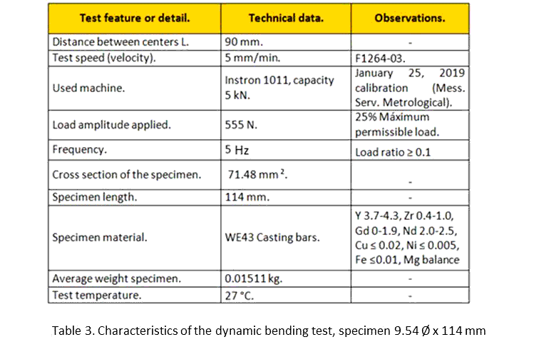
The results setup for the 3-point dynamic flexion tests was based on ASTM F1264-03 (standard specification and test methods for intramedullary fixation devices). This test was realized on two specimens with the configuration proposed in the procedure of section A1.4
These specimens were manufactured according to ASTM B 557-02 and are displayed in Figure 4a. This test determines the resistance to flexion by fatigue when placing a nail on two points and applying a dynamic load at 1 point. The full setup is described in Figure 4.
Two tests were performed where the force vs. number cycles were obtained from a 25% load amplitude (555 N). Data obtained from the simple flex test to stabilize the system, the details of the test and materials are shown in table 3. Also, a VI data acquisition system with a LabVIEW R2017 interface was used to acquire the maximum force of each of the tests and plot the force curve vs. the number of cycles.

The system shown in Figure 4 was used to perform the 3-point dynamic flexion tests on specimens with a diameter of 9.54 mm and 114 mm in length. This test consists of two supports and a lower base to place the specimen, as well as an upper support mounted on the load cell of the Instron 1011 machine which applies the load during the test to the center of the specimen. A 3D printed polypropylene chamber was prepared to allow the specimen to be immersed in Hank's balanced salts, modified without phenol red and sodium bicarbonate (H1387-1L; Batch #SLBG0073), throughout the test. It used a camera system to monitor force vs. the number of test cycles.
The acclimatization treatment that was carried out for a WE43 specimen consists of autoclave sterilization and subsequent immersion in Hank's solution handled in a chemically inert manner within a closed container using aseptic techniques according to ISO-10993-12. Acclimatization continued for 24 ± 2 h at 37 ± 1°C. Throughout the test the pH of the Hank's solution used was not manipulated to avoid any influence on the result, but at the time of its preparation the pH was 7.4. Storage conditions were validated before use.
The incubation of the WE43 specimen in the Hank´s solution was for 24 h, completed under aseptic conditions and in a sterile environment provided by a laminar flow cabinet. Although cells were not incubated in the medium, contamination in the test environment is not impossible due to the interaction of the medium with parts of the Instron machine and fixtures. The possible adhesion of particles and their progression on the surface of the material are effects that should be considered on the result of this test[10].
This acclimatization procedure provides insight for the ISO / TC 194 committee, specifically towards the new changes that are being considered on subsections a (Materials designed to degrade in the body), c (test solution), and e (immersion procedure). This is in light of the modification of the mechanical behavior of this alloy with the interaction of a medium such as Hank’s solution after sterilization[30,36].
The characteristics of the system show behavioral data from a dynamic perspective were set at an operating frequency of 5 Hz (5 cycles per second) and the applied load ratio was constant hseno completely compressed. The load applied during the fatigue test was 25% of the maximum permissible load as measured in the simple flex test for each system. This was applied in a period of 0–500,000 cycles. Figure 5 shows a load ratio condition over time, with the application of hseno load and base cycle from 0 to -1. Qizhi Chen et al, have reported the mechanical working conditions of the human body by defining the frequency per human step at 1Hz. By biomechanical convention, the operating frequency according to ASTM corresponds to 5Hz. In their work Qizhi he assumes that a person walks about 1 x 10⁷ cycles in 20 years[32].
Morphological analysis.
Specimens were prepared for microstructural analysis by polishing with 0.3µm diamond paste to a mirror finish and observation under a scanning electron microscope (SEM; Nanosem200-FEI, Netherlands) equipped with an energy dispersive spectrometer (EDS). To the samples were adhered with a graphite tape to a sample holder to obtain a in an atmosphere under vacuum at a resolution of 50 µm. With this microscope configuration, the initial flashing of the SEM filament was reviewed, selecting HV and emission current window, flash intensity in 2 was confirmed, and the specimen is inserted into the holder, proceeding to the observation. A lamella was prepared for TEM, through a beam of focused gallium ions, thinning the specimen to approximately 60 nm. Subsequently, the sample was kept in a vacuum until the day of the examination and then placed on a sample holder for analysis. A sample for transmission electron microscopy (TEM; Titan 80-300-FEI, Netherlands) was thinned to 60 nm thick by a Double Beam System (FEI QUANTA 200 3D, Netherlands) utilizing Gallium ions before being mounted on a rack and preserved in a vacuum until immediately before observation. The distribution of the elements in the micro-regions and the selected area of electron diffraction (SAED) were performed to help identify the phases. The TEM was operated at 300 keV, opening the condenser lens #2 to 150 with an exposure time of 0.5 s. The SAED in Figure 11(b) shows a crystalline structure on the diffraction pattern corresponds to the projection of the reciprocal lattice by bright field and dark field. An X-ray diffractometer (XRD; EMPYREAN, Spain), was used for phase identification using a Cu Kα radiation source, with an 8.67 s step-time scan, operated at 45 kV and 40 mA.
Citotoxicity test.
Extracts were obtained by the elution method according to ISO 10993-12:2012. Samples of WE43 were immersed into the cell culture medium for 24h at 5% CO2, 95% humidity, and 37°C with a fixed ratio of surface area to medium volume of 1.25 cm²/ml[37]. Next, extracts were collected and evaluated for impact in cell viability. For cell culture, a murine fibroblast cell line (L-929; CCL1), was purchased from the American Type Culture Collection (ATCC). Tissue culture medium, fetal bovine serum (FBS), and supplements were purchased from Gibco (IL, USA). Dyes, phosphate-buffered saline (PBS), and dimethyl sulfoxide were procured from Sigma-Aldrich (MO, USA). L-929 fibroblasts were cultured in DMEM with 10% FBS and 2mM L-glutamine in 5% CO2, 95% humidity and 37°C. For the Neutral Red Uptake assay (NRU) method, cells were seeded in 96-well plates at a density 1x10⁵ cells/well and pre-cultured for 24 hr. To allow adaptation of cells before the addition of the extracts.
For the NRU, five non serial dilutions of extracts with culture media over a range from 10% to 100% were placed on cells by quadruplicates. Treatments with standard culture media were sustained as reference (100% viability). ZDC at 0.1% was used as cytotoxic control. After a 48 h incubation period, the 96-well plate was centrifuged (210g for 10 minutes) and the medium was replaced by the neutral red stain (100 μl of a 0.2 mg/ml stock solution in cell culture medium) on each well and the plate was re-incubated at 37°C for 4 h. Afterwards, the plate was washed with PBS once and then removed, allowed to dry for 1 hr., and 100 μl of neutral red eluent (Ethanol:dH2O: acetic acid 50:49:1) were added to each well. The plate was then shaken for 1 h to dissolve the dye. After the neutral red had dissolved, the absorbance was measured using a microplate reader (xMark, BioRad, USA) at a wavelength of 540 nm with a reference wavelength of 630 nm. According to ISO 10993-5:2009, a relative metabolic activity of less than 70% was regarded as cytotoxic.
A FEM simulation was performed to visualize the behavior by mechanical integrity of the alloy and its response to failure modes. Ansys 15 software was used, through the workbench module.
