Synthesis and Characterization of PVA-Encapsulated Fe2O3-ZnO as New Composites with Tunable Optical Properties

Kahtan A. Mohammed*![]() | Shaymaa Abd AlKareem Shihab
| Shaymaa Abd AlKareem Shihab![]() | Sameer Algburi
| Sameer Algburi![]() | B. Bhavani
| B. Bhavani![]() | Ali Kareem
| Ali Kareem![]() | Mohammed Ayad Alkhafaji
| Mohammed Ayad Alkhafaji![]() | Rahman S. Zabibah
| Rahman S. Zabibah![]() | Shubham Sharma
| Shubham Sharma![]()
© 2024 The authors. This article is published by IIETA and is licensed under the CC BY 4.0 license (http://creativecommons.org/licenses/by/4.0/).
OPEN ACCESS
PVA-assisted Fe2O3-ZnO as a nanocomposite chemically prepared by in situ method. Various identification methods, including X-ray diffraction (XRD), ultraviolet-visible spectroscopy (UV-VIS), and scanning electron microscopy with energy-dispersive X-ray spectroscopy (SEM-EDX), were employed to evaluate the composite samples. The X-ray diffraction (XRD) patterns indicate that the nanocomposite exhibits the structural characteristics of hematite (α-Fe2O3), whereas no discernible influence of zinc oxide (ZnO) is observed. The (Fe2O3-ZnO)/PVA nanocomposite shows two absorption edges for both ZnO at 400nm and for Fe2O3 at 500nm and the corresponding’s bandgap was equal to 2.07 and 1.9 eV for direct and indirect transition.
Fe2O3, ZnO, nanocomposite, chemical method, physical properties
The metal oxide semiconductor is a material that plays a significant part in research due to its stability, sensitivity, adequate band gap, cost-effectiveness, strong resistance to photo-corrosion, lack of toxicity, and ease of production [1, 2]. Zinc oxide (ZnO) is a highly promising inorganic material within the category of transition metal oxides. It exhibits a diverse array of applications across various industries. In the rubber industry, ZnO is utilized as both a filler and an activator for rubber compounds. Within the pharmaceutical and cosmetics industry, it finds use in the formulation of creams, powders, and dental pastes. Additionally, ZnO serves as an effective UV radiation absorber in the textile industry. In the realm of electro technology and electronics, it is employed as a photo electronic material, field emitter, sensor, UV laser component, and solar cell. Lastly, ZnO acts as a photo catalyst in the field of photo catalysis. Zinc oxide (ZnO) exhibits various noteworthy applications, including its utilization in the synthesis of zinc silicates, its application in criminal analysis for enhancing fingerprints, and its use as a packaging material [3-5]. Iron oxides have garnered attention among various metal oxides because to their abundance, low cost, environmental friendliness, thermodynamic stability, and high surface area. The stability and ease of formation of various crystalline polymorphs of iron oxide depend on the temperature at which they are applied. Among these polymorphs, the α-Fe2O3 phase exhibits greater stability and ease of formation [6-9].
Polyvinyl alcohol (PVA) is a capping agent commonly employed in nanotechnology due to its exceptional performance characteristics. Polyvinyl acetate (PVA) is a synthetic polymer that is produced by the process of hydrolysis. It possesses notable characteristics such as high hydrophilicity, biocompatibility, and biodegradability. Polyvinyl alcohol (PVA) has been employed as a stabilizing agent in several applications, particularly for the purpose of achieving shape and size control of nanoparticles [10, 11].
In recent times, there has been a growing demand for materials that are challenging to fulfill using single-component structures. Such materials are not limited to a single class, such as organic or inorganic materials. In hybrid matrices, the integration of organic and inorganic materials occurs through a repetitive assembly process. This results in the formation of organic-inorganic hybrid systems that combine the favorable features of both types of structural blocks, as demonstrated [12].
According to our knowledge, there is no study about PVA capping of Fe2O3-ZnO nanocomposite. So, here an attempted to prepare a new PVA(Fe2O3-ZnO) nanocomposite and study its main properties.
The composite of (Fe2O3-ZnO) surrounded by PVA was created using a one-step chemical in situ process as described below: The PVA solution was prepared by dissolving 1 gram of PVA polymer with a molecular weight of 100,000 in 100 milliliters of deionized water, resulting in a solution with a concentration of 1%. This solution is referred to as solution A. The second solution, denoted as B, was created by dissolving 1 gram of ferric chloride in 50 milliliters of deionized water. A mixture of 10 ml of solution A and 10 mL of solution B was subjected to magnetic stirring at room temperature for a duration of 5 minutes, resulting in the formation of solution C. A quantity of 0.5 grams of zinc oxide (ZnO) nanoparticles was introduced into solution C, which was subsequently subjected to continuous stirring on a heated plate for a duration of 2 hours at a temperature of 50 degrees Celsius. This resulting solution is referred to as solution D. Solution D exhibited a high degree of homogeneity and clarity. A small volume of a 1M NaOH solution was gradually added to solution D. Following that, the solutions underwent thermal treatment at a temperature of 70℃ and were subjected to agitation for a period of 60 minutes on a hotplate. A colloid with a dark brown color has been successfully synthesized, indicating the synthesis of (Fe2O3-ZnO)/PVA.
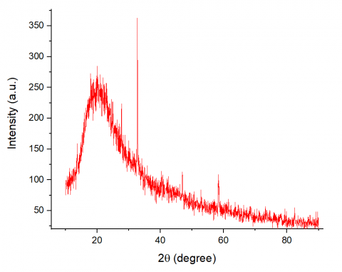
To investigate the crystal structure of the produced nanocomposite, X-ray diffraction (XRD) analysis was conducted. The resulting XRD patterns are presented in Figure 1. The diffraction peaks observed at angles of 32.7°, 45°, 58.2°, and 75.1° can be attributed to certain crystal planes of γ-Fe2O3 magnetite (PDF 39-1346). These crystal planes are identified as (220), (400), (511), and (533), respectively and there are no diffraction peaks from ZnO can be observed This assignment aligns with the findings reported by Zhang et al. in their study [13].
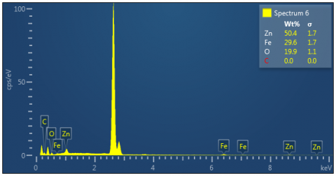
The investigation of the nanocomposite's composition was carried out by the utilization of EDX analysis. The EDX pattern is depicted in Figure 2. The EDX spectrum reveals the molar percentages of Zn, O, and Fe to be 50.4%, 19.9%, and 29.6%, respectively. This suggests that the nanocomposite primarily consists of Fe2O3 and ZnO.
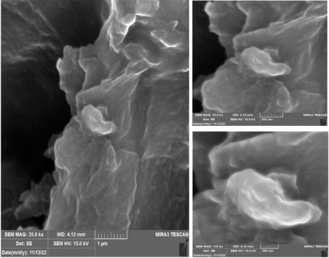
The morphology of Fe2O-ZnO capped with PVA can be seen using Scanning Electron Microscopy (SEM) with different magnification degrees shown in Figure 3. In Figure 3 can be seen there is an agglomeration for the particles inside the PVA where the PVA cover large clusters made from man-size (Fe2O3-ZnO) particles and there is no ability to identifying the individual particles.
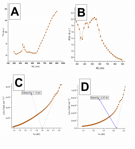
The main optical properties of prepared composite like transmittance, absorbance spectra and direct and indirect bandgaps have been tested and presented in Figure 4. Figure 4 (a) shows the transmittance spectrum of composite, from the figure can see there is no high transmission in 200-550nm wavelengths range and the material have high absorption rate here and higher than 550nm the transmission relatively increased. Absorbance spectrum presented in Figure 4 (b) which shows 2 obvious peaks first on around 400 nm belong to ZnO [14, 15] and the second one around 500 belong to Fe2O3. The energy values for both indirect and direct band gaps were determined using the Tauc relation. The band gap values were estimated using the graph shown in Figure 4 (c, d). The band gaps were determined to be 1.9 eV and 2.07 eV for indirect and direct transitions, respectively.
Figure 1. XRD peaks of (Fe2O3-ZnO)/PVA
Figure 2. EDX spectrum peaks of (Fe2O3-ZnO)/PVA
Figure 3. FESEM images of (Fe2O3-ZnO)/PVA
Figure 4. Transmittance spectrum (a), absorbance spectrum (b), indirect bandgap (c) and direct bandgap (d) of (Fe2O3-ZnO)/PVA
Fe2O3/ZnO capped nanocomposite was fabricated via a chemical in situ method. Its main physical properties like optical structural and compositional were also studied. The X-ray diffraction (XRD) patterns suggest that the nanocomposite displays the structural properties associated with hematite α-(Fe2O3), while no noticeable impact of zinc oxide (ZnO) is evident. The nanocomposite consisting of (Fe2O3-ZnO)/PVA exhibits two absorption edges, one at 400 nm for ZnO and another at 500nm for (Fe2O3. The respective bandgap values for direct and indirect transitions are determined to be 2.07 eV and 1.9 eV.
[1] Mishra, Y.K., Chakravadhanula, V.S.K., Hrkac, V., Jebril, S., Agarwal, D.C., Mohapatra, S., Avasthi, D.K., Kienle, L., Adelung, R. (2012). Crystal growth behaviour in Au-ZnO nanocomposite under different annealing environments and photoswitchability. Journal of Applied Physics, 112(6). https://doi.org/10.1063/1.4752469
[2] Chakraborty, M., Ghosh, A., Thangavel, R., Asokan, K. (2016). Conduction mechanism in mesoporous hematite thin films using low temperature electrical measurements and theoretical electronic band structure calculations. Journal of Alloys and Compounds, 664: 682-689. https://doi.org/10.1016/j.jallcom.2016.01.019
[3] Abebe, B., Zereffa, E.A., Tadesse, A., Murthy, H.A. (2020). A review on enhancing the antibacterial activity of ZnO: Mechanisms and microscopic investigation. Nanoscale Research Letters, 15: 1-19. https://doi.org/10.1186/s11671-020-03418-6
[4] Kołodziejczak-Radzimska, A., Jesionowski, T. (2014). Zinc oxide-from synthesis to application: A review. Materials, 7(4): 2833-2881. https://doi.org/10.3390/ma7042833
[5] Noukelag, S.K., Cummings, F., Arendse, C.J., Maaza, M. (2023). Physical and magnetic properties of biosynthesized ZnO/ Fe2O3, ZnO/ZnFe2O4, and ZnFe2O4 nanoparticles. Results in Surfaces and Interfaces, 10: 100092. https://doi.org/10.1016/j.rsurfi.2022.100092
[6] Valvo, M., Floraki, C., Paillard, E., Edström, K., Vernardou, D. (2022). Perspectives on iron oxide-based materials with carbon as anodes for Li-and K-ion batteries. Nanomaterials, 12(9): 1436. https://doi.org/10.3390/nano12091436
[7] Kaneti, Y.V., Tanaka, S., Jikihara, Y., Nakayama, T., Bando, Y., Haruta, M., Hossain, S.A., Golberg, D., Yamauchi, Y. (2018). Room temperature carbon monoxide oxidation based on two-dimensional gold-loaded mesoporous iron oxide nanoflakes. Chemical Communications, 54(61): 8514-8517. https://doi.org/10.1039/C8CC03639J
[8] Navrotsky, A., Mazeina, L., Majzlan, J. (2008). Size-driven structural and thermodynamic complexity in iron oxides. Science, 319(5870): 1635-1638. https://doi.org/10.1126/science.1148614
[9] Aragaw, T.A., Bogale, F.M., Aragaw, B.A. (2021). Iron-based nanoparticles in wastewater treatment: A review on synthesis methods, applications, and removal mechanisms. Journal of Saudi Chemical Society, 25(8): 101280. https://doi.org/10.1016/j.jscs.2021.101280
[10] Junaidi, J., Triyana, K., Suharyadi, E., Harsojo, H., Wu, L.Y. (2017). The roles of polyvinyl alcohol (PVA) as the capping agent on the polyol method for synthesizing silver nanowires. Journal of Nano Research, 49: 174-180. https://doi.org/10.4028/www.scientific.net/JNanoR.49.174
[11] Javed, R., Zia, M., Naz, S., Aisida, S.O., Ain, N.U., Ao, Q. (2020). Role of capping agents in the application of nanoparticles in biomedicine and environmental remediation: Recent trends and future prospects. Journal of Nanobiotechnology, 18: 1-15. https://doi.org/10.1186/s12951-020-00704-4
[12] Abou Hammad, A.B., Mansour, A.M., Elhelali, T.M., El Nahrawy, A.M. (2023). Sol-Gel/Gel casting nan architectonics of hybrid Fe2O3-ZnO/PS-PEG nanocomposites and their opt magnetic properties. Journal of Inorganic and Organometallic Polymers and Materials, 33(2): 544-554. https://doi.org/10.1007/s10904-022-02519-2
[13] Fatimah, I., Purwiandono, G., Hidayat, A., Sagadevan, S., Kamari, A. (2022). Mechanistic insight into the adsorption and photocatalytic activity of a magnetically separable γ-Fe2O3/Montmorillonite nanocomposite for Rhoda mine B removal. Chemical Physics Letters, 792: 139410. https://doi.org/10.1016/j.cplett.2022.139410
[14] Qamar, M.T., Aslam, M., Ismail, I.M., Salah, N., Hameed, A. (2016). The assessment of the photocatalytic activity of magnetically retrievable ZnO coated γ-Fe2O3 in sunlight exposure. Chemical Engineering Journal, 283: 656-667. https://doi.org/10.1016/j.cej.2015.08.002
[15] Hitkari, G., Singh, S., Pandey, G. (2018). Photoluminescence behavior and visible light photocatalytic activity of ZnO, ZnO/ZnS and ZnO/ZnS/α-Fe2O3 nanocomposites. Transactions of Nonferrous Metals Society of China, 28(7): 1386-1396. https://doi.org/10.1016/S1003-6326(18)64777-6