ABSTRACT
Aiming to provide cardiovascular morphophysiology information on the Cuniculus paca, an important neotropical rodent, eight healthy adult females of this species were evaluated three times by echocardiography under general anesthesia with isoflurane every 15 days. The exams were performed by a single experienced evaluator with the animals positioned in right and left decubitus. Posteriorly, two expert evaluators measured the cardiac chambers, walls and flow patterns, by B-mode, M-mode, and Doppler ultrasonography. The resulting values were compared among evaluators and periods by the Bland-Altman agreement test and several descriptive statistics were presented for each parameter. Echocardiographic images were obtained between the second and fifth left and right intercostal spaces, enabling the measurement of heart chambers and walls, mitral, tricuspid, aortic and pulmonary valves blood flows, and the ejection and shortening fractions calculation. None of the studied variables showed inter-observers or inter-periods variations. This study provided some normal echocardiographic variables, applicable to epidemiological, pathophysiological or case studies in the Cuniculus paca and phylogenetically close species.
Keywords:
echocardiography; rodents; neotropical; wildlife
RESUMO
Com o objetivo de fornecer informações da morfofisiologia cardiovascular da Cuniculus paca, um importante roedor neotropical, oito fêmeas adultas saudáveis dessa espécie foram avaliadas três vezes pela ecocardiografia, sob anestesia geral com isoflurano, a cada 15 dias. Os exames foram realizados por um único avaliador experiente, com os animais posicionados em decúbito direito e esquerdo. Posteriormente, dois avaliadores experientes mediram as câmaras cardíacas, as paredes e os padrões de fluxo, por meio do modo B, do modo M e da ultrassonografia Doppler. Os valores resultantes foram comparados entre avaliadores e períodos pelo teste de concordância de Bland-Altman, e várias estatísticas descritivas foram apresentadas para cada parâmetro. As imagens ecocardiográficas foram obtidas entre o segundo e o quinto espaços intercostais esquerdo e direito, o que permitiu a medição das câmaras e paredes cardíacas, do fluxo sanguíneo pelas válvulas mitral, tricúspide, aórtica e pulmonar e o cálculo das frações de ejeção e encurtamento. Nenhuma das variáveis estudadas mostrou variações entre os avaliadores ou entre os períodos. Este estudo fornece algumas variáveis ecocardiográficas normais, aplicáveis a estudos epidemiológicos, fisiopatológicos ou de casos na Cuniculus paca e em espécies filogeneticamente próximas.
Palavras-chave:
ecocardiografia; roedores; neotropical; selvagem
INTRODUCTION
The Spotted Paca (Cuniculus paca) is the second largest Neotropical rodent. In the wild, these animals have a life span of approximately 12 years, usually live alone or in pairs and are naturally distributed from the northeastern of Mexico to Paraguay and from the northern of Brazil to the southern of Argentina (Sainsbury, 2003SAINSBURY, A.W. Rodentia (rodents). In: FOWLER, M.E.; MILLER, R.E. (Eds.). Zoo and wild animal medicine. Saunders: Philadelphia, 2003. p.420-442.). Deforestation and hunting has decreased its free-life population and for this reason, clinical studies that improve preservation of this species have been fostered (Chiarello et al., 2008CHIARELLO, A.G., AGUIAR, L.M.S., CERQUEIRA, R., et al. Mamíferos ameaçados de extinção no Brasil; In: MACHADO, A.B.M., DRUMMOND, G.M. & PAGLIA, A.P. (Eds.) Livro vermelho da fauna brasileira ameaçada de extincão. MMA/Fundacão Biodiversitas. p.681-702. 2008.).
Some physiologic reference values have been established for this type of wild rodents; however, studies that aid in the understanding of physiologic responses to medical procedures are necessary (Crissey et al., 2004CRISSEY, S.D.; ANGE, K.D.; SLIFKA, K.A. et al. Serum lipid concentrations in six canid and four ursid species in four zoos. J. Zoo Wildl. Med., v.35, p.34-39, 2004. ). The cardiac morphophisiology assessment on healthy wild animals is important, because it provides a baseline for pathophysiological studies (Onuma et al., 2009ONUMA, M.; KONDO, H.; ONO, S. et al. Radiographic measurement of cardiac size in 64 ferrets. J. Vet. Med. Sci., v.71, p.355-358, 2009.; Black et al., 2011BLACK, P.A.; MARSHALL, C.; SEYFRIED, A.W.; BARTIN, A.M. Cardiac assessment of African hed gehogs, Atelerix albiventris. J. Zoo Wildl. Med., v.42, p.49-53, 2011. ). However, it is also important to consider that these animals require pharmacological restraint or anesthesia for their manipulation, and the drugs used for these procedures have varied effects on cardiovascular function (Szabuniewicz et al., 1978SZABUNIEWICZ, M.; SANCHEZ, L.; SOSA, A. et al. The electrocardiogram of the Capibara (Hydrochoerus hydrochaeris, Linné). Zentralbl. Veterinarmed., v.25, p.162-171, 1978.).
Complementary techniques are essential for evaluation of cardiovascular function. In this context, echocardiography is considered the best method for evaluation of cardiovascular morphophysiology since it is non-invasive and very accurate (Boon, 1998BOON, J.A. The echocardiographyc examination. In: _____. (Ed.). Manual of veterinary echocardiography. Baltimore: Williams and Wilkins, 1998. p.35-128.; Bonagura, 2000BONAGURA, J.D. Feline echocardiography. J. Feline Med. Surg., v.2, p.147-151, 2000.). Nonetheless, in neotropical rodents few studies have evaluated cardiovascular morphophysiology (Szabuniewicz et al., 1978SZABUNIEWICZ, M.; SANCHEZ, L.; SOSA, A. et al. The electrocardiogram of the Capibara (Hydrochoerus hydrochaeris, Linné). Zentralbl. Veterinarmed., v.25, p.162-171, 1978.; Moura et al., 2015MOURA, C.R.; NEVES. D.A.; SILVA M.L. et al. Cardiothoracic ratio and vertebral heart scale in clinically normal black-rumped agoutis (Dasyprocta prymnolopha, wagler 1831). J. Zoo Wildl. Med., v.46, p.314-9, 2015.; Diniz et al., 2013DINIZ, A.N.; SILVA JÚNIOR, J.R.; GUERRA, P.C. Electrocardiogram assessment in non-anaesthetized clinically healthy agouti (Dasyprocta primnolopha, Wagler 1831). Pesqui. Vet. Bras., v.33, Suppl.1, p.8-14. 2013. ; Diniz et al., 2017) and, in the Cuniculus paca, only electrocardiographic parameters and post mortem cardiac morphology have been described (Napier et al., 2013NAPIER, J.E.; KUTINSKY, I.B.; ARMSTRONG, D.L. et al. Evaluating echocardiogram and indirect blood pressure results in male western lowland gorillas (Gorilla gorilla gorilla) during three phases of an anesthetic protocol. J. Zoo Wildl. Med., v.44, p.875-881, 2013.; Uscategui et al., 2016USCATEGUI, R.A.; ALMEIDA, V.T.; KAWANAMI, A.E. et al. Electrocardiographic exam in female spotted pacas (Cuniculus paca). Pesqui. Vet. Bras., v.36, p.559-563, 2016.).
This study was performed trying to evaluate the feasibility and standardization of some echocardiographic parameters in the Spotted Paca, aiming to provide cardiovascular morphophysiology information of the Cuniculus paca, to contribute to the research and conservation of neotropical rodents.
MATERIAL AND METHODS
This study was approved by the Ethics Committee on Animal Use from Univ. Estadual Paulista (027420/11). Nine female adults of Spotted Paca, clinically healthy (weighting 9.2±0.9kg), from the Animal Science Department registered at Brazilian Institute of Environment and Natural Resources (IBAMA), as a Brazilian fauna specimen’s breeder for scientific purposes (482508) were enrolled in this observational prospective study.
The animals were chemically restrained using midazolam 0.5mg/kg (Dormire®, Cristalia Laboratories, Itapira, SP, Brazil) and ketamine 30mg/kg (Dopalen®, Ceva Saúde Animal Ltda, Paulínia, SP, Brazil) intramuscularly (IM) and anesthetized with isoflurane (Isoflurane®, Cristalia Laboratories, Itapira, SP, Brazil) diluted in 100% oxygen, via face mask, (immobilization and median arterial pressure above 60mmHg) for laparoscopic folicular aspiration, by Barros et al., 2016BARROS, F.F.P.C.; TEIXEIRA, P.P.M.; USCATEGUI, R.A.R. et al. Laparoscopic ovum pick-up in spotted paca (Cuniculus paca). Arq. Bras. Med. Vet. Zootec., v.68, p.858-864, 2016. associated study. Preemptive analgesia was achieved by IM administration of tramadol hydrochloride 5mg/kg (Tramadon®, Cristalia Laboratories, Itapira, SP, Brazil). Once laparoscopic procedure was finished, the animals were kept anesthetized to perform the echocardiographic examination. This exam had been performed each 15 days, totaling three different assessments used to study inter-periods variation.
Conventional B-mode, M-mode and Doppler, ultrasound exam was performed using the MyLab® VET30® (Esaote, Genova, Italy), and sector microconvex multifrequency probe (7.5-12.0MHz). The right parasternal standard window was optimized for the left atrium (LA), and left ventricle (LV,) long and short axis visualization; the left apical parasternal standard window was optimized for the LV inflow tract, longitudinal motion of the lateral and septal mitral annulus, and the LV outflow tract, data acquisition. The echocardiography exam was made by a single experiment evaluator and the storage images were analyzed off-line by two evaluators with similar experience, aiming to reduce the anesthetic time, and to determine and control the inter-observer variations. The echocardiographic exam had been performed according to American College of Veterinary Internal Medicine recommendations (Thomas et al., 1993THOMAS, W.P.; GABER, C.E.; JACOBS, G.J. et al. Recommendations for standards in transthoracic two-dimensional echocardiography in dogs and cats. J. Vet. Intern. Med., v.7, p.247-252, 1993. ) and adapted to Spotted Pacas.
Right ventricular diameter (RVD), interventricular septum (IVS), left ventricular diameter (LVD) and left ventricular wall (LVFW) were measured (mm) both at end diastole (d) and at end systole (s) by M-mode using the leading edge method from the short axis of the left ventricle in the chordal plane, with the line of M-mode aligned perpendicular to the interventricular septum and equidistant from the papillary muscles. Since simultaneous ECG was not performed the end diastole and systole were determined from valve motions and cavity sizes (Figure 1). From this evaluation, the ejection (EF), and shortening (FS) fractions were calculated using the Teichholz technique, as performed on dogs and cats.
Cardiac ultrasound image of right parasternal window in Cunicullus paca for short axis view of the left ventricle. Right ventricular diameter (RV), interventricular septum (IVS), left ventricular (LV) and left ventricular wall (PW) measured (mm) both at end diastole (d) and at end systole (s).
In the short axis view with cardiac base image (Figure 2), the relationship between the diameter of the left atrium and the aorta (LA: Ao) were measured. In a plane directed to the cardiac base, the peak velocity of the pulmonary valve (peak PV cm/s) and our pressure gradient (peak PV mmHg) were measured, with the cursor of sampling volume positioned next to the pulmonary leaflets on the face opposite to the pulmonary artery. At the mitral plan, the extent of septal separation to E point (SSPE) was obtained.
Cardiac ultrasound image of right parasternal window in Cunicullus paca for short axis view cardiac base image. Left atrium (LA) and aorta (Ao).
The five-chamber apical image was obtained through the left parasternal window (Figure 3). The cursor was aligned parallel to the aortic flow, with the sampling volume positioned next to the aortic leaflets on the face opposite to the aorta and the peak velocity of the aortic valve (peak AV cm/s) and our pressure gradient (peak AV mmHg) were measured.
Cardiac ultrasound image of left parasternal window, five-chamber apical image in Cunicullus paca for measurement the aortic peak velocity (+ m/s). Left ventricle (LV) and aorta (Ao).
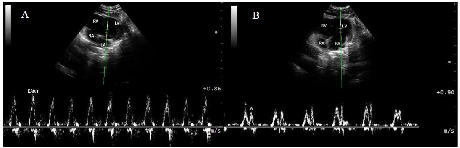
The spectral Doppler velocity curves of mitral flow were used for the analysis of the diastolic function. To obtain this flow, the cursor of volume sample was placed at the height of the commissural edges of the cusps with the mitral valve open (Figure 4). The filter and gain were adjusted to improve the image. In these curves, the peak velocity of early diastolic transmitral flow (peak E cm/s), the peak velocity of late diastolic transmitral flow (peak A cm/s), the E:A ratio, the peak velocity of early tricuspid peak velocity (peak ET cm/s) and the peak velocity of late tricuspid peak velocity (peak AT cm/s) were obtained automatically.
Cardiac ultrasound Composite image displaying summated transmitral filling waves (left A) and separated filling waves (right B) on isoflurane anesthetized female Spotted Paca (Cuniculus paca) heart rate 146±13.6bpm. Right and left ventricle and atrium (RV, LV, RA, LA respectively).
For statistical analysis, it was used Software R® (R Foundation for Statistical Computing, Vienna, Austria). Measures were initially analyzed for normality (Shapiro test) and homoscedasticity of variances (Bartlett test). Raw or transformed data were compared among evaluators and periods by the Bland-Altman agreement test, if equality was proven, the overall mean of the three examinations performed in the nine Spotted Pacas by the two evaluators were calculated and used to determine the confidence intervals (CI) and descriptive statistical. The 5% significance (P< 0.05) was fixed in all tests.
RESULTS
The echocardiographic exam proved to be a simple and applicable diagnostic technique in the Cuniculus paca. The cardiac images were obtained between the second and fifth left and right intercostal spaces near to the apex beat. Morphologic or functional abnormalities were not observed during the exam.
Although echocardiographic examination was performed by different observers, the echocardiography measurements did not show inter-observer or inter-period variations (P>0.05) and the confidence interval at 95% were considered the reference for the echocardiography parameters studied here, and were presented on Table 1 together with the measures of central tendency and variation.
The mitral and tricuspid valves, at spectral Doppler flows examination showed the E and A waves fused in approximately 80% of the cases (Figure 5) and therefore the parameters related to these waves (AM cm/sec, AM mmHg, EM cm/s, EM mmHg) could be measured only in cases in which these waves were separated. Heart rate oscillated between 115 and 197bpm and the mean was 150±17bpm.
DISCUSSION
This is the first study regarding echocardiographic exams on anesthetized Cuniculus paca. In the literature, only two cardiologic studies were found on this species, the first one described the cardiac anatomy postmortem (Ávila et al., 2010ÁVILA, B.H.P.; MACHADO, M.R.F.; OLIVEIRA, F.S. Descrição anátomo-topográfica do coração da paca (Agouti paca). Acta Sci. Vet., v.38, p.191-195, 2010.) and second one related the electrocardiography parameters on Spotted Pacas under pharmacologic restraint (Uscategui et al., 2016USCATEGUI, R.A.; ALMEIDA, V.T.; KAWANAMI, A.E. et al. Electrocardiographic exam in female spotted pacas (Cuniculus paca). Pesqui. Vet. Bras., v.36, p.559-563, 2016.).
The cardiac anatomy in the Spotted Paca was similar to that reported in domestic mammals, but differing in cranial localization by one intercostal space and two cranial cava veins, as was described in other rodents except in the Agouti a phylogenetically close rodent (Oliveira et al., 1999OLIVEIRA, P.F.N.; CARVALHO, M.A.M.; SOUZA, W.M.; MIGLINO, M.A. The right azygos vein in agouti (Dasyprocta agouti, rodentia). Braz. J. Vet. Res. Anim., v.36, p.4, 1999.; Ávila et al., 2010ÁVILA, B.H.P.; MACHADO, M.R.F.; OLIVEIRA, F.S. Descrição anátomo-topográfica do coração da paca (Agouti paca). Acta Sci. Vet., v.38, p.191-195, 2010.). The images of the cardiac chambers were made between the second and fourth intercostal spaces through right and left parasternal windows for longitudinal and transverse sections. In this species, the intercostal access for echocardiography exam is more cranial than that recommended for dogs (between the third and sixth on the right side and between the fifth and seventh on the left side by Thomas et al., 1993THOMAS, W.P.; GABER, C.E.; JACOBS, G.J. et al. Recommendations for standards in transthoracic two-dimensional echocardiography in dogs and cats. J. Vet. Intern. Med., v.7, p.247-252, 1993. ). The reduced echocardiography windows and high respiratory rate (35±12 cycles/min) made the exam more difficult; however, it was performed in all animals.
According to Ávila et al., 2010ÁVILA, B.H.P.; MACHADO, M.R.F.; OLIVEIRA, F.S. Descrição anátomo-topográfica do coração da paca (Agouti paca). Acta Sci. Vet., v.38, p.191-195, 2010. the paca’s heart is divided into fourth chambers, two atriums and two ventricles (right and left) presenting the shape of an elongated cone; however, in the echocardiographic evaluation, the heart appears rounder than elongated (Figure 4). The dimensions of the chambers and walls which were evaluated by M-mode showed a uniform distribution among the animals (Table 1). The Doppler evaluation of the valve flows resulted in 80% of E and A-waves fused, not allowing the separate measurement of these parameters (peak A cm/s, E cm/s) at the mitral and tricuspid valves.
These E and A waves fusion have been associated with tachycardia and first-degree atrioventricular block in small animals (Appleton, 1991APPLETON, C.P. Incremental changes in heart rate on mitral flow velocity: assessment in lightly sedated, conscious dogs. J. Am. Coll. Cardiol., v.17, p.227-236, 1991.). In this case, we have associated the fusion phenomenon with the higher heart rate on our anesthetized animals (150±17bpm) in relation to the values described by Estrada et al. (2009ESTRADA, A.H.; GERLACH, T.J.; SCHMIDT, M.K. et al. Cardiac evaluation of clinically healthy captive maned wolves (Chrysocyon brachyurus). J. Zoo Wildl. Med., v.40, p.478-486, 2009.) on conscious animals (86.0±7.8bpm) and may be attributed to ketamine sympathetic effect (Szabuniewicz et al., 1978SZABUNIEWICZ, M.; SANCHEZ, L.; SOSA, A. et al. The electrocardiogram of the Capibara (Hydrochoerus hydrochaeris, Linné). Zentralbl. Veterinarmed., v.25, p.162-171, 1978.) and to the disturbances caused by the anesthetic maintenance with isoflurane in laparoscopic procedures (Fuentes et al., 2004FUENTES, J.M.; HANLY, E.J.; BACHMAN, S.L. et al. Videoendoscopic endotracheal intubation in the rat: a comprehensive rodent model of laparoscopic surgery. J. Surg. Res., v.122, p.240-248, 2004.).
In cats, this alteration is commonly reported and can be controlled with maneuvers that stimulated the vagal nerve (Smith and Schober 2013SMITH, D.N.; SCHOBER, K.E. Effects of vagal maneuvers on heart and Doppler variables of left ventricular filling in healthy cats. J. Vet. Cardiol., v.15, p.33-40, 2013. ). These maneuvers may be applicable in these animals to improve the reliability of the cardiac parameters derived from the E and A-waves of mitral (peak E cm/s, peak A cm/s), tricuspid (peak E cm/s, peak A cm/s), aortic (AV cm/s and AV mmHg) and pulmonary (PV cm/s and PVmmHg) flows, resulting in high variation coefficients (>30%) in our study.
It is important to note the limitations of this study: 1) Limited sample size, due to wildlife nature of this species; 2) The effects of anesthesia were not isolated due to wild characteristic; 3) The need to reduce anesthetic time only allowed the evaluation by an observer. Despite these limitations, the exam was shown to be feasible, obtaining information which was not described in these animals. Studies evaluating the atrial and ventricular area and modern echocardiographic methods might complement the pioneering information of this research and aid in the medical conservation of the neotropical rodents.
In conclusion, echocardiographic exam was feasible on Cuniculus paca getting images of heart chambers between second and fifth intercostal spaces, via bilateral longitudinal and transverse parasternal windows. The imaging technique application allowed the visualization, evaluation and measurement of heart chambers, walls, valve flows and calculations of hemodynamic parameters as well derived from these exams. Nonetheless, Doppler evaluation of the mitral and tricuspid flows resulted on E and A-waves fusion compromising the accuracy of the parameters derived from these waves. No structural cardiac abnormalities were observed during analysis. This study provided some normal echocardiographic variables, applicable to epidemiological, pathophysiological or case studies in the Cuniculus paca and phylogenetically close species.
ACKNOWLEDGEMENTS
To the National Council for Scientific and Technological Development (CNPq), Coordination for the Improvement of Personnel of Superior Level (CAPES) and Foundation for Research Support of the State of São Paulo (Fapesp), Morphology and Animal Physiology Department, Veterinary Hospital "Governor Laudo Natel" FCAV - Unesp and Professor Dr. Julio Carlos Canola radiology service boss.
REFERENCES
- APPLETON, C.P. Incremental changes in heart rate on mitral flow velocity: assessment in lightly sedated, conscious dogs. J. Am. Coll. Cardiol., v.17, p.227-236, 1991.
- ÁVILA, B.H.P.; MACHADO, M.R.F.; OLIVEIRA, F.S. Descrição anátomo-topográfica do coração da paca (Agouti paca). Acta Sci. Vet., v.38, p.191-195, 2010.
- BARROS, F.F.P.C.; TEIXEIRA, P.P.M.; USCATEGUI, R.A.R. et al. Laparoscopic ovum pick-up in spotted paca (Cuniculus paca). Arq. Bras. Med. Vet. Zootec., v.68, p.858-864, 2016.
- BLACK, P.A.; MARSHALL, C.; SEYFRIED, A.W.; BARTIN, A.M. Cardiac assessment of African hed gehogs, Atelerix albiventris. J. Zoo Wildl. Med., v.42, p.49-53, 2011.
- BONAGURA, J.D. Feline echocardiography. J. Feline Med. Surg., v.2, p.147-151, 2000.
- BOON, J.A. The echocardiographyc examination. In: _____. (Ed.). Manual of veterinary echocardiography. Baltimore: Williams and Wilkins, 1998. p.35-128.
- CHIARELLO, A.G., AGUIAR, L.M.S., CERQUEIRA, R., et al. Mamíferos ameaçados de extinção no Brasil; In: MACHADO, A.B.M., DRUMMOND, G.M. & PAGLIA, A.P. (Eds.) Livro vermelho da fauna brasileira ameaçada de extincão. MMA/Fundacão Biodiversitas. p.681-702. 2008.
- CRISSEY, S.D.; ANGE, K.D.; SLIFKA, K.A. et al. Serum lipid concentrations in six canid and four ursid species in four zoos. J. Zoo Wildl. Med., v.35, p.34-39, 2004.
- DINIZ, A.N.; PESSOA, G.T.; MOURA, L.S. et al. Computerized electrocardiogram in agoutis (Dasyprocta prymnolopha Wagler, 1831) anesthetized with ketamine and midazolam. Pesqui. Vet. Bras., v.37, p.150-155, 2017.
- DINIZ, A.N.; SILVA JÚNIOR, J.R.; GUERRA, P.C. Electrocardiogram assessment in non-anaesthetized clinically healthy agouti (Dasyprocta primnolopha, Wagler 1831). Pesqui. Vet. Bras., v.33, Suppl.1, p.8-14. 2013.
- ESTRADA, A.H.; GERLACH, T.J.; SCHMIDT, M.K. et al. Cardiac evaluation of clinically healthy captive maned wolves (Chrysocyon brachyurus). J. Zoo Wildl. Med., v.40, p.478-486, 2009.
- FUENTES, J.M.; HANLY, E.J.; BACHMAN, S.L. et al. Videoendoscopic endotracheal intubation in the rat: a comprehensive rodent model of laparoscopic surgery. J. Surg. Res., v.122, p.240-248, 2004.
- MOURA, C.R.; NEVES. D.A.; SILVA M.L. et al. Cardiothoracic ratio and vertebral heart scale in clinically normal black-rumped agoutis (Dasyprocta prymnolopha, wagler 1831). J. Zoo Wildl. Med., v.46, p.314-9, 2015.
- NAPIER, J.E.; KUTINSKY, I.B.; ARMSTRONG, D.L. et al. Evaluating echocardiogram and indirect blood pressure results in male western lowland gorillas (Gorilla gorilla gorilla) during three phases of an anesthetic protocol. J. Zoo Wildl. Med., v.44, p.875-881, 2013.
- OLIVEIRA, P.F.N.; CARVALHO, M.A.M.; SOUZA, W.M.; MIGLINO, M.A. The right azygos vein in agouti (Dasyprocta agouti, rodentia). Braz. J. Vet. Res. Anim., v.36, p.4, 1999.
- ONUMA, M.; KONDO, H.; ONO, S. et al. Radiographic measurement of cardiac size in 64 ferrets. J. Vet. Med. Sci., v.71, p.355-358, 2009.
- SAINSBURY, A.W. Rodentia (rodents). In: FOWLER, M.E.; MILLER, R.E. (Eds.). Zoo and wild animal medicine. Saunders: Philadelphia, 2003. p.420-442.
- SMITH, D.N.; SCHOBER, K.E. Effects of vagal maneuvers on heart and Doppler variables of left ventricular filling in healthy cats. J. Vet. Cardiol., v.15, p.33-40, 2013.
- SZABUNIEWICZ, M.; SANCHEZ, L.; SOSA, A. et al. The electrocardiogram of the Capibara (Hydrochoerus hydrochaeris, Linné). Zentralbl. Veterinarmed., v.25, p.162-171, 1978.
- THOMAS, W.P.; GABER, C.E.; JACOBS, G.J. et al. Recommendations for standards in transthoracic two-dimensional echocardiography in dogs and cats. J. Vet. Intern. Med., v.7, p.247-252, 1993.
- USCATEGUI, R.A.; ALMEIDA, V.T.; KAWANAMI, A.E. et al. Electrocardiographic exam in female spotted pacas (Cuniculus paca). Pesqui. Vet. Bras., v.36, p.559-563, 2016.
Publication Dates
-
Publication in this collection
Jan-Feb 2019
History
-
Received
21 June 2017 -
Accepted
17 May 2018