The Journal of Advances in Parasitology
Research Article

Epidemiology of Ecto-Parasitic Infestation of Cattle in Milk Shed Areas of Baghabari of Shahjadpur Upazila of Sirajgonj District, Bangladesh
Muhammed Hossain1*, Md. Jamal Uddin Bhuiyan1, Md. Tariq Ibne Hai Digonto2
1Department of Parasitology, Faculty of Veterinary and Animal Science, Sylhet Agricultural University, Sylhet-3100, Bangladesh; 2Faculty of Veterinary and Animal Science, Sylhet Agricultural University, Sylhet-3100, Bangladesh.
Abstract | Epidemiological examination of 400 cattle for ecto-parasitic infestation in Baghabari milk shade area of Sirajgonj district, Bangladesh was recorded high amid the period from April 2014 to March 2015 revealed 60.00 per cent to be pervaded with several species of ticks and mites. The prevalence rate was highest in the case of Rhipicephalus sanguinus (20.00%) followed by Boophilus microplus (18.75%), Haematopinus eurysternus (11.25%) and Linognathus vituli (10.00%). The result disclosed that the infestation rate was significantly (P<0.05) higher in female compare to male. The relationship between the age of cattle and the different species of ecto-parasitic infestation communicated that, mature >5 years (64.17%) cattle were more vulnerable than that young calves <2 years (51.96%) and of adolescent dairy cattle ages 2-5 years (48.13%).The ecto-parasitic infestation was more common in weak animals than ordinary sound cattle. Seasonal prevalence showed significantly (P<0.05) higher prevalence in rainy season (74.55%) followed by summer (67.80%) and winter (42.44%). The Mean parasitic burdens were 2.12±0.13 per square inches of heavily infected area.
Keywords | Epidemiology, ecto-parasite, cattle, Sirajgonj, Prevalence
Editor | Muhammad Imran Rashid, Department of Parasitology, University of Veterinary and Animal Sciences, Lahore, Pakistan.
Received | December 01, 2015; Revised | March 22, 2016; Accepted | March 28, 2016; Published | April 13, 2016
*Correspondence | Muhammed Hossain, Department of Parasitology, Faculty of Veterinary and Animal Sciences, Sylhet Agricultural University, Sylhet-3100, Bangladesh; Email: bmhossain34sau@gmail.com
Citation | Hossain M, Bhuiyan MJU, Digonto MTHI (2016). Epidemiology of ecto-parasitic infestation of cattle in milk shed areas of Baghabari of Shahjadpur Upazila of Sirajgonj district, Bangladesh. J. Adv. Parasitol. 3(2): 56-60.
DOI | http://dx.doi.org/10.14737/journal.jap/2016/3.2.56.60
ISSN | 2311-4096
Copyright © 2016 Hossain et al. This is an open access article distributed under the Creative Commons Attribution License, which permits unrestricted use, distribution, and reproduction in any medium, provided the original work is properly cited.
Introduction
Tropical, agro-based Bangladesh has 47.51 million livestock of which 22.87 million constitute cattle (BBS, 2008). Livestock is the backbone of Bangladesh’s agricultural economy, is at risk of decline in production due to number of ecto and endo-parasites. Among various hindrances in cattle rearing malnutrition and parasitic infestation are the major limiting factors especially in Bangladesh (Jabbar and Greery, 1983) since the climate condition of Bangladesh is very conducive to a wide variety of ecto and endoparasites. Bangladesh is usually hot and humid except in winter and the climatic condition is very conducive to a wide variety of parasites as well as ticks (Razzak and Shaikh, 1969). Ecto-parasitic infestation is one of the major veterinary problems affecting livestock industries in many parts of the world (Hourrigan, 1979). Ecto-parasites including lice, ticks, mites etc. play an important role in the transmission of certain pathogens (Loomis, 1986)and are also known to cause heavy economic losses to livestock industry due to their usual habit of blood sucking, which adversely affects the economic production (Branscheid and Schroer, 1997). Among ecto-parasites, ticks have been recognized as the notorious threat due to severe irritation, allergy and toxicosis (Niyonzema and Kiltz, 1986) and have a seasonal population dynamics (Singh et al., 2000). In some cases, ticks have been reported to cause lowered productivity and mortality (Niyonzema and Kiltz, 1986) and transmit such diseases as babesiosis, theileriosis, anaplasmosis etc. (Norval et al., 1984; Jaswal et al., 2014; Salih et al., 2015). Ticks act not only as potential vectors but also as reservoirs of certain infectious agents e.g. Pasteurella multocida, Brucella abortus and Salmonella typhimurium in man and animals (Jongejan et al., 2004).
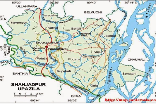
Figure 1: Map showing the study area
Lice infested animals keep poor physical condition and develop an unthrifty, anaemic appearance and discoloured greasy hair (Nelson, 1984). Lice free animals are more profitable than infested animals due to increased rate of weight gain and more feed utilization (Kettle, 1974). The situation of ticks and tick-borne diseases in animals have been partially documented in Bangladesh (Samad, 2000), but these studies was fragmented and not yet done in Sirajgonj region. Sirajgonj district is tropical type of area which actually presents in lowland and flood plain based area. Besides, this area also covered by a huge variety of floral composition. Therefore, require a quick investigation and activity for improvement in the management and control accomplishment to counteract and minimize the misfortune created by ecto-parasitic infestation in dairy cattle. Hence a need was felt to accomplish the epidemiological investigation on the ecto-parasites in cattle to know the exact status in Milk Shed Areas of Baghabari of Shahjadpur Upazila of Sirajgonj District, Bangladesh.
Materials and Methods
Ethical Issue
Before sampling informed consent was obtained from the animal owners participating in the study and cautions were taken to ensure minimums stress to the animal during sampling.
Study Area
The present research was conducted in cattle of different villages of Baghabari of Sirajgonj District of Bangladesh in which Cooperative society regularly supply their milk to Milk-Vita, Sirajgonj district which is located 130 km east to Dhaka city.
Period of Study
The study was carried out during the period of from April 2014 to April 2015.
Sample Size Calculation
The sample size was calculated by using the formula for estimating prevalence according to Houel and Toft (2004).

Where n = required sample size, pexp = expected prevalence of the disease, d = allowable error of 5% and confidence level 95%.
On the basis of information obtained through literature review the assumed prevalence of infection considered as 50% for cattle and the population size was considered as 384. We wanted to be 95% sure to detect the infection if it present. Thus therefore we considered 400 cattle.
Survey Design and Sampling
Randomly sampling was performed for this study. Cattle were examined for ecto-parasites from 20 villages from in and around Shahjadpur upazila of Sirajgonj. The investigation was carried out in several visits on three seasons (summer: March- June; Rainy: July-October and winter: November-February). A Total of 400 cattle (male 125 and female 275) were selected randomly from different areas in and around the study areas for the convenience of the study and availability of the cattle.
Collection and Preservation of Samples
The selected cattle were thoroughly investigated by close inspection, parting the hairs against their natural direction for the detection of ecto-parasites. A questionnaire including age, breed, sex was used in the study. Ecto-parasites were collected from the different parts of the body of the individual cattle by hand picking. When required, small hair brush dipped in ethanol was used for the collection of ticks. Adequate precautions were taken to preserve the mouth parts and appendages of the ecto-parasites during collection. Ecto-parasites were preserved in 70% alcohol and labelled properly.
Identification of Ecto-parasites
Morphology of ecto-parasites was studied in the laboratory with the help of dissecting (4X) and compound (10X) microscope. Ecto-parasites were identified according to the keys and descriptions given by Wall and Shearer (1997) and Soulsby (1982) by preparing permanent slides according to the procedures described by Cable (1967).
Statistical Analysis
Statistical analyses were carried out by using Statistical Package for Social Sciences (SPSS) version 11.5 for Windows (2007) using F test.
Table 1: Overall Prevalence of ecto-parasitic infestation in cattle
|
Name of the ecto-parasites |
No. of examined |
No. of infected |
Prevalence |
Parasitic burden |
|
|
Range |
Mean±SE |
||||
|
Boophilus microplus |
400 |
75 |
18.75% |
1-6 |
2.09±0.11 |
|
Rhipicephalus sanguinus |
400 |
80 |
20.00% |
1-7 |
2.18±0.04 |
|
Haematopinus eurysternus |
400 |
45 |
11.25% |
1-5 |
1.59±0.03 |
|
Linoganthus vituli |
400 |
40 |
10.00% |
1-2 |
1.12±0.02 |
|
Overall infection |
400 |
240 |
60.00% |
1-8 |
2.12±0.13 |
Result and Discussion
A total of 400 cattle examined, 240 (60.00%) were found infested with one or more species of ticks (Table 1). The findings of this study agree with the reports of Islam et al. (2009) in Sirajganj, Kamal et al. (1996) in Chittagong of Bangladesh, who recorded (65.50%) and (65.40%) prevalence of ecto-parasites in cattle, respectively. The findings of this study differ with the previous findings of some other scientists. Higher prevalence in cattle was reported by Sajid et al. (2008) in Pakistan and Nath et al. (2015) in Bangladesh.
Roy et al. (2001) reported lower (36.31%) prevalence of tick infestation in cattle at Madhupur in Bangladesh. Three species of arachnids namely, Boophilus microplus (15.82%), Rhipicephalus sanguineus (16.80%) and Haemaphysalis bispinosa (14.84%) and 2 species of lice namely, Haematopinus eurysternus (14.45%) and Linognathus vituli (09.18%) were identified.
This is similar to the findings of Islam et al. (2006) who reported B. microplus (17.40%), H. bispinosa (12.00%) and R. sanguineus (10.80%) in cattle of Bangladesh.
Age Wise Prevalence of Ecto-Parasites in Cattle
It was observed that prevalence of ecto-parasites is higher in older aged >5 years (64.17%), followed by in calves <2 years (51.67%) and grown up animals aged 3-5 years (48.13%), respectively (Table 2). However, Stuti et al. (2007) reported that calves (below two year) were the most susceptible. The probable reasons for the higher prevalence in young could be poor nutritional status and imbalance hormonal profile (Marufu, 2008). Islam et al. (2009) found that prevalence of ecto-parasitic infestation was higher in old cattle. On the other hand, Manan et al. (2007) found that resistance in the animals was building up as the age advances and the animals became more adoptable than in younger state irrespective of the farm species. It is hypothesized that the strong innate immunity and age resistance of older cattle are responsible for their less vulnerability to ecto-parasitic infestation (Sarkar, 2007) and in such way, leads to less ecto-parasitic burden.
Table 2: Age wise prevalence of ecto-parasitic infestation in cattle
|
Name of the ecto-parasites |
Ages |
||
|
Calves (<2yrs)(n=120) |
Young (2-5yrs)(n=160) |
Adult (>5yrs) (n=120) |
|
|
Boophilus microplus |
21(17.50%) |
23(14.38%) |
21(17.50%) |
|
Rhipicephalus sanguinus |
17(14.17%) |
26(16.25%) |
23(19.17%) |
|
Haematopinus eurysternus |
12(10.00%) |
18(11.25%) |
15(12.50%) |
|
Linoganthus vituli |
12(10.00%) |
10(06.25%) |
18(15.00%) |
|
Overall infection |
62(51.67%) |
77(48.13%) |
77(64.17%) |
Sex Related Prevalence of Ecto-Parasite in Cattle
The prevalence was higher in female (64.73%) than the male (52.00%) (Table 3). These results are same as of Sarkar (2007), who reported the prevalence of ecto-parasites were higher in female than male and also due to post calving stress (Marufu, 2008).
Table 3: Sex wise prevalence of ecto-parasitic infestation in cattle
|
Name of the ectoparasites |
Sex |
|
|
Male (n=125) |
Female (n=275) |
|
|
Boophilus microplus |
18(14.40%) |
57(20.73%) |
|
Rhipicephalus sanguinus |
24(19.20%) |
56(20.36%) |
|
Haematopinus eurysternus |
10(08.00%) |
35(12.73%) |
|
Linoganthus vituli |
13(10.40%) |
30(10.91%) |
|
Overall infection |
65(52.00%) |
178(64.73%) |
Season Wise Prevalence of Ecto-Parasite in Cattle
Prevalence of ecto-parasites was highest in rainy season (74.55%) followed by summer (67.80) and lowest in winter season (42.44%). In rainy and summer cattle were more susceptible to such parasitism than winter season, respectively (Table 4). It is similar with, Salih et al. (2008) found the highest number of ticks occur during the rainy season. Sanjay et al. (2007) reported the seasonal prevalence of tick infestation significantly more during the rainy and summer as compared to the winter season Biu and Nwosu (1998) found that although most of the ticks occurred in relatively low numbers throughout the year, they were generally most common from the second half of the rainy season through the dry season.
Table 4: Season wise prevalence of ecto-parasitic infestation in cattle
|
Name of the ecto-parasites |
Season |
||
|
Summer (n=118) |
Winter (n=172) |
Rainy (n=110) |
|
|
Boophilus microplus |
20(16.95%) |
24(13.95%) |
31(28.18%) |
|
Rhipiceohalus sanguinus |
26(22.03%) |
24(13.95%) |
30(27.27%) |
|
Haematopinus eurysternus |
19(16.10%) |
10(05.81%) |
11(10.00%) |
|
Linoganthus vituli |
15(12.71%) |
15(08.72%) |
10(09.09%) |
|
Overall infection |
80(67.80%) |
73(42.44%) |
82(74.55%) |
Breed Wise Prevalence of Ecto-Parasite in Cattle
Here the local breed showed lower prevalence of ecto-parasitic infestation than cross breed (Table 5). On the contrary lowest susceptibility of indigenous cattle can be attributable to the higher acquired resistance as a result of continuous exposure of ecto-parasites (Sajid et al., 2008). Resistance of indigenous breed to ecto-parasites could be related to a pre-immunity against prevailing ecto-parasites, skin hypersensitivity and increased grooming (Mattioli, 2000).
Table 5: Breed wise prevalence of ecto-parasitic infestation in cattle
|
Name of the ecto-parasites |
Breed |
|
|
Cross (n=259) |
Local (n=141) |
|
|
Boophilus microplus |
40(15.44%) |
35(24.82%) |
|
Rhipiceohalus sanguinus |
50(19.31%) |
30(21.28%) |
|
Haematopinus eurysternus |
20(07.72%) |
25(17.73%) |
|
Linoganthus vituli |
25(09.65%) |
15(10.64%) |
|
Overall infection |
135(52.12%) |
105(74.47%) |
Conclusion
In conclusion, this study provides information on prevalence of ecto-parasites species on the cattle of milk shaded area of Sirajgonj district, Bangladesh. It also confirms the endemicity of ecto-parasites and their negative impact on cattle production. Hence, very good veterinary services and management practice should be put in the place to control infestations of these valuable animals.
Conflict of interest
The Authors declares no conflict of interest.
Acknowledgement
The authors wish his acknowledge to contributions of the department of Parasitology, Sylhet Agricultural University, Bangladesh for providing all types of logistic supports and the facilities for the conduction of the experiment at the Parasitology laboratory, the author also acknowledges the contribution of the veterinary practitioner of the study areas.
Author’s contribution
The first author M Hossain examined the ecto-parasites, written the whole manuscript and made all the relevant correction during revision. MTHI Digonto partially supervised the manuscript and helped in laboratory examination. MJU Bhuiyan helped in the data management and examination of the ecto-parasites.
References