

Abstract
Highly brilliant synchrotron source is indispensable to track pressure-induced phenomena in confined crystalline samples in megabar range. In this article, a number of experimental variables affecting the quality high-pressure single-crystal x-ray diffraction data is discussed. An overview of the recent advancements in x-ray diffraction techniques at extreme conditions, in the frame of European Synchrotron Radiation Facility (ESRF)- Extremely Bright Source (EBS), is presented. Particularly, ID15b and ID27 beamlines have profited from the source upgrade, allowing for measurements of a few-micron crystals in megabar range. In case of ID27, a whole new beamline has been devised, including installation of double-multilayer mirrors and double crystal monochromator and construction of custom-made experimental stations. Two case studies from ID27 and ID15b are presented. Hypervalent CsI3 crystals, studied up to 24 GPa, have shown a series of phase transitions: Pnma → P-3c1→ Pm-3 n. First transition leads to formation of orthogonal linear iodine chains made of I3-. Transformation to the cubic phase at around 21.7 GPa leads to equalization of interatomic I–I distances and formation of homoleptic Inm- chains. The second study investigates elastic properties and structure of jadarite, which undergoes isosymmetric phase transition around 16.6 GPa. Despite a few-micron crystal size, twinning and dramatic loss of crystal quality, associated with pressure-induced phase transitions, crystal structures of both compounds have been determined in a straightforward matter, thanks to the recent developments within ESRF-EBS.
Export citation and abstract BibTeX RIS
1. Introduction
The understanding of the relation between the structure and properties of substances along with their evolution at high pressure (HP) plays a major role in the conscious design and development of pressure-responsive materials, which remains a challenge in the field of material science. Already in the beginning of 20th century, a pioneer in HP research, P.W. Bridgman recognized that crystalline solids undergo structural and phase transformations at HP, accompanied by changes of their properties [1].Various analytical methods were adapted since then to track the structural changes in materials upon load. The most frequently used technique in this field is x-ray diffraction (XRD) [2]. Diffracting samples typically profit from having high crystallinity, in order to provide suitable data quality, which allow for the exploration of the molecular geometry and reveal the underlying mechanisms of molecular activation.
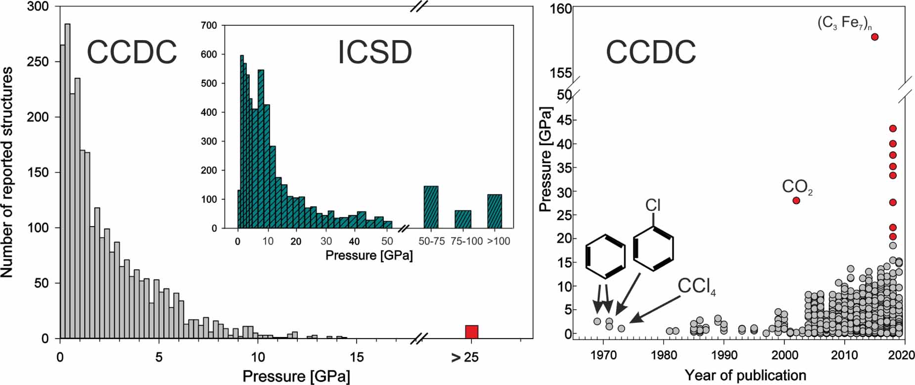
Single-crystal (SC) XRD plays an important role in the contemporary HP research, as it allows for detailed analysis of the pressure-induced conformational changes on the atomic level. More and more in situ analytical techniques become adapted for this realm, at the same time allowing for higher (megabar) pressure regimes [3]. Additionally, simultaneous application of high/low temperature, magnetic field, electric current or radiation during SCXRD experiments, opens a way to more coherent strategy for tracking the evolution of structure and properties in the wide range of crystalline materials under extreme conditions. The number of high-pressure single-crystal structures (lower pressure cut-off: 0.01 GPa) reported up to date in Inorganic Crystal Structure Database, [4] inorganic materials and Cambridge Crystallographic Data Centre [5] (CCDC, organic and coordination compounds) are ∼7500 and ∼2900, respectively (figure 1 left). The enormous increase of reported structures in the last 20 years represents the maturity of the discipline, where the major instrumental obstacles in studying molecular crystals have been overcome. In case of inorganic (mostly ionic) crystals, higher (>50 GPa) attainable pressure-range has been explored, comprising ∼4% of all of the deposited structures. It comes to view that nowadays, the major limiting factor in HP studies of the molecular single crystals, is their stability within a studied pressure regime, whereas more stable ionic inorganic materials can be explored in the megabar range.

Figure 1. Left: number of reported HP structures in inorganic crystallographic structural database (ICSD) and cambridge crystallographic data center (CCDC). Right: HP structures deposited in CCDC up to date. Multiple pressure studies count as different entries. Red points indicate deposited inorganic structures. Insets: some early examples of HP organic structures determined via SCXRD. Pressure cutoff > 0.01 GPa.
Download figure:
Standard image High-resolution imageRecent years have brought enormous technological advancements in the field of high-pressure SCXRD. Thanks to them, we can now recreate the extreme conditions of planetary inner cores, force the molecules to form species of exotic stoichiometries and explore the near-room-temperature superconductivity, to name a few [6–8]. In order to reach pressure high enough to manipulate matter in such a radical way and to have an optical access to the sample the new instruments had to be devised. Diamond anvil cell is, traditionally, the most often used device for this purpose. However, with the higher attainable pressure, the design of the device and had to be revised, effectively reducing the culet size and its design. As a result, smaller sample amount is required, further complicating SCXRD data acquisition, which relies largely on the photon-beam size. A recent upgrade at European Synchrotron Radiation Facility (ESRF) gives access to submicron-size beam with unprecedented flux and coherence [9]. Thanks to the recent upgrades, we can now characterize matter in a range of hundreds of gigapascals, in a wide range of temperature from 4 to 3000 K (as in case of laser heating) [10]. Such attainable conditions allow us to study pressure-induced superconductivity, phase transitions in the Earth's inner-core constituents and in many elements all across the Periodic Table. In this paper the recent advancements at ID15b and ID27 HP x-ray diffraction beamlines, will be discussed. The experimental aspects related to synchrotron SCXRD techniques at extreme conditions will be introduced, followed by the current research examples encompassing the studies of the pressure-induced phase transitions. The first example shows a series of pressure-induced phase transitions in photosensitive CsI3, which leads to a formation of a homoleptic In m- chains. Second example of mineral jadarite is a showcase of a poorly-scattering low-Z material with the crystallite size of a few microns, which could not be studied before the ESRF- Extremely Bright Source (EBS) upgrade.
2. Experimental considerations
SCXRD experiments carried out in diamond anvil cell (DAC) pose certain experimental difficulties [2, 11]. When high pressure is being applied, the mosaicity of the studied crystals usually increases, causing broadening of the diffraction spots and loss of intensity of high-angle reflections [12]. Therefore, excellent crystal quality is mandatory in order to collect reliable structural models. A choice of the pressure-transmitting medium plays a vital role in preserving crystal quality upon load. So-called 'hydrostatic limit' i.e. the highest attainable pressure without a significant contribution of a shear stress, has been determined for various fluids used for pressurization [13–16]. The choice of the hydrostatic medium should be not only based on the highest target pressure during the experiment, but also physicochemical properties of the medium itself. In case of the crystals containing polar molecules, alcohol mixtures (such as methanol-ethanol 4:1) may cause sample dissolution. The same is true for some non-polar molecules and a mixture of pentane-isopentane 5:1. Cryogenic loading with liquified gases (such as N2 or Ar) poses a risk of flash cooling, which may cause a single crystal to shatter. Gas-loading with He or Ne, despite an excellent hydrostaticity, may result in penetration of the gas molecules inside crystal, effectively changing its volume and physical properties [17–20]. Similar risk is posed by small organic molecules, such as alcohols, which can penetrate porous materials e.g. zeolites or metal-organic frameworks [21–23]. Additionally, alcohol mixtures should be avoided when dealing with hygroscopic materials (as they always contain a minute amounts of water) and those which could potentially react with them (e.g. carboxylic acids and respective anhydrides).
Gasket thickness and sample chamber dimensions should ensure that the crystal will not bridge between the diamond culets, nor touch the gasket wall during the experiment. As a rule of thumb, the crystal-size should be three times less than the respective gasket hole in case of liquid pressure-transmitting media. As for gas-loaded samples, the size of the crystal should ensure that threefold compression of the sample chamber volume will not cause the sample to be touched by the gasket walls. Enough space for the pressure calibrant [24] (e.g. ruby spheres) should also be envisaged. In terms of the gasket material, stainless steel is routinely used up to 10–20 GPa, and for the higher pressure ran, typically ∼250 μm Re foil is used, due to its superior mechanical properties. One should also consider potential chemical reactivity of the gasket material with the sample components. For example, some polyiodide salts were found to give off iodine vapor which subsequently reacted with a steel gasket material, forming respective iodides [25]. Experiments involving laser heating might require covering the gasket with the layer of gold, in order to prevent reaction between the heated gas and the gasket material.
Data collected at HP usually does not cover the whole Ewald sphere. Due to the opening angular aperture of circa 80°, only for cubic, hexagonal or tetragonal crystals oriented with [001] direction perpendicular to the DAC axis, the completeness can approach close to 100%. Accessible reflections fall into the toroidal region in reciprocal space [2]. As a result of an incomplete data coverage may impede space group determination, structure solution and disturb fine structural features such as shape of atomic displacement parameters or minor disorder components. Some of the ways to improve data completeness include:
- (a)Crystal pre-orientation
- (b)Loading more than one crystal in different orientations
- (c)Irradiation with shorter wavelength (larger Ewald sphere = lager accessible region)
- (d)Optimized collection strategy (scanning between many limiting positions), well executed with four-circle diffractometers
Recently, a custom-made Python3 library was released, in order to calculate the best crystal orientation in DAC for the maximum data coverage [26]. Synchrotrons benefit from the tuneable x-ray wavelength, but usually the data collection strategy is limited to a simple ω-scan, due to geometrical constraints of the heavy-duty goniometers. High-energy photons at the synchrotron source may additionally cause radiation damage, which should be checked before loading the sample in DAC [27, 28].
Recently upgraded ID15b and ID27 beamlines at ESRF are dedicated for high-pressure and low/high-temperature diffraction experiments. Both of the beamlines benefit from the high coherence and flux of the source, and they are suitable for investigation of matter in megabar regime.
3. ID27 HP beamline
The recent upgrade at ESRF EBS provided the x-ray performances of brilliance and coherence by a factor of ∼100. These improvements will allow studies to be carried out on much smaller sample volumes and on much shorter time scales. The direct impact on studies at extreme conditions is that higher pressure and temperature states that can be generated only in small volumes or in dynamic, transient processes become experimentally accessible.
The refurbished ID27 is composed of three optical hutches (OH1, OH2 and OH3), one experimental hutch with a control room, a laser laboratory and an ancillary workshop. The x-ray soured is currently composed of a small gap in-vacuum undulator of period 23 mm, at a minimum magnetic gap of 6 mm. The produced white x-ray beam has a diameter of ∼1 mm (comparing to ∼27 mm before the upgrade), which may be useful for high-pressure Laue diffraction experiments.
The first optical hutch houses the double multilayer mirror (DMM) and attenuators for the incoming white beam. DMM is placed 30 m from the source, and is used to suppress harmonics of the undulator, producing a 'pink beam' and at the same time reducing the heat load on the subsequent optical elements. Produced pink beam is characterized with a very high intensity and ΔE/E = 0.03–0.05, and can be used for time-resolved XRD, x-ray fluorescence and x-ray imaging down to the microsecond time scale.
Fixed-exit Si(111) double crystal monochromator is placed in OH2, at a distance 40 m from the source. Due to high load from the pink beam, it has to be nitrogen-cooled. Resulting monochromatic beam has ΔE/E = 10−4 in a broad energy range 15–60 keV necessary for the most of the diffraction experiments at HP.
Three Kirkpatrick-Baetz (KB) focusing mirrors are placed on three separate motorized stages (table 1), directly in the experimental hutch, 110 m down from the source. Depending on experimental requirements, the appropriate KB mirror is brought towards the beam on a motorized granite block.
Table 1. Selected specifications of KB mirrors employed at ID27 beamline.
| Focusing mirror | KB1 | KB2 | KB3 |
|---|---|---|---|
| Energy range (keV) | 15–25 | 33 | 30–60 |
| Focus (H × V) (nm) | 270 × 220 | 350 × 500 | 2000 × 2000 |
| Working distance (mm) | 180 | 430 | 470 |
| Type | No bending | No bending | Bending |
| Flux (photons s−1) monochromatic beam, ray tracing 15 keV ΔE/E = 1.5 × 10 −4 | 7 × 10¹² | 1.1 × 10¹³ | 2.2 × 10¹² |
| Flux (photons s−1) pink beam, ray tracing 15 keV ΔE/E = 2% | 5 × 10¹⁴ | 7 × 10¹⁴ | 1 × 10¹⁴ |
| Potential usage | Nano- and micro-LH-stage for DAC | Nano- and micro-LH-stage for DAC heavy duty stage | Micro-LH-stage for DAC heavy duty stage |
Up to date, KB3 mirror is used routinely for the HP experiments. The resulting focal spot diameter has 2 μm at full width half-maximum. Such small beam-diameter allows to study multiple separate single-crystal in the same DAC, in a megabar pressure range.
Three separate granite tables host three dedicated goniometers:
- (a)'heavy-duty' with a load capacity of 200 kg. It is used for standard HP experiments. Additionally, can host Paris-Edinburgh press, cryostat, resistive heating chamber, or any bulky setup which requires high mechanical stability
- (b)'laser-heating', hosts an optical system for double-sided laser heating, as described elsewhere [10].
- (c)'nano', with high-precision piezoelectric motor (sphere of confusion <1 μm). Necessary to study samples with submicron size e.g. in a megabar range, or after chemical reaction at HP.
Eiger2 X CdTe 9M photon counting detector (PCD) is employed to collect x-ray diffraction data. Detector characteristics are collected in table 2. PCDs directly convert x-rays into electronic signal (unlike e.g. charge-coupled device detectors, which have to convert x-rays to visible light first), resulting in superior special resolution and high detection efficiency. The most important features of this detector in a frame of HP experiments are: high quantum efficiency at >30 keV, high frequency and dynamic range, and short readout time. Collected data is stored in ESRF-specific hdf5 data format, which can be instantaneously converted into standard crystallographic data format such as esperanto, cbf or edf.
Table 2. Selected parameters of Eiger2 X CdTe 9 M PCD employed at ID27 and ID15b beamlines.
| Sensitive area, width × height (mm2) | 233.2 × 245.2 |
| Pixel size (μm2) | 75 × 75 |
| Count rate capability (photons/s/pixel) | 107 |
| Frame rate [Hz] | 230 |
| Data format | HDF5/NeXus |
Data frames converted to esperanto format can be directly viewed and analyzed using standard crystallographic computing programs. Currently, Crysalis software is used at ID27 for data reduction [29]. Standard structure analysis programs such as: Jana2020, Shelxle or Olex2 are available to the beamline users on dedicated data-analysis computers [30–32].
4. ID15b: dedicated high-pressure beamline
ID15b, a high-pressure dedicated beamline at the ESRF, has greatly benefited from the recent upgrade EBS, mainly because of the decrease of the factor of more than 30 in horizontal emittance. This allows to obtain a highly focused few microns meter size beam about 15 times stronger compared to the old machine. Further, the old flat panel detector was replaced by an Eiger 2X CdTe 9M photon counting detector, the same as on ID27 (vide infra), which can count up to 107 photons/sec and is made by more than nine million 75 × 75 µm pixels.
ID15b [33] is sharing a canted front end with ID15a, a beamline dedicated to materials chemistry and engineering. X-ray source is an in vacuum undulator with 20 mm period resulting in third harmonic tunable around 30 keV; the monochromator is a horizontally diffracting nitrogen cooled Si (111) single bounce Bragg monochromator [34]. ID15b operates at a fixed angle with an energy of 30 keV. Two transfocators with 200 µm diameter linear (1D) beryllium compound refractive lenses for vertical and horizontal focusing, respectively, provide a highly variable and very clean beam with a minimum spot size on the sample of less than 5 h × 6 v µm2.
The goniometer, at 44 m from the source, is mounted on an easy-accessible translation stage, and the motors allow a stable positioning and to accurate align the sample. The accessible portion of the reciprocal space is limited by the geometrical opening of the DAC. The beamline can provide standard membrane-driven DACs with a 64–80◦ opening angle. Data collections are performed by ω-scans, along the rotation axis perpendicular to the sample stage. One big advantage of the Eiger 2X (table 2) is, that it can be operated in continuous scan mode due to its vanishing readout time, saving a considerable amount of time compared to the old flat panel detector, which could only be operated in a step scan mode. For standard single crystal data collections, the usual data collection strategy consists in a pure ω-scan, for example from −32°< ω < + 32°, with 0.5° interval and 0.2–1 s exposure time per interval, whereas for powder experiments, a single ω-scan (−5°⩽ ω ⩽ + 5°) with 10 s of exposure is usually adopted. Sample alignment, data collections and pressure adjustment are controlled remotely from the control hutch and all operations are handled by a unique user-friendly interface. Calibration of the beamline, including the determination of the detector-to-sample distance, beam orientation, detector tilt with respect to the omega rotation axis and the used λ, is assured thanks to a variety of well-characterized standards as Si powder ('NIST 640 C' from the National Insitute of Standards and Technology), a single crystal of vanadinite (Pb5(VO4)3Cl, V∼ 678.2Å3) and enstatite (MgSiO3, V ∼832Å3). Data are collected in an in-house format (.h5) which is then converted by in-house scripts into Esperanto or .edf files which can be treated by software as CrysAlisPro, Fit2D or Dioptas. [35–37] ID15b is equipped with an online pressure measurement system, optimized to measure fluorescence up to 1.5 Mbar, based on the fluorescence of internal standards, such as Cr:Al2O3. (commonly referred as ruby) and Sm:SrB4O7 [24, 38]. The beamline provides a DAC gas loading system and the DACs can be loaded with noble gases (He, Ne) which assures the better hydrostatic conditions; others gases like H2, can be loaded with the gas loading system from the sample environment HP lab. These features, along with the user-friendly beamline set-up, allows to perform single-crystal and powder diffraction experiments of really challenging minerals and synthetic compounds at non-ambient conditions: cryostats and heaters for SC-measurements at low and high temperatures, respectively, are also available. The ID15b setup [33] is similar to the 'former' ID09 [39].
5. ID27 case study: phase transition and twinning in hypervalent CsI3
Cesium triiodide is an archetypical example of asymmetric trihalide [40–42]. It is one of the few inorganic trihalides stable at ambient conditions. It consists of hypervalent triiodide anions (I3 -, figure 2(a)) which have been foreseen to catenate upon compression, due to high bonding flexibility of iodine and formation of multicenter bonding architecture [43]. CsI3 semiconducting single-crystals were proposed to be implemented in x-ray detection systems, due to their high x-ray sensitivity up to 158.1 ± 6.0 μC Gy−1 cm−2 [44]. Recently, compression was shown to modulate the detection bandwidth and significant enhancements of photocurrent in this material [45]. The observed macroscopic properties were associated with enhanced I–I interactions. In fact, powder diffraction studies (in quasi-hydrostatic conditions) have shown that, above 1.3 GPa, initially layered iodine herringbone motif turns into a 3D orthogonal assembly of linear iodine chains (figure 2) [46]. Such radical change in orientation of rigid I3 - anions is accompanied with a first order phase transition from an orthorhombic Pnma to a trigonal P-3c1 space group. Further pressure increase above 22.6 GPa triggers another phase transition into the cubic Pm-3 n space group. We have previously investigated a phase diagram up to circa 16 GPa, using SCXRD, in order to elucidate the mechanism of the first (Pnma → P-3c1) transition [47]. Pressure makes the trigonal phase both thermodynamically and dynamically stable, as proved by the phonon and stabilization enthalpy calculations. The I–I bond covalency in P-3c1 polymorph increases with pressure, which can be seen as formation of infinite Im n- linear chains (figure 2(f)). Herein,we extend the studied pressure range to 25 GPa, in order to capture the second (P-3c1→ Pm-3 n) phase transition, and structurally characterize the cubic phase.

Figure 2. Shortest intermolecular contacts in orthorhombic (Pnma, a), trigonal (P-3c1, b) and cubic (Pm-3 n, c) polymorphs of CsI3, and their crystal structure: layered motif in the orthorhombic polymorph (d),(e) and the 3D motif in the trigonal and cubic polymorphs. Bond labels represent distances in [Å] rounded to a significant figure. A part of the figure is reprinted with permission from [47]. Copyright 2022 american chemical society.
Download figure:
Standard image High-resolution imageCsI3 (99%, Sigma-Aldrich) have been recrystallized from deionized water. A single crystal with a suitable size of 35 × 27 × 15 μm has been cut off from the bigger crystallite. The crystal was placed inside a ø170 μm gasket hole drilled in Re pre-indented (60 μm) gasket, fixed in ø350 μm diamond-culet DAC. The ruby has been used as a pressure-calibrant [48]. The sample was kept away from the direct laser beam, in order to avoid photodecomposition [25] Gas-loaded helium has been used as a pressure-transmitting medium [49]. The experiments have been conducted at ID27 beamline, using a monochromatic 0.3841 Å x-ray beam, with approximately 2.5 × 2.5 μm size. Wavelength has been calibrated using CeO2 (e.g. NIST SRM 674b) powder standard. Single crystal data were collected using a Rayonix M225 CCD detector through ±32° ω-scans and a step size of 0.5° around one rotation axis. After transformation into the Esperanto format, the CrysAlisPro [29] program suite was used for the indexing and data reduction. Corrections for x-ray absorption effects (by the DAC components) were applied using the semi-empirical ABSPACK routine implemented in CrysAlisPro. The structures were refined with Shelxl [50], as incorporated in OLEX2 [31]. Jana2006 was used to determine the twinning law in the high-pressure polymorph [30]. EoSFit7c software has been used to fit high-pressure volumetric data, in order to perform equation of state calculations [51]. CCDC 2194351–2194354 contain the supplementary crystallographic data for this paper. These data can be obtained free of charge via www.ccdc.cam.acuk/data_request/cif, or by emailing data_request@ccdc.cam.acuk, or by contacting The Cambridge Crystallographic Data Centre, 12 Union Road, Cambridge CB2 1EZ, UK; fax: +44 1223 336033.
Phase transition from orthorhombic to triclinic system (Pnma → P-3c1) occurs between 1.24 and 2.08 GPa in the studied single crystal (figures 2 and 3). However, unlike the powder sample compressed in silicon oil, transition in hydrostatic conditions does not show any coexistence of two phases. Transition to the trigonal phase is accompanied with a complex twinning, and deterioration of the diffraction quality (figure 3). Radical reorganization of the polyiodide chains, upon phase transition (Pnma →P-3c1), leads to high intra-crystal strain, formation of three different crystalline domains, and possible fracturation of the crystal which is accompanied with a significant broadening of diffraction spots (figure 4). The proposed mechanism of the phase transition includes rotation every third I3 - by 90° in respect to the iodine layers in the Pnma polymorph (figure 2(e)), and reorganization of the parallel in-layer I3 - fragments, in a way that they can align into linear head-to-head chains. (figure 2(b)) The determined twinning laws for the pseudo-merohedral twinning, however, do not reflect the mechanism of the phase transition. Presumably, the observed twinning is a result of the best packing of the crystallites with the different habit to the ones for the orthorhombic phase. The trigonal phase is over three times less compressible than the orthorhombic one (table 3), in accordance to the previously reported data. Notably, the high value of K' of the orthorhombic phase may point towards rapid stiffening upon load. The subsequent phase transition changes the elastic properties of the material, whereas K' = 4 in the trigonal polymorph, as the negatively-charged iodine chains are placed orthogonal to each other, minimizing the electrostatic repulsion.
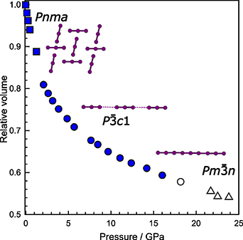
Figure 3. Volumetric compression trends and phase-stability regions for the orthorhombic (Pnma, squares), trigonal (P-3c1, circles) and cubic (Pm-3 n, triangles) polymorphs in CsI3. Blue color refers to the data published in [45], while the white color refers to data presented in this work. Insets: schematic representation of iodine chains architecture in respective polymorphs.
Download figure:
Standard image High-resolution image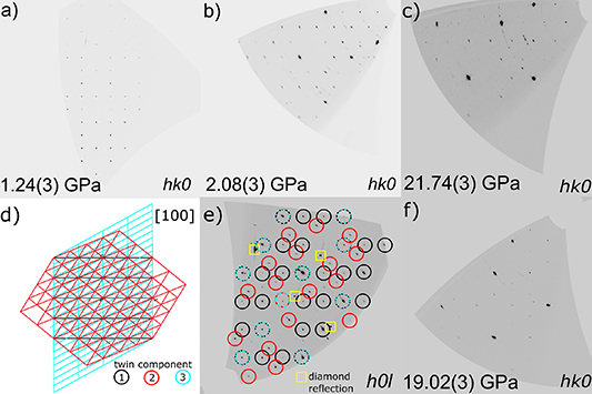
Figure 4. Precession photographs for hk0 layer before (a) and right after (b) pressure-induced phase transition in CsI3. Quality of the precession photograph for the cubic high-pressure polymorph reconstructed in orientation corresponding to the hk0 layer of the trigonal polymorph (c) is compared with the data right before the second (P-3c1 → Pm-3 n) phase transition (f). The orientation of the twin components is outlined along [001] of the first component (d), and visualized on the h0l layer of the trigonal polymorph (e). Adapted with permission from [47], 2022 American Chemical Society.
Download figure:
Standard image High-resolution imageTable 3. Refined elastic parameters pertaining to the different polymorphs of cesium triiodide, based on the isothermal II- and III-BM equation of state fits of the single-crystal and powder XRD datasets, respectively.
| Single-crystal experiment [47] | ||||
|---|---|---|---|---|
| V0, x0 (Å3, Å) | KV 0, (GPa) | K' | βV 0, x0 (GPa−1) | |
| Pnma CsI3 | 760.89(2) | 5(1) | 13(4) | 0.20(4) |
| P-3c1 CsI3 | 1013(6) | 17.9(8) | 4 | 0.0559(2) |
| Powder diffraction experiment [46] | ||||
| Pnma CsI3 | 766.4(16) | 5.6(6) | 12(2) | 0.0178(2) |
| P-3c1 CsI3 | 6.7546(3) | 17.7(9) | 3.94(12) | 0.0056(2) |
Unambiguous determination of the orientation of three twin components was possible not only due to high quality of the crystal (which anyhow deteriorated during the transition), but also due to technical capabilities of the beamline. First of all, small beam size allowed us to scan the crystal in a way, that at each measurement point, a fresh volume of the crystal was probed. This way we avoided radiolytic decomposition, which can be detected in CsI3. Secondly, combined high photon flux and high detector resolution, allowed to reliably measure diffraction intensities, even after complex twinning occurred. Even though breadth of the diffraction spots increased, it was possible not only to solve and refine the structure of the high-pressure polymorph with excellent refinement indicators (Rint= 0.029, R1= 0.063, wR1 = 0.186 S = 1.138), but also determine the twinning laws for the high-pressure phase.
Pressurization to 21.74(3) GPa leads to another phase transition to the cubic Pm-3 n polymorph, in agreement to the reported powder diffraction study (figures 2 and 3, table S1) [46]. Curiously, I–I intramolecular bond lengths undergo equalization and slight elongation (from 2.969(4) to 2.980(5)Å), with the concomitant shortening of the intermolecular I–I contacts (from 3.115(4) to 2.980(5) Å). I3 - can be seen as I2 and I- on a course of nucleophilic addition reaction. Therefore, observed elongation might stem from the higher occupation of the antibonding lowest unoccupied molecular orbital in I2 fragment and subsequent polarization, which leads to evenly-spaced centrosymmetric (I3 -)n fragments (figure 2(c)) [52]. Further pressure increase from 21.74(3) to 22.50(3) GPa causes a compression of the I–I bonds by 0.024(4)Å, whereas subsequent pressurization to 23.81(3) GPa has virtually no effect on the bond length. It can be appreciated from the minor volumetric changes in the cubic phase (figure 3). At this point, low compressibility of the evenly spaced linear In m- chain resembles the one of the covalent bonds in D∞h I3 - fragments in the trigonal polymorph.
Geometrically, the transition from the trigonal to cubic cell can be seen as a consequence of [111] direction shortening in the former, making it equivalent to the cube [53]. Additionally, the analysis of the diffraction intensities distribution clearly points out to the Pm-3 n space group (Rint = 0.032 versus Rint = 0.072 for P-3c1 space group). In conclusion, upon pressurization to 21.74(3) GPa, a new cubic polymorph of CsI3 is being formed. The polymorph contains an equally-spaced linear iodine chains, in the regular cesium cationic environment (figure 2). The system is characterized by low compressibility, pointing out to the more covalent nature of the I–I interactions within the chains.
6. ID15b case study: determination of the high-pressure polymorph of jadarite
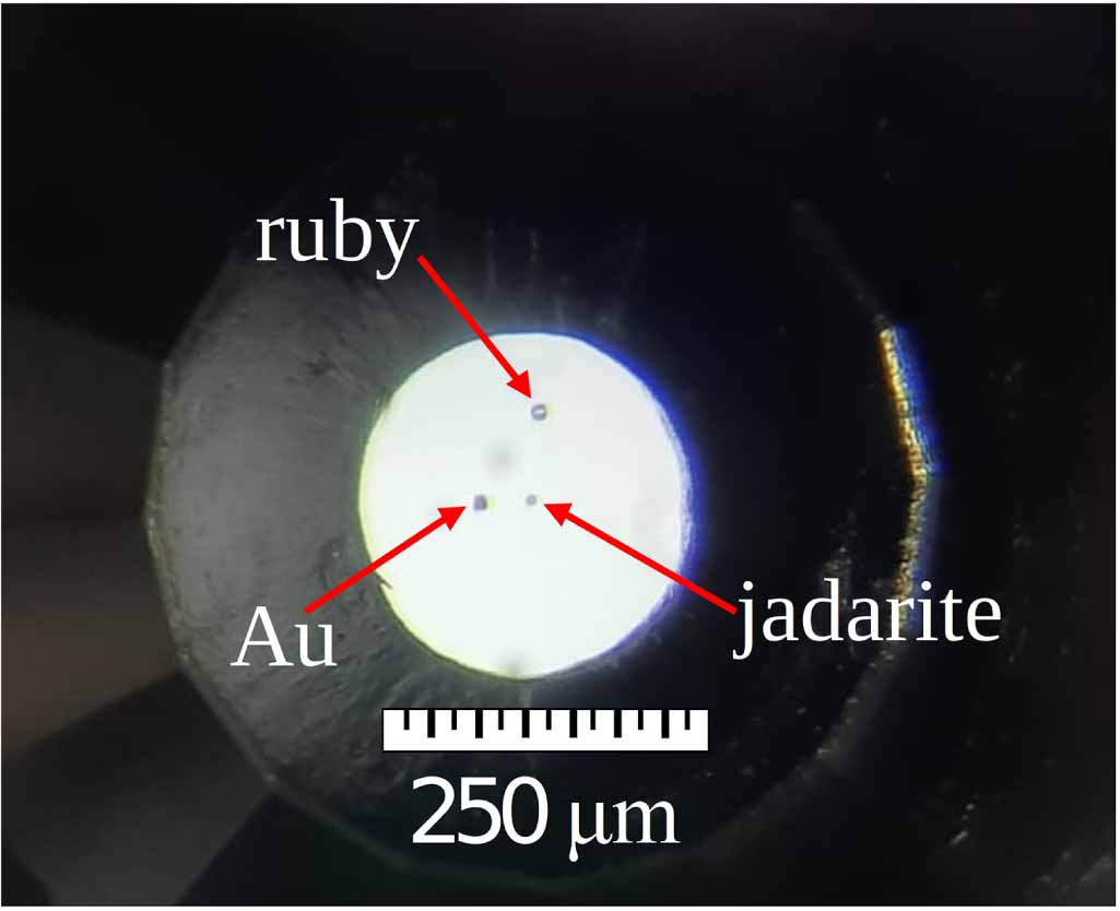
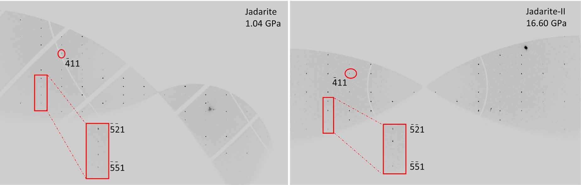
Jadarite (ideally LiNaSiB3O7(OH), space-group P21/c) is a boron-bearing mineral usually occurring in massive aggregates; only sporadic subhedral (tabular, elongate)/anhedral crystals (rarely exceeding 5 − 10 μm) were reported [54, 55]. Single crystals are extremely rare, very small (few μm, figure 5) and the low-scattering factor of jadarite (which is made essentially of very-light to light elements) has always prevented any single crystal x-ray investigation, especially in diamond anvil cells, which hinder a significant portion of the Ewald sphere. Experiments have been attempted both in in-house facilities with conventional (diffractometers) devices and in large scale facilities (ESRF, before the EBS improvements) [56].Only recently, in the framework of the aforementioned improvements linked to the coherence, brilliance and detector devices at ID15b, was possible to study this mineral at high-pressure conditions, solving even its high-pressure polymorph (jadarite-II, stable above ∼16.6 GPa) with excellent statistical parameters i.e. Rint < 0.04. The high resolution and sensitivity of the Eiger2X 9M CdTe detector was critical in order to detect and integrate also the low-intensity diffraction peaks (figure 6). A detailed description of the high-pressure deformation mechanisms and crystal structure evolution of both polymorphs has been reported in a full article [56].
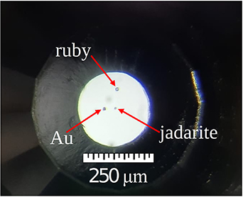
Figure 5. Picture of pressure chamber used in the experiment at 2 GPa: jadarite crystal (middle), gold (left), ruby (top).
Download figure:
Standard image High-resolution image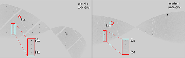
Figure 6. Reconstruction, based on the experimental x-ray diffraction data, of the hk1 reciprocal lattice planes of jadarite (left) and jadarite-II (right). Even very weak peaks were identified thanks to the high-resolution and sensitivity of the Eiger2X 9M CdTe detector (selected areas are enlarged).
Download figure:
Standard image High-resolution imageIn this study, we revaluated the structure and elastic parameters of natural jadarite. Additionally, the possibility of stochastic absorption of helium at high-pressure was studied. A new 5 × 4 × 3 μm sized jadarite crystal was loaded in membrane-driven DAC (figure 5), with 600 μm culet Boehler-Almax design anvils, along with a ruby microsphere (pressure uncertainty ± 0.05 GPa) [48] and a gold chip. Helium was used as hydrostatic pressure-transmitting fluid [13]. Lattice parameters for both polymorphs have been determined by the refinement of the orientation matrix using approximately 600 reflections, indexing of the diffraction peaks, and integration of their intensities (corrected for Lorentz-polarization effects) were performed using the CrysAlisPro package [35]. Corrections for x-ray absorption effects (by the DAC components) were applied using the semi-empirical ABSPACK routine implemented in CrysAlisPro. The so-obtained unit-cell parameters are reported in table S2 and shown in figure 7, alongside with a schematic representation of the low- and high- pressure polymorphs of jadarite (jadarite-II, figure 7). Structure refinements were performed using the package JANA2006 [30]. CCDC 2194314–2194327 contain the supplementary crystallographic data for this paper. These data can be obtained free of charge via www.ccdc.cam.acuk/data_request/cif, or by emailing data_request@ccdc.cam.acuk, or by contacting The Cambridge Crystallographic Data Centre, 12 Union Road, Cambridge CB2 1EZ, UK; fax: +44 1223 336033.

Figure 7. (left) High pressure evolution of the normalized unit cell parameters of jadarite (V in black squares, a in blue spheres, b in red diamonds and c in green upward triangles, solid markers are taken from [56] whereas empty markers are new data-points from this study) and (right) schematic view of jadarite (top) and jadarite-II (bottom); boron units in green, SiO4 tetrahedral in blue, LiO4 tetrahedral in red.
Download figure:
Standard image High-resolution image
The (isothermal) compressional behavior of the low-pressure polymorph was described using a III-Birch–Murnaghan Equation of State (BM-EoS; [57]). The BM-EoS allows to refine the bulk modulus (KV 0 or KP 0,T0, defined as −V0 (∂P/∂V)T 0 = β−1 P 0,T0, where βP 0,T0 is the volume compressibility coefficient at room conditions) and its P-derivatives (K' =∂KP 0,T0 /∂P and K'' = ∂2 KP 0,T0/∂P2). The BM-EoS parameters were refined minimizing the differences between the EoS curves and the experimental data (weighted by their uncertainties in P and V), using the EOS-FIT7-GUI software [51, 58]. Data were fitted considering an estimated uncertainty of ±0.05 GPa for pressure [48]. Elastic parameters are reported in table 4, along with the literature data from the powder diffraction experiments [56]. The single crystal data collected in this study perfectly match data collected in the previous experiment. Furthermore, no significant difference arises between the elastic behavior of powder and single crystal experiments, as the value of their compressibility is within 3σ. This further confirms that the KV of jadarite is similar to other hydrated borates (e.g. colemanite ∼67 GPa) and lies between those of other aggregates usually used in Portland concretes (e.g. quartz ∼37 GPa, calcite ∼76 GPa).
Table 4. Refined elastic parameters pertaining to the different polymorphs of jadarite, based on the isothermal III-BM Equation of State fits of the single-crystal and powder XRD datasets (powder experiment from Comboni et al [56]).
| Jadarite-I | V0, x0 (Å3, Å) | KV 0, (GPa) | K' | βV 0, x0 (GPa−1) |
|---|---|---|---|---|
| V | 596.0(4) | 58(1) | 3.6(2) | 0.0172(4) |
| a | 6.835(4) | 76(2) | 4.1(2) | 0.0044(1) |
| b | 13.813(3) | 62(2) | 2.2(2) | 0.0054(1) |
| c | 7.683(2) | 46.5(7) | 2.4(1) | 0.0072(1) |
| Single crystal experiment, III-BM EoS, P< 15.81(5) GPa | ||||
| Jadarite-I | V0, x0 (Å3, Å) | KV 0, (GPa) | K' | βV 0, x0 (GPa−1) |
| V | 594.7(2) | 55.0(5) | 3.84(9) | 0.0182 (2) |
| a | 6.7546(3) | 45(2) | 10.7(8) | 0.0074(3) |
| b | 13.809(3) | 56.0(9) | 2.8(1) | 0.0060(1) |
| c | 7.686(1) | 45.1(4) | 2.47(4) | 0.00740(6) |
| Powder experiment, III-BM EoS, P< 16.57(5) GPa | ||||
| Jadarite-I | V0, x0 (Å3, Å) | KV 0, (GPa) | K' | βV 0, x0 (GPa−1) |
| V | 594.9(3) | 56.5(9) | 3.6(2) | 0.0177(3) |
| a | 6.82(1) | 85(9) | 7(2) | 0.004(1) |
| b | 13.820(7) | 55(2) | 2.8(6) | 0.0061(2) |
| c | 7.689(2) | 45.2(6) | 2.45(7) | 0.0074(1) |
| Unique fit with powder and single crystal data | ||||
The analysis of the elastic properties has also another implication since the framework of jadarite is characterized by endless planes made by 6-mRs laying on the ab plane, stacked in such a way that a system of channels runs along the [001] crystallographic direction in which the Na atoms are hosted, which intersects another channel-like system, perpendicular to the b axis (figure 7). In such a structure, which resembles the open microporous frameworks of zeolites, it shall not be a priori excluded that He cannot be absorbed within the structure. Such phenomenon has been already observed in other microporous materials [59, 60] or even formation of He-clathrates on the surface of non-porous materials at HP [61]. The bulk moduli of single crystal and powder given in table 4, are within 3σ, suggesting that the crystal size plays no effect on the compressibility of jadarite. Since the He-adsorption should be exasperated by the higher surface/volume ratio of the powder-crystallites, given the similar time-scale of both XRD experiments at ID15b, the elastic properties comparison between single crystal and powder data, is likely to exclude such a possibility. Permeation with He could not be detected via analysis of Fourier maps of the host structure for the single-crystal sample, nor via analysis of the precession photographs.
7. Conclusions
Recent developments in the frame of EBS allow for HP SCXRD experiments exploring wide range of materials in a broad range of scattering power and pressure regimes. Herein, we presented two border cases: boron-containing jadarite and cesium triiodide. Both crystals undergo pressure-induced phase transitions. In case of jadarite, which was measured at ID15b beamline, a few-micron crystal yielded data good enough to track the changes in the molecular structure, and determine the crystallographic architecture in the high-pressure polymorph. Highly-absorbing CsI3 also undergoes a phase transition which results in symmetry raise, which results in a radical change of the initially layered structure into a 3D assembly. A whole process is coupled with a complex twinning and a high crystal strain. Excellent beam parameters at ID27 beamtime allowed, despite compromised crystal quality and low completeness, to solve and refine the structure from the highly mosaic crystal, revealing underlying twin relations in the high-pressure polymorph. Apart from solely pressure-induced phenomena, single crystals at both beamlines can be additionally subjected to low temperatures (down to 4 K), high temperatures and laser radiation. Technological advancements, in particular new high-frequency detectors, open a new way to HP time-resolved diffraction studies which can give insights into kinetics of pressure-induced phase transitions and formation of short-life metastable polymorphs. Currently, such experiments can be conducted on a few-micron sized crystals in megabar pressure regimes.
Data availability statement
The data that support the findings of this study are available upon reasonable request from the authors.
Supplementary data (0.1 MB PDF)