

Abstract
This paper describes a new plasmonic platform made of lithographic gold nanorectangles array coated by a thermosensitive polymer layer poly( isopropylacrylamide). This polymer layer responds to temperature variations by conformational changes and is therefore able to vary the distance between the gold surface and the molecular probes. Hence, this stimulable substrate can be used as a tunable device for surface-enhanced Raman scattering (SERS) using Nile blue A as a molecular probe. We show that an increase of the external temperature reversibly induces a significant enhancement of the Nile blue A SERS signal. This was attributed to a stronger interaction between the gold nanorectangles and Nile blue A. The thermosensitive plasmonic devices developed in this paper thus provide a dynamic SERS platform for sensing application.
Export citation and abstract BibTeX RIS

Original content from this work may be used under the terms of the Creative Commons Attribution 3.0 licence. Any further distribution of this work must maintain attribution to the author(s) and the title of the work, journal citation and DOI.
1. Introduction
Since its discovery in 1977 surface-enhanced Raman scattering (SERS) is a powerful vibrational spectrocopic technique for ultrasensitive analyzing the composition of materials (solids, liquids and gases) without special preparation of the sample [1–3]. The SERS technique allows to strongly increase the Raman signals of molecules adsorbed onto plasmonic (gold, silver...) nanostructured surfaces [4–6]. The aggregates formation is requisite to receive strongly active areas where the electromagnetic field is enormous due to the interaction of interparticle plasmon [7]. However, aggregation causes heterogeneous surfaces with the random distribution in density and shape of the hot spots. Thus, the quantitative detection is impossible. To solve this issue, two-dimensional (2D) substrates of plasmonic nanostructures were fabricated. Recent reports described the 2D structures based on silver nanoparticles on silicon for trace detection of the paraquat [8, 9]. However, the size of the particles and the distance between them were not uniform. Another report developed 2D arrays of gold nanodots by sputtering Au using anodic aluminum oxide template which can control the size of particles and the distance between them [10]. In our study we produced lithographic gold nanostructures, which have uniform size, shape and density.
One of the requirements to receive strong SERS relies on the analytes because SERS signals are strong in the first-layer. Hence, analysts that have good affinity toward the gold surface lead to high SERS signals. However, the analytes with low or no affinity commonly do not have SERS effect [7]. To solve this issue, gold nanostructures have been grafted with various materials that can attract the analyte close to the gold surface or increase the analyte concentration [11, 12]. Poly-N-isopropylacrylamide (pNIPAM) coating has the capacity of trapping efficiently hydrophobic analytes close to the gold surface, leading to the enhancement of their Raman signal [13, 14].
In this work a stimulable device is designed by lithographic gold nanorectangles (AuNRs) array coated with a thermoresponsive pNIPAM. This polymer has the ability to change the distance between the gold nanorectangles surface and the molecular probes. We used pNIPAM as a spacer due to its phase transition behaviour. Indeed, pNIPAM brush is swollen and become hydrophilic state in water at the temperature below the lower critical solution temperature (LCST  ). Above the LCST, pNIPAM brush is collapsed hydrophobic state [15]. These conformational changes from the swollen state to the collapsed state of pNIPAM are expected to modify the properties of the substrate as a function of temperature and thus modify the trapping functionality to the substrate of the molecular probes.
2. Experiment
2.1. Chemicals
 isopropylacrylamide monomer (NIPAM) 99% and
  pentamethyldiethyltriamine (PMDETA) were purchased from Acros Organics. Triethylamine (TEA) 99% was purchased from Merck. Tert-butyl nitrite 90%, 2-(4-aminophenyl)ethanol 98%, CuBr 98%, and nile blue A dye were purchased from Sigma Aldrich. 2-bromoisobutyryl bromide 97% was bought from Alfa Aesar. The 4-(2-hydroxyethyl)-benzene diazonium tetrafluoroborate salt, termed HEBDT
  was chemically synthesized following the approach described in our previous report [16].
2.2. Gold nanorectangles functionalization by pNIPAM
The lithographic gold nanorectangles (AuNRs) array produced by electron-beam lithography (EBL) was grafted pNIPAM by following the multi-steps strategy relying on three major steps (figure 1): (i) the grafting of the aryl group derived from 4-hydroxyethylbenzene diazonium tetra-fluorborate salt (HEBDT). In this step, the clean AuNRs array was incubated in the HEBDT salt solution 3 mM at room temperature for 5 h; (ii) Then, the obtained AuNRs array with terminal hydroxyl groups was immersed in the toluene solution containing 0.1 M 2-bromoisobutyryl bromide and 0.12 M TEA for 5 min. The terminal groups of bromo ester were thus obtained onto the gold surface (Au-Br); (iii) pNIPAM brushes were grafted from Au-Br surface by atomic transfer radical polymerization (ATRP). A catalyst solution was produced by injection a 3 ml solution of PMDETA (0.3 M) in methanol to 20 mg of CuBr. Then, the ATRP was carried out by injecting the catalyst solution to 9 ml of deionized water containing 2.0 g NIPAM monomer under an argon stream. Then, the obtained solution of ATRP was transferred to a Schlenk flask containing the substrate of functionalized gold nanorectangles (Au-Br). After stirring for 15 min under the argon stream at room temperature, the substrate was taken out of the flask and carefully washed with water and ethanol. Finally, the substrate was dried under the argon flush.
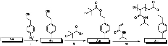
Figure 1. Multi-steps strategy of functionalization of AuNRs by pNIPAM.
Download figure:
Standard image High-resolution image2.3. Instrumentation
The extinction spectra were recorded by the LOT ORIEL spectrometer model MS 260i, combined with an optical microscope (OLYMPUS BX 51). Tapping-mode atomic force microscopy (AFM) of the samples before and after the modification was performed using SPM Nanoscope III (Veeco, Bruker) set-up in water. The SERS spectra were measured on a Jobin-Yvon LABRAM HR 800 Raman micro spectrometer with the excitation laser at 632.8 nm.
3. Results and discussion
3.1. Characterization of AuNR@pNIPAM array
Recent efforts have been focused on identifying appropriate nanoplasmonic materials which have sensitive spectra plasmon upon a variation of the local environment. Wherein, AuNRs are interesting materials due to their optical properties with the plasmon wavelength that can be finely tuned by modification of AuNRs aspect ratio. Therefore, we focused our attention on AuNRs which have unique optical properties with two localized surface plasmon (LSP) bands. The longitudinal and transversal LSP band corresponds to the oscillation of the electrons parallel to the major and the short axis, respectively. AuNRs remain popular for plasmonic applications due to the adjustable ability of the LSP resonance wavelength by varying the aspect ratio, defined as the long axis (length) divided by the short axis (width) of a rod-shaped particle, their strong scattering efficiency and a small LSP damping.
In this work we consider an AuNRs array with a small aspect ratio  (200 nm long, 167 nm wide and 30 nm high). The AFM images of these AuNRs are shown in figure 2(a). The extinction band around 596 nm corresponds to the transversal LSP mode (black dashed line in figure 2(c)). The other extinction band occurs at higher wavelengths around 616 nm corresponding to the longitudinal LSP mode (black dashed line in figure 2(d)). For such anisotropic particles, a strong red-shift is expected when the refractive index changes. The surrounding medium of AuNRs is changed by grafting the polymer film. The AFM images of AuNRs array before and after polymer grafting show that the pNIPAM brushes are grafted only on the AuNRs and not on the substrate (figures 2(a) and (b)). The dry polymer thickness in the air was
  . The presence of polymer layer on the AuNRs leads to the location of peak absorption red-shift in wavelength of LSP due to an increase of refractive index:
  for the transversal LSP mode, and
  for the longitudinal LSP mode (figures 2(c) and (d)).
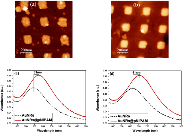
Figure 2. The AFM images of AuNRs before (a) and after (b) pNIPAM grafting. (c) Transversal extinction spectra recorded in the air of AuNRs array before (black dashed line) and after pNIPAM grafting (red line). (d) Longitudinal extinction spectra recorded in the air of AuNRs array before (black dashed line) and after pNIPAM grafting (red line).
Download figure:
Standard image High-resolution imageThe dynamic swelling ratio  of pNIPAM was determined as
  (where
  and
and  are the thickness of pNIPAM brush at swelling and dry states, respectively). The swelling ratio was deduced from our report to
  [17]. Thus, the pNIPAM thickness in the swollen state can be deduced to
  . When the AuNRs were coated by pNIPAM, the phase transition of the polymer can change the refractive index of the surrounding medium of the AuNRs and lead to the optical properties of plasmonic nanostructures modified. The pNIPAM thickness can decrease from 10 to 5 nm and the refractive index can increase from 1.37 to 1.43 by raising the temperature [18]. In order to characterize the thermosensitive properties of this system, we considered its optical response as a function of temperature. The longitudinal mode of AuNRs is highly sensitive in wavelength to the local environment. Thus, in optical studies, the longitudinal LSP mode of AuNRs has received main attention. Extinction spectra on the longitudinal LSP mode of the AuNR@pNIPAM sample recorded in water at 20 and
  are shown in figure 3(a). From these spectra, it is clear that red-shift of the LSPR band is observed
  upon an increase of the temperature, due to a change in the medium index in vicinity of AuNRs by the conformational change of the pNIPAM. The LSP red-shift as a function of temperature is plotted in figure 3(b). The sigmoidal curve fitting (red solid line) based on the experimental data (black dot) showed a sharp transition between two plateau areas below and above the LCST of pNIPAM.

Figure 3. (a) Extinction spectra on longitudinal LSP mode of the AuNR@pNIPAM sample recorded in water at  (blue curve) and
  (red curve), and (b) the longitudinal LSP red-shift upon the variation of temperature.
Download figure:
Standard image High-resolution image3.2. SERS activities of AuNR@pNIPAM subtrate
The SERS activity based on the synthesized hybrid substrate was determined by Nile blue A (NBA) dye as a probed molecule. Firstly, the substrate was immersed in NBA 0.73 ppb solution for 5 min. Next, it was washed with water and ethanol, and then dried. The temperature dependence of the SERS activity of AuNR@pNIPAM substrate was investigated by measurement their Raman spectra below and above the LCST of pNIPAM. SERS spectra of NBA recorded in water at 20 and  were shown in figure 4. The enhanced Raman signals of NBA probe were characterized by bands from C–C–C and C–N–C deformations at
  and
and  ; C–C–C and N–C–C in-plane bending at
  ; C–H bending at 1256 and
  ; and ring stretching vibrations at 1643, 1542, 1493, 1435, 1419 and
  . The bands are agreed upon by the literature [19].

Figure 4. SERS spectra of NBA molecules at concentration of 0.73 ppb adsorbed on AuNR@pNIPAM array substrate recorded at temperatures from 20 to  and back to
  .
.
Download figure:
Standard image High-resolution imageRecently, the sensitivity studies of several types of SERS substrates based on silver/gold nanostructres on 2D substrates demonstrated that the detection limit can be down to 0.01 ppm for parquat [8, 9] and 1 ppb for methamidophos [10]. In our report, hybrid substrate based on lithographic gold nanorectangles array and pNIPAM presents a high sensitivity which allows to detecting the NBA molecules at ultra-low concentration (0.73 ppb). Attentively, the results demonstrate an increase of the SERS response of NBA molecules at the temperature above the LCST of pNIPAM (compared to the SERS spectra registered below the LCST). The band intensities increase up to three-fold. This can be explained by an increase of the local field intensity when the distance between the AuNRs surface and the analyte is decreasing. Hence, the distance between the AuNRs surface and the molecular probes have influence on the SERS signal. It is due to the pNIPAM conformation change upon the temperature variation. Interestingly, figure 4 shows that SERS signal decreases back to the first spectra recorded at  when cooling down the substrate to below the LCST (
 ). Especially, the modifications of the Raman signal intensity of NBA obtained for AuNR@pNIPAM substrate are fully reversible upon the variation of external temperature.
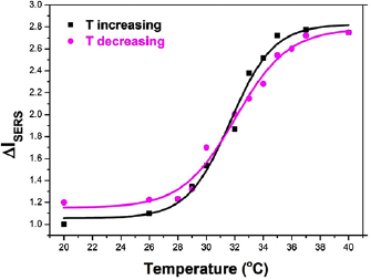
The temperature dependence of the intensity variation of Raman band at  was plotted in figure 5. A steep increase was observed when the temperature increases from 28 to
  corresponding to the phase transition zone of pNIPAM. It is remarkable that this thermo-driven variation was fully reversible during repeating cycles of the temperature decrease from 40 and
  . Interestingly, the intersection between two curves with increasing and decreasing temperatures is located around
  , the LCST of pNIPAM. The full reversibility of the system indicates that our thermoresponsive system allows us to dynamically tune the distance between the AuNRs surface and analyte.
Figure 5. SERS intensity variations of peak at  of NBA (0.73 ppb) on AuNR@pNIPAM substrate with an increasing and decreasing of temperature.
Download figure:
Standard image High-resolution image
4. Conclusions
In conclusion, an interesting hybrid nanostructure consisting of thermoresponsive pNIPAM and AuNRs array was successfully fabricated. By our strategy of functionalization, the polymer thickness can be easily estimated by AFM. The synthesized hybrid plasmonic nanostructure was used as a platform for trace detection of Nile blue A by Raman spectroscopy. A high SERS effect was observed on the AuNR@pNIPAM substrate. On the other hand, the results demonstrated that the SERS signal can be dynamically and reversely tuned as a function of temperature. Above the transition phase temperature of pNIPAM, the hybrid substrate produced an enormous SERS signal because of the collapse of polymer brushes lead to decrease of the distance between probe molecule and gold surface. Our obtained results in this works demonstrate the huge promising of stimulable polymer-coated nanostructures as effective devices for sensing applications.
Acknowledgments
This work was supported by Hanoi University of Science and Technology (HUST) under grant number T2017-TT-007.