Maternal Behavior: Why mother rats protect their children
The presence of the hormone oxytocin in the central amygdala makes a mother rat willing to put her life in danger in order to protect her offspring.
- Nencki Institute of Experimental Biology, Poland
When an animal encounters a threat it has to analyze both the threat (what type of threat is it? how close is it?) and also the local environment (can I escape? is there anywhere to hide?). The animal must then choose from a range of possible responses: it can, for example, try to fight the threat or to escape (Blanchard et al., 2011). Another possibility, when there is no safe escape route, is to 'freeze' and hope that a predator will not notice you (Blanchard and Blanchard, 1969). Freezing can be a viable strategy when an animal is on its own, but it is not an option for a mother looking after young offspring (Figure 1). With very young progeny that cannot run, the only choice is to confront the threat. When the offspring are a bit older, it might be possible to usher them to safety.
Figure 1
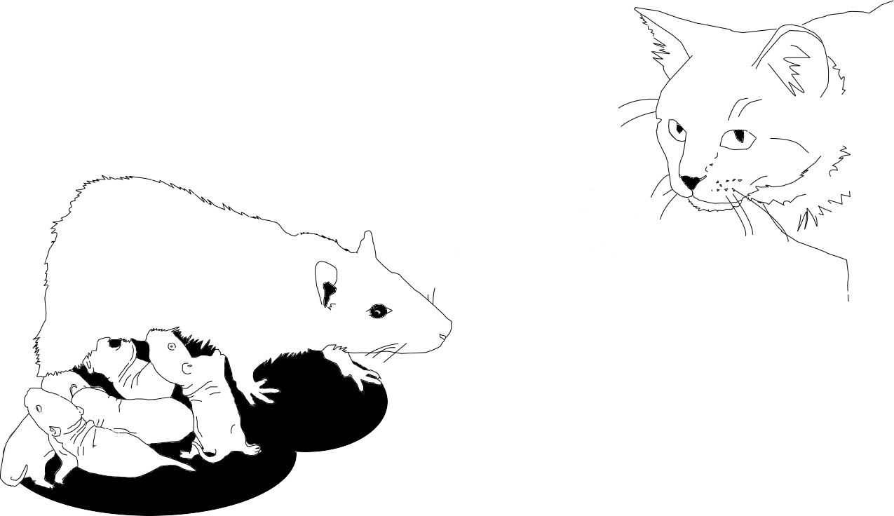
The way a mother rat responds to a threat depends on the age of her pups.
Illustration: Karolina Rokosz.
While the neural mechanisms that give rise to different defensive responses are relatively well understood, the vast majority of studies reported to date have been based solely on experiments on male subjects. Moreover, the parental status of the animals was often neglected. Female rats are generally considered less territorial than males, but their behavior changes once they become mothers (also known as 'dams') and they can become aggressive towards potentially dangerous intruders, even if this will also place the dam in danger (Bosch, 2013). How does the brain switch between self-defense and offspring-defense modes? Is the defensive repertoire affected by the age of the pups? Now, in eLife, Marta Moita and colleagues – including Elizabeth Rickenbacher as first author, Rosemarie Perry and Regina Sullivan – report that, in the presence of pups, self-defense responses are suppressed by the presence of a hormone called oxytocin in a region of the brain called the central amygdala (Rickenbacher et al., 2017).
Oxytocin is a well known hormone that promotes social bonding and causes contractions of the uterus and cervix during sexual intercourse and childbirth, as well as milk ejection during breastfeeding. More recently it was discovered that oxytocin can also control freezing behavior (Knobloch et al., 2012). In general oxytocin is released into the blood. However, in the case of the fear response, oxytocin is secreted directly into the central amygdala, which is one of the structures in the brain that controls freezing behavior (Wilensky et al., 2006).
In a series of elegant experiments Rickenbacher et al. show that, in the presence of pups, dams do not freeze when confronted with a threat (here, a noxious stimulus delivered along with an odor). Moreover, their response depends on the age of the pups. With very young pups (between four and six days old), the dams approach the threat. However, if the pups are older (between 19 and 21 days old), the dams turn towards the pups and huddle with them (probably because older pups are capable of running away if necessary).
This pattern of behavior changes dramatically if an oxytocin antagonist is very precisely injected into the central amygdala. When the oxytocin is blocked by the antagonist, the dams start behaving as if the pups were not present and they often freeze when exposed to threatening situations.
This change in the behavior of the dams has important implications for the pups. In a normal situation, a dam does not freeze in the presence of her pups and instead displays an array of active defensive behaviors. During this process, the pups learn to associate the noxious stimulus and odor, which are intended for the mother, with something unpleasant. If the mothers freeze, this emotional information is not transferred from the dam to the pups.
The work of Rickenbacher et al. – who are based at the Champalimaud Neuroscience Programme in Portugal and New York University School of Medicine – addresses some important questions, and also raises new ones. Is it just the oxytocin in the central amygdala that suppresses freezing of the dams? How do the pups learn about the danger? Answering these questions will keep neuroscientists (and rats) busy for years to come.
References
-
Crouching as an index of fearJournal of Comparative and Physiological Psychology 67:370–375.https://doi.org/10.1037/h0026779
-
Risk assessment as an evolved threat detection and analysis processNeuroscience & Biobehavioral Reviews 35:991–998.https://doi.org/10.1016/j.neubiorev.2010.10.016
-
Maternal aggression in rodents: brain oxytocin and vasopressin mediate pup defencePhilosophical Transactions of the Royal Society B: Biological Sciences 368:20130085.https://doi.org/10.1098/rstb.2013.0085
Article and author information
Author details
Publication history
- Version of Record published: June 13, 2017 (version 1)
Copyright
© 2017, Meyza et al.
This article is distributed under the terms of the Creative Commons Attribution License, which permits unrestricted use and redistribution provided that the original author and source are credited.
Metrics
-
- 4,957
- views
-
- 209
- downloads
-
- 2
- citations
Views, downloads and citations are aggregated across all versions of this paper published by eLife.
Download links
A two-part list of links to download the article, or parts of the article, in various formats.
Downloads (link to download the article as PDF)
Open citations (links to open the citations from this article in various online reference manager services)
Cite this article (links to download the citations from this article in formats compatible with various reference manager tools)
Maternal Behavior: Why mother rats protect their children
eLife 6:e28514.
https://doi.org/10.7554/eLife.28514
Further reading
-
- Neuroscience
Obstructive sleep apnea (OSA) is a prevalent sleep-related breathing disorder that results in multiple bouts of intermittent hypoxia. OSA has many neurological and systemic comorbidities, including dysphagia, or disordered swallow, and discoordination with breathing. However, the mechanism in which chronic intermittent hypoxia (CIH) causes dysphagia is unknown. Recently, we showed the postinspiratory complex (PiCo) acts as an interface between the swallow pattern generator (SPG) and the inspiratory rhythm generator, the preBötzinger complex, to regulate proper swallow-breathing coordination (Huff et al., 2023). PiCo is characterized by interneurons co-expressing transporters for glutamate (Vglut2) and acetylcholine (ChAT). Here we show that optogenetic stimulation of ChATcre:Ai32, Vglut2cre:Ai32, and ChATcre:Vglut2FlpO:ChR2 mice exposed to CIH does not alter swallow-breathing coordination, but unexpectedly disrupts swallow behavior via triggering variable swallow motor patterns. This suggests that glutamatergic–cholinergic neurons in PiCo are not only critical for the regulation of swallow-breathing coordination, but also play an important role in the modulation of swallow motor patterning. Our study also suggests that swallow disruption, as seen in OSA, involves central nervous mechanisms interfering with swallow motor patterning and laryngeal activation. These findings are crucial for understanding the mechanisms underlying dysphagia, both in OSA and other breathing and neurological disorders.
-
- Neuroscience
The central tendency bias, or contraction bias, is a phenomenon where the judgment of the magnitude of items held in working memory appears to be biased toward the average of past observations. It is assumed to be an optimal strategy by the brain and commonly thought of as an expression of the brain’s ability to learn the statistical structure of sensory input. On the other hand, recency biases such as serial dependence are also commonly observed and are thought to reflect the content of working memory. Recent results from an auditory delayed comparison task in rats suggest that both biases may be more related than previously thought: when the posterior parietal cortex (PPC) was silenced, both short-term and contraction biases were reduced. By proposing a model of the circuit that may be involved in generating the behavior, we show that a volatile working memory content susceptible to shifting to the past sensory experience – producing short-term sensory history biases – naturally leads to contraction bias. The errors, occurring at the level of individual trials, are sampled from the full distribution of the stimuli and are not due to a gradual shift of the memory toward the sensory distribution’s mean. Our results are consistent with a broad set of behavioral findings and provide predictions of performance across different stimulus distributions and timings, delay intervals, as well as neuronal dynamics in putative working memory areas. Finally, we validate our model by performing a set of human psychophysics experiments of an auditory parametric working memory task.


