
Subscribe to RSS
DOI: 10.5999/aps.2020.01340
Primary cutaneous mucormycosis of the scalp

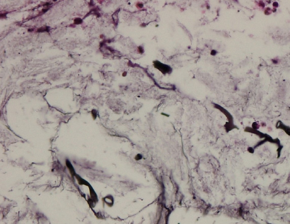
Mucormycosis is a fungal infection caused by the Zygomycetes class and genus of Mucorales, which usually reside in soil or plants. While daily exposure is common, infection has only been reported in immunocompromised patients. Because the route of exposure is usually fungal spores via air, human mucormycosis usually occurs in the form of pulmonary or rhinocerebral infections. Primary cutaneous mucormycosis is a rare form of the entity, reported to have rapid progression and high mortality. An 83-year-old male who had finished his sixth cycle of chemotherapy for small cell lung cancer, presented with pus-like discharge from two openings on the scalp and swelling of his left periorbital region, with chemosis of his conjunctiva and decreased ocular motility in all directions but no diplopia ([Fig. 1]). Magnetic resonance imaging showed myositis of the superior rectus muscle and optic neuritis, but no brain involvement ([Fig. 2]). Wound cultures were negative for microorganisms and there was no significant response to intravenous antibiotics or debridement. A biopsy was performed and histopathology revealed thick-walled non-septate hyphae, with irregular wide-angle branches, consistent with the diagnosis of primary cutaneous mucormycosis ([Figs. 3], [4]). We immediately administered systemic amphotericin B (1 mg/kg/day) and his wounds and the orbital lesion healed rapidly within 20 days. Primary cutaneous mucormycosis occurs uncommonly, and is usually caused by direct inoculation by fungal spores into the skin in immunocompromised patients. Intravenous amphotericin B is an effective first-line therapy. While the presentation of cutaneous mucormycosis may resemble cellulitis or cutaneous abscesses of variable causes, the physician should have a high degree of suspicion for early diagnosis and proper, effective treatment.


NOTES
Ethical approval
The study was approved by the Institutional Review Board of the Catholic Medical Center (IRB No. OC20ZISI0012) and performed in accordance with the principles of the Declaration of Helsinki. Written informed consent was obtained.
#
Patient consent
The patient provided written informed consent for the publication and the use of his images.
#
Author contribution
Conceptualization: BF Seo. Data curation: JH Seo.
Formal analysis: JH Seo. Methodology: G Yoo.
Visualization: JH Seo. Writing - original draft: BF Seo.
Writing - review & editing: G Yoo.
#
#
Publication History
Received: 22 June 2020
Accepted: 23 July 2021
Article published online:
19 March 2022
© 2021. The Korean Society of Plastic and Reconstructive Surgeons. This is an open access article published by Thieme under the terms of the Creative Commons Attribution-NonCommercial License, permitting unrestricted noncommercial use, distribution, and reproduction so long as the original work is given appropriate credit. Contents may not be used for commercial purposes. (https://creativecommons.org/licenses/by-nc/4.0/)
Thieme Medical Publishers, Inc.
333 Seventh Avenue, 18th Floor, New York, NY 10001, USA