Published online Jun 15, 2000. doi: 10.3748/wjg.v6.i3.417
Revised: February 3, 2000
Accepted: March 14, 2000
Published online: June 15, 2000
- Citation: Huang Y, Lu SJ, Dong JX, Li F. New proof of neuro-endocrine-immune network expression of islet amyloid polypeptide in plasma cells in gastric mucosa of peptic ulcer patients. World J Gastroenterol 2000; 6(3): 417-418
- URL: https://www.wjgnet.com/1007-9327/full/v6/i3/417.htm
- DOI: https://dx.doi.org/10.3748/wjg.v6.i3.417
Peptic ulcer, as a common disease, seriously affected people's, work and life. Its occurrence, development and change have close relationship with the change of people's moods. Animal experiment proved that significant changes occurred in the endocrine system of the gastric ulcer rats[1]. Recent study also showed that the number of lymphocytes increased markedly in the gastric mucosa of peptic ulcer patients[2]. All the above indicated that peptic ulcer is closely related neuro-endocrine-immune system. IAPP, a novel islet hormone, not only takes part in the regulation of blood glucose[3], but also protects gastric mucosa[4]and regulates gastrointestinal movements[5]. On the basis of previous studies, we observed the expression change of IAPP and explored the relationship between the endocrine and the immune system in gastric mucosa of peptic ulcer patients, so as to provide morphologic data on the existence of neuro-endocrine-immune network and the changes in peptic ulcer.
Twenty-one samples, including 6 cases from normal human stomach, 15 cases from gastrectomy of gastric ulcer patients, were collected. The paraffin sections were prepared as usual. Immunohistochemical PAP method was used to show IAPP-IR cells. Briefly five-micron sections were placed on glass slides deparaffinized in xylene, rinsed in ethanol, and brought to PBS through a series of descending concentration of ethanol; endogenous peroxidase activity was blocked with methanol-H2O2 at room temperature for 30 min; rabbit anti-IAPP serum (peninsula, USA) was diluted 1/6000 with PBS, and the sections were incubated overnight at 4 °C. Goat anti-rabbit IgG (Huamei, Beijing) (1/50), peroxidase-anti-peroxidase (Capital Medical University) (1/100) and DAB kit (Zhongshan, Beijing) were used for staining. As the negative control, the primary antiserum was replaced by PBS and other steps were the same as stated above. All the sections were counterstained with Mayer hematoxylin.
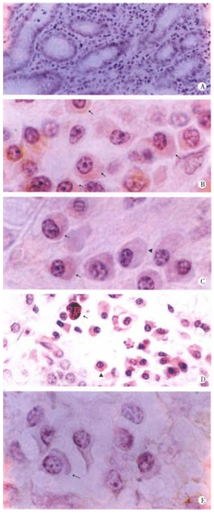
The IAPP-IR cell was not observed in the gastric mucosa of normal subject (Figure 1A). In comparison, a great number of plasma cells IAPP-IR were found in the gastric mucosa of peptic ulcer patients (Figure 1B, Figure 1C). Most of IAPP-IR plasma cells were weak and only a few were strong for IAPP staining (Figure 1D). Of the negative control sections, no immunoreactive product to IAPP was found in plasma cells (Figure 1E).
The gastric mucosa, in which there are a lot of neurons, endocrine cells and immunocytes that may interact with each other, is an important field for the study of neuro-endocrine-immune network. It will undoubtedly provide valuable data for the study on this network by exploring the change of immune-endocrine of gastric mucosa of peptic ulcer patients. Based on the observation of T and B lymphocytes which increase obviously in the gastric mucosa of peptic ulcer patients[2] and the action of IAPP, a novel islet hormone, which inhibits gastric acid secretion[6] and protects gastric mucosa[4], we further studied the expression change of IAPP in the gastric mucosa. Unexpectedly, it was found that the plasma cells of gastric mucosa increase d in number, moreover most of them expressed IAPP to some degree. Firstly, the specificity of the above findings should be confirmed because there was no IAPP expression in the plasma cells on the negative control sections; and there were also IAPP-IR negative plasma cells around the positive ones. Secondly, the significance of IAPP expression in plasma cells should be studied. IAPP is mainly secreted by islet B cells[7]. Recent study indicated that besides regulating blood glucose, IAPP could inhibit gastric acid secretion[4], and protect gastric mucosa[5]. IAPP -IR cells of islet were markedly increased during the healing process of rat gastric ulcer[8]. The above-mentioned studies all suggested that IAPP is beneficial to ulcer healing. As it is known, the plasma cells of gastric mucosa come from B lymphocytes and they respond by synthesizing and secreting IgA. It is observed, for the first time, that plasma cells in gastric mucosa of peptic ulcer patients not only increased in the number, but also expressed IAPP. Combined with our previous observation that T and B lymphocytes of gastric mucosa increased in peptic ulcer patients, it is reasonable to infer that some plasma cells in gastric mucosa of peptic ulcer patients may transform to ones expressing IAPP so as to maintain the high level of IAPP in the gastric mucosa and help promote ulcer healing just as a growth factor[9].
It is found, for the first time, that IAPP was expressed in plasma cells of gastric mucosa of peptic ulcer patients, which provides morphologic evidence for the existence of neuro-endocrine-immune network.
Dr. Yan Huang, graduated from Beijing Medical University as a PhD in 1995, now professor and postgraduate tutor, majoring in neuroendocrine research, having 18 papers published.
Edited by Zhu LH
proofread by Sun SM
| 1. | Zheng ZT. Digestive Ulcer. Beijing: The People's Health Publis hing House 1998; 306-325. [Cited in This Article: ] |
| 2. | Huang Y, Lu SJ, Dong JX, Li F. Changes of T and B lymphocytes in gastric mucosa of peptic ulcer patients. Shijie Huaren Xiaohua Zazhi. 1999;7: 648. [Cited in This Article: ] |
| 3. | Ludvik B, Kautzky-Willer A, Prager R, Thomaseth K, Pacini G. Amylin: history and overview. Diabet Med. 1997;14 Suppl 2:S9-13. [PubMed] [Cited in This Article: ] |
| 4. | Clementi G, Caruso A, Cutuli VM, Prato A, de Bernardis E, Amico-Roxas M. Effect of amylin in various experimental models of gastric ulcer. Eur J Pharmacol. 1997;332:209-213. [PubMed] [DOI] [Cited in This Article: ] [Cited by in Crossref: 14] [Cited by in F6Publishing: 16] [Article Influence: 0.6] [Reference Citation Analysis (0)] |
| 5. | Gedulin BR, Young AA. Hypoglycemia overrides amylin-mediated regulation of gastric emptying in rats. Diabetes. 1998;47:93-97. [PubMed] [DOI] [Cited in This Article: ] [Cited by in Crossref: 28] [Cited by in F6Publishing: 30] [Article Influence: 1.2] [Reference Citation Analysis (0)] |
| 6. | Rossowski WJ, Jiang NY, Coy DH. Adrenomedullin, amylin, calcitonin gene-related peptide and their fragments are potent inhibitors of gastric acid secretion in rats. Eur J Pharmacol. 1997;336:51-63. [PubMed] [DOI] [Cited in This Article: ] [Cited by in Crossref: 58] [Cited by in F6Publishing: 61] [Article Influence: 2.3] [Reference Citation Analysis (0)] |
| 7. | Huang Y, Li ZC, Shi AR. Morphometric study on coexistence of islet amyloid polypeptide with insulin in rat pancreatic islet cells during postnatal development. Acta Anatomica Sinica. 1995;26: 305-308. [Cited in This Article: ] |
| 8. | Li ZC, Shi AR. The morphometric study on IAPP-IR cells and Ins-IR cells of islet during the healing process of rat experimental gastric ulcer. J Beijing Med Univ. 1995;27:333-335. [Cited in This Article: ] |
| 9. | Wookey PJ, Tikellis C, Nobes M, Casley D, Cooper ME, Darby IA. Amylin as a growth factor during fetal and postnatal development of the rat kidney. Kidney Int. 1998;53:25-30. [PubMed] [DOI] [Cited in This Article: ] [Cited by in Crossref: 29] [Cited by in F6Publishing: 31] [Article Influence: 1.2] [Reference Citation Analysis (0)] |


