
Antibiotic Resistance and Species Profile of Enterococcus Species in Dogs with Chronic Otitis Externa


1
Laboratory of Aquatic Biomedicine, College of Veterinary Medicine and Research Institute for Veterinary Science, Seoul National University, Seoul 08826, Korea
2
Department of Veterinary Internal Medicine, Jeonbuk National University, Iksan 54596, Korea
*
Authors to whom correspondence should be addressed.
†
These authors contributed equally to this work.
Vet. Sci. 2022, 9(11), 592; https://doi.org/10.3390/vetsci9110592
Submission received: 28 September 2022
/
Revised: 20 October 2022
/
Accepted: 21 October 2022
/
Published: 27 October 2022
(This article belongs to the Special Issue Advances in Veterinary Clinical Microbiology)
Abstract
:Simple Summary
Otitis externa (OE) is a common disease in dogs and can be induced by various causes. After the primary causes that induced the ear canal issue, microbial infections occur secondly. As the main treatment strategies are primary cause correction and antibiotic administration, prolonged treatment is likely to induce the emergence of antibiotic resistance bacteria. Here, we describe the Enterococcus bacteria, one of the main infection agents of OE. The bacterial genus showed several species distributions and antibiotic resistance. This fact clarifies the importance of appropriate antibiotic selection and prudent antibiotic administration. As companion animals share lots of space with humans, pathogen transmissions between humans and companion animals are likely to occur. This study contributes not only to treatment strategies for Enterococcus infections but can also be used as a comparable index of antibiotic resistance of Enterococcus in the future.
Abstract
Otitis externa, a common disease in dogs, has different etiologies. Enterococcus is a Gram-positive bacterium that frequently causes opportunistic ear infections. Here, we determined the distribution of Enterococcus in canine otitis externa via time-of-flight mass spectrometry and biochemical tests and evaluated their resistance patterns to 10 commonly used antibiotics. Among the 197 Enterococcus isolates, E. faecalis (48.7%; 96/197) was the most common, followed by E. faecium (21.3%; 42/197), E. casseliflavus (11.7%; 23/197), E. hirae (10.7%; 21/197), E. avium (3.6%; 7/197), E. gallinarum (2.5%; 5/197), E. canintestini (1.0%; 2/197), and E. durans (0.5%; 1/197). All isolates were tested for antibiotic resistance using the Kirby–Bauer disc diffusion method. Enterococcus faecalis strains were highly resistant to erythromycin (45.8%) and rifampin (34.3%) but were generally susceptible to penicillin class antibiotics. In contrast, E. faecium isolates were highly resistant to penicillin class antibiotics (ampicillin, 61.9%; penicillin, 71.4%). Most importantly, E. faecium demonstrated high resistance to most of the antibiotics used in this study. Multidrug resistance was found in 28.4% of the isolates (56/197). This study shows prevalence and antibiotics resistance profiles of Enterococcus species in canine chronic otitis externa. The results can contribute to establish therapeutic strategies of Enterococcus infections and be used as a comparable index of antibiotic resistance of Enterococcus in the future.
1. Introduction
Otitis externa, inflammation of the external ear canal, has various etiologies and is a relatively common disease with an incidence of 7.5–16.5% in canines [1]. The causes of otitis externa can be broadly divided into primary and secondary causes. The primary causes are those that induce inflammation in the normal ear, such as allergies, autoimmune diseases, and foreign bodies [2,3]. Secondary infections occur when the primary cause alters the composition of the aural environment [2,3].
The external ear canal of dogs with otitis externa is colonized by diverse microorganisms. Among these, Gram-positive enterococci are frequently encountered but are rarely studied as representatives of the external ear canal flora in association with other symbiotic microorganisms. Enterococci are principally commensals of the bowel and are causative agents of opportunistic infections, such as wound infections, mastitis in cattle, and infections of the urethra and ears in dogs [3].
Enterococci are intrinsically resistant to several antimicrobial agents. In humans, multidrug-resistant (MDR) enterococci are among the most important pathogens causing serious nosocomial infections [4]. Enterococcus faecalis is reportedly a reservoir of antimicrobial-resistance genes for pathogenic or potentially pathogenic bacteria [5]. Vancomycin is one of the few effective drugs available for the treatment of such infections [6]. However, vancomycin-resistant strains of enterococci have emerged in Europe with the initiation of feeding avoparcin, another glycopeptide, to food-producing animals [6]. In the United States, the injudicious use of vancomycin in human hospitals has resulted in the same effect; that is, an increase in selective pressure resulting in increased colonization of vancomycin-resistant enterococci (VRE) (especially in hospitals) [7]. The prevention and treatment of enterococci infections entails huge expenditures in public health [8,9,10]. Moreover, as farm animals were found to be one of the reservoirs of VRE, enterococci surveillance was expanded to veterinary fields [11,12,13].
Single or repeated exposure(s) to antibiotics could increase the level of resistance in pathogenic bacteria in humans and animals. The level of acquired resistance in bacteria can be considered an indicator of selection pressure due to antibiotic usage in a population and resistance-related problems are expected in pathogens [14]. Regular monitoring of the level of resistance in pathogens and indicator bacteria of the normal flora in both humans and animals is recommended [15]. This monitoring is important because it allows the comparison of the prevalence and evolution of resistance patterns [16]. Knowledge of antibiotic resistance in bacteria in companion animals can help identify potential risks to owners in close contact with companion animals and select optimistic therapeutic drugs in clinical practices. In this study, we aimed to determine the phenotypic resistance patterns of enterococci in the external ear canal of dogs that received antimicrobial treatment.
2. Materials and Methods
2.1. Enterococcus Isolation and Growth Conditions
Enterococcus strains were isolated from external ear canal swab samples collected from dogs with chronic otitis externa from animal hospitals in Seoul and Gyeongsangnam-do, South Korea. The swab samples were maintained in Amies transport medium (YUHAN LAB TECH, Seoul, Korea) until processing. The samples were placed in a 1.5 mL centrifuge tube containing phosphate-buffered saline (PBS) and vortexed vigorously. The supernatants were spread on Columbia blood agar (5% sheep blood; Oxoid, Hamspire, UK) and incubated overnight at 37 °C. Suspected Enterococcus colonies were subcultured on fresh tryptic soy agar (TSA; Difco, MI, USA) and incubated overnight at 37 °C. The subculture step on TSA was repeated thrice. After isolating the purified colonies, the isolates were identified via time-of-flight mass spectrometry using a matrix-assisted laser desorption/ionization Biotyper (Bruker Daltonics, Bremen, Germany), according to the manufacturer’s instructions. A total of 197 isolates belonged to the genus Enterococcus; however, only 89 strains were identified. The bacteria were stored at −70 °C in tryptic soy broth (Difco) containing 15% glycerol until biochemical tests were performed.
2.2. Biochemical Test for Enterococcus Species Confirmation
To confirm and identify the species of all Enterococcus isolates, a biochemical test was performed using the API rapid ID 32 STREP kit (bioMérieux SA, Craponne, France) according to the manufacturer’s instructions. The results were visually read by referring to the reading table provided in the product manual. Results were interpreted using an online database (V4.0; https://apiweb.biomerieux.com/ accessed on 20 September 2022).
2.3. Antibiotic Susceptibility Test
The Kirby–Bauer disk diffusion method, as described by the Clinical and Laboratory Standards Institute [17], was used to examine the susceptibility of the Enterococcus species to 10 commonly used antibiotics: ampicillin (10 µg), penicillin (10 units), vancomycin (30 µg), doxycycline (30 µg), ciprofloxacin (5 µg), levofloxacin (5 µg), linezolid (30 µg), erythromycin (15 µg), chloramphenicol (30 µg), and rifampin (5 µg). Briefly, overnight cultured colonies of isolates were suspended in sterile PBS to a 0.5 McFarland standard. The suspensions were spread on Mueller–Hinton agar, the antibiotic discs were placed, and incubated at 35 °C for 18 h. For quality confirmation of antibiotic discs, the Staphylococcus aureus strain ATCC® 25923 was used. Enterococcus isolates resistant to more than three classes of antimicrobials were considered MDR isolates [18].
2.4. Statistical Analysis
Differences in resistance prevalence between E. faecalis and E. faecium were analyzed by the Fischer’s exact test using Microsoft Excel software. A significance level of α 0.05 was used.
3. Results
3.1. Enterococcus Species Distribution
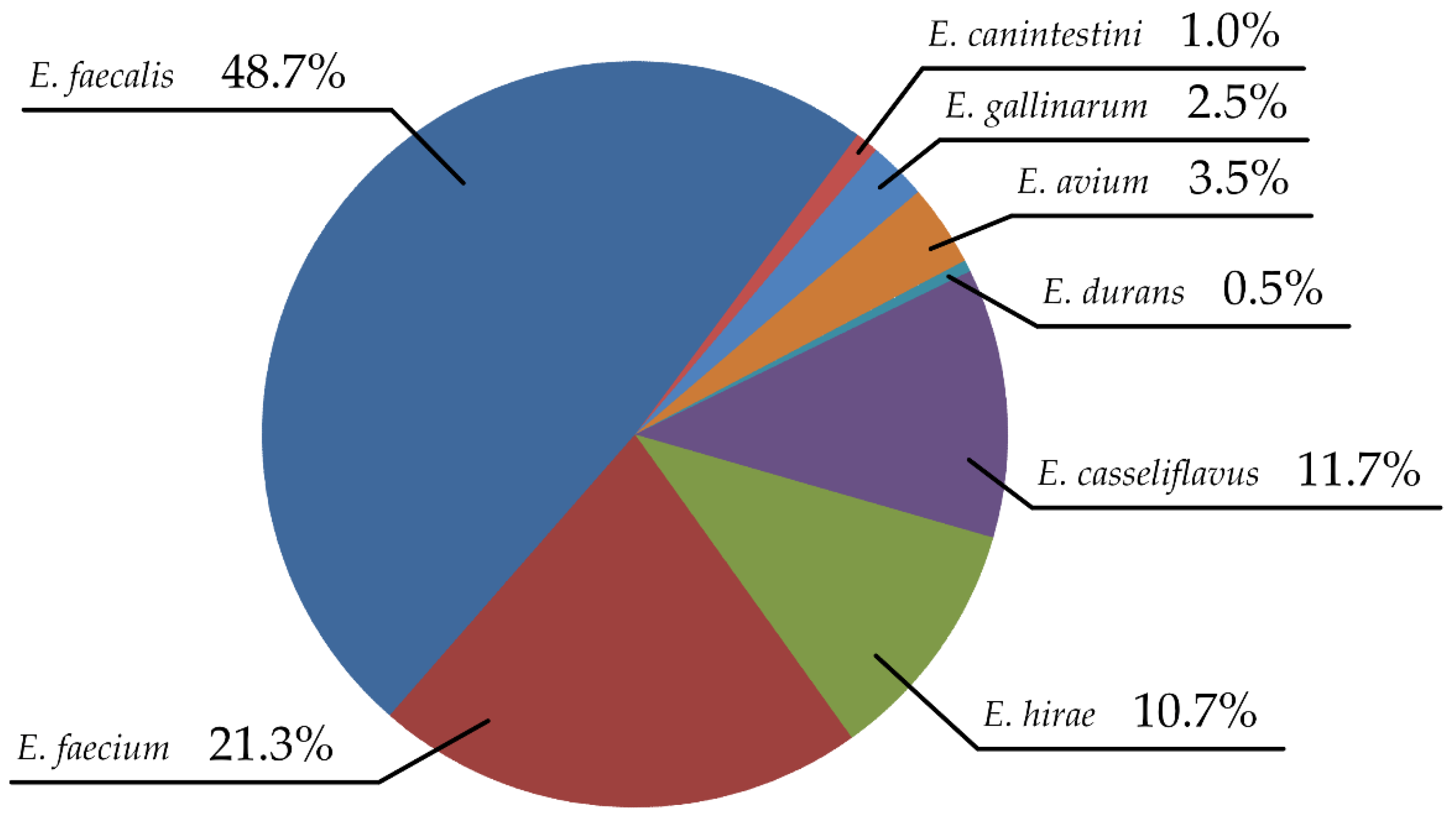
In this study, 214 bacterial colonies were suspected to be Enterococcus based on colony morphology. Of these, 197 species were confirmed using time-of-flight mass spectrometry and biochemical tests. Seven species were identified: E. faecium (96/197), E. faecalis (42/197), E. casseliflavus (23/197), E. hirae (21/197), E. avium (7/197), E. gallinarum (5/197), E. canintestini (2/197), and E. durans (1/197) (Figure 1). Enterococcus faecalis was the most frequently isolated species (96 strains; 48.7%), followed by E. faecium (42 strains; 21.3%). These two species accounted for approximately 70% of all isolates.
3.2. Antibiotic Resistance Profile
Antibiotic susceptibility tests of the Enterococcus isolates demonstrated significant resistance of the isolates to different classes of antibiotics. The E. faecalis isolates showed the highest rate of resistance against erythromycin (45.8%; 44/96), followed by rifampin (34.4%; 33/96), ciprofloxacin (27.1%; 26/96), levofloxacin (25.0%; 24/96), doxycycline (19.8%; 19/96), linezolid (13.5%; 13/96), chloramphenicol (10.4%; 10/96), and other antibiotics. The large portion of E. faecium isolates were resistant to penicillin (71.4%; 30/42), followed by ciprofloxacin (69.0%; 29/42), levofloxacin (66.7%; 28/42), ampicillin (61.9%; 26/42), rifampin (54.8%; 23/42), erythromycin (50.0%; 21/42), doxycycline (38.1%; 16/42), linezolid (23.8%; 10/42), and other antibiotics (Table 1). Five vancomycin-resistant E. faecalis (5.2%; 5/96) and E. faecium (11.9%; 5/42) strains were identified.
Other Enterococcus species showed a relatively low ratio of antibiotic resistance. E. hirae isolates were resistant to rifampin in 28.5%; 6/21, followed by linezolid (23.8%; 5/21), doxycycline (19%; 4/21), and ciprofloxacin (19%; 4/21). E. casseliflavus strains were mostly resistant to penicillin, ciprofloxacin, linezolid, and rifampin in a 13% ratio (3/23). The other Enterococcus species strains also showed resistant strains to several antibiotics.
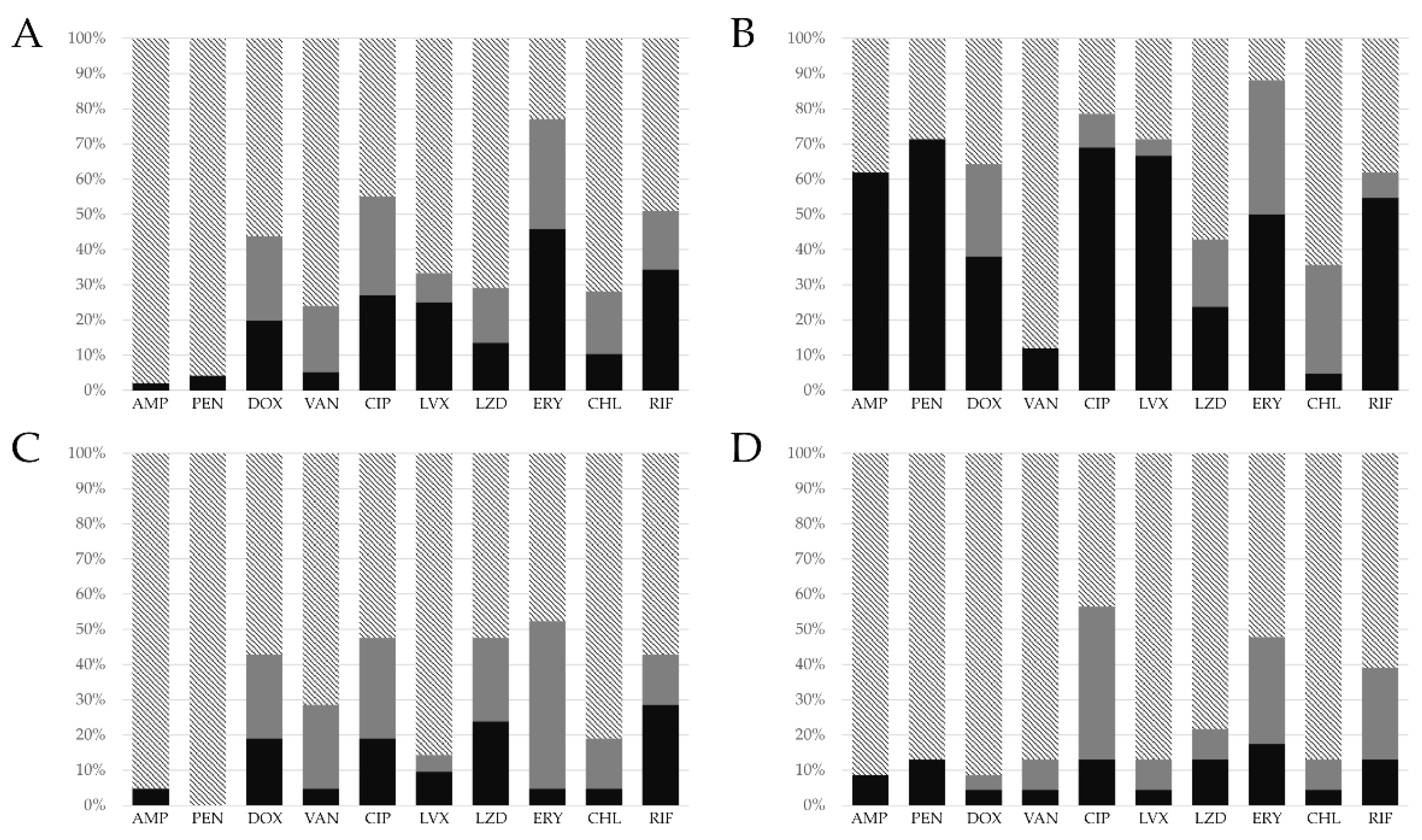
The antibiotic susceptibility test results of four Enterococcus species that accounted for over 10% of the total number of isolates were categorized as resistant, intermediate, and susceptible (Figure 2). Enterococcus faecalis showed a high percentage of intermediate resistance to erythromycin (31.2%; 30/96), followed by ciprofloxacin (28.1%; 27/96), doxycycline (23.9%; 23/96), and vancomycin (18.7%; 18/96). Enterococcus faecium showed a high intermediate resistance rate to erythromycin (38.1%; 16/42). Other Enterococcus species showed a relatively low percentage of antibiotic resistance.
A total of 16 vancomycin-resistant Enterococcus isolates (5 E. faecalis, 5 E. faecium, 2 E. avium, 2 E. gallinarum, 1 E. casseliflavus, and 1 E. hirae) were identified, and 27 bacterial strains demonstrated intermediate vancomycin resistance.
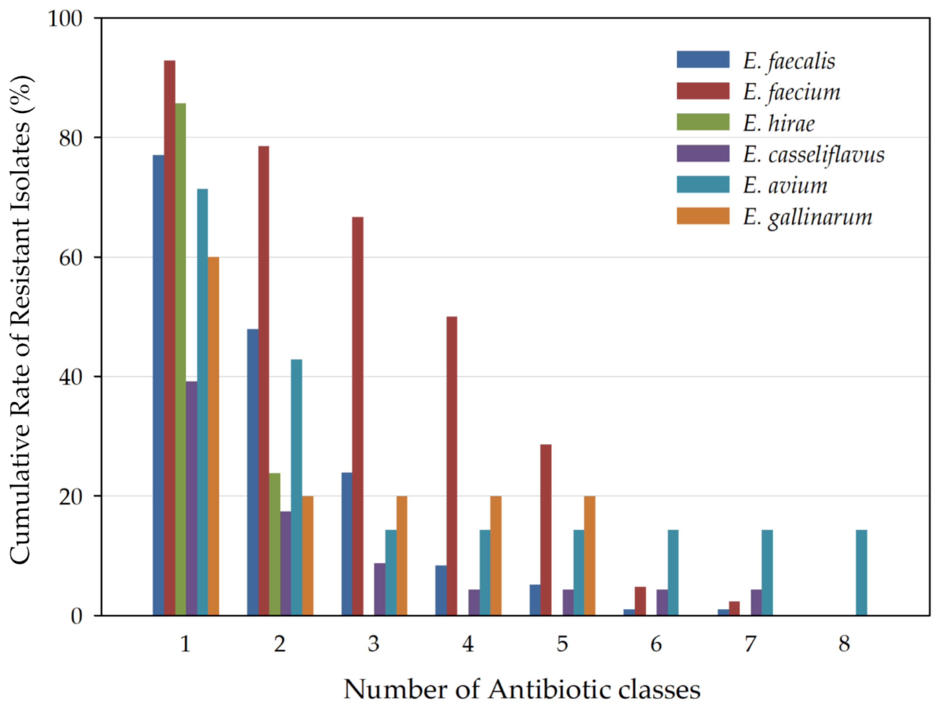
Multidrug-resistance features of the Enterococcus isolates are presented for each species (Figure 3), showing the cumulative percentage of strains that were resistant to one or more antibiotic classes. A total of 28.4% (56/197) of the isolates were found to be MDR. Multidrug resistance was observed more in E. faecium than in E. faecalis. Among E. faecalis isolates, 23/96 (23.9%) were designated as MDR strains. However, 28/42 (66.7%) of E. faecium isolates were MDR. E. faecium significantly showed a high resistance rate in four antibiotics classes, including penicillin, tetracycline, quinolone, and rifampin (p < 0.05). MDR isolates were also presented in the other Enterococcus species, E. avium (1/7; 14.3%), E. casseliflavus (2/23; 8.7%), and E. canintestini (1/2; 50%).
4. Discussion
Enterococcus, along with Staphylococcus, is an opportunistic pathogenic genus frequently detected in the external ear canal of dogs [19]. Several studies have reported antibiotic resistance in enterococci; most have focused on the composition and resistance patterns of Enterococcus collected from fecal, food, and environmental samples [20,21,22,23]. In some studies on Enterococcus from the canine ear canal, E. faecalis was the most dominant species [19,24,25]. Similarly, in this study, E. faecalis was the predominant species. This study is one of the few to include results on the proportion of other species such as E. hirae, E. casseliflavus, E. avium, E. gallinarum, E. canintestini, and E. durans, in addition to the major Enterococcus strains collected from dog ears.
E. hirae infections have been reported as exceedingly rare cases in human medicine, but the bacterial species has been regarded as a probable causative agent in veterinary medicine [26,27,28,29,30,31,32]. The infections were reported to induce septicemia, enteritis, and endocarditis in animals, such as chickens, rats, dogs, and pigs [24,25,31,32]. Although E. hirae is not a main infectious microbe, infection cases in the immunocompetent have been reported, and these cases were severe and life-threatening.
E. gallinarum and E. casseliflavus are intrinsically vancomycin-resistant enterococci (VRE) [33,34,35,36,37]. These species possess the glycopeptide resistance gene C (vanC) in a highly conserved genome region [33,34]. These species are resistant to low levels of vancomycin due to the vanC gene [33,34]. In the case of otitis externa, this resistance feature can be crucial. Considering the low-level antibiotic resistance, the target site must be reached at a sufficient antibiotic concentration for effective treatment. However, because high antibiotic concentrations are difficult to maintain in ear canals, from the perspective of treatment strategy, these antibiotic-resistant bacteria can likely contribute to resistance arousal and bacterial reinfection.
The resistance patterns of the strains to 10 commonly used antibiotics belonging to eight classes (penicillins, glycopeptides, macrolides, tetracyclines, fluoroquinolones, phenicols, oxazolidinones, and anamycins) were examined. The prevalence of E. faecalis was higher than that of E. faecium. However, E. faecium had the highest antibiotic resistance among the species. The resistance percentage of E. faecium to four antibiotic classes was significantly higher than that of E. faecalis (penicillin, tetracycline, quinolone, and rifamycin classes; p < 0.05), which is in accordance with Huycke et al. (1998) [38]. Huycke et al. (1998) mentioned an alarming increase in the antibiotic resistance level of E. faecium. Recent research has also represented the distributions of Enterococcus species and antibiotic resistance profiles [19,39,40]. Each study showed different results in E. faecalis (21.3–87.7%) and E. faecium (12.1–59.6%) prevalence and resistance patterns of isolates, but consistency in the significant resistance rate of E. faecium (over 40%) against the penicillin class.
Previous research has found that E. faecium has a high rate of antibiotic resistance [41]. In particular, the studies focused on its resistance to cell wall inhibitory agents. According to reports, approximately 30% of clinical isolates of E. faecium were resistant to penicillin and even combinations with aminoglycoside antibiotics [41,42]. Compared to this study, the resistance ratio towards penicillin class antibiotics is twice as high as reported. That is because not only of the characteristics of E. faecium species, but also of repetitive and prolonged antibiotic treatment.
Vancomycin resistant enterococci (VRE) are important microorganisms in clinical practice [6,43,44]. Therapeutic alternatives against VRE are limited to recently introduced antibiotics, such as daptomycin, linezolid, and quinupristin/dalfopristin [43]. Considering the intermediate antibiotic resistance, the target site must be reached at the maximum antibiotic concentration for effective treatment [45,46]. However, high drug concentrations are difficult to maintain in ear canals because ear canal skin has been damaged, and exudates and waxy materials in the ear canal hinder achieving enough concentrations. From the perspective of treatment strategy, intermediate antibiotic-resistant bacteria can likely contribute to bacterial reinfection [45,46].
Enterococcus faecalis and E. faecium showed apparent differences in penicillin class antibiotic (ampicillin and penicillin) resistance (p < 0.05). Enterococcus faecalis showed a resistance percentage of 2.1% to ampicillin and 4.1% to penicillin, whereas E. faecium showed a resistance percentage of 61.9% to ampicillin and 71.4% to penicillin. In a previous study in humans, infections by ampicillin-resistant Enterococcus have increased [47], and this is alarming as E. faecium causes bacteremia with a higher mortality rate than E. faecalis [47,48]. Ampicillin-resistant enterococci were found to be carried not only by humans but also by companion animals [12,49,50,51]. Several studies described a high prevalence of ampicillin-resistant enterococci in companion animals, and even lineages between human infections [49,50].
Of the 197 isolates, 56 (28.4%) were found to be MDR strains. Previous studies mainly discussed MDR Staphylococcus and Pseudomonas species as the main pathogens of otitis externa [25,52,53]. However, some studies, including the present study, demonstrated Enterococcus bacteria and their multidrug resistance patterns [19,53]. These findings provide a warning and highlight the emergence of antibiotic resistance in another bacterial genus. The MDR Entericoccus can cause infections, sometimes outbreaks, and prolong therapeutic lapses depending on infection control agents. These serial steps cause high mortality and high costs in medical care. Nelson et al. (2022) showed MDR infections cost nearly USD 1.9 billion and resulted in over 10,000 deaths in the United States in 2017 [54]. Therefore, it is important to monitor the resistance of Enterococcus species as an indicator of antibiotic abuse or misuse.
Companion animals have a social function in modern times. They share living environments with human beings, and participate in several activities. However, as human and companion animal relations are getting closer, it is more likely to transmit zoonotic diseases [55,56]. In terms of One Health, the zoonotic aspect is becoming a big issue. The main topic in this section was vector-borne infectious diseases, but recently pathogenic bacterial transmission has received attention [57]. Antimicrobial resistance bacterial infections in companion animals are examples of this [58]. Therefore, it is important to assess microbiological risks. The risks can be transmission of infectious agents and resistance gene transfer [59].
In this study, we evaluated the antimicrobial resistance patterns of Enterococcus species isolated from dogs with chronic otitis externa. With the emergence of antibiotic resistance being one of the greatest risks to public health, it is important to clarify the antibiotic resistance of microbes and establish appropriate therapeutic strategies. The results of this study can not only contribute to treatment strategies for Enterococcus infections but also be used as a comparable index of antibiotic resistance for Enterococcus in the future.
Author Contributions
Conceptualization, J.K. and S.C.P.; methodology, H.J.K. and M.H.Y.; software, J.K. and H.J.K.; validation, S.C.P. and C.P.; formal analysis, J.K.; investigation, J.K.; resources, J.K.; data curation, J.K. and H.J.K.; writing—original draft preparation, H.J.K.; writing—review and editing, J.K.; visualization, H.J.K. and M.H.Y.; supervision, S.C.P.; project administration, S.C.P.; funding acquisition, S.C.P. All authors have read and agreed to the published version of the manuscript.
Funding
This research was funded by the Cooperative Research Program of the Center for Companion Animal Research (PJ013985032022) of the Rural Development Administration of the Republic of Korea.
Institutional Review Board Statement
All animal procedures were approved by the Seoul national university IACUC (approval number SNU-180718-3).
Informed Consent Statement
Informed consent was obtained from all animal owners in the study.
Data Availability Statement
Not applicable.
Conflicts of Interest
The authors declare no conflict of interest.
References
- Harvey, R.G. Ear Diseases of the Dog and Cat; CRC Press: London, UK, 2005; ISBN 9781840765274. [Google Scholar]
- Rosser, E.J. Causes of otitis externa. Vet. Clin. N. Am. Small Anim. Pract. 2004, 34, 459–468. [Google Scholar] [CrossRef] [PubMed]
- Quinn, P.J.; Markey, B.K.; Leonard, F.C.; Hartigan, P.; Fanning, S.; Fitzpatrick, E.S. Veterinary Microbiology and Microbial Disease, 2nd ed.; Wiley-Blackwell: Chichester, UK, 2011; ISBN 9781405158237. [Google Scholar]
- Vergis, E.N.; Hayden, M.K.; Chow, J.W.; Snydman, D.R.; Zervos, M.J.; Linden, P.K.; Wagener, M.M.; Schmitt, B.; Murder, R.R. Determinants of vancomycin resistance and mortality rates in enterococcal bacteremia: A prospective multicenter study. Ann. Intern. Med. 2001, 135, 484–492. [Google Scholar] [CrossRef] [PubMed]
- Van den Bogaard, A.E.; Stobberingh, E.E. Epidemiology of resistance to antibiotics: Links between animals and humans. Int. J. Antimicrob. Agents 2000, 14, 327–335. [Google Scholar] [CrossRef]
- Cetinkaya, Y.; Falk, P.; Mayhall, C.G. Vancomycin-resistant enterococci. Clin. Microbiol. Rev. 2000, 13, 686–707. [Google Scholar] [CrossRef]
- McVey, D.S.; Kennedy, M.; Chengappa, M. Veterinary Microbiology; Wiley-Blackwell: Hoboken, NJ, USA, 2013; ISBN 9780470959497. [Google Scholar]
- García Martínez de Artola, D.; Castro, B.; Ramos, M.J.; Díaz Cuevas, Z.; Lakhwani, S.; Lecuona, M. Outbreak of vancomycin-resistant enterococcus on a haematology ward: Management and control. J. Infect. Prev. 2017, 18, 149–153. [Google Scholar] [CrossRef] [Green Version]
- Zhou, X.; Willems, R.J.; Friedrich, A.W.; Rossen, J.W.; Bathoorn, E. Enterococcus faecium: From microbiological insights to practical recommendations for infection control and diagnostics. Antimicrob. Resist. Infect. Control 2020, 9, 130. [Google Scholar] [CrossRef]
- Said, M.S.; Tirthani, E.; Lesho, E. Enterococcus Infections; StatPearls Publishing: Treasure Isalnd, FL, USA, 2021. Available online: https://www.ncbi.nlm.nih.gov/books/NBK567759/ (accessed on 20 October 2022).
- Dolka, B.; Gołębiewska-Kosakowska, M.; Krajewski, K.; Kwieciński, P.; Nowak, T.; Szubstarski, J.; Wilczyński, J.; Szeleszczuk, P. Occurrence of Enterococcus spp. in poultry in Poland based on 2014–2015 data. Med. Weter. 2017, 73, 220–224. [Google Scholar] [CrossRef] [Green Version]
- Ruzauskas, M.; Virgailis, M.; Šiugždinienė, R.; Sužiedėlienė, E.; Šeputienė, V.; Daugelavičius, R.; Zienius, D.; Šengaut, J.; Pavilonis, A. Antimicrobial resistance of Enterococcus spp. isolated from livestock in Lithuania. Vet. Arhiv. 2009, 79, 439–449. [Google Scholar]
- Hamilton, E.; Kaneene, J.B.; May, K.J.; Kruger, J.M.; Schall, W.; Beal, M.W.; Hauptman, J.G.; DeCamp, C.E. Prevalence and antimicrobial resistance of Enterococcus spp and Staphylococcus spp isolated from surfaces in a veterinary teaching hospital. J. Am. Vet. Med. Assoc. 2012, 240, 1463–1473. [Google Scholar] [CrossRef]
- Murray, B.E. Problems and dilemmas of antimicrobial resistance. Pharmacotherapy 1992, 12, 86S–93S. [Google Scholar]
- Martel, J.L.; Tardy, F.; Sanders, P.; Boisseau, J. New trends in regulatory rules and surveillance of antimicrobial resistance in bacteria of animal origin. Vet. Res. 2001, 32, 381–392. [Google Scholar] [CrossRef] [PubMed] [Green Version]
- Herrero, I.A.; Fernández-Garayzábal, J.F.; Moreno, M.A.; Domínguez, L. Dogs should be included in surveillance programs for vancomycin-resistant enterococci. J. Clin. Microbiol. 2004, 42, 1384–1385. [Google Scholar] [CrossRef] [PubMed]
- Performance Standards for Antimicrobial Susceptibility Testing, 32nd ed.; Clinical and Laboratory Standard Institute: Wayne, PA, USA, 2022.
- Magiorakos, A.P.; Srinivasan, A.; Carey, R.B.; Carmeli, Y.; Falagas, M.E.; Giske, C.G.; Harbarth, S.; Hindler, J.F.; Kahlmeter, G.; Olsson-Liljequist, B.; et al. Multidrug-resistant, extensively drug-resistant and pandrug-resistant bacteria: An international expert proposal for interim standard definitions for acquired resistance. Clin. Microbiol. Infect. 2012, 18, 268–281. [Google Scholar] [CrossRef] [PubMed] [Green Version]
- Jo, H.J.; Chae, H.S.; Kim, H.J.; Kim, M.J.; Park, G.N.; Kim, S.H.; Chang, K.S. High prevalence of Enterococcus spp. From dogs with otitis externa. Korean J. Vet. Serv. 2012, 35, 99–104. [Google Scholar] [CrossRef] [Green Version]
- Klare, I.; Konstabel, C.; Badstübner, D.; Werner, G.; Witte, W. Occurrence and spread of antibiotic resistances in Enterococcus faecium. Int. J. Food Microbiol. 2003, 88, 269–290. [Google Scholar] [CrossRef]
- Golob, M.; Pate, M.; Kušar, D.; Dermota, U.; Avberšek, J.; Papić, B.; Zdovc, I. Antimicrobial resistance and virulence genes in Enterococcus faecium and Enterococcus faecalis from humans and retail red meat. Biomed Res. Int. 2019, 2019, 2815279. [Google Scholar] [CrossRef] [Green Version]
- Jamet, E.; Akary, E.; Poisson, M.A.; Chamba, J.F.; Bertrand, X.; Serror, P. Prevalence and characterization of antibiotic resistant Enterococcus faecalis in French cheeses. Food Microbiol. 2012, 31, 191–198. [Google Scholar] [CrossRef]
- Kim, Y.B.; Seo, K.W.; Shim, J.B.; Hyun Son, S.; Noh, E.B.; Lee, Y.J. Molecular characterization of antimicrobial-resistant Enterococcus faecalis and Enterococcus faecium isolated from layer parent stock. Poult. Sci. 2019, 98, 5892–5899. [Google Scholar] [CrossRef]
- De Graef, E.M.; Devriese, L.A.; Baele, M.; Vancanneyt, M.; Swings, J.; Haesebrouck, F.; Decostere, A. Identification of enterococcal, streptococcal and Weissella species in the faecal flora of individually owned dogs. J. Appl. Microbiol. 2005, 99, 348–353. [Google Scholar] [CrossRef]
- Hariharan, H.; Coles, M.; Poole, D.; Lund, L.; Page, R. Update on antimicrobial susceptibilities of bacterial isolates from canine and feline otitis externa. Can. Vet. J. 2006, 47, 253. [Google Scholar]
- Oh, J.H.; Cho, A.Y.; Kim, Y.S.; Lee, K.Y.; Sun, I.O. A rare case of urinary tract infection caused by Enterococcus hirae in an elderly man with benign prostate hyperplasia. Chonnam Med. J. 2019, 55, 177–178. [Google Scholar] [CrossRef] [PubMed] [Green Version]
- Dicpinigaitis, P.V.; De Aguirre, M.; Divito, J. Enterococcus hirae bacteremia associated with acute pancreatitis and septic shock. Case. Rep. Infect. Dis. 2015, 123852. [Google Scholar]
- Chan, T.S.; Wu, M.S.; Suk, F.M.; Chen, C.N.; Chen, Y.F.; Hou, Y.H.; Lien, G.S. Enterococcus hirae-related acute pyelonephritis and cholangitis with bacteremia: An unusual infection in humans. Kaohsiung J. Med. Sci. 2012, 28, 111–114. [Google Scholar] [CrossRef] [PubMed] [Green Version]
- Lee, G.H.; Lee, H.W.; Lee, Y.J.; Park, B.S.; Kim, Y.W.; Park, S. Acute pyelonephritis with Enterococcus hirae and literature review. Urogenit. Tract Infect. 2017, 12, 49–53. [Google Scholar] [CrossRef]
- Brayer, S.; Linn, A.; Holt, S.; Ellery, K.; Mitchell, S.; Williams, J. Enterococcus hirae bacteremia in an infant: Case report and review of the literature. J. Pediatr. Infect. Dis. 2019, 8, 571–573. [Google Scholar] [CrossRef]
- Chadfield, M.S.; Christensen, J.P.; Juhl-Hansen, J.; Christensen, H.; Bisgaard, M. Characterization of Enterococcus hirae outbreaks in broiler flocks demonstrating increased mortality because of septicemia and endocarditis and/or altered production parameters. Avian Dis. 2005, 49, 16–23. [Google Scholar] [CrossRef]
- Devriese, L.A.; Chiers, K.; De Herdt, P.; Vanrompay, D.; Desmidt, M.; Ducatelle, R.; Haesebrouck, F. Enterococcus hirae infections in psittacine birds: Epidemiological, pathological and bacteriological observations. Avian Pathol. 1995, 24, 523–531. [Google Scholar] [CrossRef]
- Toye, B.; Shymanski, J.; Bobrowska, M.; Woods, W.; Ramotar, K. Clinical and Epidemiologic Significance of Enterococci Intrinsically Resistant to Vancomycin (Possessing the van C Genotype). J. Clin. Microbiol. 1998, 36, 1469. [Google Scholar] [CrossRef] [Green Version]
- Leclercq, R.; Dutka-Malen, S.; Duval, J.; Courvalin, P. Vancomycin resistance gene vanC is specific to Enterococcus gallinarum. Antimicrob. Agents Chemother. 1992, 36, 2005–2008. [Google Scholar] [CrossRef] [Green Version]
- Schouten, M.A.; Hoogkamp-Korstanje, J.A.A.; Meis, J.F.G.; Voss, A. Prevalence of vancomycin-resistant enterococci in Europe. Eur. J. Clin. Microbiol. 2000, 19, 816–822. [Google Scholar] [CrossRef]
- Choi, S.H.; Lee, S.O.; Kim, T.H.; Chung, J.W.; Choo, E.J.; Kwak, Y.G.; Kim, M.N.; Kim, Y.S.; Woo, J.H.; Ryu, J.; et al. Clinical features and outcomes of bacteremia caused by Enterococcus casseliflavus and Enterococcus gallinarum: Analysis of 56 cases. Clin. Infect. Dis. 2004, 38, 53–61. [Google Scholar] [CrossRef] [PubMed] [Green Version]
- Monticelli, J.; Knezevich, A.; Luzzati, R.; Di Bella, S. Clinical management of non-faecium non-faecalis vancomycin-resistant enterococci infection. Focus on Enterococcus gallinarum and Enterococcus casseliflavus/flavescens. J. Infect. Chemother. 2018, 24, 237–246. [Google Scholar] [CrossRef] [PubMed] [Green Version]
- Huycke, M.M.; Sahm, D.F.; Gilmore, M.S. Multiple-drug resistant enterococci: The nature of the problem and an agenda for the future. Emerg. Infect. Dis. 1998, 4, 239. [Google Scholar] [CrossRef] [PubMed] [Green Version]
- Bang, K.; An, J.U.; Kim, W.; Dong, H.J.; Kim, J.; Cho, S. Antibiotic resistance patterns and genetic relatedness of Enterococcus faecalis and Enterococcus faecium isolated from military working dogs in Korea. J. Vet. Sci. 2017, 18, 229–236. [Google Scholar] [CrossRef]
- Hwang, I.Y.; Ku, H.O.; Lim, S.K.; Park, C.K.; Jung, G.S.; Jung, S.C.; Nam, H.M. Species distribution and resistance patterns to growth-promoting antimicrobials of enterococci isolated from pigs and chickens in Korea. J. Vet. Diagn. Investig. 2009, 21, 858–862. [Google Scholar] [CrossRef] [Green Version]
- Herman, D.J.; Gerding, D.N. Antimicrobial resistance among enterococci. Antimicrob. Agents Chemother. 1991, 35, 1–4. [Google Scholar] [CrossRef] [Green Version]
- Bush, L.M.; Calmon, J.; Cherney, C.L.; Wendeler, M.; Pitsakis, P.; Poupard, J.; Levison, M.E.; Johnson, C.C. High-level penicillin resistance among isolates of enterococci: Implications for treatment of enterococcal infections. Ann. Intern. Med. 1989, 110, 515–520. [Google Scholar] [CrossRef]
- Werner, G.; Coque, T.M.; Hammerum, A.M.; Hope, R.; Hryniewicz, W.; Johnson, A.; Klare, I.; Kristinsson, K.G.; Leclercq, R.; Lester, C.H.; et al. Emergence and spread of vancomycin resistance among enterococci in Europe. Euro Surveill. 2008, 13, 19046. [Google Scholar] [CrossRef]
- Nilsson, O. Vancomycin resistant enterococci in farm animals–occurrence and importance. Infect. Ecol. Epidemiol. 2012, 2, 16959. [Google Scholar]
- Hombach, M.; Böttger, E.C.; Roos, M. The critical influence of the intermediate category on interpretation errors in revised EUCAST and CLSI antimicrobial susceptibility testing guidelines. Clin. Microbiol. Infect. 2013, 19, E59–E71. [Google Scholar] [CrossRef] [Green Version]
- Molechan, C.; Amoako, D.G.; Abia, A.L.K.; Somboro, A.M.; Bester, L.A.; Essack, S.Y. Molecular epidemiology of antibiotic-resistant Enterococcus spp. from the farm-to-fork continuum in intensive poultry production in KwaZulu-Natal, South Africa. Sci. Total Environ. 2019, 692, 868–878. [Google Scholar] [CrossRef] [PubMed]
- Lester, C.H.; Sandvang, D.; Olsen, S.S.; Schønheyder, H.C.; Jarløv, J.O.; Bangsborg, J.; Hansen, D.S.; Jensen, T.G.; Frimodt-Møller, N.; Hammerum, A.M.; et al. Emergence of ampicillin-resistant Enterococcus faecium in Danish hospitals. J. Antimicrob. Chemother. 2008, 62, 1203–1206. [Google Scholar] [CrossRef] [PubMed] [Green Version]
- Ghanem, G.; Hachem, R.; Jiang, Y.; Chemaly, R.F.; Raad, I. Outcomes for and risk factors associated with vancomycin-resistant Enterococcus faecalis and vancomycin-resistant Enterococcus faecium bacteremia in cancer patients. Infect. Control Hosp. Epidemiol. 2007, 28, 1054–1059. [Google Scholar] [CrossRef] [PubMed]
- Damborg, P.; Top, J.; Hendrickx, A.P.; Dawson, S.; Willems, R.J.; Guardabassi, L. Dogs are a reservoir of ampicillin-resistant Enterococcus faecium lineages associated with human infections. Appl. Environ. Microbiol. 2009, 75, 2360–2365. [Google Scholar] [CrossRef] [Green Version]
- Tremblay, C.L.; Charlebois, A.; Masson, L.; Archambault, M. Characterization of hospital-associated lineages of ampicillin-resistant Enterococcus faecium from clinical cases in dogs and humans. Front. Microbiol. 2013, 4, 245. [Google Scholar] [CrossRef]
- Van den Bunt, G.; Top, J.; Hordijk, J.; De Greeff, S.C.; Mughini-Gras, L.; Corander, J.; van Pelt, W.; Bonten, M.J.M.; Fluit, A.C.; Willems, R.J.L. Intestinal carriage of ampicillin-and vancomycin-resistant Enterococcus faecium in humans, dogs and cats in The Netherlands. J. Antimicrob. Chemother. 2018, 73, 607–614. [Google Scholar] [CrossRef]
- Lyskova, P.; Vydrzalova, M.; Mazurova, J. Identification and antimicrobial susceptibility of bacteria and yeasts isolated from healthy dogs and dogs with otitis externa. J. Vet. Med. A Physiol. Pathol. Clin. Med. 2007, 54, 559–563. [Google Scholar] [CrossRef]
- Park, S.; Bae, S.; Kim, J.; Oh, T. Identification and antimicrobial susceptibility of bacteria isolated from dogs with chronic otitis externa. J. Vet. Clin. 2017, 34, 23–26. [Google Scholar] [CrossRef]
- Nelson, R.E.; Hyun, D.; Jezek, A.; Samore, M.H. Mortality, Length of Stay, and Healthcare Costs Associated with Multidrug-Resistant Bacterial Infections among Elderly Hospitalized Patients in the United States. Clin. Infect. Dis. 2022, 74, 1070–1080. [Google Scholar] [CrossRef]
- Loeffler, A.; Lloyd, D.H. Companion animals: A reservoir for methicillin-resistant Staphylococcus aureus in the community? Epidemiol. Infect. 2010, 138, 595–605. [Google Scholar] [CrossRef]
- Overgaauw, P.A.; Vinke, C.M.; van Hagen, M.A.; Lipman, L.J. A one health perspective on the human–companion animal relationship with emphasis on zoonotic aspects. Int. J. Environ. Health Res. 2020, 17, 3789. [Google Scholar] [CrossRef] [PubMed]
- Day, M.J. One health: The importance of companion animal vector-borne diseases. Parasit. Vectors 2011, 4, 49. [Google Scholar] [CrossRef] [PubMed] [Green Version]
- Buma, R.; Maeda, T.; Kamei, M.; Kourai, H. Pathogenic bacteria carried by companion animals and their susceptibility to antibacterial agents. Biocontrol Sci. 2006, 11, 1–9. [Google Scholar] [CrossRef] [PubMed] [Green Version]
- Pomba, C.; Rantala, M.; Greko, C.; Baptiste, K.E.; Catry, B.; Van Duijkeren, E.; Mateus, A.; Moreno, A.M.; Pyörälä, S.; Ružauskas, M.; et al. Public health risk of antimicrobial resistance transfer from companion animals. J. Antimicrob. Chemother. 2017, 72, 957–968. [Google Scholar] [CrossRef] [PubMed]
Figure 1.
Distribution of Enterococcus strains isolated from dogs with chronic otitis externa.

Figure 2.
Antibiotic resistance profiles of Enterococcus strains isolated from dogs with otitis externa. (A) E. faecalis. (B) E. faecium. (C) E. hirae. (D) E. casseliflavus. AMP, ampicillin; PEN, penicillin; DOX, doxycycline; VAN, vancomycin; CIP, ciprofloxacin; LVX, levofloxacin; LZD, linezolid; ERY, erythromycin; CHL, chloramphenicol; and RIF, rifampin. Striped, susceptible; gray, intermediate; and black, resistant.

Figure 3.
Cumulative percentages of multidrug resistance in Enterococcus isolates of six species. E. faecalis (23/96; 23.9%), E. faecium (28/42; 66.7%), E. avium (1/7; 14.3%), E. casseliflavus (2/23; 8.7%), E. canintestini (1/2; 50%), and E. hirae (0/21; 0%).
Figure 3.
Cumulative percentages of multidrug resistance in Enterococcus isolates of six species. E. faecalis (23/96; 23.9%), E. faecium (28/42; 66.7%), E. avium (1/7; 14.3%), E. casseliflavus (2/23; 8.7%), E. canintestini (1/2; 50%), and E. hirae (0/21; 0%).


Table 1.
The prevalence of antimicrobial resistance in Enterococcus strains isolated from chronic otitis externa in dogs.
Table 1.
The prevalence of antimicrobial resistance in Enterococcus strains isolated from chronic otitis externa in dogs.
| Antibiotics | E. faecalis | E. faecium | E. casseliflavus | E. hirae | E. avium | E. gallinarum | E. canintestini | E. durans |
|---|---|---|---|---|---|---|---|---|
| n = 96 | n = 42 | n = 23 | n = 21 | n = 7 | n = 5 | n = 2 | n = 1 | |
| AMP | 2 (2.1%) | 26 (61.9%) | 2 (8.7%) | 1 (4.8%) | 2 (28.5%) | 1 (20%) | 1 (50%) | - |
| PEN | 4 (4.2%) | 30 (71.4%) | 3 (13%) | - | 2 (28.5%) | 1 (20%) | - | - |
| DOX | 19 (19.8%) | 16 (38.1%) | 1 (4.3%) | 4 (19%) | 1 (14.3%) | - | - | - |
| VAN | 5 (5.2%) | 5 (11.9%) | 1 (4.3%) | 1 (4.8%) | 2 (28.5%) | 2 (40%) | - | - |
| CIP | 26 (27.1%) | 29 (69%) | 3 (13%) | 4 (19%) | 1 (14.3%) | 1 (20%) | 1 (50%) | - |
| LVX | 24 (25%) | 28 (66.7%) | 1 (4.3%) | 2 (9.5%) | 1 (14.3%) | 1 (20%) | 1 (50%) | - |
| LZD | 13 (13.5%) | 10 (23.8%) | 3 (13%) | 5 (23.8%) | 2 (28.5%) | - | - | - |
| ERY | 44 (45.8%) | 21 (50%) | 4 (17.4%) | 1 (4.8%) | 3 (42.8%) | 1 (20%) | - | - |
| CHL | 10 (10.4%) | 2 (4.7%) | 1 (4.3%) | 1 (4.8%) | 1 (14.3%) | - | 1 (50%) | - |
| RIF | 33 (34.4%) | 23 (54.7%) | 3 (13%) | 6 (28.6%) | 2 (28.5%) | 2 (40%) | 1 (50%) | - |
AMP, ampicillin; PEN, penicillin; DOX, doxycycline; VAN, vancomycin; CIP, ciprofloxacin; LVX, levofloxacin; LZD, linezolid; ERY, erythromycin; CHL, chloramphenicol; RIF, rifampin.
Publisher’s Note: MDPI stays neutral with regard to jurisdictional claims in published maps and institutional affiliations. |
© 2022 by the authors. Licensee MDPI, Basel, Switzerland. This article is an open access article distributed under the terms and conditions of the Creative Commons Attribution (CC BY) license (https://creativecommons.org/licenses/by/4.0/).
Share and Cite
MDPI and ACS Style
Kwon, J.; Ko, H.J.; Yang, M.H.; Park, C.; Park, S.C. Antibiotic Resistance and Species Profile of Enterococcus Species in Dogs with Chronic Otitis Externa. Vet. Sci. 2022, 9, 592. https://doi.org/10.3390/vetsci9110592
AMA Style
Kwon J, Ko HJ, Yang MH, Park C, Park SC. Antibiotic Resistance and Species Profile of Enterococcus Species in Dogs with Chronic Otitis Externa. Veterinary Sciences. 2022; 9(11):592. https://doi.org/10.3390/vetsci9110592
Chicago/Turabian StyleKwon, Jun, Hyoung Joon Ko, Myoung Hwan Yang, Chul Park, and Se Chang Park. 2022. "Antibiotic Resistance and Species Profile of Enterococcus Species in Dogs with Chronic Otitis Externa" Veterinary Sciences 9, no. 11: 592. https://doi.org/10.3390/vetsci9110592
Note that from the first issue of 2016, this journal uses article numbers instead of page numbers. See further details here.