Article

Two Novel Yersinia pestis Bacteriophages with a Broad Host Range: Potential as Biocontrol Agents in Plague Natural Foci

1
National Institute for Communicable Disease Control and Prevention, China CDC, Beijing 102206, China
2
State Key Laboratory of Infectious Disease Prevention and Control, Beijing 102206, China
3
Department of Public Health, Medical Institute of Qinghai University, Xining 810016, China
4
Yunnan Institute for Endemic Disease Control and Prevention, Dali 671000, China
5
Yunnan Provincial Key Laboratory for Zoonosis Control and Prevention, Dali 671000, China
6
Qinghai Institute for Endemic Disease Control and Prevention, Xining 811602, China
*
Authors to whom correspondence should be addressed.
These authors contributed equally to this work.
Viruses 2022, 14(12), 2740; https://doi.org/10.3390/v14122740
Submission received: 11 November 2022 / Revised: 2 December 2022 / Accepted: 5 December 2022 / Published: 8 December 2022
(This article belongs to the Special Issue Bacteriophage Bioinformatics)

Abstract

:
Bacteriophages (phages) have been successfully used as disinfectors to kill bacteria in food and the environment and have been used medically for curing human diseases. The objective of this research was to elucidate the morphological and genomic characteristics of two novel Yersinia pestis phages, vB_YpeM_ MHS112 (MHS112) and vB_YpeM_GMS130 (GMS130), belonging to the genus Gaprivervirus, subfamily Tevenvirinae, family Myoviridae. Genome sequencing showed that the sizes of MHS112 and GMS130 were 170507 and 168552 bp, respectively. A total of 303 and 292 open reading frames with 2 tRNA and 3 tRNA were predicted in MHS112 and GMS130, respectively. The phylogenetic relationships were analysed among the two novel Y. pestis phages, phages in the genus Gaprivervirus, and several T4-like phages infecting the Yersinia genus. The bacteriophage MHS112 and GMS130 exhibited a wider lytic host spectrum and exhibited comparative temperature and pH stability. Such features signify that these phages do not need to rely on Y. pestis as their host bacteria in the ecological environment, while they could be based on more massive Enterobacteriales species to propagate and form ecological barriers against Y. pestis pathogens colonised in plague foci. Such characteristics indicated that the two phages have potential as biocontrol agents for eliminating the endemics of animal plague in natural plague foci.

1. Introduction

Plague is an acute infectious disease caused by Yersinia pestis that is mainly found in wild rodents, and parasitic fleas are considered transmitting vectors. To date, public health measures and effective antibiotic treatments have led to a drastic decrease in plague worldwide [1]; however, the disease has still not been eradicated. In fact, more risk for the plague is the endemic threat posed to local residents or travellers in natural plague foci, especially in the background that natural plague foci are distributed widely in the Americas, Asia, and Africa [1]. In addition, multiple drug-resistant strains of Y. pestis have been isolated from patients with bubonic plague in Madagascar [2,3]. For streptomycin resistance in Y. pestis isolates, plasmid-mediated resistance mechanisms [2,3] and rpsL gene mutation mechanisms have also been reported [4,5]. Currently, bacteriophages have been successfully used as disinfectors to kill bacteria in the food, environmental, and agricultural fields and have been used for medical applications in curing human diseases [6].
Many bacteriophages (phages) are capable of lysing Y. pestis, and some of them have once been used for the antibacterial treatment of plague [7]. In addition, corresponding phage receptors have been identified in different parts of the LPS core [8,9,10] and/or outer membrane proteins (ail and OmpF) [9]. Several phages have been shown to be highly specifically lytic to Y. pestis and are routinely used for the diagnosis of plague. They include lytic phages that resemble members of Podoviridae in morphology, such as the Pokrovskaya [11], A1122 [12], Yep-phi [13], etc. A filamentous phage acquired by Y. pestis, YpfΦ [14], had the capacity to infect another pathogenic Yersinia species (Yersinia enterocolitica and Yersinia pseudotuberculosis), as well as Escherichia coli strains. In addition, Y. pestis P2-like temperate phages, such as L-413C (NC_004745) [15], vB_YpM_22, vB_YpM_46, and vB_YpM_50 [16], were also isolated from various environmental backgrounds. A few T4-like phages that can infect Y. pestis have been described to date, such as YpsP-PST (KF208315.1) [11], fPS-2 (LR215722), fPS-65 (LR215724), fPS-90 (LR215723) [17], fD1 (HE956711) [18], and JC221 [19].
Two novel Y. pestis phages (vB_YpeM_MHS112 and vB_YpeM_GMS130) were isolated in this study. The two phages belong to the genus Gaprivervirus, subfamily Tevenvirinae, family Myoviridae, and the initial characterisations were illustrated using the host range test, one-step growth curve analysis, and stability tests under various stress conditions. The genomes of the two Gaprivervirus phage isolates were analysed and compared with phages from the same genus through annotation and alignment. In addition, the corresponding phylogenetic relationships among phages in the genus Gaprivervirus and several T4-like phages, including those infecting Yersinia, were illustrated in this study.

2. Materials and Methods

2.1. Isolation and Identification of Phages

Two phages (MHS112 and GMS130) were isolated in two different counties that once belonged to the Rattus flavipectus plague focus in Yunnan, China. MHS112 was isolated from the intestinal contents of shrews (Suncus murinus) in Menghai County. GMS130 was isolated from stool samples of R. flavipectus in Gengma County (Lincang City, Yunnan). Phages MHS112 and GMS130 were isolated with the Y. pestis EV strain as the host indicator using similar standard isolation protocols. Briefly, after centrifugation for 10 min at 9000× g, the supernatant was filtered through a 0.45 µm filter and incubated with the host strains for 24 h at 37 °C. Luria-Bertani (LB) broth or LB agar plates were used to cultivate Y. pestis. SM buffer (5.8 g/L of NaCl, 2.0 g/L of MgSO4, 50 mL/L of 1 M Tris, pH 7.5, and 5 mL/L of presterilised 2% gelatine) was used for phage storage and dilutions. Phage plaque assays were performed by the double-layer agar method, as described in [19]. Following appropriate dilution, the suspensions were plated for single plaque formation. Then, after three rounds of single plaque isolation, MHS112 and GMS130 were isolated, and their plaque characteristics were recorded. Phage propagation was performed according to classic procedures [20].

2.2. Transmission Electron Microscopy

For electron microscopy, the purified phages were spotted onto 400 mesh carbon-coated grids and negatively stained with 2% phosphotungstic acid (pH 6.5). The grids were observed using a transmission electron microscope (Hitachi HT7700, Tokyo, Japan, 80 kV) with a Gatan 832.10 W CCD camera (Gatan, Pleasanton, CA, USA) operating at Gatan Digital Micrograph software. The dimensions of three viral particles of each phage were measured, and the values were averaged.

2.3. Host Range Analysis

Host range analysis was performed using the spot testing method. The host range assays also tested more representative strains of family Enterobacteriaceae other than Y. pestis, and a total of 114 other genera or species of Enterobacteriaceae were included in the host range analysis, including the genera Yersinia, Escherichia, Salmonella, Shigella, Cronobacter, Klebsiella, Enterobacter, Enterococcus, Cronobacter, Serratia, Proteus, Providencia, Morganella, Citrobacter, Pantoea, etc. (Supplementary Table S2).

2.4. Growth Characteristics and Thermo, pH Stability Tests

The one-step growth curve of the phage was determined according to the procedures in the previous literature [19] with slight modification. Briefly, 1 mL phage and 1 mL host bacterium (Y. pestis EV76 or E. coli-CMCC43201) were mixed at an optimal multiplication of infection. The virion samples were taken at different time points (Supplementary Figures S1 and S2), and the titre was measured by the double-layer plate method after serial dilution 10-fold. Three replicates were performed. The measurement of phage temperature and pH stability tests was carried out according to previous literature [21].

2.5. Phage Genome Sequencing, Assembly, Annotation, and Comparison

Genomic DNA was extracted from phage propagation picked from a single plaque. Bacterial nucleotides were removed from the phage lysates using DNase 1 and RNase A. Genomic phage DNA was extracted using a Phage Genomic DNA Extraction Kit (ABigen Corp., Beijing, China) and proteinase K according to the manufacturer’s instructions. Phage genomes were sequenced using an Illumina library kit (DNA Library Prep Kit, San Diego, CA, USA) and Illumina NovaSeq PE150 sequencing platform, resulting in >400-fold coverage. The raw sequence data were assembled using SOAPdenovo [22]. The putative genes in phage genomes were identified by the RAST (https://rast.nmpdr.org/, accessed on 14 October 2022) and Galaxy (https://usegalaxy.org/, accessed on 14 October 2022) web services and visualised using Geneious Prime 2022 (https://www.geneious.com, accessed on 21 October 2022). The comparative genetic map was constructed using EasyFig 2.2.5 [23].

2.6. Alignments of the Inferred Amino Acid Sequences of gp23

Twenty-two phage genomes in the genus Gaprivervirus were acquired from NCBI (Supplementary Table S1). In addition, 32 T4-like phage genomes acquired from NCBI were also included in the phylogenetic analysis. The alignments of the inferred amino acid sequences of the structure gene gp23 (major capsid protein) were performed according to previous research [21,24]. BLASTP (https://blast.ncbi.nlm.nih.gov/Blast.cgi, accessed on 18 October 2022) was used to compare the gp23 gene products of MHS112 and GMS130 with other phages in the genus Gaprivervirus or T4-like phages [6] (Supplementary Table S1). MEGA11 was used to construct the phylogenetic tree [25].

2.7. Accession Number

The whole genome sequences of the phages were deposited in GenBank under the accession numbers vB_YpeM_MHS112 (OP750247) and vB_YpeM_GMS130 (OP750248).

3. Results

3.1. Isolation and Morphology of the MHS112 and GMS130 Phages

The two isolated phages (MHS112 and GMS130) formed clear plaques of 2–3 mm diameter in 24 h in the double-agar layer. According to electron microscopy morphological observations, MHS112 and GMS130 had hexagonal outline heads, indicating their icosahedral symmetry, and had long contractile tails. Such characteristics suggested that they belonged to the family Myoviridae under the Caudovirales order. The size of the head for MHS112 ranged from 73 to 74 nm in diameter and 93 to 101 nm in length, with tail lengths of 110 nm and 15 nm in width. The size of the head for GMS130 ranged from 59 to 63 nm in diameter and 85 to 89 nm in length, with tail lengths of 85 nm and 15 nm in width (Figure 1). Such elongated heads indicated that the two phages have the characteristics of T4-like phages [18] (coined the genus Tequatroviruses according to the taxonomy of prokaryotic viruses updated from the ICTV Bacterial and Archaeal Viruses Subcommittee) [26].

3.2. Host Range Determination of MHS112 and GMS130 Phages

MHS112 and GMS130 phage isolates can infect four representative biovars of Y. pestis (antiqua, mediaevalis, orientalis, and microtus) in China. MHS112 and GMS130 are also sensitive to Y. enterocolitica and Y. pseudotuberculosis. In addition, the two phages exhibited relatively wide host ranges in a few species of the family Enterobacteriaceae, such as MHS112 and GMS130, which can both lyse E. coli, Shigella sonnei, Salmonella typhimurium, and Salmonella blidan. In the collection of bacterial species in this study, collectively, phage GMS130 showed a broader host range than MHS112 because GMS130 had the ability to infect more non-Yersinia strains, including nonpathogenic E. coli (ATCC 25,922 and FC 7792), enteroaggregative E. coli (EAEC), enterohemorrhagic E. coli (EHEC), Shigella dysenteriae, Shigella boydii, and Enterobacter cloacae. Correspondingly, there were some species strains that only MHS112 can infect, such as Shigella flexneri, E. coli (ATCC8739, ATCC41446, and MG1655), and Salmonella cholerasuis (Supplementary Table S2).

3.3. Growth Characteristics and Stability Assessment

The one-step growth study revealed that MHS112 and GMS130 had similar latent periods of 20 min, with different burst sizes of approximately 42 and 62 virions per infected bacterium, respectively. When exposed to 28, 37, and 50 °C for 1 h, MHS112 and GMS130 both had high survival rates. The thermostability began to decrease when the temperature was above 60 °C. Assessment of the pH stability test revealed that MHS112 and GMS130 generally survived well after incubation at pH 3–10 in a one-hour period (Supplementary Figures S1 and S2).

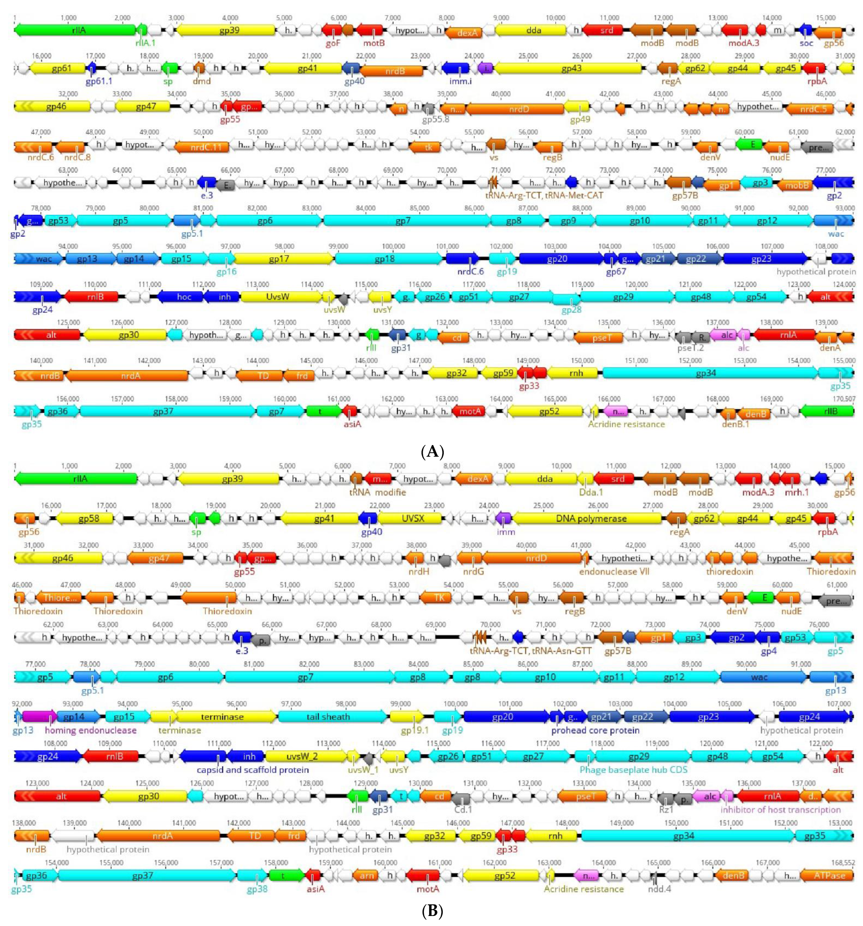
3.4. Genomic Features of MHS112 and GMS130 Phages

After genome sequencing and data processing, high-quality sequences were obtained. The phage genomes of MHS112 and GMS130 consist of a linear dsDNA sequence, 170,507 (MHS112) and 168,552 (GMS130) in size, and the G+C contents of MHS112 and GMS130 are all 40.5%; this rate is different from that of T4 (34.5%) [27]. Phage MHS112 encodes 303 CDSs, of which 259 lie on the reverse strand and 44 lie on the forward strand. The phage GMS130 encoded 292 CDSs, 247 on the reverse strand and 45 on the forward strand (Supplementary Table S3). Two tRNA genes (tRNA-Arg, tRNA-Met) and three tRNA genes (tRNA-Arg, tRNA-Met, tRNA-Asn) were identified in phage MHS112 and GMS130, respectively. MHS112 and GMS130 do not harbour virulence-related genes or antibiotic resistance genes in the two phage genomes, and the absence of lysogenic genes supports the conclusion that they belong to lytic nature phages. The proteins identified for the two phages could be categorised into eleven functional classes [17], i.e., transcription, translation, nucleotide metabolism, virion structure proteins (including head, neck, tail, chaperonins/assembly catalysts), DNA replication/recombination/repair/packaging and processing, lysis, host or phage interactions, host alteration/shutoff, homing endonucleases and homologues, predicted integral membrane or periplasmic proteins, and unknown function hypothetical proteins. The structural and functional arrangement of CDSs is illustrated in Figure 2.

3.5. Alignments of MHS112 and GMS130 with Phage SP18 and Phage vB_EcoM_VR20

Genome alignment at the DNA level demonstrated that the phage genomes of MHS112 and GMS130 are 96.77% identical (coverage 87%). In addition, genome comparisons of MHS112 and GMS130 with other representative phages in the genus Gaprivervirus revealed that MHS112, at the DNA level, had 95.30% sequence identity to SP18 [28] and 96.61% identity to the low-temperature T4-like coliphage vB_EcoM_VR20 [29], respectively. GMS130 shared 97.35% identity with SP18 and 95.59% identity with vB_EcoM_VR20 (Figure 3A). Comparative genomics revealed that MHS112 and GMS130 phages have very limited nucleotide similarity to previous T4-like (Tequatrovirus) phage isolates (JC221) [19] from Yunnan Province (data not shown). In addition, Figure 3B shows the small nonmatched gene cluster (red frame in Figure 3A) containing two genes encoding a long tail fibre distal subunit (gp37) and a chaperone protein (gp38) [17] among MHS112, GMS130, SP18, and vB_EcoM_VR20, and such a mismatch maybe, to some extent, could explain the difference in host lysis ranges.

3.6. Phylogenetic Relationship Analysis of Phages in the Genus Gaprivervirus and T4-Like Phages Infecting the Genus Yersinia

The major capture structural protein gp23 genes in the MHS112 and GMS130 genomes all comprise 1560 bp and encode 519 amino acid (aa) residues. Comparing these inferred amino acid sequences of gp23 among phages in the genus Gaprivervirus (such as p479 [6], UPEC01, UPEC07 [30], etc.) and several representative T4-like phages in the Myoviridae family, including those infecting the genus Yersinia T4-like phages (such as YpsP-PST [11], fPS-2, fPS-65, fPS-90 [17], fD1 [18], JC221 [19], etc.). The two novel phage isolates (MHS112 and GMS130) were clustered into the genus Gaprivervirus. With the exception of SP18 (a Shigella phage), all phages in Gaprivervirus use Escherichia as a host. In addition, T4-like phages infecting the genus Yersinia were mostly grouped into Tequatrovirus, with the exception of previously isolated Y. pestis phage JC221 [19] from Yunnan Province (Figure 4).

4. Discussion

4.1. Morphological Properties with T4-Like Phage Characteristics

Because the two phages, MHS112 and GMS130, have elongated heads and contractile tails with base plates (Figure 1), these morphological properties were characterised as T4-like phage properties [18]. Therefore, the two phages in this study were primarily considered T4-like phages (or newly coined as Tequatrovirus in subfamily Tevenvirinae, family Myoviridae). Through genome comparison with other phages in the genus Gaprivervirus, MHS112 and GMS130 were found to have above 95% sequence identity with phages in the genus Gaprivervirus (Figure 3A). However, there was only 13% collinear nucleotide similarity to the T4 phage (data not shown). According to the classification standard, phage genome sequence similarity greater than 50% is considered the same genus [31], so MHS112 and GMS130 should belong to the genus Gaprivervirus, subfamily Tevenvirinae, and family Myoviridae. Because phages in the genus Gaprivervirus all used E. coli or Shigella sonnei as primer hosts, they were the first phages took Y. pestis as one of the hosts and characterised with the genus Gaprivervirus properties.
T4-like phages are a diverse group of lytic bacterial Myoviruses that share genetic homologies and morphological similarities with the well-studied coliphage T4 [32,33]. In fact, T4-type Myoviruses were previously classified into four subgroups based on sequence comparison of three major structural proteins (gp18, gp19 and gp23), i.e., the T-evens, pseudo-T-evens, shizo-T-evens, and exo-T-evens [34]. In addition, with increasing divergence from T4-like phages, the T4-type phage superfamily further comprised at least 15 distinct subgroups [29]. Approximately 90% of the known T4-like phages grow on E. coli [24,35,36] or other enterobacteria, but the remaining 10% grow on phylogenetically more distant bacteria (Aeromonas [24], Vibrio [37,38], cyanobacteria, etc.) [37]. Some T4-like phages infecting the genus Yersinia have been identified thus far, such as YpsP-PST [11], phiJA1 [10], fPS-2, fPS-65, and fPS-90 isolated from pig stools in Finland [17]. The phiD1 (fD1) isolated from the sewage of Turku in Finland belongs to the T4-like phage [18]. Previous research found that the phages fPS-2, fPS-65, and fPS-90 also infected Y. pseudotuberculosis and Y. pestis through LPS and OmpF as receptors [17].

4.2. Associated Ecological Function of Phages with Wider Host Ranges

Phages are extremely abundant in the environment and can outnumber their prokaryotic hosts by several orders of magnitude [39]. T4-like phages, as a large, virulent, aggregated phage population, significantly influence microbial ecology and, consequently, affect the entire ecosystem [40]. Phages MHS112 and GMS130 were isolated with Y. pestis as the indicator host. However, the two phages exhibited relatively wide host ranges, not only infecting the Yersinia genus, such as Y. pseudotuberculosis and Y. enterocolitica, but also infecting some species in the order Enterobacteriales, such as the genera Escherichia, Shigella, and Salmonella.
Many phages are capable of lysing Y. pestis. For example, T7-related phages generally have specific lytic characteristics to Y. pestis, so they are routinely used for plague diagnostic purposes, such phages as Pokrovskaya [11], A1122 [12], Yep-phi, and YpP-G [7]. However, T7-related virulent phages are not suitable for diminishing the endemicity of animal plague due to their narrow host lysis ranges because once the host bacteria disappear, such phages cannot persist in the environment. For the Y. pestis P2-related phages, such as L-413C [15], L-413C was not able to infect 27 Y. pestis strains. More importantly, P2-related phages have lysogenic characteristics, and they can coexist with host strains in lysogenic conditions, so P2-related phages are generally not suitable for the purpose of eliminating epizootic endemics in natural plague foci. Similar to T4-like phages (Tequatrovirus) in subfamily Tevenvirinae, our newly isolated phages MHS112 and GMS130 belong to genus Gaprivervirus, subfamily Tevenvirinae. The lytic nature endowed the two novel bacterial phages with potential candidates for ecological control in natural plague foci.

4.3. The Ecological Barrier Function Endowed Phages with a Wider Host Range in Natural Plague Foci

Our newly isolated Y. pestis phages MHS112 and GMS130 exhibited virulent and wider host range characteristics, even though they were primitively isolated with Y. pestis as an indicator host. In fact, in the ecological environment, they do not need to rely on Y. pestis as their host bacteria, while they could be based on more massive Enterobacteriales species to propagate. In addition, the two phages appeared to reproduce more efficiently in E. coli strains (CMCC43201) than in Y. pestis (Supplementary Figure S1), similar to results observed in previous research in T4-like phages [18]. Correspondingly, a large number of phages are inhibited in the ecological environment in natural plague foci because they have the capability to lyse Y. pestis, so such lysing phages can become an ecological barrier against the Y. pestis pathogen colonised in plague foci.

5. Conclusions and Perspectives

In this study, two novel Y. pestis Gaprivervirus phages (MHS112 and GMS130) were identified, and their genomes were compared with those of closely related phages. The two Gaprivervirus phages infect both Y. pestis and more Enterobacteriales species. Together with comparatively larger burst sizes per infected Y. pestis bacterium (approximately 42 and 62 virions per bacterium) and stability characteristics in common factors in the environment, these characteristics indicated that they have potential as microecological biocontrol agents used to diminish the endemicity of plague in natural foci in the future. However, such practice should rely on more scientific research, such as measuring the reduction rate on bacterial load, assessing the inhibition effectiveness in natural plague foci, and developing more practicable phage cocktail schemes, such as more distinct phages, for example, Tequatrovirus phages, which were contained in schemes. In addition, by changing various host specificity-associated genes via genetic engineering, phages with a wider host range could be developed, which will greatly benefit from the biocontrol of animal plague in natural plague foci [41].

Supplementary Materials

The following supporting information can be downloaded at: https://www.mdpi.com/article/10.3390/v14122740/s1, Table S1. Genomic Features of Gaprivervirus bacteriophages and representative T4-like bacteriophages infecting Yersinia in this study. Table S2. Host range specificities of MHS112 and GMS130 bacteriophage. Table S3. Annotation of the MHS112 and GMS130 bacteriophage genomes. Figure S1. Growth characteristics and stability test of MHS112 (A) and GMS130 (B).

Author Contributions

Methodology, H.J., Y.Z., X.S., C.L. and W.L.; software, H.J. and Y.W.; validation, Y.H. and Y.Z.; formal analysis, W.L., H.J. and Y.W.; investigation, H.J., Y.Z., C.Z. and J.G.; resources, Y.Z., W.L. and P.W.; data curation, H.J. and Y.W.; writing—original draft preparation, W.L.; writing—review and editing, W.L. and P.W.; visualisation, H.J.; supervision, W.L.; project administration, H.X. and W.L. All authors have read and agreed to the published version of the manuscript.

Funding

This research was funded by the National Key Research and Development Plan (No. 2021YFC1200205), the National Natural Science Foundation of China (82002197), and the Key Scientific and Technology Project of Inner Mongolia Autonomous Region (2021ZD0006).

Institutional Review Board Statement

Not applicable.

Informed Consent Statement

Not applicable.

Data Availability Statement

Not applicable.

Acknowledgments

We thank Yuanhai You from the National Institute for Communicable Disease Control and Prevention (ICDC), China CDC, for providing, technical support on bioinformation analyses, as well as Haijian Zhou, Yanwen Xiong, and Meiying Yan from ICDC, Dongke Chen from the Beijing Hospital for providing some of the bacteria used in the phage host range analysis.

Conflicts of Interest

The authors declare no conflict of interest. The founding sponsors had no role in the design of the study; in the collection, analyses, or interpretation of data; in the writing of the manuscript, and in the decision to publish the results.

References

  1. Morelli, G.; Song, Y.; Mazzoni, C.J.; Eppinger, M.; Roumagnac, P.; Wagner, D.M.; Feldkamp, M.; Kusecek, B.; Vogler, A.J.; Li, Y.; et al. Yersinia pestis genome sequencing identifies patterns of global phylogenetic diversity. Nat. Genet. 2010, 42, 1140–1143. [Google Scholar] [CrossRef] [PubMed]
  2. Guiyoule, A.; Gerbaud, G.; Buchrieser, C.; Galimand, M.; Rahalison, L.; Chanteau, S.; Courvalin, P.; Carniel, E. Transferable plasmid-mediated resistance to streptomycin in a clinical isolate of Yersinia pestis. Emerg. Infect. Dis. 2001, 7, 43–48. [Google Scholar] [CrossRef] [PubMed] [Green Version]
  3. Galimand, M.; Guiyoule, A.; Gerbaud, G.; Rasoamanana, B.; Chanteau, S.; Carniel, E.; Courvalin, P. Multidrug resistance in Yersinia pestis mediated by a transferable plasmid. N. Engl. J. Med. 1997, 337, 677–680. [Google Scholar] [CrossRef]
  4. Dai, R.; He, J.; Zha, X.; Wang, Y.; Zhang, X.; Gao, H.; Yang, X.; Li, J.; Xin, Y.; Wang, Y.; et al. A novel mechanism of streptomycin resistance in Yersinia pestis: Mutation in the rpsL gene. PLoS Negl. Trop. Dis. 2021, 15, e0009324. [Google Scholar] [CrossRef] [PubMed]
  5. Andrianaivoarimanana, V.; Wagner, D.M.; Birdsell, D.N.; Nikolay, B.; Rakotoarimanana, F.; Randriantseheno, L.N.; Vogler, A.J.; Sahl, C.M.; Somprasong, N.; Cauchemez, S.; et al. Transmission of Antimicrobial Resistant Yersinia pestis during a Pneumonic Plague Outbreak. Clin. Infect. Dis. 2022, 74, 695–702. [Google Scholar] [CrossRef] [PubMed]
  6. Li, P.; Wang, H.; Li, M.; Qi, W.; Qi, Z.; Chen, W.; Dong, Y.; Xu, Z.; Zhang, W. Characterization and genome analysis of a broad lytic spectrum bacteriophage P479 against multidrug-resistant Escherichia coli. Virus Res. 2022, 308, 198628. [Google Scholar] [CrossRef]
  7. Zhao, X.; Skurnik, M. Bacteriophages of Yersinia pestis. Adv. Exp. Med. Biol. 2016, 918, 361–375. [Google Scholar] [CrossRef]
  8. Filippov, A.A.; Sergueev, K.V.; He, Y.; Huang, X.Z.; Gnade, B.T.; Mueller, A.J.; Fernandez-Prada, C.M.; Nikolich, M.P. Bacteriophage-resistant mutants in Yersinia pestis: Identification of phage receptors and attenuation for mice. PLoS ONE 2011, 6, e25486. [Google Scholar] [CrossRef] [Green Version]
  9. Zhao, X.; Cui, Y.; Yan, Y.; Du, Z.; Tan, Y.; Yang, H.; Bi, Y.; Zhang, P.; Zhou, L.; Zhou, D.; et al. Outer membrane proteins ail and OmpF of Yersinia pestis are involved in the adsorption of T7-related bacteriophage Yep-phi. J. Virol. 2013, 87, 12260–12269. [Google Scholar] [CrossRef] [Green Version]
  10. Filippov, A.A.; Sergueev, K.V.; He, Y.; Nikolich, M.P. Bacteriophages capable of lysing Yersinia pestis and Yersinia pseudotuberculosis: Efficiency of plating tests and identification of receptors in escherichia coli K-12. Adv. Exp. Med. Biol. 2012, 954, 123–134. [Google Scholar] [CrossRef]
  11. Rashid, M.H.; Revazishvili, T.; Dean, T.; Butani, A.; Verratti, K.; Bishop-Lilly, K.A.; Sozhamannan, S.; Sulakvelidze, A.; Rajanna, C. A Yersinia pestis-specific, lytic phage preparation significantly reduces viable Y. pestis on various hard surfaces experimentally contaminated with the bacterium. Bacteriophage 2012, 2, 168–177. [Google Scholar] [CrossRef] [Green Version]
  12. Kiljunen, S.; Datta, N.; Dentovskaya, S.V.; Anisimov, A.P.; Knirel, Y.A.; Bengoechea, J.A.; Holst, O.; Skurnik, M. Identification of the lipopolysaccharide core of Yersinia pestis and Yersinia pseudotuberculosis as the receptor for bacteriophage phiA1122. J. Bacteriol. 2011, 193, 4963–4972. [Google Scholar] [CrossRef] [PubMed] [Green Version]
  13. Zhao, X.; Wu, W.; Qi, Z.; Cui, Y.; Yan, Y.; Guo, Z.; Wang, Z.; Wang, H.; Deng, H.; Xue, Y.; et al. The complete genome sequence and proteomics of Yersinia pestis phage Yep-phi. J. Gen. Virol. 2011, 92, 216–221. [Google Scholar] [CrossRef] [PubMed]
  14. Chouikha, I.; Charrier, L.; Filali, S.; Derbise, A.; Carniel, E. Insights into the infective properties of YpfPhi, the Yersinia pestis filamentous phage. Virology 2010, 407, 43–52. [Google Scholar] [CrossRef] [PubMed] [Green Version]
  15. Garcia, E.; Chain, P.; Elliott, J.M.; Bobrov, A.G.; Motin, V.L.; Kirillina, O.; Lao, V.; Calendar, R.; Filippov, A.A. Molecular characterization of L-413C, a P2-related plague diagnostic bacteriophage. Virology 2008, 372, 85–96. [Google Scholar] [CrossRef] [PubMed] [Green Version]
  16. Qi, Z.; Meng, B.; Wei, X.; Li, X.; Peng, H.; Li, Y.; Feng, Q.; Huang, Y.; Zhang, Q.; Xu, X.; et al. Identification and characterization of P2-like bacteriophages of Yersinia pestis. Virus Res. 2022, 322, 198934. [Google Scholar] [CrossRef] [PubMed]
  17. Salem, M.; Pajunen, M.I.; Jun, J.W.; Skurnik, M. T4-like Bacteriophages Isolated from Pig Stools Infect Yersinia pseudotuberculosis and Yersinia pestis Using LPS and OmpF as Receptors. Viruses 2021, 13, 296. [Google Scholar] [CrossRef] [PubMed]
  18. Skurnik, M.; Jaakkola, S.; Mattinen, L.; von Ossowski, L.; Nawaz, A.; Pajunen, M.I.; Happonen, L.J. Bacteriophages fEV-1 and fD1 Infect Yersinia pestis. Viruses 2021, 13, 384. [Google Scholar] [CrossRef]
  19. Yuan, Y.; Xi, H.; Dai, J.; Zhong, Y.; Lu, S.; Wang, T.; Yang, L.; Guan, Y.; Wang, P. The characteristics and genome analysis of the novel Y. pestis phage JC221. Virus Res. 2020, 283, 197982. [Google Scholar] [CrossRef]
  20. Anand, T.; Vaid, R.K.; Bera, B.; Barua, S.; Riyesh, T.; Virmani, N.; Yadav, N.; Malik, P. Isolation and characterization of a bacteriophage with broad host range, displaying potential in preventing bovine diarrhoea. Virus Genes 2015, 51, 315–521. [Google Scholar] [CrossRef]
  21. Liao, Y.T.; Zhang, Y.; Salvador, A.; Harden, L.A.; Wu, V.C.H. Characterization of a T4-like Bacteriophage vB_EcoM-Sa45lw as a Potential Biocontrol Agent for Shiga Toxin-Producing Escherichia coli O45 Contaminated on Mung Bean Seeds. Microbiol. Spectrum 2022, 10, e0222021. [Google Scholar] [CrossRef] [PubMed]
  22. Luo, R.; Liu, B.; Xie, Y.; Li, Z.; Huang, W.; Yuan, J.; He, G.; Chen, Y.; Pan, Q.; Liu, Y.; et al. SOAPdenovo2: An empirically improved memory-efficient short-read de novo assembler. Gigascience 2012, 1, 18. [Google Scholar] [CrossRef] [PubMed]
  23. Sullivan, M.J.; Petty, N.K.; Beatson, S.A. Easyfig: A genome comparison visualizer. Bioinformatics 2011, 27, 1009–1010. [Google Scholar] [CrossRef] [PubMed] [Green Version]
  24. Tetart, F.; Desplats, C.; Kutateladze, M.; Monod, C.; Ackermann, H.W.; Krisch, H.M. Phylogeny of the major head and tail genes of the wide-ranging T4-type bacteriophages. J Bacteriol. 2001, 183, 358–366. [Google Scholar] [CrossRef] [PubMed] [Green Version]
  25. Tamura, K.; Stecher, G.; Kumar, S. MEGA11: Molecular Evolutionary Genetics Analysis Version 11. Mol. Biol. Evol. 2021, 38, 3022–3027. [Google Scholar] [CrossRef]
  26. Lefkowitz, E.J.; Dempsey, D.M.; Hendrickson, R.C.; Orton, R.J.; Siddell, S.G.; Smith, D.B. Virus taxonomy: The database of the International Committee on Taxonomy of Viruses (ICTV). Nucleic Acids Res. 2018, 46, D708–D717. [Google Scholar] [CrossRef] [Green Version]
  27. Miller, E.S.; Kutter, E.; Mosig, G.; Arisaka, F.; Kunisawa, T.; Ruger, W. Bacteriophage T4 genome. Microbiol. Mol. Biol. Rev. 2003, 67, 86–156. [Google Scholar] [CrossRef] [Green Version]
  28. Kim, K.H.; Chang, H.W.; Nam, Y.D.; Roh, S.W.; Bae, J.W. Phenotypic characterization and genomic analysis of the Shigella sonnei bacteriophage SP18. J. Microbiol. 2010, 48, 213–222. [Google Scholar] [CrossRef]
  29. Kaliniene, L.; Klausa, V.; Truncaite, L. Low-temperature T4-like coliphages vB_EcoM-VR5, vB_EcoM-VR7 and vB_EcoM-VR20. Arch. Virol. 2010, 155, 871–880. [Google Scholar] [CrossRef]
  30. Kazibwe, G.; Ndekezi, C.; Alinaitwe, R.; Alafi, S.; Nanteza, A.; Kimuda, M.P.; Nakavuma, J.L. Genome Sequences of Bacteriophages UPEC01, UPEC03, UPEC06, and UPEC07 Infecting Avian Pathogenic Escherichia coli. Microbiol. Resour. Announc. 2022, 11, e0081121. [Google Scholar] [CrossRef]
  31. Adriaenssens, E.; Brister, J.R. How to Name and Classify Your Phage: An Informal Guide. Viruses 2017, 9, 70. [Google Scholar] [CrossRef] [PubMed] [Green Version]
  32. Ackermann, H.W.; Krisch, H.M. A catalogue of T4-type bacteriophages. Arch. Virol. 1997, 142, 2329–2345. [Google Scholar] [CrossRef] [PubMed]
  33. Chibani-Chennoufi, S.; Canchaya, C.; Bruttin, A.; Brussow, H. Comparative genomics of the T4-Like Escherichia coli phage JS98: Implications for the evolution of T4 phages. J. Bacteriol. 2004, 186, 8276–8286. [Google Scholar] [CrossRef] [PubMed] [Green Version]
  34. Ignacio-Espinoza, J.C.; Sullivan, M.B. Phylogenomics of T4 cyanophages: Lateral gene transfer in the 'core' and origins of host genes. Environ. Microbiol. 2012, 14, 2113–2126. [Google Scholar] [CrossRef] [PubMed]
  35. Desplats, C.; Krisch, H.M. The diversity and evolution of the T4-type bacteriophages. Res. Microbiol. 2003, 154, 259–267. [Google Scholar] [CrossRef] [PubMed]
  36. Denou, E.; Bruttin, A.; Barretto, C.; Ngom-Bru, C.; Brussow, H.; Zuber, S. T4 phages against Escherichia coli diarrhea: Potential and problems. Virology 2009, 388, 21–30. [Google Scholar] [CrossRef] [Green Version]
  37. Comeau, A.M.; Bertrand, C.; Letarov, A.; Tetart, F.; Krisch, H.M. Modular architecture of the T4 phage superfamily: A conserved core genome and a plastic periphery. Virology 2007, 362, 384–396. [Google Scholar] [CrossRef] [Green Version]
  38. Miller, E.S.; Heidelberg, J.F.; Eisen, J.A.; Nelson, W.C.; Durkin, A.S.; Ciecko, A.; Feldblyum, T.V.; White, Q.; Paulsen, L.T.; Nierman, W.C.; et al. Complete genome sequence of the broad-host-range vibriophage KVP40: Comparative genomics of a T4-related bacteriophage. J. Bacteriol. 2003, 185, 5220–5233. [Google Scholar] [CrossRef] [Green Version]
  39. Labrie, S.J.; Samson, J.E.; Moineau, S. Bacteriophage resistance mechanisms. Nat. Rev. Microbiol. 2010, 8, 317–327. [Google Scholar] [CrossRef]
  40. Hamdi, S.; Rousseau, G.M.; Labrie, S.J.; Tremblay, D.M.; Kourda, R.S.; Ben Slama, K.; Moineau, S. Characterization of two polyvalent phages infecting Enterobacteriaceae. Sci. Rep. 2017, 7, 40349. [Google Scholar] [CrossRef]
  41. Lee, H.; Ku, H.J.; Lee, D.H.; Kim, Y.T.; Shin, H.; Ryu, S.; Lee, J.-H. Characterization and Genomic Study of the Novel Bacteriophage HY01 Infecting Both Escherichia coli O157:H7 and Shigella flexneri: Potential as a Biocontrol Agent in Food. PLoS ONE 2016, 11, e0168985. [Google Scholar] [CrossRef] [PubMed]
Figure 1. Morphological observations of MHS112 (left) and GMS130 (right) bacteriophages from transmission electron microscopy.
Viruses 14 02740 g001
Figure 2. Genomic organisation and structure of Y. pestis Gaprivervirus bacteriophages MHS112 (A) and GMS130 (B). The red and brown arrows indicate transcription- and translation-associated genes, respectively; the green arrows indicate lysis-associated genes; and the yellow arrows indicate genes with corresponding DNA replication, recombination, repair, packaging, and processing functions. The dark blue, medium blue, and light blue arrows indicate the head, neck, and tail genes, respectively, and the stippled blue arrows indicate chaperonins/assembly catalysts genes. The purple and pink arrows indicate host or bacteriophage interactions and host alteration/shut off genes, respectively. The peach arrows indicate homing endonucleases and homologous genes. The grey arrows indicate predicted integral membrane or periplasmic proteins, and the white arrows indicate genes with unknown function.
Viruses 14 02740 g002
Figure 3. Comparative genomic analysis of bacteriophages MHS112 and GMS130 with bacteriophages SP18 and vB_EcoM_VR20. (A) Whole genome alignment of four phage genomes. The red frame indicates a small nonmatched gene cluster. (B) Organisation of the small nonmatched gene cluster from the red frame in (A).
Viruses 14 02740 g003aViruses 14 02740 g003b
Figure 4. Unrooted phylogenetic tree of the structural gene gp23 in the genus Gaprivervirus bacteriophages and representative T4-like phages infecting Yersinia.
Viruses 14 02740 g004
Publisher’s Note: MDPI stays neutral with regard to jurisdictional claims in published maps and institutional affiliations.

